


















A patient whose eyelid droops at the end of the day, whose voice fades as the conversation goes on, who swallows the first mouthful well and chokes on the tenth. This is not fatigue: it is fatigability, and it has a precise mechanism at the neuromuscular junction. The distinction is not academic. It decides what you do in the session, because strengthening taken to exhaustion can make this disease worse, and because its acute worsening still kills.
- Updated 15 August 2026
- Level Clinical summary
- Sources 52 verified references
- ICD-11 8C60
Myasthenia gravis in three figures
A rare disease, very largely seropositive, whose acute complication remains lethal.
Sources: prevalence, systematic review of 94 studies published between 1952 and 20228 ; antibody distribution, German myasthenia gravis registry, 1,432 patients with complete serology11 ; mortality, Chinese multicentre prospective cohort of 277 patients after crisis22.
In brief
- Fatigability is not fatigue. The cardinal sign is worsening of the deficit with repeated effort and its recovery at rest. An assessment done in the morning can be strictly normal: it is in the evening, or after ten repetitions, that the disease shows itself.
- Conventional strengthening can decompensate the patient. The only trial that compared progressive resistance with aerobic training recorded adverse effects in both arms, including bulbar symptoms in two patients35. Rehabilitation exists and is dosed: submaximal intensity, short sets, clear recovery, monitoring of bulbar and respiratory signs.
- Myasthenic crisis is a life-threatening emergency. Breathlessness, retained secretions, a nasal voice, choking episodes that are getting worse: the session stops and the patient is referred. In-hospital mortality is still 5 %22.
- Common drugs make the disease worse, certain antibiotics first among them27. It is not for the physiotherapist to prescribe, but knowing that the phenomenon exists changes how an abrupt deterioration is read.
- The rehabilitation literature is scarce and small in size. The meta-analyses cover handfuls of patients, and several of their pooled results do not cross the threshold of significance30. Saying so is more useful than extrapolating from other neuromuscular diseases.
In this article
- What is myasthenia gravis, and why is fatigability not fatigue?
- How do you recognise myasthenic fatigability when the patient is in front of you?
- What are the diagnostic tests worth, and which one fits in a clinic?
- Which antibodies, and what do they change for the physiotherapist?
- Does the ocular form generalise, and how fast?
- When do you have to stop the session and refer urgently?
- Which common drugs make myasthenia worse?
- What does the rehabilitation literature really say, and how far does it go?
- How do you dose a session without decompensating the patient?
- What is the role of the respiratory physiotherapist?
- What does thymectomy change for rehabilitation?
- What do two published cases teach us?
- How do you apply this in the clinic?
- Frequently asked questions
What is myasthenia gravis, and why is fatigability not fatigue?
An autoimmune disease of the neuromuscular junction, whose whole clinical behaviour follows from a single fact: transmission holds at rest and gives way with repetition.
Myasthenia gravis is an autoimmune disease in which autoantibodies attack the acetylcholine receptor or other proteins of the postsynaptic membrane of the neuromuscular junction6. The name, inherited from Latin, myasthenia gravis, means severe muscle weakness. It dates from a time when the disease killed half of those it struck; that is no longer the case, and the French rare disease registry now gives a survival of 82.7 % at ten years across 3,963 adult patients9.
The mechanism is easy to remember and it explains all the rest. With each action potential, the nerve terminal releases a quantum of acetylcholine; in normal conditions the amount released far exceeds the threshold needed to trigger contraction, and that margin is called the safety margin of the junction. Myasthenia destroys that margin. The antibodies reduce the number of available receptors, block those that remain and damage the folds of the postsynaptic membrane. Now, acetylcholine release physiologically declines during the first stimuli of a train, in everyone. In a healthy subject that decline has no consequence because the margin absorbs the dip. In the myasthenic patient the margin no longer exists: the same decline takes some junctions below threshold, and fibres stop contracting although the nerve is still commanding them.
The myasthenic muscle is not permanently weak. It becomes weak because it has been asked to work, and it becomes strong again because it has been allowed to fall silent.
Hence the cardinal sign, which no other condition reproduces as clearly: the deficit worsens with repeated effort and recovers at rest. That is why a patient climbs two flights without difficulty and stops on the third, why they speak clearly for three minutes and become unintelligible by the tenth, why they eat lunch properly and choke at dessert. It is also why an assessment carried out in the morning, in a rested patient, can be perfectly normal.
Four benchmarks on the disease
What recent registries and systematic reviews establish.
Sources: incidence8 ; age at onset, French rare disease registry9 ; thymoma, systematic review of 49 studies10 ; fatigue, German registry42.
Fatigue and fatigability: two things that are not treated in the same way
The confusion is constant, including in clinical letters. It has a direct practical consequence: general fatigue and muscle fatigability do not respond to the same levers, and one of the two does not improve with training.
The fatigability described here is a measurable, objective and localised phenomenon: strength falls with repetition, and it returns at rest. It is a sign. Fatigue is a subjective complaint of exhaustion, generalised, which does not necessarily translate into a measurable loss of strength. It is a symptom, and a common one: the German registry finds it in 59 % of 1,464 patients, correlated with severity scores and associated with a history of exacerbation or crisis as well as with a diagnostic delay of more than a year42. The Dutch-Belgian registry gives severe fatigue in 62 % of 420 patients, with the same association with severity43.
What matters for us is that Gilhus's review, which gathered together all the available training studies, concludes explicitly that fatigue not directly linked to weakness is little influenced by physical training, whereas strength and daily function do improve29. In other words: promising a patient that reconditioning will lift their fatigue is a promise the data do not support. Promising that they will gain strength and functional capacity is reasonable.
| What you observe | Myasthenic fatigability | General fatigue |
|---|---|---|
| Nature | Objective sign, measurable | Subjective symptom, reported |
| Location | A precise muscle or group, often asymmetrical | Global sensation, not systematised |
| How it is brought out | Repetition of a standardised effort: the deficit appears | Questionnaire, fatigue scale |
| At rest | Clear recovery, sometimes complete | Partial and inconsistent improvement |
| During the day | Characteristic worsening towards the evening | Variable, often present from the morning |
| With training | Strength and function improve29 | Little influenced29 |
| Other avenues | Adjustment of the background treatment | Sleep, mood: depression 36 %, anxiety 33 %48 ; sleep disorders common49 |
Key points
A myasthenic patient who says “I am tired” may be talking about two distinct things. Ask them to be specific: does the strength come back after a few minutes sitting down? If so, it is fatigability, and it can be worked on. If the sensation persists at rest and was already there on waking, it is fatigue, and reconditioning is not the main tool.
What myasthenia is not
Three neighbours share the neuromuscular ground and are not managed in the same way.
The Lambert-Eaton myasthenic syndrome is also a disorder of the junction, but presynaptic : the antibodies target the voltage-gated calcium channels of the nerve terminal, and not the postsynaptic receptors. The clinical consequence is the reverse of myasthenia: strength can increase transiently during the first contractions, the facilitation phenomenon, before declining. It is frequently associated with small cell lung cancer and involves autonomic dysfunction.
The congenital myasthenic syndromes are genetic and not autoimmune in origin. They appear separately in the international classification of diseases, under code 8C61 where autoimmune myasthenia carries 8C60. They are among the most frequent misdiagnoses in a seronegative picture, along with myopathies and functional disorders4.
The polyradiculoneuropathies, finally, are disorders of the peripheral nerve and not of the junction. The deficit there is permanent and not fatigable, and the tendon reflexes are abolished, whereas they are preserved in myasthenia. We devote a separate article to them, on chronic inflammatory demyelinating polyradiculoneuropathy : the reasoning on dosage is a cousin of this one, but the mechanism and the diagnostic criteria are quite different.
How do you recognise myasthenic fatigability when the patient is in front of you?
The disease is not visible on a static assessment. It shows itself when you provoke it, and it hides when you question a rested patient.
The physiotherapist holds a particular position here. They see the patient over a long period, often at fixed times, and they see them in action. That is exactly the situation in which a fatigable deficit becomes visible, whereas a fifteen-minute consultation in the morning can miss it entirely.
Provoking fatigability rather than waiting for it
The principle of the fatigability manoeuvres is always the same: ask for a sustained or repeated effort, and observe whether performance degrades. Three can be used without equipment.
The sustained upward gaze test : the patient fixes a point above their field of vision for one to two minutes, without blinking more than necessary. An eyelid that gradually drops during the test is a fatigable ptosis. Comparing the two sides counts as much as the measurement: asymmetry is common.
The count-out-loud test : the patient counts from 1 to 50, without taking more breaths than needed. You listen to the voice. If the timbre turns nasal, if articulation falls apart over the last twenty numbers, bulbar fatigability is demonstrated. It is a decisive manoeuvre, because bulbar involvement is what drives the prognosis.
The repeated shoulder abduction test or repeated squatting: you count the number of repetitions before range or speed drops, you let the patient rest for a minute, you count again. A patient who loses half their performance on the second set and recovers at rest is not deconditioned, they are fatigable.
The timing trap
An assessment carried out at 9 a.m. in a patient who has slept well can be normal. That is not an argument against the diagnosis. If the complaint concerns the end of the day, you have to assess at the end of the day, or induce fatigue before measuring. Always note the time of the assessment and the interval since the last pyridostigmine dose: without those two pieces of information, two assessments are not comparable with each other.
Topography, and what it announces
The disease has muscles it prefers. Ocular involvement, ptosis and diplopia, opens the picture in a large proportion of patients. Bulbar involvement affects swallowing, phonation and chewing. Axial involvement produces the dropped head, sometimes as the first sign. Limb involvement predominates proximally. And respiratory involvement, the most feared, can stay silent for a long time.
Dysphagia deserves particular attention because it is both common and deceptive. A recent scoping review, based on instrumental assessments, shows that the impairment affects every phase of swallowing, oral, pharyngeal and oesophageal, that its severity follows the MGFA classification, and above all that silent aspiration is common there: among patients who aspirate without coughing, 75 % develop aspiration pneumonia45. A patient who does not cough is not a patient who is not aspirating.
What fatigability does to strength, and what rest gives back to it
Schematic representation of the behaviour described in the literature. The values illustrate the principle, they do not come from a measured series.
Teaching diagram, with no numerical data behind it. The behaviour shown, worsening with sustained effort and recovery at rest, is the one described as the cardinal sign of the disease629.
Grading severity: the scales you will read in clinical letters
Three tools come up constantly in neurology letters, and it is useful to know what they measure, because they do not measure the same thing.
The MGFA classification, drawn up by a task force of the Myasthenia Gravis Foundation of America, ranks patients from class I to class V according to the topography and the severity of the involvement3. It describes a state, not a course.
The QMG score (Quantitative Myasthenia Gravis) is an examiner-rated measure, scored from 0 to 39, which includes timed fatigability tests. It is the primary endpoint of the MGTX trial19 and the one on which the rehabilitation meta-analyses find their clearest signal.
The MG-ADL is an eight-item patient-reported questionnaire, scored from 0 to 24, covering activities of daily living. It is the one that speaks most directly of what we work on, and paradoxically the one on which the rehabilitation evidence is weakest, as we shall see.
| Class | What it describes | What it implies in the session |
|---|---|---|
| I | Ocular weakness only, with or without ptosis. Normal strength elsewhere. | Ordinary physical activity possible. Watch for the appearance of non-ocular signs. |
| II | Mild weakness beyond the ocular muscles. IIa with limb or axial predominance, IIb with bulbar or respiratory predominance. | The ground of most training studies. Work possible, dosed, with closer monitoring if IIb. |
| III | Moderate weakness. Same split between IIIa and IIIb. | Clear caution. Short sessions, functional goals, no seeking of exhaustion. |
| IV | Severe weakness. Same split between IVa and IVb. | Management coordinated with the neurologist. Respiratory physiotherapy takes first place. |
| V | Intubation, with or without mechanical ventilation. That is the definition of crisis. | Critical care setting. Early mobilisation has been described there, under strict monitoring46. |
The letters a and b count for us more than the numbers. A patient who is IIb or IIIb, with bulbar or respiratory predominance, is a patient in whom the safety margin is thinnest, and that is where session monitoring has to be tightest.
Red flags in this chapter
These elements do not fall to physiotherapy and require a medical opinion without delay:
- Appearance or worsening of choking episodes, especially on liquids, or of a voice that turns nasal within a few days.
- Dropped head that is new, or an inability to hold the head up at the end of the day.
- Breathlessness at rest, orthopnoea, or a need to sleep sitting up, in a patient who did not do so before.
- A cough that has become ineffective : the patient can no longer clear their secretions.
- General worsening in the days that follow the introduction or the increase of a corticosteroid: this is a known phenomenon, documented in the chapter on crisis.
What are the diagnostic tests worth, and which one fits in a clinic?
The diagnosis belongs to the neurologist. But one screening test fits in a pocket, and knowing what the others are worth avoids misreading a report.
The diagnosis rests on three pillars: the clinical picture, serology and electrophysiology. Faced with typical symptoms and a positive antibody test it is straightforward; it is in seronegative patients that a detailed clinical and neurophysiological examination becomes decisive6.
The ice pack test, and what it is really worth
The principle is physiological: cooling slows acetylcholinesterase activity, so prolongs the presence of acetylcholine in the synaptic cleft, so transiently improves transmission. In practice, cold is applied to the ptotic eyelid for two minutes and the palpebral fissure is measured before and after. A clear improvement makes the test positive.
A class I study compared it with single-fibre electromyography in 155 patients referred for ptosis, 102 of whom actually had ocular myasthenia. The ice pack test gives a sensitivity of 86 % (95 % CI 79 to 93) and a specificity of 79 % (95 % CI 68 to 90); single-fibre electromyography, a far more demanding examination, gives 94 % and 79 %14. The two examinations have statistically comparable diagnostic accuracy. And the most useful result in practice lies in this sentence: when both tests are negative, the negative predictive value reaches 94 %, which points strongly towards another diagnosis.
A positive test is not a diagnosis
A specificity of 79 % means that about one patient in five without myasthenia will nonetheless have a positive ice pack test14. One published case describes a positive test in a patient with a schwannoma of the oculomotor nerve, without associated myasthenia15. The test points, it does not conclude. What a physiotherapist does with it: an argument to support a request for an opinion, never an announcement to the patient.
The examinations you will read about in the report
The serology is run in sequence: anti-acetylcholine receptor antibodies, then anti-MuSK, and sometimes anti-LRP4. The techniques have improved: fixed cell-based assays, now available, have shown in routine practice a positive predictive value of 99 % across 770 patients tested, with a single false positive retained12.
The repetitive nerve stimulation test looks for a decrement in the amplitude of the muscle response during a train of low-frequency stimuli. It is the exact electrophysiological translation of what the fatigability manoeuvre shows clinically.
Thoracic imaging is routine, looking for a thymoma, present in about 21 % of myasthenias10. It is a search for a tumour, not a formality: all thymomas are regarded as malignant, and their resection is considered independently of myasthenic status51.
Key points
Three things are enough for the physiotherapist. One: a normal assessment in the morning rules nothing out. Two: the ice pack test is a good orientation tool, with a sensitivity of 86 % and a specificity of 79 %, and it replaces no examination. Three: faced with clear, documented fatigability, the useful action is to write a precise, time-stamped report describing the test performed and the deficit observed, and to refer.
Which antibodies, and what do they change for the physiotherapist?
Serological status is not just a line of laboratory data: it announces a topography, a treatment response, and for one of them a drug contraindication.
The German myasthenia gravis registry gives the most precise distribution available today. Of 3,319 patients, 1,432 had complete serology for the three antibodies. The distribution is as follows: 63.6 % anti-AChR positive, 2.4 % anti-MuSK, 4.8 % anti-LRP4 of whom half in association with anti-AChR, 0.8 % double positive for MuSK and AChR, and 28.4 % triple seronegative11.
Distribution of antibodies across 1,432 complete serologies
German prospective myasthenia gravis registry. Patients with no identified antibody form the second largest group.
Source: German prospective longitudinal myasthenia gravis registry, 1,432 patients tested for the three antibodies11. The value labels are placed outside the bars so that they stay legible whatever their length.
Anti-AChR: the most common form
This is the majority subgroup and the one on which most therapeutic trials rest. The antibodies there activate the complement cascade, which explains the efficacy of the complement inhibitors developed in recent years. It is also the subgroup in which thymectomy has been evaluated by randomised trial, and the only one to which its conclusions apply directly19.
Anti-MuSK: the subgroup where vigilance changes in nature
Anti-MuSK antibody myasthenia is rare but it behaves differently, and it is the subgroup whose particularities a physiotherapist has to know. Clinically, it preferentially affects the bulbar, facial, shoulder and neck muscles13. In other words, it concentrates its involvement exactly where the risk of respiratory failure and of aspiration is highest.
Above all, one point of treatment has direct consequences for what we observe: anticholinesterase drugs must be avoided in anti-MuSK myasthenia, because of a risk of clinical worsening and of cholinergic crisis13. An anti-MuSK patient will therefore generally not be on pyridostigmine, and the ritual question “did you take your treatment before the session?” does not have the same meaning for them. Corticosteroids are less effective there, with a need for higher and more prolonged doses, while rituximab has proved particularly effective and is now often used early.
Seronegative and LRP4: do not conclude too quickly
Nearly one patient in three has none of the three antibodies. That does not mean the disease is less severe, nor that the diagnosis is doubtful. The international ENMC workshop devoted to seronegative myasthenia stresses on the contrary the difficulty of these situations, the absence of a specific test, and the fact that the most frequent errors are myopathies, congenital myasthenic syndromes and functional disorders4.
As for patients with isolated anti-LRP4, the German registry shows that they carry the heaviest disease burden of all the subgroups: MG-ADL 2.5 points higher than in anti-AChR patients (95 % CI 1.2 to 3.9) and markedly worse quality of life, with heavy use of escalation and rescue treatments11. The German registry also finds in them the most severe fatigue in the corpus42.
| Subgroup | Frequency | Topography | What it changes in the session |
|---|---|---|---|
| Anti-AChR | 63.6 % | Ocular then generalised | The reference frame. Thymectomy evaluated by randomised trial in this subgroup. |
| Triple seronegative | 28.4 % | Variable | Diagnosis sometimes debated: do not relax monitoring on the pretext of a blank serology. |
| Anti-MuSK | 2.4 % | Bulbar, facial, shoulders, neck | Bulbar and respiratory risk to the fore. Anticholinesterase drugs avoided: no post-pyridostigmine “window” to exploit. |
| Isolated anti-LRP4 | 2.4 % | Variable | Highest disease burden and fatigue. Goals to be set low at the start. |
| Double positive | 0.8–2.4 % | Variable | LRP4 and AChR patients behave like the other subgroups. |
Key points
One question only to ask at the start of care: which antibody? If the answer is MuSK, the session is built around bulbar and respiratory risk, and the patient is probably not on an anticholinesterase. If the answer is “none”, nothing is reassuring for all that. If the answer is isolated LRP4, set the goals lower.
Does the ocular form generalise, and how fast?
A myasthenia that starts with a ptosis rarely stays ocular. Knowing the window in which it switches helps you interpret a new symptom.
The ocular form is defined by involvement limited to the extraocular muscles and to the levator of the eyelid: ptosis, diplopia, without weakness elsewhere. It is class I of the MGFA classification3. The subject, for a physiotherapist, is not the management of diplopia but conversion: a patient followed for some entirely different reason and carrying an ocular myasthenia may develop a generalised form during the period in which you see them.
The figures vary with the cohorts, the populations and the treatments received, but the message converges. A Thai cohort of 200 patients finds 39 % conversion, with a median delay of 16 months and a two-year rate of 25.5 %16. A Chinese cohort of 435 adult patients finds 54.5 % conversion, with rates of 31.7 %, 49.8 % and 65.4 % at 6, 12 and 24 months17.
Conversion from the ocular form to the generalised form
Two cohorts, two different results. What the two share: the switch happens early.
Sources: Chinese cohort of 435 adult-onset ocular myasthenias, conversion rates at 6, 12 and 24 months17 ; Thai cohort of 200 patients, 2-year rate of 25.5 % and median time to conversion of 16 months16. The intermediate points of the Thai curve are interpolated between the origin and the published value at 24 months.
The gap between the two cohorts is explained in part by the treatments received, and that is the interesting point. Immunosuppression strongly reduces the risk of generalisation: a meta-analysis of ten observational studies covering 1,458 participants gives an odds ratio of 0.23 (95 % CI 0.15 to 0.38) in favour of treatment18. The 2020 international consensus also added a specific recommendation on early immunosuppression in the ocular form1.
The factors associated with faster conversion, in the Thai cohort, are anti-AChR positivity (adjusted HR 2.88; 95 % CI 1.79 to 4.63), thymic abnormalities (HR 2.30; 95 % CI 1.41 to 3.74) and smoking (HR 1.78; 95 % CI 1.04 to 3.03)16. Smoking is the only one of these three factors on which a rehabilitation team can act, if only by mentioning it.
An ocular myasthenia is not a mild myasthenia. It is often a generalised myasthenia that has not yet started.
Red flags in this chapter
In a patient known to have ocular myasthenia, these elements signal a possible generalisation and warrant a prompt opinion:
- The appearance of limb weakness, especially proximal, or of a new difficulty climbing the stairs or getting up from a chair.
- A voice that changes in timbre during a conversation, or chewing that becomes laborious at the end of a meal.
- Any swallowing difficulty, even intermittent, even only on liquids.
- Breathlessness on exertion out of proportion to the patient's usual level.
When do you have to stop the session and refer urgently?
This is the chapter that on its own justifies the existence of this text. Myasthenic crisis is acute respiratory failure, it occurs in a substantial share of patients, and it still kills.
Myasthenic crisis is defined by weakness of the respiratory muscles or of the upper airway muscles sufficient to require ventilation, invasive or not. The international consensus set its definition, as well as that of impending crisis2. It corresponds to class V of the MGFA classification3.
Its frequency is not anecdotal. The Chinese prospective cohort puts it in 10 to 20 % of myasthenic patients22 ; a classic review speaks of about one patient in five, most often in the first year of the disease23, and another of up to 27 %24.
The prognosis has been transformed: mortality, which reached 75 % four decades ago, is now below 5 %23. But “below 5 %” is not “nil”. The prospective cohort of 277 patients followed for one year after a crisis gives 5.05 % in-hospital mortality and 15.16 % all-cause mortality over the follow-up period, the leading causes of death being septic shock (35.7 %) and multiple organ failure (21.4 %)22.
Red flag box: recognising and acting on a myasthenic crisis
What must make you interrupt the session immediately:
- Breathlessness at rest, or being short of breath when speaking, or an inability to finish a sentence without pausing for breath.
- Orthopnoea : the patient can no longer tolerate lying flat, or reports that they have been sleeping sitting up for a few nights.
- Ineffective cough and retained secretions: the patient can hear their secretions and can no longer shift them.
- Choking episodes that are getting worse, especially on liquids, or poorly managed saliva.
- A voice that turns nasal or weak within a few hours or a few days.
- Recruitment of the accessory inspiratory muscles, indrawing, paradoxical breathing, or a respiratory rate that rises with no obvious cause.
- Agitation, sweating, tachycardia : non-specific warning signs which, on this ground, often precede decompensation.
What you do, in this order:
- Stop the effort. No further exercise, no “let us finish the set”.
- Sit the patient up in a sitting or half-sitting position, whichever they tolerate best. Do not lay a breathless patient flat.
- Give nothing to swallow. Neither water nor tablet, as long as swallowing has not been assessed.
- Call 15. A myasthenic crisis is a life-threatening emergency that belongs to intensive care. Do not send the patient home advising them to see a doctor.
- Pass on the useful information : known diagnosis of myasthenia, antibody if you know it, current treatment, what was done in the session, and the precise chronology of the worsening.
Oxygen saturation is not a good safety criterion here: it falls late in respiratory failure of neuromuscular origin. A myasthenic patient can be decompensating with a still-normal saturation. It is the clinical picture and the chronology that decide.
What triggers a crisis
The most frequently reported precipitating factors are respiratory infections, aspiration, sepsis, surgery, too rapid a taper of immunomodulation, the start of corticosteroid therapy, exposure to aggravating drugs and pregnancy23.
Two of these items deserve to be developed because they come as a surprise.
Starting corticosteroids can make things worse. This is not a theoretical rarity: a retrospective study of 119 patients with bulbar symptoms started on corticosteroids finds initial worsening in 52.1 % of them, severe in 13.5 %, with a median onset on the third day and a median duration of 3.5 days26. The intravenous bolus route was more at risk than the oral route (74.2 % against 44.3 % initial worsening). The independent factors for severe worsening included a reduced cough strength (OR 11.56; 95 % CI 1.39 to 95.94). In practical terms: a patient who deteriorates in the week following a change of corticosteroid therapy is not necessarily failing rehabilitation.
Sepsis is the leading cause of death in crisis, and it is partly avoidable. A multicentre study of 57 crises shows that sepsis lengthens the intensive care stay and the duration of ventilation, and that its predictors are age over 50 years (OR 7.74; 95 % CI 1.29 to 46.53) and a prednisone dose of at least 60 mg per day during the stay (OR 13.07; 95 % CI 2.17 to 86.73)25. That is one more argument for taking the prevention of aspiration seriously upstream.
An aspiration pneumonia in a myasthenic patient is not just one more complication. It is one of the known routes to intensive care, and the only one on which a rehabilitation team can weigh upstream.
What happens next, and why it concerns us
Management of the crisis combines intensive care and rapid immunomodulation: plasma exchange, intravenous immunoglobulin, intravenous methylprednisolone, with a delay to action and an inconsistent efficacy that explain the interest in recent targeted treatments52.
The functional prognosis after a crisis is rather good when the patient gets through it: at one year, 79.76 % of the patients in the Chinese cohort had a favourable course, with MG-ADL down from a mean of 21.3 to 2.822. In other words, a patient coming out of a crisis has ahead of them a recovery trajectory that fully justifies rehabilitation, and that is where we come in.
What to do in the session faced with a warning sign
Decision tree. The right-hand branch admits no delay.
Tree built from the consensus definition of crisis and of impending crisis2, from the precipitating factors identified23 and from the course observed after crisis22. It organises a course of action, it does not replace a medical opinion.
Which common drugs make myasthenia worse?
Prescribing is not our business. But a patient who deteriorates abruptly a fortnight after starting an antibiotic is information that someone has to pick up.
The phenomenon is real, documented and classified. A review distinguishes two separate mechanisms that are worth not confusing27.
The first group brings together the drugs that induce a de novo myasthenia by altering immune homeostasis: immune checkpoint inhibitors, penicillamine, tyrosine kinase inhibitors, interferons. Myasthenia induced by checkpoint inhibitors is recognised enough for the 2020 international consensus to have devoted a dedicated recommendation to it1. An analysis of the American pharmacovigilance database shows that it occurs more in older people (adjusted odds ratio 2.4; 95 % CI 1.84 to 3.13) and without the female predominance of idiopathic myasthenia28. The international classification recognises it under a code of its own, 8C60.0.
The second group brings together the drugs that impair neuromuscular transmission in a patient who already has myasthenia: certain antibiotics, certain antiarrhythmics, anaesthetics and muscle relaxants27. These are the ones most often met in community practice.
| Family | Mechanism | What the physiotherapist does with this |
|---|---|---|
| Immune checkpoint inhibitors | Induction of a de novo myasthenia through immune dysregulation27 | In an oncology patient, a new fatigability is not necessarily cancer-related asthenia. Mention it. |
| Penicillamine, interferons, tyrosine kinase inhibitors | Induction of a de novo myasthenia27 | The same: the temporal link with the start of treatment is the useful information. |
| Certain antibiotics | Blockade of neuromuscular transmission27 | The most frequent situation in community practice. A deterioration after an infection may come from the infection or from its treatment. |
| Certain antiarrhythmics | Blockade of neuromuscular transmission27 | Spot recent changes to cardiology prescriptions. |
| Anaesthetics and muscle relaxants | Blockade of neuromuscular transmission27 | Perioperative setting. Explains in part the postoperative vulnerability of these patients. |
| Corticosteroids, at the start of treatment | Known transient worsening26 | 52.1 % initial worsening in bulbar patients, median onset on the 3rd day. Do not conclude that rehabilitation has failed. |
The physiotherapist's exact role, and its limit
We do not prescribe, we do not deprescribe, and we do not advise stopping a treatment. What we do is simpler and more useful: when a myasthenic patient deteriorates, ask what has changed in the last two to four weeks, prescription included, and pass the answer to the doctor with the chronology. A dated deterioration is usable information; a deterioration noted without a date is not.
Key points
Three questions open the session in a myasthenic patient who is doing less well: a recent infection? A new or changed treatment? A change in corticosteroid dose? These three questions cover a large share of the precipitating factors identified23 and take less than thirty seconds.
What does the rehabilitation literature really say, and how far does it go?
It says that exercise is safe in the stable patient, and that its benefits are more modest and more uncertain than article abstracts suggest. Both halves of that sentence count.
Let us start with the fact that has changed most. For a long time, exercise was advised against in myasthenia, on the logical grounds that sustained use of a muscle makes weakness worse there. Gilhus's review settled that point: after identifying ten intervention studies covering 159 patients with a generalised form, three of them on the respiratory muscles, the conclusion is that physical training is safe in myasthenia, that it improves muscle strength and daily function, and that a minimum of 150 minutes of exercise a week is recommended for mild to moderate forms29. The reference primer goes the same way in writing that active physical training should be encouraged6.
So much for permission. There remains the question of what is gained, and there you have to look at the figures rather than at the conclusions.
What the most recent meta-analysis actually measured
The most recent systematic review with meta-analysis included 20 studies, of which only 7 randomised trials, the others being non-randomised trials, quasi-experimental studies and observational work; 10 studies could be pooled quantitatively30. Its abstract announces improvements in symptoms, in quality of life and in activities of daily living. Its detailed results are more nuanced, and it is that nuance that should guide what we promise a patient.
What training changes, with its confidence intervals
Pooled effects from the 2026 meta-analysis. An interval that crosses the line of zero means the benefit is not demonstrated.
Source: systematic review with meta-analysis of 20 studies, of which 7 randomised trials30. Each row carries its own scale, set on the widest bound of its interval: the position relative to no effect is readable row by row, but the lengths cannot be compared from one outcome to another.
The hurried reader will take away that “exercise improves”. The attentive reader will take away three more useful things.
The strongest signal concerns the QMG score, with a mean difference of 1.61 points in favour of training (95 % CI 0.74 to 2.49) and zero heterogeneity between the three pooled studies30. It is the cleanest result in this whole literature. To be set against a scale that runs from 0 to 39.
The MG-ADL, for its part, does not move in a demonstrated way. The standardised mean difference is 0.40 points in favour of training, but its confidence interval runs from 0.94 in favour of training to 0.14 against it: it crosses zero, with a heterogeneity of 74 %30. That is precisely the outcome that measures activities of daily living, and therefore the one our patients ask us to improve. It has to be said.
The six-minute test and forced vital capacity are in the same position : intervals that cross zero, with a heterogeneity of 86 % for vital capacity30. The two respiratory outcomes that come out significant, forced expiratory volume in one second and maximal inspiratory pressure, do so with heterogeneities of 94 % and 91 %, that is to say at the limit of what can be interpreted.
A confidence interval that crosses zero does not say that exercise is useless. It says that those studies, with those sample sizes, could not demonstrate it. The nuance is the one that separates a promise from a reasonable bet.
The other syntheses converge, and on the same weakness
There are several of them, and their agreement is instructive: they all conclude in the same place, with the same reservations.
A 2020 systematic review had retained 11 studies out of 365, identifying three approaches, physical training, respiratory training and balance work, all improving functional outcomes without any being recommendable over another for want of comparative studies, and with a methodological quality judged low to moderate34.
A 2026 systematic review covering eight controlled studies published between 2015 and 2025 concludes that the interventions are well tolerated, without lasting worsening of symptoms, with consistent gains in functional capacity, muscle strength and activities of daily living for aerobic and combined programmes, and more heterogeneous results for respiratory training31. Its stated limitations: small samples, short follow-up, heterogeneity.
A 2025 meta-analysis retained only 6 studies out of 445 identified, with a single one of high methodological quality (level 1b on the OCEBM scale) and five of moderate quality; its five conclusions are graded B33.
Finally, a systematic review devoted to respiratory muscle training alone retained 7 studies for 223 participants of mean age 57.5 years, of which only 2 randomised trials, with intensities ranging from 15 to 75 % of maximal respiratory pressures and frequencies of 3 to 10 sessions a week; heterogeneity was such that a descriptive synthesis had to replace any quantitative pooling32.
Two randomised trials worth reading in full
The first is the one that documents the adverse effects, and it is the most important for safety. Fifteen patients of MGFA class II to IV were randomised to 20 sessions over 8 weeks, either progressive resistance or aerobic training. Twelve completed, with 95 % adherence. The QMG score did not change in either group, and maximal strength and functional capacity increased in the resistance group. But both groups reported adverse effects: bulbar symptoms in two patients, increased fatigue in three, and one dropout was judged possibly related to resistance training35. The authors conclude that eight weeks of moderate to high intensity training is feasible in most patients with mild myasthenia. The restriction is in the original conclusion, it is not invented.
The second is a French trial, MGEX, and it is instructive through its negative result. Forty-five patients with stabilised generalised myasthenia were randomised between usual care alone and usual care plus exercise, the latter consisting of a home rowing programme unsupervised, three 40-minute sessions a week for three months. The exercise was well tolerated. But the intention-to-treat analysis showed no improvement in quality of life : adjusted between-group difference of 0.8 points on the MGQOL-15-F scale, with an interval running from 5.4 in favour of exercise to 3.7 against it36. A detail that counts for safety: the two patients admitted to hospital for an exacerbation during the trial belonged to the usual care group.
Conversely, the RESTOREX trial randomised 40 patients of mild to moderate severity between 30 minutes of daily walking and rest, and finds a better quality of life (p = 0.02) and a better distance on the six-minute test (p = 0.007) in the exercise arm, with no adverse effect at all37. A simple programme, of low intensity, daily.
Modalities and level of evidence
The table below gathers what each modality actually has behind it. The levels are assigned according to GRADE logic, taking into account the type of study, the sample sizes, the heterogeneity and the consistency between syntheses. No rehabilitation modality in myasthenia reaches a high level today, and it would be dishonest to present one as such.
Aerobic training
of moderate intensity
What supports it. Several concordant randomised trials on tolerance; gain on the QMG with zero heterogeneity in the meta-analysis30 ; quality of life and walking distance improved in RESTOREX with no adverse effect37 ; recommendation of a minimum of 150 minutes a week29.
What limits it. Samples of a few dozen patients, almost exclusively mild to moderate forms. MGEX, the most rigorous trial, is negative on its primary outcome36.
Respiratory
training
What supports it. Randomised trial after a hospital admission showing forced vital capacity up by 0.21 L against a fall of 0.06 L in the control group (p = 0.001)38 ; preoperative trial of 80 patients with better postoperative volumes and better independence on the 5th day (p = 0.001)39 ; low-intensity cycle ergometer trial improving vital capacity (p = 0.003)40.
What limits it. Seven studies for 223 participants, of which two randomised trials, with intensities ranging from 15 to 75 % of maximal pressures: no single protocol can be drawn from them32.
Resistance
strengthening
What supports it. Maximal strength and functional capacity increased in the resistance arm of the feasibility trial, without any change in the QMG score35 ; muscle activation higher than in the control group in a trial included in the meta-analysis30.
What limits it. A single dedicated trial, fifteen patients randomised and twelve analysed. It is also the arm from which the dropout judged possibly related to the intervention came, and one of the two arms in which bulbar symptoms appeared35. Dosing counts more than modality here.
Swallowing
rehabilitation
What supports it. High prevalence of dysphagia and documented seriousness of silent aspiration, with 75 % of pneumonias in silent aspirators45 ; published case of improvement after 12 months of load-controlled rehabilitation in an anti-MuSK patient47.
What limits it. No randomised trial. Swallowing rehabilitation strategies in this setting are not established, by the admission of the authors of the case themselves47.
Balance
work
Early mobilisation
during the crisis
What supports it. One published case of progressive mobilisation begun on the 3rd day in intensive care under monitoring of vital signs and of fatigability, with standing on the 12th and walking on ventilation on the 17th, with no adverse event46.
What limits it. A single patient. The authors write explicitly that no reference standard exists for physiological assessment at the time of mobilisation, nor for the content and intensity of exercise in severe myasthenia46.
Key points
What can honestly be said to a patient: exercise will not make your disease worse if it is dosed, and it should give you back strength and capacity. It may not change your general fatigue, and the data on everyday tasks are uncertain. That sentence is supported. “Exercise will improve your daily life” is not, for want of a confidence interval showing it.
How do you dose a session without decompensating the patient?
This is the central question, and the literature does not answer it with a protocol. It answers it with limits, monitoring markers and a rule of progression.
We have to start with this uncomfortable observation: there is no validated rehabilitation protocol for myasthenia. The 2020 systematic review says so plainly, no modality can be recommended over another for want of comparative studies34. The one devoted to the respiratory muscles says so too, with intensities ranging from 15 to 75 % of maximal pressures depending on the study32. And the intensive care mobilisation case says it again, writing that no reference standard exists for exercise intensity in severe myasthenia46.
What can be built, from the programmes that were tested without incident, is a framework. It rests on five principles.
First principle: submaximal, always
Physiological reasoning dictates it. Since the disease destroys the safety margin of the junction, and since weakness appears through progressive exhaustion of transmission across contractions, deliberate exhaustion is exactly what must not be sought. Strengthening taken to failure, which is the norm in many orthopaedic protocols, has no justification here and exposes the patient to what the feasibility trial observed: bulbar symptoms and increased fatigue35.
The practical corollary: a set is stopped before the quality of the movement degrades, not once it has degraded. The last repetition of a set should look like the first.
Second principle: short sets, clear recovery
Since recovery at rest is the counterpart of fatigability, it is the most specific therapeutic tool we have. Work broken into short sets separated by real rests exploits the mechanism of the disease exactly, where continuous work meets it head on. The best tolerated programme in this whole literature, that of RESTOREX, is also the most modest: 30 minutes of walking a day, with no adverse effect reported37.
Third principle: use the pharmacological window, unless anti-MuSK
In a patient on pyridostigmine, strength is better in the hours that follow the dose. Scheduling the session in that window is a common-sense decision, provided it is recorded : two assessments made at different intervals are not comparable. A reminder from the chapter on antibodies: in an anti-MuSK patient that window generally does not exist, since anticholinesterase drugs are avoided there13.
Fourth principle: monitor the bulbar and the respiratory, not only the legs
It is the monitoring that distinguishes a myasthenia session from an ordinary reconditioning session. The signs to watch for are not in the muscle being worked: they are in the voice, in swallowing and in breathing. The published intensive care mobilisation case describes precisely this approach, progressive exercise with monitoring of vital signs and of muscle fatigability at every step46.
Fifth principle: progress slowly, and maintain
Gilhus's review contains an observation that is little discussed and that changes the design of a programme: continuing the training is necessary to maintain the gain29. An eight-week programme followed by a stop leaves little behind. The target of 150 minutes a week proposed for mild to moderate forms should therefore be understood as a cruising regime, not as a course of treatment.
| Parameter | What has been tested without incident | Source |
|---|---|---|
| Weekly volume | 150 minutes a week, as a target for mild to moderate forms | Review of 10 studies, 159 patients29 |
| Most modest programme | 30 minutes of walking a day, no adverse effect reported | Randomised trial, 40 patients37 |
| Aerobic frequency | 3 sessions of 40 minutes a week, well tolerated but with no gain in quality of life | MGEX randomised trial, 45 patients36 |
| Structured programme | 20 sessions over 8 weeks, 95 % adherence, with adverse effects in both arms | Feasibility trial, 15 patients35 |
| Inspiratory training | 6 weeks of moderate intensity in intervals, after a hospital admission | Randomised trial, 54 patients38 |
| Respiratory intensity | From 15 to 75 % of maximal pressures depending on the study: no consensus | Review of 7 studies, 223 participants32 |
| Low-intensity aerobic training | Cycle ergometer for 8 weeks, vital capacity improved | Randomised trial, 17 patients40 |
| Population concerned | Almost exclusively MGFA classes I to III, stabilised | All the syntheses343133 |
The sample sizes, written out in full
The numbers in the middle column deserve a look: 40 patients, 45 patients, 15 patients, 17 patients, 54 patients. The whole intervention literature in myasthenia rehabilitation fits into a few hundred subjects, and the most recent review counts only 7 randomised trials among its 20 studies30. That is not a reason to do nothing, it is a reason to monitor instead of applying a protocol.
Red flags in this chapter
These situations require you to suspend progression, not merely to adapt it:
- The deficit produced by the session does not recover after the usual rest, or persists the next day.
- New bulbar symptoms appear during or after the session: voice, chewing, swallowing. This is the adverse effect observed in the feasibility trial35.
- Overall fatigue increases from session to session instead of stabilising.
- The patient has been admitted to hospital, has changed background treatment, or has had an infection: the previous level is no longer valid, you resume lower.
What is the role of the respiratory physiotherapist?
Two distinct roles: strengthening a failing respiratory pump, and protecting the airway of a patient who aspirates without coughing. The second saves more lives than the first.
The expiratory muscle, often forgotten, often deficient
A study of 28 patients of MGFA class II to III finds that nearly 40 % have expiratory strength below the lower limit of normal, and that maximal expiratory pressure expressed as a percentage of predicted is an independent determinant of the distance covered on the six-minute test (R² = 0.493; p < 0.001)41. In other words, in these patients the limitation to walking runs partly through the respiratory pump, and not only through the limb muscles.
Expiratory strength is also what conditions the efficiency of the cough, and therefore the ability to protect oneself from aspiration. It is the direct link between respiratory assessment and safety.
What respiratory training has shown
Three randomised trials are usable, each on a different situation.
After a hospital admission, a six-week programme combining inspiratory muscle training and moderate-intensity interval aerobic exercise was compared with standard care in 54 patients. Forced vital capacity rose by 0.21 L in the trained group and fell by 0.06 L in the control group (p = 0.001); the Borg scale and walking distance also improved. Note that the gain in maximal inspiratory pressure did not reach significance between groups (p = 0.18)38.
Before a thymectomy, 80 patients were randomised between chest physiotherapy alone and chest physiotherapy plus moderately intense respiratory training and aerobic exercise. Postoperative volumes (vital capacity, forced vital capacity, forced expiratory volume in one second, peak expiratory flow) were higher in the trained group, as was independence on the fifth postoperative day (p = 0.001)39. It is a concrete result: preparing before the operation changes what happens afterwards.
In outpatients, a low-intensity cycle ergometer programme over eight weeks in 17 patients of class I to IIb improved forced vital capacity in the treated group (p = 0.003) whereas it did not move in the control group40.
Caution remains in order: the dedicated systematic review stresses that this work rests on seven studies for 223 participants, of which two randomised trials, with a heterogeneity that ruled out any quantitative pooling32. And in the general meta-analysis, the respiratory outcomes that reach significance do so with heterogeneities of 86 to 94 %30.
Protecting the airway
This is where the physiotherapist has the clearest and best justified role, and it rests on one figure: among myasthenic patients who aspirate without coughing, 75 % develop aspiration pneumonia45. And aspiration is a recognised precipitating factor for myasthenic crisis23, and sepsis is the leading cause of death after a crisis22.
The chain is therefore explicit: unrecognised dysphagia, silent aspiration, pneumonia, crisis, intensive care, sepsis. Every link in this chain is documented, and the first two are accessible to a rehabilitation team.
In practical terms: ask at each session whether any choking episodes have appeared, whether they concern liquids, whether they occur rather at the end of a meal. Listen to the voice after swallowing, a wet voice signalling stasis. And above all, do not rely on the absence of coughing. The scoping review insists on the need for instrumental assessments, videofluoroscopy or nasendoscopy, to identify these disorders45 : our role is to spot and to refer, not to conclude.
Key points
The respiratory assessment of a myasthenic patient is not limited to inspiration. Expiratory strength conditions the cough, and therefore the protection of the airway, and it is deficient in nearly 40 % of class II to III patients41. Before a planned thymectomy, preoperative training has shown a benefit on postoperative volumes and independence39 : this is a window of intervention not to be missed.
What does thymectomy change for rehabilitation?
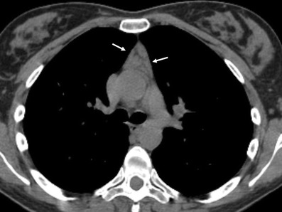
CT scanAxial chest CT: symmetrical thymic hyperplasia in a patient with myasthenia gravis, arrows on the enlarged gland.
Source : Juanpere et al., Insights into imaging, 2013, figure 16 · CC BY
A randomised trial has settled a controversy several decades old. Its conclusions hold for a precise patient profile, and they open two windows for us, one before the operation and one after.
The thymus is implicated in the disease, and its removal had long been performed without formal proof of benefit. The MGTX trial compared extended transsternal thymectomy plus prednisone against prednisone alone in 126 randomised patients with non-thymomatous generalised myasthenia, anti-AChR positive, aged 18 to 65, with less than five years of disease and MGFA class II to IV19.
The result: the time-weighted mean QMG score over three years was 6.15 in the thymectomy group against 8.99 on prednisone alone, with lower corticosteroid exposure as well19. A two-year extension prolonged follow-up to five years on the same outcomes20.
What MGTX does not say
The inclusion criteria are the limit of the result. MGTX covered patients who were anti-AChR positive, non-thymomatous, under 65, with less than five years of disease. Nothing that it establishes transfers automatically to an anti-MuSK, seronegative or elderly patient, or to one whose disease has been progressing for fifteen years. The 2020 international consensus did update its recommendations on thymectomy in the light of this trial, without extending them beyond it1.
Thymoma is a different case and does not follow the same logic: about 20 to 30 % of people with a thymoma develop myasthenia, the thymoma-associated form is almost always anti-AChR positive, most often generalised and more refractory to treatment, and surgical resection is considered for any thymoma, independently of myasthenic status, all thymomas being regarded as malignant51.
The two windows for the physiotherapist
Before. Preoperative respiratory preparation has been the subject of a randomised trial in 80 patients scheduled for extended thymectomy, with postoperative volumes and independence on the fifth day higher in the trained group39. It is a rare finding in this condition: a measured benefit, on a functional outcome, in a randomised trial of decent size.
After. The postoperative period carries risk. A meta-analysis of 25 studies identified the independent risk factors for myasthenic crisis after thymectomy: a history of crisis, preoperative bulbar symptoms, preoperative Osserman stage, preoperative pyridostigmine dose, preoperative respiratory function, postoperative pulmonary infection and the presence of a thymoma21. Three of these seven factors are respiratory or bulbar, which puts respiratory physiotherapy back at the centre of the perioperative period.
A myasthenic patient who has bulbar symptoms before their thymectomy is not merely more troubled. They are statistically more exposed to a crisis after the operation.
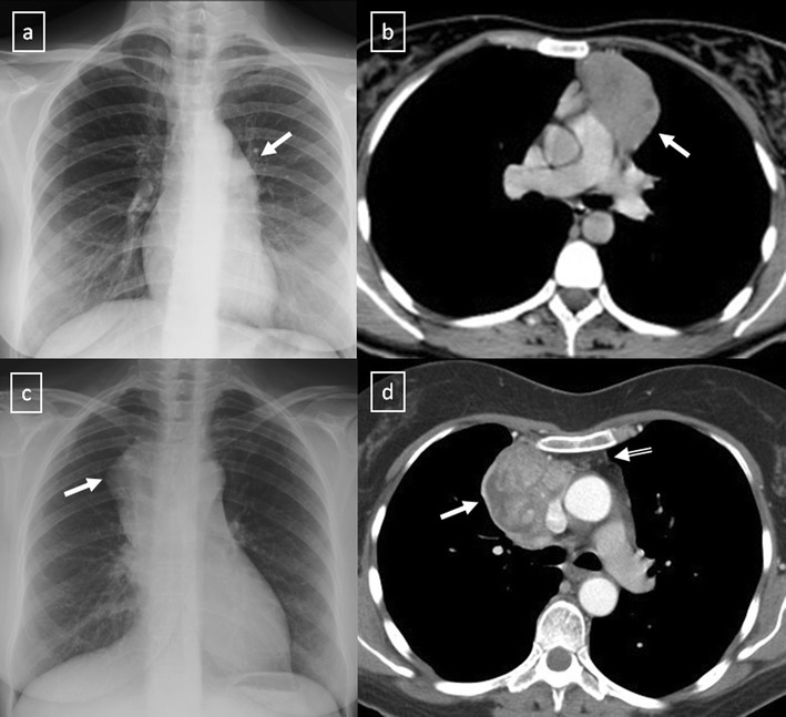
CT scanChest radiograph and CT: stage II then stage III thymoma in two patients with myasthenia gravis, arrows on the anterior mediastinal mass.
Source : Juanpere et al., Insights into imaging, 2013, figure 18 · CC BY
What do two published cases teach us?
Two observations reported in the indexed literature, one in intensive care during a crisis, the other in the community over twelve months of swallowing rehabilitation. They prove nothing on their own, and they show what no meta-analysis shows: the conduct, step by step.
Case 1: mobilisation started in intensive care during a myasthenic crisis
A 67-year-old woman presents with a dropped head, walking difficulties and respiratory failure. Myasthenia is diagnosed the same day. Still on that same day, she develops hypoxaemia, is intubated and admitted to intensive care on mechanical ventilation.
Exercise is started on the third day of admission, with the load increased gradually under monitoring of vital signs and of muscle fatigability. Standing begins on the twelfth day. Walking on mechanical ventilation is introduced on the seventeenth day, with no adverse event. Weaning from ventilation is achieved on the twenty-fourth day. After bolus corticosteroid therapy and thymectomy, function improves: independent walking with a stick on the sixty-fourth day, discharge on the seventy-eighth.
What this case brings. It documents that graded mobilisation is possible at the most severe stage, provided it is guided by continuous monitoring rather than by a protocol. The authors write explicitly that no reference standard exists today for physiological assessment at the time of mobilisation, nor for the content and intensity of exercise in severe myasthenia.
What it does not bring. One case. The authors themselves conclude that larger studies are needed. No protocol can be drawn from it, only a proof of feasibility and a method of monitoring.
Source: Okamoto A, Sasaki Y, Mizutani Y, et al. Case Report: Early rehabilitation initiated in the intensive care unit for myasthenic crisis. Front Rehabil Sci. 2026;7:1791183.46
Case 2: twelve months of swallowing rehabilitation in anti-MuSK myasthenia
A 67-year-old woman with anti-MuSK antibody myasthenia presents with severe dysphagia. She received intravenous bolus corticosteroid therapy, high-dose intravenous immunoglobulin and plasma exchange; she required ventilation and a tracheostomy, then had repeated pneumonias, leading to rituximab being given in the face of an insufficient response to the earlier treatments.
The disease appears stabilised, but the severe dysphagia persists, with silent aspiration, a delayed swallow reflex and pharyngeal stasis. Intensive and prolonged rehabilitation is set up over about twelve months: regular swallowing assessments, progressive muscle strengthening exercises at controlled load levels, and interferential current for the sensory disturbances. Result: return to oral feeding, with no myasthenic crisis and no recurrence of aspiration pneumonia.
What this case brings. Three things. First, the demonstration that keeping the load controlled is the discriminating parameter, including for the swallowing musculature. Then the duration: twelve months, to be compared with the six to eight weeks of the limb training trials. Finally, an illustration of the anti-MuSK profile described above, bulbar involvement to the fore and recourse to rituximab13.
What it does not bring. The authors themselves state that effective swallowing rehabilitation strategies in this population are not established. Rehabilitation was combined with background medical treatment, and it is impossible to separate the two.
Source: Oshima Y, Nanto T, Eimoto K, et al. Long-Term Swallowing Rehabilitation for Muscle-Specific Tyrosine Kinase Antibody-Positive Myasthenia Gravis: A Case Report. Cureus. 2025;17(3):e80544.47
What the two cases have in common
In both observations, what is described is not an intensity but a mode of conduct : assess, load a little, reassess, and move up only if the previous step has been absorbed. It is the only thing the myasthenia literature allows us to recommend consistently, and it is also the most transferable to the clinic.
How do you apply this in the clinic?
What this changes at the first session, in the assessment, in progression and in the letter.
At the first session
- Establish the status. Which antibody? Which MGFA class? Which treatment, at what time? Has there been a crisis, and when? A history of crisis is a recognised risk factor for a later crisis in a surgical setting21, and it is associated with more severe fatigue42.
- Fix the time of the sessions and stick to it. Comparisons from one assessment to the next only make sense at a constant time of day and a constant interval after treatment.
- Do a fatigability assessment, not only a strength assessment: sustained gaze, counting out loud, a repeated series of a functional movement. Record the number of repetitions before deterioration, not only maximal strength.
- Ask about swallowing and breathing systematically, even if the reason for referral is orthopaedic. A patient referred for a shoulder may have a dysphagia that nobody has looked for.
- Identify the alert route : who to call, which department, which neurologist. Before you need it.
Building the session
- Start low. A stabilised class II patient can often do more than you think; a IIb or III patient calls for a cautious start. The first step is there to measure, not to progress.
- Break it up. Short sets, real rests between the sets. Rest is not wasted time in this disease, it is the therapeutic mechanism.
- Stop before the deterioration. The last repetition should look like the first.
- Listen to the voice during the session. It is the simplest bulbar fatigability sensor we have, and it is free.
- Finish below the limit. A patient should leave able to get through their day, not emptied.
Progressing
The rule is simple: you only move up a step if the previous one has been absorbed without later deterioration. That means asking the question at the next session, systematically: how was the end of the day? And the next day? Were there any choking episodes, did the voice change?
The medium-term target, for a stabilised mild to moderate form, is that minimum of 150 minutes a week29, all activities combined, including what the patient does outside the clinic. And since the gain is lost when training stops29, building independence is not an extra: it is the goal.
| What we do | What we avoid | Why |
|---|---|---|
| Short sets, clear rests | Continuous work to exhaustion | The safety margin of the junction is exhausted by repetition and restored by rest6 |
| Submaximal intensity | Strengthening taken to failure | Bulbar symptoms and increased fatigue observed in a trial35 |
| Session at a fixed time, after the dose | Comparing two assessments made at different times | The deficit varies through the day and with the pharmacology29 |
| Monitor voice, swallowing, breathing | Monitoring only the muscle being worked | Bulbar and respiratory involvement drives the prognosis2245 |
| Ask what has changed when there is a deterioration | Concluding straight away that there has been too much rehabilitation | Infection, corticosteroids and drugs are recognised precipitating factors232627 |
| Respiratory work, inspiratory and expiratory | Neglecting expiration | Expiratory strength deficient in nearly 40 % of classes II to III, and a determinant of walking41 |
| Prepare before a planned thymectomy | Waiting for the postoperative period to act | Postoperative volumes and independence improved in a randomised trial39 |
| Aim for a lasting regime | An 8-week programme with no follow-on | Continuing is necessary to maintain the gain29 |
The letter
A report that is useful to the neurologist fits in five lines and contains: the time of the session and the interval since the last dose of treatment, the fatigability test performed with its numerical result, the load level reached, the tolerance at a distance reported by the patient, and any new bulbar or respiratory sign. That is what nobody else is in a position to observe.
Frequently asked questions
Can a patient with myasthenia do strength training?
Yes, under conditions. Physical training is considered safe in myasthenia and improves strength and daily function29, and the reference primer states that active training should be encouraged6. But the only trial dedicated to progressive resistance training randomised fifteen patients and recorded bulbar symptoms in two of them and increased fatigue in three, across both groups35. The practical answer is therefore: yes, submaximal, in short sets, without seeking failure, and with monitoring of bulbar signs.
Should the session come before or after the pyridostigmine dose?
After, in the window where the drug is working, which gives the patient their best working capacity. Above all, the point is to keep the same interval from one session to the next, otherwise successive assessments are not comparable. Important exception: in anti-MuSK antibody myasthenia, anticholinesterase drugs are avoided because of a risk of worsening and of cholinergic crisis13, so the question does not arise in the same way.
How do you tell normal fatigue from the start of decompensation?
By the recovery and by the topography. Expected fatigability concerns the muscle that has been worked and recovers after a few minutes of rest. Decompensation affects muscles that have not been worked, bulbar and respiratory ones in particular, persists beyond the session and worsens from one day to the next. Faced with breathlessness, a choking episode or an ineffective cough, you do not wait to decide: you stop and you call 15.
Is oxygen saturation enough to monitor a patient with myasthenia?
No, and this is a classic trap. In respiratory failure of neuromuscular origin, desaturation is a late phenomenon : a patient can be decompensating with a still-normal saturation. The useful indicators are clinical: the ability to finish a sentence, tolerance of lying flat, cough efficiency, recruitment of the accessory muscles, management of saliva.
Does a patient with purely ocular myasthenia need monitoring?
Yes. Conversion to a generalised form concerns 39 % of patients in a Thai cohort, with a median delay of 16 months16, and 54.5 % of a Chinese cohort, with 49.8 % of conversions within twelve months17. The switch happens early. Any new limb weakness, change of voice or swallowing difficulty in a patient labelled “ocular” warrants prompt review.
Can rehabilitation reduce the patient's fatigue?
Probably not directly, and this is a nuance best stated from the outset. The review of training studies concludes that the sense of fatigue not directly linked to muscle weakness is little influenced by physical training, unlike strength and daily function29. Fatigue in myasthenia is common, 59 % in the German registry42, and it often coexists with depressive symptoms and sleep disorders4849, which call for other levers.
Can a patient in intensive care for a myasthenic crisis be mobilised?
It has been described, in one published case, with exercise begun on the third day, standing on the twelfth and walking on mechanical ventilation on the seventeenth, with no adverse event46. But this is a single observation, and the authors stress that no reference standard exists for exercise intensity at this stage. The decision belongs to the intensive care team, and the approach described rests on continuous monitoring of vital signs and of fatigability, not on a protocol.
What is the difference with Lambert-Eaton syndrome?
The site of the lesion. Myasthenia is postsynaptic, the antibodies targeting the acetylcholine receptor or associated proteins6. Lambert-Eaton myasthenic syndrome is presynaptic, the antibodies targeting the calcium channels of the nerve terminal. Clinical consequence: strength can increase transiently there during the first contractions, the facilitation phenomenon, whereas myasthenia only declines. The context differs too, with a frequent association with small cell lung cancer and with autonomic involvement.
Does a normal assessment rule out the diagnosis?
No. That is the particularity of this disease: because the deficit is fatigable, an assessment carried out in the morning in a rested patient can be strictly normal. If the complaint concerns the end of the day, you have to assess at the end of the day or induce fatigue before measuring. Note, however, that where ocular myasthenia is suspected, a negative ice pack test combined with a negative single-fibre electromyography has a negative predictive value of 94 % and points strongly towards another diagnosis14.
Does thymectomy improve every patient?
It has been shown to be beneficial in a precise profile. The MGTX trial covered anti-AChR positive patients, without thymoma, aged 18 to 65, with less than five years of disease and MGFA class II to IV: in that group, the time-weighted mean QMG score over three years was 6.15 with thymectomy against 8.99 on prednisone alone19, a result extended to five years20. These conclusions do not automatically extend to other profiles. Thymoma follows a different logic: resection is considered for any thymoma, all of them being regarded as malignant51.

