
















CIDP is one of the few peripheral neuropathies that can be treated, and one of the few where the prognosis depends directly on the date the name was given. Between two neurology appointments three months apart, the physiotherapist is often the only professional to see the patient every week: it is the physiotherapist who notices that the patient can no longer rise from a chair without using their arms, that the foot catches on the stair carpet, that the handshake has weakened. This article sets out what needs to be recognised, what the EAN/PNS 2021 criteria changed, and what rehabilitation actually contributes alongside immunomodulatory treatment. One caveat from the outset: the rehabilitation literature specific to CIDP amounts to a single trial of 18 patients. We will say so rather than fill the gap with data borrowed from Guillain-Barré syndrome.
- Updated 14 August 2026
- Level Clinical summary
- Sources 31 verified references
- ICD-11 8C01.3
CIDP in three figures
A rare disease, often recognised late, and often wrongly named.
Sources: prevalence, Gable 2026, United States claims data3 ; delay, Arvin-Berod 2025, survey of 542 European patients8 ; overdiagnosis, Broers 2021, 96 patients referred to the Erasmus centre in Rotterdam7.
In brief
- The clinical signature fits into one sentence : motor weakness that is at once proximal and distal, broadly symmetrical, with diffuse areflexia and sensory involvement, progressing or relapsing over at least 8 weeks.
- Proximal weakness is the sign that settles it. In the Rotterdam series, 100 % of the CIDP cases that had been missed had proximal weakness, and 48 % of the patients wrongly labelled as CIDP had none7.
- The 8 weeks separate CIDP from Guillain-Barré. Guillain-Barré reaches its nadir in under 4 weeks and then improves; CIDP carries on. Between the two, acute-onset CIDP reveals itself by deteriorating again after the initial treatment9.
- It is a treatable disease. Intravenous immunoglobulin improves disability with a high level of evidence: relative risk 2.40 against placebo, 4 patients to treat for one additional benefit12.
- Rehabilitation replaces nothing. The only exercise trial conducted in CIDP involves 18 patients, all on immunoglobulin: 12 weeks of aerobic training gained 11.0 % of VO2 max, 12 weeks of resistance training 13.8 % of isokinetic strength16.
- The gain is lost on stopping. A year later, the six patients who had stopped had lost 13.0 % of strength again; the four who had carried on had kept everything17.
- On the dose, nobody knows. No study conducted in CIDP has compared a heavy load with a light one. What we have is an absence of any signal of harm at the doses tested, which is not a guarantee of safety above them.
- The most useful role of the physiotherapist may well be to measure. A dated, repeated MRC sum score and I-RODS scale turn an impression into an argument for the neurologist.
Contents
- What is CIDP, and why did its name change in 2021?
- How do you recognise the clinical picture in the clinic?
- Why does the 8-week threshold separate CIDP from Guillain-Barré?
- What do the EAN/PNS 2021 criteria say, and what did they change?
- Why is CIDP so often misdiagnosed?
- What do disease-modifying treatments do, and what do they not replace?
- What rehabilitation for CIDP, and at what level of evidence?
- How hard can you load without overdoing it?
- How do you measure, so as to see deterioration before the neurologist does?
- What do published clinical cases teach us?
- How do you apply this in the clinic?
- Frequently asked questions
What is CIDP, and why did its name change in 2021?
An autoimmune disease of the peripheral nerve, whose nomenclature was overhauled by the 2021 European guidelines. Understanding what changed avoids reading a neurology report with the vocabulary of the previous decade.
A sheath under attack, not a diseased muscle
Chronic inflammatory demyelinating polyradiculoneuropathy is a dysimmune neuropathy: the immune system attacks the myelin surrounding the fibres of the peripheral nerve and, in certain forms, the proteins that structure the node of Ranvier. The nerve conducts more slowly, sometimes not at all across a segment. The muscle itself is healthy: it simply no longer receives the command properly.
This distinction is not academic for a physiotherapist. Denervation weakness is not deconditioning weakness, and it does not respond to the same reasoning. The deficit varies with the rhythm of inflammation and treatment, not with the rhythm of training. A session that appears to make a patient worse may simply coincide with a relapse. A session that appears to make them better may coincide with an immunoglobulin infusion. We will return to this at length: it is the methodological crux of this whole chapter.
The term "polyradiculoneuropathy" says something important: the disease affects the roots as much as the nerve trunks. That is precisely what explains the proximal weakness, atypical for a neuropathy, and it is where recognition of the picture is won or lost.
How many people are affected?
CIDP is rare, and published figures vary by a factor of four depending on the method. An analysis of United States claims data covering 2016 to 2023 puts the prevalence at 23.3 per 100,000 (95 % CI 23.1 to 23.5) and the incidence at 2.8 per 100,000 per year (95 % CI 2.7 to 2.9), that is around 77,000 people living with the disease in the United States3. The authors themselves flag the limitation of their source: a billing code is not a validated diagnosis, and the overdiagnosis documented elsewhere is mechanically reflected in the figure.
A population study conducted in Birmingham, in the United Kingdom, on patients actually under follow-up gives much lower values: prevalence of 6.18 per 100,000 (95 % CI 4.66 to 8.05) and mean incidence of 0.54 per 100,000 per year (95 % CI 0.404 to 0.713)4. That same study reports a lower prevalence in people of Indian, Pakistani or Bangladeshi origin after the age of 50 (2.64 against 10.15 per 100,000), with no difference between 18 and 49 years.
The gap between those two figures is not a methodological detail: it says that we do not know precisely how many people have CIDP, because we do not know precisely to whom the diagnosis is correctly assigned. For practice, keep the order of magnitude in mind: a community practice will not see several CIDP cases a year. It may see one, and perhaps before the neurologist does.
Two methods, two orders of magnitude
Estimates per 100,000 people. The light bars are annual incidences, the dark ones prevalences.
Sources: Gable 2026, Inovalon closed claims database, 8,697 prevalent patients3 ; Rajabally 2025, clinical census as of 10 July 20254. The two measures do not contradict each other: they count different things.
Four markers for placing the patient
Sources: age, Gable 20263 ; wrong diagnosis, Arvin-Berod 20258 ; variants and maintenance, Nomura 2026, single-centre series of 33 patients in Kumamoto5. The proportion of variants reflects the recruitment of a referral centre, not the general population: the authors say so explicitly.
What 2021 changed in the vocabulary
The joint guidelines of the European Academy of Neurology and the Peripheral Nerve Society, published in 2021, revised the 2010 consensus and introduced two changes of vocabulary that can be read directly in neurology reports1.
The first: the phrase "atypical CIDP" has gone in favour of "CIDP variants". The reason is explicit in the text: these forms are well-characterised entities, not irregularities. There are five: multifocal, focal, distal, motor, sensory.
The second: the levels of diagnostic certainty have gone from three to two. Where definite, probable and possible CIDP used to be distinguished, only "CIDP" and "possible CIDP" now are. The justification is methodological and worth knowing: the diagnostic accuracy of the criteria for "probable" and "definite" did not differ significantly, so the distinction carried no information.
These variants are not rare. In a consecutive Japanese series of 33 patients diagnosed between 2020 and 2025 using the 2021 criteria, 36.4 % were typical CIDP and 63.6 % variants : distal in 39.4 % of cases, multifocal in 15.2 %, sensory in 6.0 %, motor in 3.0 %5. This is a referral centre, so recruitment is biased towards difficult cases, and the authors say as much. But the lesson holds: a picture that does not look like the textbook portrait does not rule out CIDP.
| Form | Distribution of motor weakness | What sets it apart | Relative frequency |
|---|---|---|---|
| Typical CIDP | Proximal AND distal, symmetrical, in all four limbs | Sensory involvement in at least two limbs, reflexes absent or reduced throughout, course over at least 8 weeks | 36.4 % of the Kumamoto series |
| Distal variant (DADS) | Distal, symmetrical, predominantly in the lower limbs | Distal sensory loss to the fore; to be distinguished from a length-dependent polyneuropathy | 39.4 % |
| Multifocal variant (Lewis-Sumner syndrome, MADSAM) | Asymmetrical, often in the upper limbs, in the territory of a nerve trunk | Sensory and motor involvement in the same territory; this is the form with the longest diagnostic delay | 15.2 % |
| Focal variant | Limited to one limb or one plexus | Rare; raises the question of compression or a nerve trunk lesion | not observed in this series |
| Motor variant | Purely motor, with no sensory complaint | Immunoglobulin is offered first line rather than corticosteroids | 3.0 % |
| Sensory variant | No motor weakness to the fore | Proprioceptive ataxia possible; motor weakness may appear later | 6.0 % |
Key points
CIDP is an autoimmune disease of the myelin of the peripheral nerve and of the roots, which is what explains its proximal weakness. Since 2021, we no longer say "atypical CIDP" but "CIDP variant", and there are now only two levels of certainty instead of three. Variants account for a substantial share of cases at a specialist centre: a picture that does not match the classic portrait does not exclude the diagnosis.
How do you recognise the clinical picture in the clinic?
The physiotherapist does not make the diagnosis of CIDP. But they see the patient every week, they see them stand up, walk, grasp. This chapter describes what, in what they observe, should prompt a letter.
Proximal weakness, the signature
Most of the polyneuropathies met in practice are length-dependent: they begin with the longest fibres, so with the toes, and slowly work upwards. The patient describes tingling in a stocking distribution, then a glove one. Strength, when it is affected, is affected distally: the foot dorsiflexors, the intrinsic muscles of the hand.
CIDP does not work that way. Because it affects the roots, it strikes the proximal at the same time as the distal. The patient can no longer rise from a low chair without using their hands, struggles to climb stairs, can no longer reach an object above their head. And they also have numb toes and weak dorsiflexors. It is this coexistence that should raise the alarm, because no ordinary polyneuropathy produces it.
Proximal weakness is not one sign among others: it is the sign whose absence led nearly half of the false positives to be wrongly labelled, and whose presence was constant in every missed true positive.
This is not a clinical hunch, it is a numerical result. In the retrospective study conducted at the Erasmus centre in Rotterdam between 2011 and 2017, among patients referred with a CIDP label that turned out to be wrong, 48 % had no proximal weakness and 29 % had distal weakness only. Conversely, among patients referred with another diagnosis and in whom CIDP was ultimately retained, 100 % had proximal weakness7.
Symmetry, areflexia, the sensory side
Three further features complete the picture of the typical form.
Symmetry. Weakness is broadly symmetrical. In the same Rotterdam series, 20 % of the weaknesses in overdiagnosed patients were asymmetrical7. One caution, though: asymmetry does not rule out CIDP, it points towards the multifocal variant, which is precisely the one that takes longest to recognise.
Areflexia. Tendon reflexes are reduced or absent, and not only at the ankles: in all four limbs. A brisk knee jerk in a patient suspected of CIDP is an inconsistency worth flagging.
Sensory involvement. It affects at least two limbs in the typical form. It is often in the background of the patient's account, which starts with strength, but it has to be sought: vibration sense at the malleolus, position sense of the big toe, discrimination. Proprioceptive ataxia may dominate the picture, and the patient then says they "lose their balance in the dark" or in the shower.
What the physiotherapist sees and the consultation does not
A patient is seen in neurology every three to six months. They are seen in physiotherapy two to three times a week. That difference in frequency is the physiotherapist's main asset in this condition, and it is underused.
What is visible in a session and not in a consultation: the speed of a sit-to-stand, the number of steps before stopping, the fact that a patient now takes the handrail with both hands, an object dropped from the right hand that they put down to tiredness. It is these observations, if they are dated and quantified, that make it possible to tell the neurologist not "he seems less well to me" but "his MRC sum score has gone from 56 to 51 in six weeks, and he no longer does the five sit-to-stands he was doing in March".
Fatigue deserves a mention of its own. It is common in CIDP and is not only motor. A study comparing 47 patients with 31 controls finds total, motor and cognitive fatigue significantly higher in the patients, the motor component being the most marked. The only predictors of both components are functional disability and depressive symptoms, with no link to the progression biomarker tested26. A recent review broadens this finding to a whole set of non-sensorimotor symptoms: dysautonomia, circadian rhythm disturbance, mood, subtle cognition27. An exhausted patient is therefore not necessarily a patient who is deconditioning.
What should prompt a letter
Referral tree. It replaces no examination: it says when to write.
Built from the EAN/PNS 2021 clinical criteria1 and the pitfalls identified by Broers 20217. None of these branches amounts to a diagnosis: they determine a referral.
Red flags: what calls for a rapid opinion
- Proximal motor weakness worsening week by week, whatever label the notes carry.
- Breathlessness, a voice that runs out of air, swallowing that catches : involvement of the respiratory and bulbar muscles is rare in CIDP but possible, and it is an emergency.
- A patient labelled Guillain-Barré who starts deteriorating again after a period of improvement, particularly beyond the eighth week.
- Clear worsening after an infection or surgery, with no mechanical explanation found on imaging.
- Muscle wasting that sets in quickly : it signals axonal loss, which does not recover the way demyelination does.
- A known CIDP that drops off between two infusions : the maintenance schedule may need reviewing, and that is the neurologist's decision.
Key points
What distinguishes CIDP from ordinary neuropathies comes down to one word: proximal. Weakness that affects both the girdle and the extremity, symmetrical, with diffuse areflexia and a sensory component, is not an ordinary polyneuropathy. The physiotherapist sees this patient ten times more often than the neurologist does: their added value is to date and quantify what they observe.
Why does the 8-week threshold separate CIDP from Guillain-Barré?
The two diseases share a mechanism, dysimmune demyelination, and much of the clinical picture. What separates them is a curve over time. And that is exactly what weekly follow-up allows you to plot.
The nadir, not the onset
Guillain-Barré syndrome is an acute, monophasic disease: worsening is rapid, maximum severity (the nadir) is reached in under 4 weeks, and recovery then begins. CIDP, in its typical form, progresses or relapses over at least 8 weeks : that is the temporal criterion of the European guidelines16.
So it is not the speed of onset that settles it, it is the date at which the patient stops getting worse. A picture that sets in over ten days may be either. A picture still worsening in the third month is not a Guillain-Barré.
The grey zone: acute-onset CIDP
There is a form of CIDP that begins like a Guillain-Barré, with a nadir reached in under 8 weeks: acute-onset CIDP. A recent review reports a proportion of the order of 16 % of CIDP cases in one of the series it cites6, while a European survey reports, citing the earlier literature, an acute onset in under 4 weeks in up to 13 % of patients8. The two figures come from different sources and do not measure quite the same thing; what they agree on is that this is not anecdotal.
This form reveals itself after the initial treatment. The patient receives immunoglobulin for their presumed Guillain-Barré, improves, then deteriorates again. Two readings are then possible: a treatment-related fluctuation, where the effect of the immunoglobulin wears off before the disease burns out, or a CIDP resuming its course. A literature review devoted to this distinction concludes that it remains difficult, that the electrophysiological, ultrasound and immunological criteria proposed have not settled it, and that the very concept of recurrent Guillain-Barré is disputed9.
The stakes are not nosological. Guillain-Barré is treated with a short course; CIDP requires long-term immunosuppression. Getting it wrong means letting a patient deteriorate for want of maintenance treatment. Seen from the other side of that border, the figure is of the same order: the European guideline devoted to Guillain-Barré syndrome asks that the diagnosis be reconsidered in favour of acute-onset CIDP when progression continues beyond eight weeks, which happens in about 5 % of patients initially labelled Guillain-Barré. Our article on that disease covers its recovery phase, its residual fatigue and the two deteriorations a physiotherapist may observe in it.
A patient who has come through a Guillain-Barré, referred for rehabilitation, and who starts declining again past the eighth week, does not need a programme adjustment. He needs a letter.
Three curves, three diseases
Severity of the deficit over time. Teaching diagram: individual trajectories vary.
Diagram built from the EAN/PNS 2021 temporal criteria1 and Inan's 2024 review of the distinction between recurrent Guillain-Barré and acute-onset CIDP9. The vertical axis is qualitative.
| Criterion | Typical CIDP | Guillain-Barré syndrome | Length-dependent polyneuropathy (diabetic, for instance) |
|---|---|---|---|
| Time course | Progression or relapses over more than 8 weeks | Nadir in under 4 weeks, then improvement | Onset over years |
| Motor weakness | Proximal AND distal | Often ascending, proximal possible | Distal, late, often subtle |
| Symmetry | Symmetrical (except the multifocal variant) | Symmetrical | Symmetrical |
| Reflexes | Reduced or absent in all four limbs | Absent | Ankle jerks lost first |
| Sensory involvement | Present, at least two limbs | Present | To the fore, stocking then glove |
| What should cast doubt on the label | Clear, lasting improvement without treatment | Deterioration resuming after 8 weeks | Proximal weakness, a course measured in weeks, asymmetry |
| Physiotherapist's approach | Measure, date, pass it on | Measure, date, pass it on | Prevention of the at-risk foot, education |
The last line of this table deserves a word. Diabetic polyneuropathy is the context in which a CIDP most easily goes unnoticed, because the patient already has an explanation for their tingling. Yet diabetes is also the most frequent comorbidity in patients with CIDP in the American cohort3. Having diabetes does not immunise you against CIDP: it merely makes it more tempting to stop looking. Our article on peripheral neuropathy, notably diabetic sets out the length-dependent picture against which the comparison has to be made.
Key points
The discriminator between CIDP and Guillain-Barré is temporal: nadir before 4 weeks and recovery on one side, progression or relapses beyond 8 weeks on the other. Between the two, acute-onset CIDP is recognised by its relapse after an initial improvement. It is a curve, and weekly follow-up is the best tool for plotting it.
What do the EAN/PNS 2021 criteria say, and what did they change?
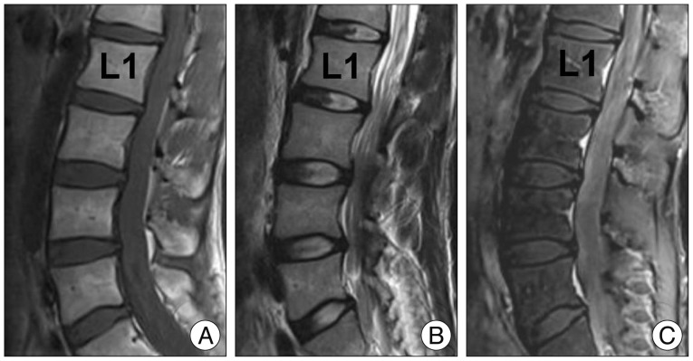
MRISagittal lumbar MRI (T1, T2, contrast-enhanced T1): thickening of the cauda equina nerve roots, a sign suggestive of CIDP.
Source : Lee et al., Journal of Korean Neurosurgical Society, 2014, figure 2 · CC BY-NC
These criteria are the ones that appear in the neurologist's report. Reading them correctly means understanding why a patient is classed as "possible CIDP" and what that implies for what follows.
How the guidelines were built
The 2021 text is the work of a joint task force: seventeen experts, a patient representative and two Cochrane methodologists, who framed twelve questions in PICO format and then graded the evidence using the GRADE method1. The same text was published simultaneously in two journals, the Journal of the Peripheral Nervous System and European Journal of Neurology2 : a reference to either points to the same document.
Diagnosis rests on three pillars
A compatible clinical picture (the one described above), electrophysiological abnormalities of demyelination in at least two motor nerves, and supportive findings when the first two are not enough: raised CSF protein, hypertrophy of the roots or trunks on imaging or ultrasound, sensory abnormalities on nerve conduction studies, and above all the objective response to treatment.
This last point is central and often misunderstood. Improvement after immunoglobulin is not proof if it is not objectively documented : the 2025 report on unmet needs in CIDP stresses that there is no consensus definition of what constitutes a "response", a "refractory" patient, a "remission" or a "relapse", and that this lack of shared vocabulary is detrimental to treatment decisions11. That is one more reason for the physiotherapist to bring figures: they are missing from the file.
What these criteria are worth, in figures
A study published in 2026 compared the diagnostic accuracy of the EFNS/PNS 2010 and EAN/PNS 2021 criteria in 339 patients with CIDP and 339 matched controls referred for suspected chronic dysimmune neuropathy who received another diagnosis10.
The results, including the "possible" category: sensitivity of 92 % for the 2010 criteria and 94 % for those of 2021, specificities of 67 % and 66 % respectively. Restricting to the full category (probable or definite in 2010, "CIDP" in 2021): sensitivity of 91 % against 83 %, specificity of 78 % against 86 %.
In other words, the 2021 revision traded sensitivity for specificity. It labels fewer patients wrongly, and misses more of them. Given the massive overdiagnosis documented elsewhere, that choice is defensible. It does, however, have a practical consequence: a "possible CIDP" patient is not a patient without CIDP, and a patient who does not meet the criteria but responds objectively to treatment remains a patient to be treated.
One last result from this study bears directly on the care pathway: a unilateral nerve conduction study would have missed 77 diagnoses of possible CIDP10. How far the testing extends matters as much as the grid used to read it.
What the 2021 revision traded
Full category only (probable or definite in 2010, "CIDP" in 2021). 339 patients, 339 matched controls.
Source: van Doorn 2026, matched case-control study of 678 patients10. Including the "possible" category, the two sets of criteria are comparable (92 % against 94 % sensitivity).
The trap not to fall into again
Reading "possible CIDP" as "probably not a CIDP" is a reasoning error. The "possible" category exists because the electrophysiological criteria do not capture every form, particularly the multifocal variants, where demyelinating abnormalities are rarer: in the Kumamoto series, these were the patients whose mean diagnostic delay reached 4.6 years5. A patient classed as "possible" who responds objectively to treatment is a patient who gets treated.
Key points
The EAN/PNS 2021 criteria combine a clinical picture, electrophysiological demyelination in at least two motor nerves, and supportive findings including the objective response to treatment. They have two levels of certainty instead of three, and are more specific but less sensitive than those of 2010 (86 % against 78 % specificity, 83 % against 91 % sensitivity). Nerve conduction studies that are too limited defeat them.
Why is CIDP so often misdiagnosed?
Overdiagnosis and underdiagnosis coexist, and they do not have the same causes. Knowing the five recorded pitfalls tells you, in the session, which of the two is happening.
A third of labels are wrong, in both directions
The reference study on this question went back over the records of patients referred to the Erasmus centre in Rotterdam between 2011 and 2017, either with a diagnosis of CIDP or with another diagnosis later revised to CIDP, and reclassified them using the EFNS/PNS 2010 criteria7.
The diagnosis of CIDP was overturned in 32 % of patients who arrived with that label, that is 31 out of 96. In the other direction, of 81 patients who really did have CIDP, 16, that is 20 %, were referred with another diagnosis.
The two errors do not cost the same. Overdiagnosis exposes the patient to expensive immunotherapy that is not without risk, for a disease they do not have, and it delays the real diagnosis. Underdiagnosis lets a treatable disease progress towards axonal loss, which does not recover.
What separates the false positives from the missed true positives
Proportions within each of the two groups. Proximal weakness separates the two populations on its own.
Source: Broers 2021, retrospective series from the Erasmus centre in Rotterdam, 2011 to 20177. The "proximal weakness present" and "criteria met" proportions among the overdiagnosed are the complements of the published figures (48 % without proximal weakness, 65 % not meeting the criteria).
The five pitfalls, named
The authors of that study set out the mechanisms of the error explicitly. They are worth knowing because three of them can be spotted without any additional investigation.
- Paying no attention to proximal weakness, when it is the hallmark of the disease.
- Failing to recognise clinically atypical phenotypes : half the underdiagnosed patients had a variant.
- Overrating CSF protein. A raised protein in the cerebrospinal fluid is not specific: 74 % of overdiagnosed patients had one.
- Misinterpreting nerve conduction studies.
- Not applying the electrodiagnostic criteria : 65 % of the overdiagnosed did not meet them.
To which an implicit sixth can be added: 97 % of overdiagnosed patients did indeed have a neuropathy, of another kind. The problem was therefore not seeing a neuropathy, but stopping there.
Seven months, nineteen investigations
A survey conducted between September 2022 and April 2023 among 542 patients and their neurologists in the United Kingdom, France, Germany, Italy and Spain gives the measure of the pathway8. Mean age 54.0 years (standard deviation 12.4), 62 % men. Mean number of diagnostic procedures per patient: 19.6 (standard deviation 9.4). Median time between first symptoms and diagnosis: 7.0 months, with an interquartile range of 3.2 to 13.0 months.
37 % of patients had received at least one wrong diagnosis along the way, and the most frequent of those wrong diagnoses was Guillain-Barré syndrome, in 37 % of cases. Three factors lengthened the delay: mild symptoms at the outset, a variant form, and having been misdiagnosed.
That last point deserves emphasis for a physiotherapist. A patient referred with subtle symptoms, an atypical form and a first label that does not fit is precisely the patient who will spend months in rehabilitation without anyone questioning the diagnosis. That is where the physiotherapist's place matters most, and where it is least often taken.
Three situations where a further opinion must be sought
- A patient in rehabilitation for more than two months for a "neuropathy" that keeps getting worse. A length-dependent neuropathy does not progress over weeks.
- A patient who is not recovering the way a Guillain-Barré should recover. The expected trajectory is improvement; its absence is information.
- A patient whose weakness is asymmetrical and follows a nerve trunk, labelled compression or sequelae, with no lesion found. That is the presentation of the multifocal variant, whose mean diagnostic delay reached 4.6 years in the Kumamoto series5.
What do disease-modifying treatments do, and what do they not replace?
This chapter is not here so that the physiotherapist may prescribe anything. It is here so that they know what the treatment does, at what rhythm, and why a session held the day after an infusion does not mean the same thing as a session held the day before.
Intravenous immunoglobulin, the foundation
The Cochrane review updated in 2024 retained nine randomised trials totalling 372 participants12. Compared with placebo, intravenous immunoglobulin increases the probability of a significant improvement in disability within six weeks of starting treatment: relative risk 2.40 (95 % CI 1.72 to 3.36), that is 4 patients to treat for one additional benefit (95 % CI 3 to 5). The level of evidence is high.
On the Rankin scale, the improvement is 0.26 points (95 % CI 0.05 to 0.48), also high-quality evidence. On the INCAT scale at 24 weeks, it reaches 0.80 points (95 % CI 0.23 to 1.37), moderate evidence. The authors conclude that further placebo-controlled trials would probably not change these conclusions.
Compared with oral prednisolone, immunoglobulin probably makes little difference to the probability of improvement at four weeks (relative risk 0.91, 95 % CI 0.50 to 1.68). Compared with intravenous methylprednisolone, it probably increases the probability of improvement at two weeks, with a wide confidence interval.
Corticosteroids, widely used and barely proven
This is the most striking paradox in the file. The Cochrane review devoted to corticosteroids found only one unblinded randomised trial comparing prednisone with no treatment, in 35 eligible patients, which did not even measure the review's primary outcome13. The impairment score improved in 12 of the 19 patients on prednisone against 5 of the 16 with no treatment, a relative risk of 2.02 (95 % CI 0.90 to 4.52), with a very low.
The authors write that corticosteroids are commonly used in practice on the strength of very low quality observational data, and that those same observational data document serious adverse effects in the long term. A second trial comparing monthly high-dose oral dexamethasone with standard-dose daily prednisolone in 40 participants finds little difference in efficacy.
Despite that evidence base, the EAN/PNS 2021 guidelines place immunoglobulin or corticosteroids as initial treatment, with a strong recommendation for either1. This is not an inconsistency: the GRADE method allows a strong recommendation backed by weak evidence when the expected benefits clearly outweigh the harms, which is the case for a disabling disease with limited alternatives.
Plasma exchange, effective and short-lived
The Cochrane review devoted to plasma exchange retains two small trials. The first, a cross-over trial in 18 participants, shows at four weeks an improvement of 2 points (95 % CI 0.8 to 3.0) more than sham exchange on an 11-point disability scale. But 8 of the 12 patients who improved then deteriorated rapidly14. Adverse effects occur, according to non-randomised data, in 3 to 17 % of procedures, sometimes serious ones.
The 2021 guidelines logically reserve plasma exchange for situations where immunoglobulin and corticosteroids have failed, again with a strong recommendation1.
| Treatment | Demonstrated effect | Level of evidence | What it changes in the session |
|---|---|---|---|
| Intravenous immunoglobulin | Improvement in disability against placebo: RR 2.40 (1.72 to 3.36), 4 patients to treat | High | Cyclical effect: the patient is often at their best a few days after the infusion and drops off before the next one. Always schedule assessments at the same point in the cycle. |
| Subcutaneous immunoglobulin | Recommended for maintenance by the 2021 text | See guidelines | Administered at home, with a steadier effect: less of a trough before the next dose. |
| Corticosteroids | A single trial against no treatment: RR 2.02 (0.90 to 4.52) | Very low | Steroid myopathy possible in the long term: new proximal weakness on corticosteroids is not necessarily the CIDP progressing. |
| Plasma exchange | 2 points (0.8 to 3.0) on an 11-point scale at 4 weeks | Moderate to high, on two small trials | Brief improvement: 8 of the 12 patients who improved deteriorated rapidly. Do not credit rehabilitation with a gain that follows an exchange. |
| Supervised treatment withdrawal | 8 of the 12 candidate patients stayed stable for more than 12 months without treatment | Series of 125 patients, non-randomised | A taper decided on by the neurologist is not an abandonment: a third of stable patients can do without treatment for the long term. |
A point that defeats intuition: some patients can stop
A significant share of patients go into remission, whether spontaneous or treatment-related. A London team offered supervised withdrawal to clinically stable patients on immunoglobulin: of 125 people, 45 met the stability criteria, 36 were eligible for the withdrawal trial, 12 agreed, and 8 of them, that is 66.7 %, stayed stable for at least 12 months without treatment. Every patient who deteriorated returned to their previous level once treatment was restarted15.
For a physiotherapist, this means two things. First, a patient who is offered withdrawal is not being abandoned: it is a documented, reversible process. Second, it is precisely during these periods off treatment that close clinical monitoring is most valuable, and the physiotherapist is the professional who sees the patient most often.
What rehabilitation does not do
No rehabilitation modality has ever been compared with an immunomodulatory treatment in CIDP, and the only available exercise trial involved patients who were all on subcutaneous immunoglobulin. Rehabilitation is added to the treatment; at no point does it take its place.
This has to be said plainly because the opposite is sometimes said. Autoimmune demyelination is not corrected by exercise. What exercise addresses is the consequences: deconditioning, loss of strength in the muscles still innervated, apprehension when walking, cardiorespiratory deconditioning. That is useful, measurable, and it is not the same thing.
What rehabilitation for CIDP, and at what level of evidence?
The short answer is: very little evidence, and it needs saying. The long answer consists in examining exactly what we have, what can be drawn from it, and where the boundary with extrapolation lies.
The only exercise trial conducted in CIDP
A Danish team enrolled 18 patients with CIDP, all treated with subcutaneous immunoglobulin, in a protocol of three 12-week periods16 :
- 12 weeks without exercise (reference period);
- 12 weeks of aerobic training, three times a week on a cycle ergometer;
- 12 weeks of resistance training, three times a week, unilateral work in knee and elbow flexion and extension.
Results. During the exercise-free period, maximal oxygen uptake and strength did not move significantly (respectively 4.9 % ± 10.3, p = 0.80 and 3.7 % ± 10.1, p = 0.17, downwards). Aerobic training increased VO2 max by 11.0 % ± 14.7 (p = 0.02). Resistance training increased combined isokinetic strength by 13.8 % ± 16.0 (p = 0.0004).
The exercise-free reference period is what makes this trial interpretable. It shows that the disease was neither improving nor deteriorating spontaneously in these patients over the preceding twelve weeks: the gain that follows is therefore not the natural course. It is the best methodological protection available in the absence of a control group, and it is also its limitation: without randomisation, order effects and expectation effects are not controlled for.
What is lost when you stop
A year after the end of the trial, 10 of the 17 eligible patients agreed to be reassessed17. Six had stopped training: their combined isokinetic strength had fallen by 13.0 % (range 25.8 to 2.9, p = 0.03) and their VO2 max by 16.6 % (range 18.8 to 12.6, p = 0.06). Four had carried on, three with aerobic training and one with resistance training: in them, strength and aerobic capacity were preserved.
The numbers are tiny and the comparison is not randomised: those who carry on are not comparable to those who stop. But the message is consistent with what we know about training in general, and it has a direct practical consequence: what is not maintained is lost, and a CIDP programme has to be conceived as something lasting, not as a course of thirty sessions.
The only exercise trial conducted in CIDP
Percentage changes. 18 patients, all on subcutaneous immunoglobulin. Three successive 12-week periods, then follow-up at one year in 10 patients.
Sources: Markvardsen 2018 for the first three periods16, Markvardsen 2019 for the one-year follow-up17. The heights are proportional to the published percentages.
What the broader literature says, and why it is not enough
For want of trials in CIDP, one is tempted to look around. Three sources widen the field, each with its limitations.
Strengthening in polyneuropathies in general. A scoping review surveyed studies of strength training at a load of at least 70 % of the repetition maximum in patients with polyneuropathy: of 362 articles screened and 101 read in full, eight studies were retained, covering Charcot-Marie-Tooth disease, CIDP and diabetic polyneuropathy. Five randomised trials, two case series, one cross-over trial. Methodological quality was judged fair to poor for seven of them. The conclusion is explicit: strengthening may improve strength, but no recommendation can be made18.
Promoting physical activity in neuromuscular diseases. A Cochrane review retained 13 studies and 795 randomised participants, covering nine inherited or acquired neuromuscular diseases. No meta-analysis could be carried out. Ten of the thirteen studies were at high or unclear risk of bias in the collection of the physical activity outcome. The authors' conclusion is that they remain uncertain about the effectiveness of these interventions. One point deserves to be noted separately: they found no evidence that supervised activity increases the risk of a serious adverse event, while noting that the collection of such events was incomplete19.
Balance. A systematic review of six studies and 133 participants with non-diabetic peripheral neuropathies reports improvements in static and dynamic balance and a reduction in fall risk with stabilometry platforms combined with virtual reality and feedback, with good adherence. The authors stress the variability of the protocols and the small number of studies21. These populations include a range of neuropathies and not specifically CIDP.
What cannot be borrowed from Guillain-Barré
There is a distinctly richer rehabilitation literature on Guillain-Barré syndrome: a 2025 scoping review retained 16 articles and concludes that exercise can improve strength, reduce fatigue and support functional independence20. It does not transfer. Guillain-Barré is a monophasic disease in recovery : the nerve remyelinates and the programme accompanies a trajectory that rises. CIDP is a chronic disease fluctuating under treatment : the programme accompanies a trajectory that can fall again at any moment without rehabilitation having anything to do with it. Using the protocols of one for the other also exposes you to crediting the session with an effect that belongs to the disease.
Modalities and levels of evidence
The table below summarises what each modality can claim in CIDP. The GRADE levels given are our own appraisal from the sources cited, applying the usual downgrading criteria: small numbers, absence of randomisation, indirectness of the population.
| Modality | Observed effect | Population studied | Level of evidence in CIDP |
|---|---|---|---|
| Resistance training, 3 sessions a week, 12 weeks | Combined knee and elbow isokinetic strength: +13.8 % ± 16.0 (p = 0.0004) | 18 CIDP patients on immunoglobulin | Very low (single trial, no randomised control arm) |
| Aerobic reconditioning, cycle ergometer, 3 sessions a week, 12 weeks | VO2 max: +11.0 % ± 14.7 (p = 0.02) | The same 18 patients | Very low (same trial) |
| Continuing training in the long term | Gains preserved at one year in 4 patients; loss of 13.0 % of strength in the 6 who stopped | 10 CIDP patients reassessed | Very low (observational, non-randomised comparison) |
| High-load strengthening (at least 70 % of 1RM) | May improve strength; direct comparisons impossible | 8 studies, Charcot-Marie-Tooth, CIDP and diabetic polyneuropathy | Very low ; the authors conclude that no recommendation is possible |
| Balance work on a platform with feedback | Improvement in static and dynamic balance, reduction in fall risk | 6 studies, 133 patients, non-diabetic neuropathies | Very low and indirect (non-specific population) |
| Promotion of physical activity supervised | Uncertain effectiveness; no signal of excess risk of a serious event | 13 studies, 795 participants, neuromuscular diseases | Low to very low, indirect |
| Ankle-foot orthosis, assistive devices | No study identified specific to CIDP | None | Absence of evidence ; individual clinical decision |
| Electrical stimulation aimed at nerve regeneration | Results mainly preclinical; harmful effects of stimulation given too early after nerve repair have been reported | 60 publications, mostly animal | No evidence in humans in this indication |
medical treatment
Intravenous immunoglobulin against placebo on improvement in disability. Relative risk 2.40 (95 % CI 1.72 to 3.36), 4 patients to treat, 5 trials and 269 participants12.
It is the only element in the management of CIDP that reaches this level. It has nothing to do with rehabilitation.
medical treatment
Plasma exchange on short-term improvement in disability and motor conduction, from two small trials at low risk of bias, with rapid deterioration afterwards14.
rehabilitation
All rehabilitation for CIDP sits here. One non-randomised trial of 18 patients, an observational follow-up of 10, a scoping review of 8 studies of which 7 were of fair to poor quality.
This does not mean rehabilitation is useless. It means that the size of its effect, its optimal dose and its safety limits are not established, and that a practitioner who claims otherwise is going beyond their data.
and not evidence of absence
Orthoses, assistive devices, education in energy conservation, home adaptation. No study in CIDP. These interventions remain justified by clinical reasoning and by the documented functional impact of the disease: the fact that they have not been evaluated is not an argument against them.
How hard can you load without overdoing it?
This is the question every physiotherapist asks in front of a demyelinating neuropathy, and it is the one on which the literature is poorest. This chapter sets out what is documented, what is not, and an approach that takes account of both.
What do we mean when we say "overwork"?
The notion of overwork weakness, or overwork weakness, is an old one in neuromuscular rehabilitation. It describes a lasting loss of strength said to follow overly intense exercise in a partially denervated or dystrophic muscle, as opposed to the transient fatigue of normal exercise. The term is still used in the recent case literature: a Japanese team describes a patient with Lambert-Eaton myasthenic syndrome in whom independence in activities of daily living was achieved through low-intensity, high-repetition endurance work, with monitoring of subjective intensity over time, with no overwork weakness appearing31.
Two honest caveats are needed about that reference. First, it is a single case: one case demonstrates nothing about how often a phenomenon occurs. Second, Lambert-Eaton syndrome is a disease of the neuromuscular junction, not a demyelinating neuropathy: the transposition is not direct. We cite it because it illustrates a method of monitoring, not because it constitutes evidence in CIDP.
What the data allow you to say
Three findings are firmly established.
First finding: at the doses tested, no worsening has been reported. In the Danish trial, three weekly resistance sessions over twelve weeks in patients with CIDP increased strength, and the authors report no deterioration16. That is an observation, not a predefined safety outcome.
Second finding: no signal of excess risk in neuromuscular diseases in general. The Cochrane review on promoting physical activity concludes that it found no evidence that supervised activity increases the risk of a serious adverse event, while noting that the collection of such events was incomplete in the included studies19. An absence of signal in an incomplete collection is not proof of safety.
Third finding: a signal of harm does exist, but elsewhere. A systematic review of 60 publications on exercise and electrical stimulation in nerve repair concludes that an early active programme broadly favours axonal regeneration, while reporting that harmful effects of exercise or stimulation given too early after nerve repair have been recorded. The authors specify that human research is too limited to conclude22. That body of work is mostly animal and concerns traumatic nerve injury, not autoimmune demyelination.
What the data do not allow you to say
We have to be explicit, because this is the point where it is most tempting to fill the gap with conviction.
- No study conducted in CIDP has compared a high load with a moderate one. There is therefore no threshold beyond which we would know not to go.
- No study has measured overwork weakness as an outcome in CIDP. We know neither its frequency, nor its reversibility, nor its risk factors in this disease.
- The available trial included only treated, stable patients. Nothing is known about exercise during a relapse, or in an untreated patient.
We do not have evidence that loading heavily is safe. We have an absence of any signal of harm at one specific dose, in eighteen treated, stable patients. That is not the same sentence, and the difference is exactly what separates cautious practice from extrapolation.
An approach that takes account of the uncertainty
What follows is not evidence but clinical reasoning applied to a context where evidence is lacking. We flag it as such.
- Start from the documented dose rather than inventing one. Three sessions a week, segmental work in knee and elbow flexion and extension, a cycle ergometer for the aerobic component: that is the protocol that produced the only published results in this disease16. Departing from it upwards is a personal choice, not an application of the data.
- Make monitoring measurable instead of leaving it subjective. Weakness that is getting worse shows up on a repeated MRC sum score, not on an end-of-session impression. The useful reference point is the next day and the day after, not the minute that follows.
- Distinguish fatigue from loss of strength. Fatigue is intrinsic to the disease and affects a majority of patients26. A patient who is tired after a session is not necessarily an overworked patient; a patient who has lost two MRC points on a muscle group is.
- Do not credit the session with what belongs to the treatment cycle. Deterioration that always occurs at the end of the interval between two infusions is telling you about maintenance, not about training. Record the date of the last infusion at every assessment.
- Progress slowly and one variable at a time. Increase the load or the volume or the frequency, never all three together, so that any deterioration can be attributed.
- Set yourself a stopping rule written down in advance. For example: progression suspended and reassessment if the MRC sum score falls by more than two points, or if a benchmark functional capacity deteriorates over two consecutive assessments.
Key points
On dose, the state of knowledge is as follows: one specific protocol produced a gain with no reported worsening in 18 treated, stable patients, there is no signal of excess risk in neuromuscular diseases in general, and there are no data above that dose. The reasonable approach is to start from the documented protocol, progress one variable at a time, and make monitoring numerical rather than felt.
How do you measure, so as to see deterioration before the neurologist does?
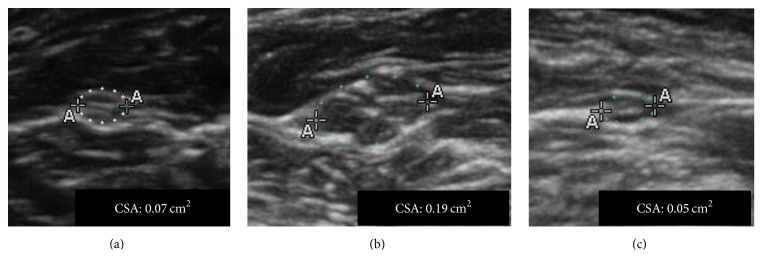
UltrasoundAxial ultrasound of the median nerve in patients with CIDP, with measurement of the nerve cross-sectional area according to the degree of impairment.
Source : Merola et al., Neurology research international, 2016, figure 2 · CC BY
This is probably the physiotherapist's most concrete contribution in this disease. It does, however, require using the tools that the CIDP literature itself uses, and knowing which ones have been tested and set aside.
The two measures to keep
The MRC sum score. It is the sum of the manual muscle testing grades over a standardised set of muscle groups. It appears as an outcome in CIDP trials and in the Cochrane review on immunoglobulin12, and it serves as a benchmark of clinical stability in treatment withdrawal protocols15. It requires no equipment.
The I-RODS scale. It is a patient-reported disability questionnaire built on the Rasch model, specific to inflammatory neuropathies. It is used to characterise clinical stability in CIDP cohorts15, and it is among the disability scales for which a recent review considers that the minimal clinically important difference has demonstrated its validity and clinical relevance25.
That same review adds an important and counter-intuitive qualification: the minimal clinically important differences established for strength scores, walking tests, sensory scales and electrophysiological measures lack reliability and direct clinical relevance in relation to the notion of clinically meaningful benefit, whereas those of the INCAT, ONLS and I-RODS disability scales have proved themselves25. In other words: measuring strength remains useful for following a trend, but the scale that tells you whether the patient is better is the disability one.
What has been tested and set aside
Posturography on a force platform looks attractive for objectifying a balance disorder. It has been evaluated in dysimmune neuropathies, in 52 patients with CIDP and 13 patients with neuropathy associated with a monoclonal IgM. Centre-of-pressure path length was 25 % greater in patients complaining of a balance problem (p = 0.03), and reliability between successive measurements was excellent. But correlations with disability and quality of life scales were weak, and changes over time were not consistently associated with changes in the other outcomes. The authors' conclusion is unambiguous: despite excellent reliability, validity and responsiveness are poor, and its use in clinical practice or in trials cannot be recommended24.
That is a lesson useful beyond CIDP: a reliable instrument, one that gives you the same figure twice running, may be measuring nothing that matters.
Grip strength
Grip dynamometry is widely used in the follow-up of CIDP. A 2025 review sets out its value for quantifying weakness, following its course and assessing the response to treatment, while stressing the need to standardise measurement protocols and the value of pairing it with clinical scales23. It is a quick, reproducible and sensitive measure, provided you always use the same dynamometer, the same position and the same number of attempts.
| Measure | What it captures | Suggested rhythm | What it lets you write |
|---|---|---|---|
| MRC sum score | Summed segmental strength, proximal and distal | Every 4 to 6 weeks, always at the same point in the treatment cycle | "Down from 56 to 51 between 3 March and 14 April, the loss involving the hip flexors and the foot dorsiflexors" |
| I-RODS | Patient-reported disability, specific to inflammatory neuropathies | Every 4 to 8 weeks | "Can no longer climb a flight of stairs or carry his shopping, when he could in February" |
| Grip strength by dynamometer | Distal upper limb weakness, sensitive and quick | At every assessment | "28 kg on the right in March, 21 kg in May, same dynamometer and same position" |
| 30-second sit-to-stand | Proximal lower limb function, the most relevant here | At every assessment | "9 sit-to-stands in March, 5 in May" |
| Date of the last infusion | Position in the treatment cycle | At every assessment | Makes it possible to tell an end-of-interval drop-off from underlying worsening |
| Fatigue scale | Motor and cognitive components, common and underrated | Every 8 weeks | Documents a symptom the consultation rarely covers |
| Platform posturography | Postural sway | Not recommended | Poor validity and responsiveness despite excellent reliability |
What do published clinical cases teach us?
Three real, published and referenced observations. None involves a rehabilitation programme: that kind of publication does not exist in CIDP, and we will not fabricate a case to fill the gap. What they do illuminate precisely is what the physiotherapist is in a position to see.
A postoperative deficit that was not a surgical complication
Published case report28
A 61-year-old man, under follow-up for sarcoidosis, presents with low back pain and undergoes dome laminoplasty at L2-L3, L3-L4 and L4-L5, with no known operative complication. Four hours after the operation, he develops bilateral lower limb weakness, predominantly in tibialis anterior and extensor hallucis longus (L4 to S1 territories), with saddle anaesthesia. Magnetic resonance imaging shows no acute compression.
Electromyography, carried out six months after the operation, finds absent F waves in the fibular and tibial nerves along with reduced amplitudes. Referred for a second neurological opinion, the patient is given the diagnosis of CIDP. Intravenous immunoglobulin is started and he reports an improvement in his symptoms.
What this case teaches the physiotherapist. A deficit that appears in the aftermath of spinal surgery is spontaneously put down to the surgery, and the patient ends up in postoperative rehabilitation. Here, six months went by before the electromyogram. Normal imaging in the face of motor weakness that does not fit the operated territory should prompt the question to be asked again. Source: Jones C, Hughes AP. Cureus. 2024;16(7):e64578. PMID 39144896
Weakness attributed to lupus for months
Published case report29
A woman in her fifties presents with progressive generalised weakness, weight loss, muscle wasting and paraesthesia. The initial diagnosis retained is systemic lupus erythematosus, but she continues to deteriorate despite treatment. A broad differential is reopened, including lupus, a paraneoplastic syndrome, CIDP, amyotrophic lateral sclerosis, multiple sclerosis and Guillain-Barré syndrome. After serological studies, imaging, nerve conduction studies and electromyography, the diagnosis retained is Lewis-Sumner syndrome, the multifocal variant of CIDP.
The authors stress that lupus and Lewis-Sumner syndrome overlap clinically but differ on identifiable points: the latter is typically asymmetrical, demyelinating and confined to the peripheral nerves, whereas the former is systemic, more often axonal and includes signs of vasculitis. They conclude that a wrong diagnosis delays effective treatment and worsens the prognosis.
What this case teaches the physiotherapist. This is the multifocal variant scenario: an asymmetrical patient, with a known systemic disease that "explains" everything, and deterioration that continues despite properly conducted treatment. Deterioration under well-conducted treatment is in itself information to pass on. Source: Khan S, Wennberg B, Hooda F, Witkowska M. AME Case Rep. 2025;9:111. PMID 41210444
Involvement of a single limb, over years
Published case report30
A 27-year-old woman is admitted for recurrent numbness and weakness of the left lower limb. Her history includes episodes of left peripheral facial palsy and of numbness with lower limb weakness, which had improved after a short course of oral corticosteroids. Neurological examination finds, besides the sensorimotor involvement of the left lower limb, atrophy of the tongue with deviation to the left. Cerebrospinal fluid analysis and magnetic resonance imaging are normal. Electromyography shows motor and sensory demyelination of the left lower limb, and sural nerve biopsy demyelination with axonal degeneration.
The involvement gradually extends to both lower limbs, which leads to the diagnosis of MADSAM. After corticosteroids and tacrolimus fail, rituximab improves the symptoms and reduces the recurrences.
What this case teaches the physiotherapist. Recurrent unilateral involvement of one limb, in a young person, with earlier episodes that "resolved", looks like anything but CIDP in a practitioner's mind. The repetition and the gradual extension are the two things that tip the balance. A log of the episodes, kept by the physiotherapist, beats a memory. Source: Gui M, Lin J, Li Y, Chen B, Bu B, Li Z. Am J Case Rep. 2026;27:e950084. PMID 41656713
Why there is no rehabilitation case here
We looked for published case reports describing a rehabilitation programme carried out in a patient with CIDP. Searches on PubMed and Europe PMC essentially return only cases of rehabilitation in Guillain-Barré syndrome. We prefer to flag that gap rather than present a Guillain-Barré case while letting readers believe it sheds light on CIDP, or compose a plausible case. That too is useful information for the reader: on this specific point, there is no reference to cite.
How do you apply this in the clinic?
A five-stage approach, from the first assessment to the letter to the neurologist. It owns the distinction between what is grounded in data and what is clinical reasoning on undocumented ground.
Stage 1: establish a numerical baseline
Before anything else, set down the markers that will make it possible to say, in six weeks' time, whether anything has changed. MRC sum score, grip strength by dynamometer, 30-second sit-to-stand, I-RODS, date of the last infusion. Five measures, some twenty minutes, and a dated table. Without that baseline, everything that follows is impression.
Stage 2: set the rhythm to the treatment cycle
Intravenous immunoglobulin acts in courses spaced out over time, with an effect that wanes before the next infusion. A patient assessed three days after their infusion and reassessed the day before the next one will appear to have deteriorated when nothing will have changed underneath. Always assess at the same point in the cycle, and record it. Subcutaneous immunoglobulin, given at home and more frequently, smooths this out.
Stage 3: build the programme on the documented dose
The only protocol to have produced a published result in CIDP rests on three sessions a week, with segmental resistance work for the knee and elbow flexors and extensors, and aerobic reconditioning on a cycle ergometer16. Two additions are matters of clinical reasoning rather than evidence: balance work, consistent with the proprioceptive deficit and documented in non-diabetic neuropathies21, and fitting an ankle-foot orthosis for foot drop, which has never been evaluated in CIDP but whose indication arises on the same grounds as in other neuropathies.
Stage 4: monitor with a rule written in advance
Decide before you start what will trigger a suspension of progression and a reassessment. A simple rule, written in the notes and explained to the patient, beats a judgement made after the event: a fall of more than two points in the MRC sum score, or deterioration of a benchmark functional capacity over two consecutive assessments, or the appearance of one of the red flags listed above.
Stage 5: write to the neurologist with figures
This is the act with the greatest value and the lowest cost. A useful letter does not say "the patient seems less well to me". It says:
Model handover
"Patient seen twice a week since 3 March. MRC sum score 56 on 3 March, 54 on 12 April, 51 on 20 May, the loss involving the hip flexors and the foot dorsiflexors bilaterally. Right grip strength down from 28 to 21 kg on the same dynamometer. 30-second sit-to-stand down from 9 to 5. All three assessments were carried out between the third and fifth day after the infusion. The patient reports increasing fatigue towards the end of the interval."
That paragraph is what the consultation cannot produce, because it sees the patient only once. It supplies exactly the kind of material whose absence the report on unmet needs in CIDP flags: objective data for deciding whether the treatment is working, whether the schedule needs adjusting, or whether this is a relapse11.
Key points
Five stages: a numerical baseline, an assessment rhythm set to the treatment cycle, a programme built on the documented dose, a stopping rule written in advance, and a letter to the neurologist containing numbers and dates. It is that last point which separates useful rehabilitation from maintenance rehabilitation.
Frequently asked questions
How do you tell CIDP from Guillain-Barré syndrome?
By the time course. Guillain-Barré reaches its maximum severity in under 4 weeks and then improves. Typical CIDP progresses or relapses over at least 8 weeks. Between the two, a CIDP may begin acutely and reach its nadir in under 8 weeks: that is acute-onset CIDP, which reveals itself by deteriorating again after the initial treatment. A patient labelled Guillain-Barré who is still deteriorating in the third month should be referred back to the neurologist.
Which clinical sign should make you think of CIDP rather than diabetic polyneuropathy?
Proximal motor weakness. A length-dependent polyneuropathy starts at the toes and works upwards: it is distal and mainly sensory. CIDP affects the proximal and the distal at the same time, symmetrically, with diffuse areflexia. In the Broers 2021 series, 100 % of the patients whose CIDP had been missed had proximal weakness, whereas 48 % of those wrongly labelled CIDP had none.
Can rehabilitation replace immunoglobulin?
No. Intravenous immunoglobulin acts on the immune mechanism of the disease, with a high level of evidence: the 2024 Cochrane review reports a relative risk of improvement of 2.40 compared with placebo, that is 4 patients to treat for one additional benefit. Rehabilitation has never been compared with an immunomodulatory treatment, and the only exercise trial conducted in CIDP involved 18 patients all on subcutaneous immunoglobulin. It is added to the treatment, it does not replace it.
Can intensive strength training make CIDP worse?
No study conducted in CIDP has compared a heavy load with a light one: the honest answer is that nobody knows. What is documented: in the only available trial, strengthening at 3 sessions a week for 12 weeks improved strength with no reported worsening, and the Cochrane review on physical activity in neuromuscular diseases found no signal of excess risk of a serious adverse event, while stressing that the collection of such events was incomplete. Above those doses, there are no data.
Which scale should be used to follow a CIDP patient in practice?
The MRC sum score for segmental strength and the I-RODS scale for patient-reported functional impact. These are the two measures the CIDP literature uses, and I-RODS is among the disability scales for which the minimal clinically important difference has been established. Force platform posturography, by contrast, has been tested in dysimmune neuropathies and is not recommended: its reliability is excellent but its validity and responsiveness to change are poor.
How long does it take for CIDP to be diagnosed?
7 months on median between the first symptoms and the diagnosis, according to a survey of 542 patients and their neurologists in France, the United Kingdom, Germany, Italy and Spain. A quarter of patients exceed 13 months. 37 % received at least one wrong diagnosis along the way, most often that of Guillain-Barré. Variant forms and quiet initial pictures are the ones that wait longest.
Can a CIDP patient stop treatment one day?
Yes, some of them can. A London team offered supervised withdrawal to clinically stable patients on immunoglobulin: of 12 patients who agreed, 8 stayed stable for at least 12 months without treatment, and all those who deteriorated returned to their previous level once treatment was restarted. That decision belongs to the neurologist. The physiotherapist's role during that period is to step up numerical monitoring, since they are the professional who sees the patient most often.
Is my patient's fatigue due to deconditioning?
Not necessarily. Fatigue is a constitutive symptom of CIDP. A study comparing 47 patients with 31 healthy controls finds total, motor and cognitive fatigue significantly higher in the patients, the motor component being the most marked. Its only identified predictors are functional disability and depressive symptoms. A review broadens this finding to other non-sensorimotor symptoms: dysautonomia, sleep disturbance, mood, cognition. Treating that fatigue as simple deconditioning risks loading a patient whose complaint is not of muscular origin.

