















In brief
Diabetic peripheral neuropathy (DPN), or distal sensorimotor polyneuropathy, is the most common chronic complication of diabetes; it stems from chronic hyperglycaemia that damages the axons and the vasa nervorum. It produces distal, symmetrical "stocking" involvement, striking the small fibres first (burning, pain) then the large fibres (loss of protective sensation, which exposes the foot to ulcers). Diagnosis is clinical (10 g monofilament, 128 Hz tuning fork, ankle reflexes), with nerve conduction studies reserved for atypical presentations. Management rests on strict glycaemic control, foot-care education, daily foot inspection and multimodal exercise. Up to 50% of people with diabetes are affected during their lifetime.
Evidence-based clinical review built on the ADA Standards of Care 2025, IWGDF 2023 and AAN 2022 Practice Guideline recommendations together with recent meta-analyses: screening, foot risk stratification, multimodal exercise and prevention of complications.
Clinical review
- The diabetic peripheral neuropathy (DPN) is the most common chronic complication of diabetes, affecting up to 50% of patients during their lifetime (Hicks & Selvin 2019). The main modifiable risk factors are glycaemic control (HbA1c), high blood pressure, dyslipidaemia, obesity and smoking.
- The pathophysiology rests on chronic hyperglycaemia which activates toxic metabolic pathways (polyol, AGEs, oxidative stress) that damage the axons and the vasa nervorum. The course is a distal retrograde axonopathy that begins in the feet and strikes the small fibres first (pain/burning) then the large fibres (loss of protective sensation).
- Diagnosis is clinical in a patient with diabetes and a typical presentation (ADA Standards 2025): history of positive and negative symptoms in a stocking distribution, 10 g monofilament (Semmes-Weinstein), 128 Hz tuning fork, ankle reflexes. Nerve conduction studies are reserved for atypical presentations.
- The IWGDF 2023 stratification (Schaper 2024) of foot risk sorts patients into 4 categories (0 to 3) according to loss of protective sensation, peripheral arterial disease, deformities and history: it sets how often they are reviewed (yearly to three-monthly).
- The Charcot foot (neuroarthropathy) affects 0.1-13% of people with diabetes, and up to 29-35% when neuropathy is present. Its acute form (a hot, red, swollen foot without the pain those signs would predict) is a medical emergency, often mistaken for cellulitis or thrombosis.
- The 5-year mortality after a diabetic foot ulcer reaches 30.5 % and 56.6% after major amputation: comparable to breast cancer (9%) and lung cancer (80%) according to Armstrong 2020. These figures justify aggressive prevention.
- The strict glycaemic control approach is the only strategy proven to slow the progression of DPN (DCCT/EDIC, ACCORD). It is the cornerstone of prevention.
- The structured foot-care education programme reduces the risk of ulceration by roughly 50% (Lincoln 2008; SR Schmidt 2018). Daily foot inspection is the single most effective preventive measure.
- Multimodal exercise (aerobic + strengthening + balance + flexibility) improves nerve function, balance and quality of life, with a moderate to large effect (Streckmann 2022 SR/MA, Sports Med). It is a first-line therapy, safe even when moderate neuropathy is present.
- The Tai Chi practice significantly improves postural control in patients with DPN (Mao 2023 SR/MA, PMID 37297699), without, however, showing clear superiority over other balance interventions.
- Pharmacotherapy for neuropathic pain (AAN 2022 Practice Guideline, Price 2022) recommends: gabapentinoids (gabapentin, pregabalin), SNRIs (duloxetine), tricyclics (amitriptyline), sodium channel blockers (valproate). To be avoided: opioids (risk of misuse, little long-term efficacy).
- The TENS modality has low-quality evidence for chronic neuropathic pain (Gibson 2019 Cochrane). It can serve as a short-term adjunct, but must not replace active approaches.
- The red flags that are specific here: acute Charcot foot (heat, redness, oedema without proportionate pain), ulcer with fever or discharge (osteomyelitis), asymmetrical or rapidly progressive motor deficit (suspect CIDP, mononeuritis), unexplained weight loss with neuropathy (paraneoplastic causes).
- The psychological factors (depression, anxiety, catastrophising) amplify the perception of pain and undermine adherence to foot care. Systematic screening and referral for bio-psycho-social management are indicated.
- Self-management rests on 4 pillars: glycaemic control, daily foot inspection, suitable footwear (orthoses if IWGDF ≥ 2), skin moisturising. All carry robust cumulative evidence for the prevention of ulcers and amputations.
- Return to activity must be gradual and supervised, above all when protective sensation has been lost. Low-impact activities (swimming, stationary cycling, Tai Chi) come first; avoid running on uneven ground in IWGDF 2-3 patients.
- A critical point: the small fibre neuropathy is under-diagnosed by the standard tests (monofilament, tuning fork), which explore the large fibres. The "gold standard" (IENFD skin biopsy) remains invasive and hard to access.
- Case studies carry a level of evidence 5 (the lowest). They illustrate an approach but do not demonstrate efficacy. Always rank the evidence: meta-analyses (1a) > RCTs (1b) > cohorts (2) > case-control studies (3) > series (4) > case reports (5).
- Triaging red flags and interprofessional collaboration (diabetologist, podiatrist, education nurse, psychologist) are core clinical skills for the physiotherapist working in direct access.
- Measuring outcomes with validated PROMs (pain NRS, Brief Pain Inventory, Neuropathy Disability Score, NTSS-6) makes it possible to quantify progress, set MCID targets and support shared decision-making.
- Overcoming the barriers to EBP (time, training, support) calls for multi-faceted strategies: pre-appraised resources (PEDro, guidelines), continuing education, mentoring and organisational leadership (Scurlock-Evans 2014, Greenhalgh 2014).
Contents
- What are the fundamentals to know about diabetic peripheral neuropathy?
- How can DPN be assessed and diagnosed with confidence?
- How should foot risk be stratified and the Charcot foot recognised?
- Which treatment strategies are the most effective?
- How do you secure lasting recovery and prevent flares?
- What do real clinical cases teach us?
- How can these recommendations be applied in practice?
What are the fundamentals to know about diabetic peripheral neuropathy?
Definition, epidemiology and risk factors
The diabetic peripheral neuropathy (DPN), or more precisely diabetic distal sensorimotor polyneuropathy (DSPN), covers a set of lesions of the peripheral nervous system linked to diabetes mellitus. It is the most common chronic complication of diabetes and affects up to 50% of patients with diabetes during their lifetime¹. A 2023 systematic review of 19 Pakistani studies (n = 8,487) found a pooled prevalence of 43.2%², illustrating the geographical variability tied to diagnostic criteria. Among older people without diabetes, 25 to 32% have a peripheral neuropathy³, underlining that ageing is a risk factor in its own right.
Sources: Hicks & Selvin 2019¹ (Curr Diab Rep); SR Pakistan 2023² (Sci Rep); Stino & Smith 2017 prediabetes⁴ (J Diabetes Investig); McDermott/Hicks 2024³ (JAGS); SR painful DPN 2025⁵ (Diabetes Res Clin Pract). Prevalence varies with the definition used (clinically defined vs electrophysiological vs symptomatic).
The non-modifiable risk factors are the duration of diabetes, age and genetic predisposition. The main modifiable factors are:
- The glycaemic control measured by HbA1c: a linear relationship holds between a raised HbA1c and the incidence of DPN (DCCT/EDIC for type 1, ACCORD for type 2)⁶.
- High blood pressure, the dyslipidaemia (raised triglycerides in particular: Pop-Busui 2024 Neurology⁷), the obesity and the metabolic syndrome.
- The smoking and the excessive alcohol consumption.
It is essential to remember that a neuropathy can be detected as early as the prediabetes stage⁴, which justifies early screening and targeting of cardiometabolic risk factors well before diabetes is formally diagnosed. Also worth noting are the non-diabetic causes of neuropathy that must be ruled out systematically: B12 deficiency (often worsened by long-term metformin), hypothyroidism, alcohol consumption, chemotherapy, inflammatory neuropathies (CIDP) and paraneoplastic causes⁸.
Pathophysiology and natural history
The pathophysiology of DPN is multifactorial, but chronic hyperglycaemia is its main driver⁹. Excess intracellular glucose activates several damaging metabolic pathways that injure the neurons, the Schwann cells and the nourishing microvessels (vasa nervorum) :
- Polyol pathway : conversion of glucose into sorbitol by aldose reductase, osmotic accumulation, depletion of myo-inositol and NADPH.
- Advanced glycation end-products (AGEs) : non-enzymatic binding of glucose to proteins, structural damage and activation of RAGE receptors (chronic inflammation).
- Oxidative stress : overproduction of reactive oxygen species (ROS) that overwhelm antioxidant defences and cause mitochondrial dysfunction.
- PKC activation and vascular disturbance (nerve ischaemia).
The natural history follows a "dying-back" retrograde axonopathy⁹: the longest axons are hit first, which explains the typical distal, symmetrical "stocking" distribution pattern. Progression runs from the feet to the legs and then, later, from the fingers to the hands ("stocking and glove").
Early small fibre involvement explains the intense (nociceptive) pain; later large fibre involvement produces the loss of protective sensation (LOPS), a major risk factor for ulceration and amputation. Source: Feldman 2019 Nat Rev Dis Primers⁹.
This two-phase course carries major clinical weight: a patient can move from a painful symptomatic phase to a pain-free one (the "burning feet" that go quiet), falsely reassuring but in fact more dangerous, because it heralds the loss of protective sensation and exposure to injuries that go unnoticed. The 5-year mortality after a diabetic foot ulcer reaches 30.5%, and 56.6% after major amputation¹⁰: figures comparable to several cancers (breast 9%, lung 80%, all cancers pooled 31%).
Key points
- DPN affects up to 50% of people with diabetes during their lifetime. Its duration and the glycaemic control are the main determinants.
- Chronic hyperglycaemia activates 4 toxic pathways (polyol, AGEs, oxidative stress, PKC) that damage axons and microvessels at the same time.
- The course is a distal retrograde axonopathy : the feet first, the hands later. The small fibres (pain) are affected before the large ones (touch, vibration, proprioception).
- Pain fading away is not the same as recovery: it heralds the loss of protective sensation and exposure to silent ulcers.
- The 5-year mortality after a diabetic ulcer reaches 30.5%: comparable to several cancers. That figure justifies aggressive, structured prevention.
References
- Hicks CW, Selvin E. Epidemiology of Peripheral Neuropathy and Lower-Extremity Disease in Diabetes. Curr Diab Rep. 2019;19(10):86. PMID 31456118.
- Mussa BM, Srivastava A, Verberne AJM, et al. The prevalence of peripheral neuropathy among the patients with diabetes in Pakistan: a systematic review and meta-analysis. Sci Rep. 2023;13:11744. PMC 10359406.
- McDermott K, Schrack JA, Windham BG, et al. Peripheral neuropathy, gait speed, and lower extremity function in community-dwelling older adults with and without diabetes. J Am Geriatr Soc. 2024;72(10):3242-3251. PMID 38994636.
- Stino AM, Smith AG. Peripheral neuropathy in prediabetes and the metabolic syndrome. J Diabetes Investig. 2017;8(5):646-655. doi:10.1111/jdi.12650.
- Iqbal Z, Azmi S, Yadav R, et al. Diabetic Peripheral Neuropathy: Epidemiology, Diagnosis, and Pharmacotherapy. Clin Ther. 2018;40(6):828-849. PMID 29709457.
- Pop-Busui R, Boulton AJM, Feldman EL, et al. Diabetic Neuropathy: A Position Statement by the American Diabetes Association. Diabetes Care. 2017;40(1):136-154. PMID 27999003.
- Andersen ST, Witte DR, Andersen H, et al. Development and Progression of Polyneuropathy Over 5 Years in Patients With Type 2 Diabetes. Neurology. 2024;102(11):e209652. doi:10.1212/WNL.0000000000209652.
- American Diabetes Association Professional Practice Committee. 12. Retinopathy, Neuropathy, and Foot Care: Standards of Care in Diabetes — 2025. Diabetes Care. 2025;48(Suppl 1):S252-S265. Standards 2025.
- Feldman EL, Callaghan BC, Pop-Busui R, et al. Diabetic neuropathy. Nat Rev Dis Primers. 2019;5(1):41. PMID 31197183.
- Armstrong DG, Swerdlow MA, Armstrong AA, Conte MS, Padula WV, Bus SA. Five year mortality and direct costs of care for people with diabetic foot complications are comparable to cancer. J Foot Ankle Res. 2020;13:16. PMID 32209136.
How can DPN be assessed and diagnosed with confidence?
History: characterising symptoms and risk factors
The history is the first step in diagnosing DPN, and often the most productive one. It must explore several dimensions systematically¹.
Characterising the symptoms: ask for a precise description of the sensations, distinguishing two categories:
- The positive symptoms (small fibre involvement, which comes earlier): burning, tingling, pins and needles, electric shocks, allodynia (pain from a normally non-painful stimulus), often worse at night².
- The negative symptoms (large fibre involvement, which comes later): numbness, "cotton wool" sensations or a feeling of walking on cotton wool, unsteadiness when walking, distal muscle weakness, unexplained falls³.
Distribution and timing: typical DPN presents with a distal, symmetrical, ascending "stocking" distribution. The first symptoms affect the toes then climb upwards; at a more advanced stage the hands are involved ("stocking and glove"). Any atypical presentation (asymmetrical, predominantly motor, rapid progression over a few weeks, proximal involvement) must raise the suspicion of another cause⁴.
Risk factors and history: always establish the following:
- Duration of diabetes, recent HbA1c, type of antidiabetic treatment.
- Cardiometabolic comorbidities: hypertension, dyslipidaemia, obesity.
- Alcohol intake, smoking, occupation (toxic exposure).
- Past history: chemotherapy, bariatric surgery (B12 deficiency), kidney disease (uraemia), thyroid disorders, autoimmune disease.
- Foot history: ulcers, amputations, usual footwear.
Clinical examination: monofilament, tuning fork and differential diagnosis
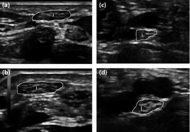
UltrasoundHigh-resolution transverse ultrasound: median nerve of a control subject (a) and a type 2 diabetic patient (b), posterior tibial nerve of a control (c) and a diabetic patient (d), nerve cross-section outlined.
Source : Ishibashi et al., Journal of diabetes investigation, 2015, figure 1 · CC BY-NC-ND
In a patient with diabetes and typical symptoms, DPN can be diagnosed clinically without resorting to electromyography (EMG), in line with the ADA Standards of Care 2025⁵. The minimum recommended examination comprises:
| Clinical test | What it assesses | Method | Performance |
|---|---|---|---|
| Monofilament 10 g (Semmes-Weinstein) | Protective sensation (large Aβ fibres) | 1st, 3rd, 5th plantar metatarsal heads, 3 applications per site, 1 sham | Validated Sensitivity 53-93%, specificity 75-100% for predicting ulcer risk⁶ |
| 128 Hz tuning fork | Vibration sense (large Aβ fibres) | Medial malleolus or head of the 1st metatarsal, tested on both sides | Validated Often an early abnormality, a good marker of progression⁷ |
| Ankle reflex | S1 reflex arc | Tendon hammer, knee flexed | Validated Frequently abolished, and abolished early |
| Pinprick / cold test | Small Aδ / C fibres | Cocktail stick, cold compress, side-to-side comparison | Good Detects small fibre involvement, which is often isolated |
| EMG | Motor and sensory nerve conduction | Specialised electrophysiological test | Reference standard Reserved for atypical or doubtful presentations⁵ |
| Skin biopsy (IENFD) | Intra-epidermal nerve fibre density | 3 mm punch, immunohistochemistry | Gold standard small fibres, but invasive and hard to access |
The ADA recommends annual screening of every patient with type 2 diabetes from diagnosis onwards, and 5 years after diagnosis for type 1 diabetes⁵. Loss of protective sensation detected with the monofilament is a major risk factor for ulceration and must trigger intensified preventive measures.
Mandatory differential diagnosis: before concluding that this is DPN, rule out:
- Vitamin B12 deficiency (very common with long-term metformin: measuring the level is essential)⁸.
- Hypothyroidism (TSH).
- Alcoholic neuropathy (targeted questioning, GGT, MCV).
- Chronic inflammatory demyelinating polyradiculoneuropathy (CIDP): suspect it if the presentation is asymmetrical or motor, if progression runs beyond 8 weeks, or if there is a proximal deficit.
- Toxic neuropathies : chemotherapy (oxaliplatin, vincristine, taxanes), antiretrovirals.
- Autoimmune diseases : Sjögren, lupus, sarcoidosis, vasculitis.
- Paraneoplastic causes : if there is unexplained weight loss or an inflammatory syndrome.
- Chronic kidney failure with uraemia.
A minimum laboratory work-up in the presence of a polyneuropathy comprises: full blood count, electrolytes, creatinine, blood glucose and HbA1c, TSH, B12, plasma protein electrophoresis⁵.
Classification: Toronto, NDS and IWGDF
The Toronto classification (Tesfaye et al. 2010)⁹ stages DPN in four categories: possible, probable, confirmed, subclinical. This stratification has prognostic and therapeutic value:
- Possible DPN : symptoms OR clinical signs.
- Probable DPN : symptoms AND clinical signs (2 or more).
- Confirmed DPN : signs + electrophysiological abnormality.
- Subclinical DPN : abnormal nerve conduction with no sign or symptom.
The Neuropathy Disability Score (NDS) is a clinical score combining vibration, pain, temperature and ankle reflexes (0-10) that predicts ulcer risk⁵. But for physiotherapy practice in France, it is the IWGDF foot risk stratification system that structures management best: that is the subject of the next chapter.
Red flags when assessing DPN
- Asymmetrical presentation or unilateral presentation → mononeuropathy, radiculopathy, focal lesion.
- Rapid progression (weeks) or a predominantly motor deficit → suspect CIDP, vasculitis or a paraneoplastic cause.
- Unexplained weight loss with progressive neuropathy → screen for malignancy.
- Proximal involvement with wasting (thighs) → diabetic amyotrophy of Bruns-Garland (DLRPN), which needs a neurological opinion.
- Hot, red, swollen foot with no wound in a person with diabetes and neuropathy → suspect an acute Charcot foot and this is an emergency (see chapter 3).
- Plantar ulcer + fever or discharge → suspect osteomyelitis → imaging + urgent specialist opinion.
- Severe autonomic signs (disabling orthostatic hypotension, gastroparesis, early erectile dysfunction) → cardiac autonomic neuropathy, cardiological work-up.
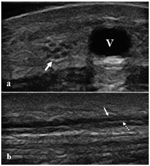
UltrasoundTransverse (a) and longitudinal (b) ultrasound of the sural nerve, arrows on the nerve, a technique used to assess diabetic cutaneous neuropathy.
Source : Liu et al., PloS one, 2012, figure 1 · CC BY
Key points
- The history must characterise the positive vs negative symptoms and their symmetrical, ascending "stocking" distribution.
- The minimum clinical examination: 10 g monofilament, 128 Hz tuning fork, ankle reflexes, pinprick test.
- EMG is reserved for atypical presentations (asymmetrical, motor, rapid).
- Always rule out the non-diabetic causes : B12, hypothyroidism, alcohol, CIDP, paraneoplastic causes.
- Classifying (Toronto, NDS) makes it possible to give a prognosis and stratify ulcer risk.
References
- Pop-Busui R, Boulton AJM, Feldman EL, et al. Diabetic Neuropathy: A Position Statement by the American Diabetes Association. Diabetes Care. 2017;40(1):136-154. PMID 27999003.
- Pop-Busui R, Ang L, Boulton AJM, et al. Diagnosis and Treatment of Painful Diabetic Peripheral Neuropathy. ADA Clinical Compendia. 2022;2022(1):1-32. doi:10.2337/db2022-01.
- McDermott K, Schrack JA, Windham BG, et al. Peripheral neuropathy, gait speed, and lower extremity function in community-dwelling older adults with and without diabetes. J Am Geriatr Soc. 2024;72(10):3242-3251. PMID 38994636.
- Feldman EL, Callaghan BC, Pop-Busui R, et al. Diabetic neuropathy. Nat Rev Dis Primers. 2019;5(1):41. PMID 31197183.
- American Diabetes Association Professional Practice Committee. 12. Retinopathy, Neuropathy, and Foot Care: Standards of Care in Diabetes — 2025. Diabetes Care. 2025;48(Suppl 1):S252-S265. Standards 2025.
- Boulton AJM, Armstrong DG, Albert SF, et al. Comprehensive Foot Examination and Risk Assessment: A report of the Task Force of the Foot Care Interest Group of the American Diabetes Association. Diabetes Care. 2008;31(8):1679-1685. PMID 18663232.
- Selvarajah D, Kar D, Khunti K, et al. Diabetic peripheral neuropathy: advances in diagnosis and strategies for screening and early intervention. Lancet Diabetes Endocrinol. 2019;7(12):938-948. doi:10.1016/S2213-8587(19)30081-6.
- Iqbal Z, Azmi S, Yadav R, et al. Diabetic Peripheral Neuropathy: Epidemiology, Diagnosis, and Pharmacotherapy. Clin Ther. 2018;40(6):828-849. PMID 29709457.
- Tesfaye S, Boulton AJM, Dyck PJ, et al. Diabetic neuropathies: update on definitions, diagnostic criteria, estimation of severity, and treatments. Diabetes Care. 2010;33(10):2285-2293. PMID 20876709.
How should foot risk be stratified and the Charcot foot recognised?
IWGDF 2023 stratification: a major clinical tool
The International Working Group on the Diabetic Foot (IWGDF) has published the international reference recommendations for the prevention and management of the diabetic foot since 1999. The IWGDF 2023 update¹ (published in 2024 in Diabetes/Metabolism Research and Reviews) introduces a foot risk stratification in four categories that guides how often patients are reviewed and how intensive preventive measures should be.
LOPS = Loss of Protective Sensation (protective sensation lost, as detected with the 10 g monofilament). PAD = peripheral arterial disease (pulse palpation, ABPI < 0.9). Source: Bus et al. IWGDF 2023, Diabetes Metab Res Rev²; Schaper 2024¹. The follow-up interval and the intensity of preventive measures (education, therapeutic footwear, orthoses) increase with the category.
This classification has two major clinical uses:
- Adapting the follow-up interval for physiotherapy and podiatry to the real level of risk: avoiding over-medicalisation of risk 0 and concentrating resources on risk 2-3.
- Justifying therapeutic footwear and insoles: essential from IWGDF 2 onwards, they can cut plantar pressures by 30% or more³ and prevent ulcer recurrence⁴.
Daily foot self-inspection is non-negotiable from IWGDF 1 onwards and must be taught systematically, with a mirror for the sole of the foot when mobility is reduced. The cumulative evidence for structured therapeutic education shows a reduction of about 50% in ulcer risk⁵ (RR 0.54; 95% CI 0.29-1.00).
The Charcot foot: an emergency that is often missed
The Charcot neuroarthropathy is a potentially devastating complication of DPN, marked by progressive destruction of the bones, joints and soft tissues of the foot. Its prevalence in people with diabetes is estimated at between 0.1% and 13%, rising to 29-35% when severe neuropathy is present⁶. The acute form is an emergency: missing it leads to irreversible joint destruction and exposes the patient to amputation.
Typical clinical presentation of acute Charcot :
- The hot foot (temperature difference > 2 °C compared with the other foot, easy to measure with an infrared thermometer).
- The red and swollen foot.
- Pain is moderate or absent and strikingly out of proportion to the inflammatory signs, which is the major diagnostic trap.
- A history of minor trauma (sometimes) or often no trigger at all.
- Diabetes usually poorly controlled, neuropathy established.
The differential diagnosis includes: cellulitis or erysipelas (more marked pain, inflammatory syndrome, raised white cell count), deep vein thrombosis, osteomyelitis, gout. Further imaging (MRI first choice, technetium bone scan if MRI is contraindicated) confirms the diagnosis and defines its extent⁷.
Red flags for the Charcot foot and the infected diabetic foot
- Hot, red, swollen foot with no wound in a person with diabetes and DPN → acute Charcot until proved otherwise. Immediate total offloading (total contact cast, TCC) and a specialist opinion within 24-48 h.
- Temperature difference > 2 °C on infrared thermometry between the two feet → a sensitive early sign of incipient Charcot.
- Ulcer + fever + purulent discharge → osteomyelitis or cellulitis: urgent surgical opinion, bacteriological samples, imaging.
- Positive "probe-to-bone test" (a sterile probe in the depth of the wound touching bone) → strong suspicion of osteomyelitis.
- Critical ischaemia (rest pain, black wound with sharp edges, absent pulses, ABPI < 0.5) → vascular emergency.
- Gangrene (dry or wet) → immediate surgical opinion, debridement, antibiotic therapy.
- Limited mouth opening in a child with diabetes with TMJ involvement → think of juvenile idiopathic arthritis (a rare but documented differential diagnosis).
Initial physiotherapy management of acute Charcot :
- Total offloading is imperative using a total contact cast (TCC) or a removable CROW boot, for 3 to 6 months on average (until the temperature difference has normalised)⁷.
- Regular monitoring of temperature, of oedema and of inflammatory markers.
- After the acute phase: progressive rehabilitation of weight-bearing, adaptation of footwear (custom therapeutic shoes if there are deformities), compensatory proprioceptive work.
- Long-term monitoring: risk of contralateral recurrence is high (up to 30% at 5 years)⁸.
Key points
- The IWGDF 2023 stratification into 4 categories (0 to 3) guides the follow-up interval and the intensity of foot prevention.
- Daily self-inspection and the therapeutic footwear (from IWGDF 2 onwards) are the pillars of ulcer prevention.
- The acute Charcot foot presents as a hot, red, swollen foot without proportionate pain: an emergency often mistaken for cellulitis.
- Measuring the temperature difference (> 2 °C) with an infrared thermometer between the two feet is a sensitive early sign.
- Initial management of acute Charcot: total offloading is imperative (TCC) for 3-6 months + imaging + specialist opinion.
References
- Schaper NC, van Netten JJ, Apelqvist J, Bus SA, Fitridge R, Game F, Monteiro-Soares M, Senneville E; IWGDF Editorial Board. Practical guidelines on the prevention and management of diabetes-related foot disease (IWGDF 2023 update). Diabetes Metab Res Rev. 2024;40(3):e3657. PMID 37243927.
- Bus SA, Sacco ICN, Monteiro-Soares M, et al. Guidelines on the prevention of foot ulcers in persons with diabetes (IWGDF 2023 update). Diabetes Metab Res Rev. 2024;40(3):e3651. doi:10.1002/dmrr.3651.
- Bus SA, Armstrong DG, Crews RT, et al. Guidelines on offloading foot ulcers in persons with diabetes (IWGDF 2023 update). Diabetes Metab Res Rev. 2024;40(3):e3647. PMID 37226568.
- Armstrong DG, Boulton AJM, Bus SA. Diabetic Foot Ulcers and Their Recurrence. N Engl J Med. 2017;376(24):2367-2375. PMID 28614678.
- Lincoln NB, Radford KA, Game FL, Jeffcoate WJ. Education for secondary prevention of foot ulcers in people with diabetes: a randomised controlled trial. Diabetologia. 2008;51(11):1954-1961. PMID 18758747.
- Wukich DK, Raspovic KM, Hobizal KB, Sadoskas D. Charcot neuroarthropathy in persons with diabetes: It's time for a paradigm shift in our thinking. Diabetes Metab Res Rev. 2024;40(3):e3754. doi:10.1002/dmrr.3754.
- Boulton AJM, Whitehouse RW. The Diabetic Foot. In: Feingold KR, Anawalt B, Blackman MR, et al., editors. Endotext [Internet]. South Dartmouth (MA): MDText.com, Inc.; 2023. NBK409609.
- Armstrong DG, Swerdlow MA, Armstrong AA, Conte MS, Padula WV, Bus SA. Five year mortality and direct costs of care for people with diabetic foot complications are comparable to cancer. J Foot Ankle Res. 2020;13:16. PMID 32209136.
Which treatment strategies are the most effective?
Hierarchy of recommended interventions
The management of DPN rests on a clear hierarchy validated by the international guidelines (ADA 2025, AAN 2022, IWGDF 2023)¹⁻³. The aim is not only to relieve pain but also to slow the progression and to prevent the devastating complications (ulcers, amputations, Charcot).
The pyramid has 5 levels: glycaemic control (base), education, multimodal exercise, targeted pharmacotherapy, adjuncts. The figures in brackets show the position in the hierarchy by population impact + level of evidence. Sources: ADA 2025¹, AAN 2022², IWGDF 2023³.
1. Optimal glycaemic control: the DCCT/EDIC (type 1) and ACCORD/VADT (type 2) trials showed that improving glycaemic control reduces the incidence of DPN and slows its progression⁴. It is the only intervention whose effect on the progression of the nerve disease is clearly established. Every patient with DPN must have regular diabetology review with optimisation of HbA1c targets.
2. Annual foot assessment and education: in line with the ADA Standards 2025¹, every person with diabetes must have an annual foot assessment (monofilament, tuning fork, pulse palpation, search for deformities). Structured therapeutic education (daily inspection, choice of shoes, skin hygiene, warning signs) reduces the risk of ulceration by about 50%⁵.
Multimodal exercise: the therapeutic mainstay
Physical exercise is now a first-line intervention validated by recent meta-analyses⁶,⁷. The meta-analysis by Streckmann et al. (2022, Sports Medicine)⁶ confirms a moderate to large effect on:
- The neuropathic pain (significant reduction on the NRS and BPI scales).
- Static and dynamic balance: a reduction in the risk of falling.
- The walking speed and endurance.
- The nerve function (improved nerve conduction, and even intra-epidermal fibre density in some patients).
Current consensus favours a multimodal approach combining 4 components:
The multimodal programme combines 4 components (A+S+B+F) with complementary aims. Streckmann 2022⁶: the aerobic + balance combination currently looks like the best non-pharmacological option. Mao 2023⁷: Tai Chi significantly improves postural control.
Safety of exercise when DPN is present: contrary to an old belief, exercise is not contraindicated when DPN is present, including when protective sensation has been lost. The safety principles are:
- Foot inspection before and after every session.
- Suitable footwear (sports shoes, seamless socks).
- Skin moisturising to prevent cracks.
- Prefer low-impact activities (swimming, stationary bike, cross-trainer) when IWGDF ≥ 2 or deformities are present.
- Avoid running on uneven ground when LOPS is marked.
- Initial supervision by a physiotherapist to confirm safety and progress gradually.
Adjuncts, pharmacotherapy and psychological factors
Pharmacotherapy for neuropathic pain: the official recommendation of the American Academy of Neurology (AAN 2022 Practice Guideline Update, Price et al.)² brings together the best available data:
| Class | Examples | Level of evidence | Comment |
|---|---|---|---|
| Gabapentinoids | Gabapentin, pregabalin | Recommended (moderate) | First line. Watch for drowsiness and oedema |
| SNRIs | Duloxetine, venlafaxine | Recommended (moderate) | Particularly useful with comorbid depression |
| Tricyclics (TCAs) | Amitriptyline, nortriptyline | Recommended (low to moderate) | Effective but anticholinergic effects |
| Sodium channel blockers | Valproate, carbamazepine | Recommended (low) | Liver and blood monitoring |
| Topical capsaicin 8% | Qutenza patch | Recommended (low) | Applied in a specialist setting |
| Opioids | Tramadol, tapentadol | To be avoided | Misuse, dependence, limited long-term efficacy |
The AAN explicitly recommends not prescribing opioids as first-line treatment for diabetic neuropathic pain, because of the limited long-term benefit and the risks of misuse and dependence². Prescribing remains a medical responsibility, but the physiotherapist must know the principles in order to fit in with the treatment and work with the patient's GP.
Adjunctive physical modalities: several modalities are used in clinical practice, but their level of evidence varies:
- TENS : the Gibson 2019 Cochrane meta-analysis⁸ concludes that the evidence is of low quality for chronic neuropathic pain. It can be a short-term adjunct, without replacing active approaches.
- Photobiomodulation (low-level laser) : heterogeneous results, low to moderate level of evidence; no strong recommendation.
- Manual therapy / neural mobilisation : limited evidence, isolated trials; clinical use justified only when there are associated mobility restrictions.
- Focal or platform mechanical vibration : promising preliminary trials, but low quality of evidence.
The psychological dimension: chronic pain, fear of falling and loss of function are associated with high levels of anxiety, depression and catastrophising⁹. These factors amplify the perception of pain and undermine adherence to foot care, creating a vicious circle. Systematic screening (HADS, PHQ-9) and referral for Cognitive behavioural therapy (CBT) or a mindfulness-based approach are indicated in patients with a marked psychological component¹⁰.
Key points
- The treatment hierarchy is clear: 1. Glycaemic control → 2. Foot-care education → 3. Multimodal exercise → 4. Targeted pharmacotherapy → 5. Adjuncts.
- Multimodal exercise (aerobic + strength + balance + flexibility) is a safe first-line therapy, even when LOPS is present.
- The AAN 2022 pharmacotherapy : gabapentinoids, SNRIs, TCAs, sodium channel blockers. Avoid opioids.
- The TENS modality has low-quality evidence (Cochrane Gibson 2019); adjunctive short-term use only.
- The psychological screening and CBT are indicated in patients with a marked anxiety-depression component.
References
- American Diabetes Association Professional Practice Committee. 12. Retinopathy, Neuropathy, and Foot Care: Standards of Care in Diabetes — 2025. Diabetes Care. 2025;48(Suppl 1):S252-S265. Standards 2025.
- Price R, Smith D, Franklin G, et al. Oral and Topical Treatment of Painful Diabetic Polyneuropathy: Practice Guideline Update Summary: Report of the AAN Guideline Subcommittee. Neurology. 2022;98(1):31-43. doi:10.1212/WNL.0000000000013038.
- Schaper NC, van Netten JJ, Apelqvist J, et al. Practical guidelines on the prevention and management of diabetes-related foot disease (IWGDF 2023 update). Diabetes Metab Res Rev. 2024;40(3):e3657. PMID 37243927.
- Pop-Busui R, Boulton AJM, Feldman EL, et al. Diabetic Neuropathy: A Position Statement by the American Diabetes Association. Diabetes Care. 2017;40(1):136-154. PMID 27999003.
- Lincoln NB, Radford KA, Game FL, Jeffcoate WJ. Education for secondary prevention of foot ulcers in people with diabetes: a randomised controlled trial. Diabetologia. 2008;51(11):1954-1961. PMID 18758747.
- Streckmann F, Balke M, Lehmann HC, et al. Exercise and Neuropathy: Systematic Review with Meta-Analysis. Sports Med. 2022;52(5):1043-1065. doi:10.1007/s40279-021-01596-6.
- Mao W, Wang T, Sun M, Zhang F, Li L. Effects of Tai Chi on Postural Control in People with Peripheral Neuropathy: A Systematic Review with Meta-Analysis. Healthcare (Basel). 2023;11(11):1559. PMID 37297699.
- Gibson W, Wand BM, Meads C, Catley MJ, O'Connell NE. Transcutaneous electrical nerve stimulation (TENS) for chronic pain — an overview of Cochrane Reviews. Cochrane Database Syst Rev. 2019;4(4):CD011890. doi:10.1002/14651858.CD011890.pub3.
- Khdour MR. Treatment of diabetic peripheral neuropathy: a review. J Pharm Pharmacol. 2020;72(7):863-872. doi:10.1111/jphp.13241.
- Pop-Busui R, Ang L, Boulton AJM, et al. Diagnosis and Treatment of Painful Diabetic Peripheral Neuropathy. ADA Clinical Compendia. 2022;2022(1):1-32. doi:10.2337/db2022-01.
How do you secure lasting recovery and prevent flares?
Making the patient an active partner through self-management
Patient empowerment is a central pillar for preventing the worsening of DPN and the dreaded diabetic foot ulcer. Moving from passive care to active self-management rests on structured therapeutic education programmes that have proved effective not only in improving knowledge but also in reducing the incidence of ulcers and amputations¹.
The four pillars of self-management :
- Tight glycaemic control: the most effective measure for slowing the progression of DPN. It means regular HbA1c monitoring and close collaboration with the diabetologist².
- Daily foot inspection: the most effective preventive act. Look for redness, blisters, cuts and skin changes. Use a mirror if mobility does not allow the sole to be seen. Teaching this practice reduces the risk of ulceration by about 50%¹.
- Suitable footwear: essential from IWGDF 2 onwards. Custom therapeutic shoes and personalised orthoses can cut peak plantar pressures by 30% or more³. Teach the basic rules: check inside the shoe before putting it on, seamless socks in natural fibres, never walk barefoot.
- Moisturising and skin care: apply a moisturising cream daily (avoiding the spaces between the toes to limit maceration). This preserves skin elasticity and barrier function⁴.
The effectiveness of these programmes depends on how far they are personalised (health literacy, socio-cultural context, cognitive capacity) and on how far they are repeatedly reinforced over time. One-off education is not enough: a sustained approach with regular reminders is needed⁵.
Safe return to sport and activity
Physical activity is a major therapeutic intervention for patients with DPN. Beyond glycaemic control, it has direct effects on nerve function, balance and quality of life⁶,⁷. The return must, however, be gradual, supervised and tailored to avoid iatrogenic injury, particularly in patients who have lost protective sensation.
Four steps to a safe return :
- A full preliminary assessment: quantify the sensory loss (monofilament), assess muscle strength, joint stability, static and dynamic balance (Berg Balance Scale, Timed Up & Go, Tinetti). Stratify the foot risk (IWGDF). Examine the feet (callus, deformities, previous ulcers).
- A multimodal programme: combine aerobic + strengthening + balance + flexibility (see chapter 3). Favour low-impact activities when IWGDF ≥ 2 (swimming, stationary bike, cross-trainer, Tai Chi, adapted yoga). Mao 2023 SR/MA⁷ confirms that Tai Chi improves postural control.
- Gradual, monitored progression: the principle of progressive overload, increasing duration or intensity in steps of no more than 10% per week. Watch for pain or skin lesions. Inspect the feet before and after every session.
- A focus on balance and fall prevention: patients with DPN have roughly a threefold higher risk of falling (Hicks 2023⁸). Specific exercises on unstable surfaces (under supervision), Tai Chi and adapted yoga have proved effective⁷.
Activities to favour vs to avoid :
| To favour | To do with care | To avoid if IWGDF 2-3 |
|---|---|---|
| Swimming, aqua aerobics | Walking on level ground (suitable shoes) | Running on uneven ground |
| Stationary bike | Outdoor cycling (saddle adjusted) | Pivoting sports (skiing, football) |
| Tai Chi, adapted yoga | Strengthening with light loads | Contact sports |
| Cross-trainer | Supervised balance exercises | Barefoot (yoga without protection) |
Critique and controversies
Although education and exercise command consensus, several challenges remain. The first is long-term adherence : many studies show a significant fall in adherence to self-care behaviour after a few months⁹. The move from supervised education to independent self-management remains the weak link. Reinforcement strategies (telephone reminders, apps, telemedicine) are promising but their scientific validity is uneven.
The second concerns the "optimal dose" of exercise. The multimodal recommendations command consensus, but the ideal frequency, intensity and duration are still debated. More intensive programmes (HIIT) have shown marked metabolic benefits, but they raise the risk of injury in an at-risk population; the benefit-risk balance must be reassessed for each individual⁶.
Finally, the emergence of wearable technologies and health apps (pressure sensors, connected insoles, home infrared thermometers) opens promising prospects for remote monitoring and the early detection of incipient Charcot, but the risk is of creating a digital divide that excludes the less tech-savvy patients, who are often the most at risk⁵.
Key points
- Self-management rests on 4 pillars: glycaemic control, daily inspection of the feet, suitable footwear, skin moisturising.
- Structured therapeutic education reduces the risk of ulceration by about 50 %.
- Return to activity must be gradual (+10%/week maximum), multimodal and supervised.
- The fall risk is three times higher when DPN is present: specific balance exercises (Tai Chi) are essential.
- Adapting activity to the IWGDF category: low impact (swimming, cycling, Tai Chi) if IWGDF 2-3, and avoid running on uneven ground.
References
- Lincoln NB, Radford KA, Game FL, Jeffcoate WJ. Education for secondary prevention of foot ulcers in people with diabetes: a randomised controlled trial. Diabetologia. 2008;51(11):1954-1961. PMID 18758747.
- Pop-Busui R, Boulton AJM, Feldman EL, et al. Diabetic Neuropathy: A Position Statement by the American Diabetes Association. Diabetes Care. 2017;40(1):136-154. PMID 27999003.
- Bus SA, Sacco ICN, Monteiro-Soares M, et al. Guidelines on the prevention of foot ulcers in persons with diabetes (IWGDF 2023 update). Diabetes Metab Res Rev. 2024;40(3):e3651. doi:10.1002/dmrr.3651.
- Schaper NC, van Netten JJ, Apelqvist J, et al. Practical guidelines on the prevention and management of diabetes-related foot disease (IWGDF 2023 update). Diabetes Metab Res Rev. 2024;40(3):e3657. PMID 37243927.
- Boulton AJM, Whitehouse RW. The Diabetic Foot. In: Endotext [Internet]. South Dartmouth (MA): MDText.com; 2023. NBK409609.
- Streckmann F, Balke M, Lehmann HC, et al. Exercise and Neuropathy: Systematic Review with Meta-Analysis. Sports Med. 2022;52(5):1043-1065. doi:10.1007/s40279-021-01596-6.
- Mao W, Wang T, Sun M, Zhang F, Li L. Effects of Tai Chi on Postural Control in People with Peripheral Neuropathy: A Systematic Review with Meta-Analysis. Healthcare (Basel). 2023;11(11):1559. PMID 37297699.
- Hicks CW, Wang D, Schneider ALC, et al. The association of peripheral neuropathy detected by monofilament testing with risk of falls and fractures in older adults. J Am Geriatr Soc. 2023;71(7):2185-2195. doi:10.1111/jgs.18338.
- Iqbal Z, Azmi S, Yadav R, et al. Diabetic Peripheral Neuropathy: Epidemiology, Diagnosis, and Pharmacotherapy. Clin Ther. 2018;40(6):828-849. PMID 29709457.
What do real clinical cases teach us?
A "classic" case: DPN with a plantar ulcer
A representative case is reported by Waghe and Athawale (2024) in Cureus¹: a man aged 62 years, with diabetes for 25 years, presents with pain and blisters on the right foot that progressed to an ulcer once they burst. Examination finds reduced joint range and muscle strength in the affected foot, on a background of distal sensory neuropathy.
Multimodal management :
- Targeted strengthening exercises for the foot and ankle.
- Infrared radiation therapy (adjunct).
- Sensory integration to stimulate the altered perception.
- Therapeutic foot-care education (inspection, footwear, hygiene).
Outcomes measured on the NRS (Numeric Pain Rating Scale), the WHO-QOL and the DFSQ-UMA (Diabetic Foot Self-Care Questionnaire) showed substantial improvement by the end of the programme¹. This case illustrates the value of a multimodal approach combining exercise, physical modalities and education.
Complex case: Charcot neuroarthropathy with equinovarus deformity
The case of Chitlange, Padmawar and Phansopkar (2023) in Cureus² illustrates the management of a patient aged 38 years with a Charcot foot and equinovarus deformity following earlier fractures and K-wire stabilisation. Rehabilitation comprised:
- Initial phase: mandatory total offloading (TCC) to stabilise the inflammatory process.
- Recovery phase: progressive rehabilitation of weight-bearing, joint work at the ankle and hindfoot, tailored muscle strengthening.
- Adaptation of therapeutic footwear and of orthoses to prevent ulcers over the new bony deformities.
- Intensive proprioceptive work to compensate for the new shape of the foot.
This case brings out three major lessons: (1) the total offloading is non-negotiable in the acute phase; (2) post-acute rehabilitation must be very gradual; (3) secondary prevention (footwear, orthoses) is essential, because the Charcot foot carries a high risk of ulcers and of contralateral recurrence.
Hierarchy of evidence and the limits of case studies
Case studies are valuable for illustrating an approach, flagging rare presentations or prompting reflection. But their level of evidence is the lowest (n=1, level 5 of the Oxford CEBM hierarchy; very low quality according to GRADE). They can never demonstrate efficacy.
Simplified GRADE / Oxford CEBM hierarchy. Practical implication: where an appealing case study and a meta-analysis diverge, the decision must follow the meta-analysis. Case studies remain valuable for generating hypotheses, flagging rare presentations or illustrating clinical reasoning.
Critique and controversy
Case studies must be read critically. Their main limitation is their low level of evidence (n=1): no generalisation is possible. Positive results may be due to the placebo effect, to the natural course of the disease, or to features specific to the patient (motivation, comorbidities, family support).
There is a major publication bias at work: cases reporting spectacular therapeutic successes or atypical presentations are far more likely to be published than those describing failures or unremarkable courses. This can create a falsely optimistic picture of how effective some interventions are.
Finally, the fabrication bias also exists: the "grey" literature and some journals with limited peer review publish fictitious case studies or extrapolated data. Always check the source (PMID, DOI, PMC) before citing a case as a clinical example.
Key points
- The Waghe 2024 Cureus case illustrates a plantar ulcer in a 62-year-old patient with 25 years of diabetes, treated with a multimodal approach (exercise, infrared, sensory integration, education).
- The Chitlange 2023 Cureus case illustrates the management of a Charcot foot with equinovarus deformity: mandatory total offloading in the acute phase, then progressive rehabilitation and suitable footwear.
- Case studies carry a level of evidence 5 (the lowest). They illustrate but never demonstrate anything.
- Where there is divergence from a meta-analysis, follow the meta-analysis (level 1).
- Be wary of publication bias and the fabrication bias : always check the PMID/DOI.
References
- Waghe VR, Athawale V. Physiotherapeutic Interventions in Diabetic Foot Ulcer Management: A Case Report. Cureus. 2024;16(2):e55244. PMC 10981459.
- Chitlange NM, Padmawar S, Phansopkar P. Comprehensive Rehabilitation of Charcot Foot With Equinovarus Deformity: A Case Report. Cureus. 2023;15(11):e48431. PMC 10702148.
- Boulton AJM, Whitehouse RW. The Diabetic Foot. In: Endotext [Internet]. South Dartmouth (MA): MDText.com; 2023. NBK409609.
- Wukich DK, Raspovic KM, Hobizal KB, Sadoskas D. Charcot neuroarthropathy in persons with diabetes: It's time for a paradigm shift in our thinking. Diabetes Metab Res Rev. 2024;40(3):e3754. doi:10.1002/dmrr.3754.
- Streckmann F, Balke M, Lehmann HC, et al. Exercise and Neuropathy: Systematic Review with Meta-Analysis. Sports Med. 2022;52(5):1043-1065. doi:10.1007/s40279-021-01596-6.
- Mao W, Wang T, Sun M, Zhang F, Li L. Effects of Tai Chi on Postural Control in People with Peripheral Neuropathy: A Systematic Review with Meta-Analysis. Healthcare (Basel). 2023;11(11):1559. PMID 37297699.
- Schaper NC, van Netten JJ, Apelqvist J, et al. Practical guidelines on the prevention and management of diabetes-related foot disease (IWGDF 2023 update). Diabetes Metab Res Rev. 2024;40(3):e3657. PMID 37243927.
How can these recommendations be applied in practice?
Red flags, yellow flags and referral
One of the physiotherapist's most important skills is to recognise the limits of their scope of practice and to identify the situations that call for further expertise. Effective triage is a guarantee of safety and efficiency.
Red flags specific to DPN: beyond the general framework of spinal red flags (Finucane 2020 IFOMPT)¹, several signs specific to DPN must trigger prompt medical referral:
Red flags specific to diabetic neuropathy
- Hot, red, swollen foot with no wound in a person with diabetes and neuropathy → suspect an acute Charcot foot. Total offloading and a specialist opinion within 24-48 h.
- Ulcer + fever + purulent discharge or positive "probe-to-bone test" → suspected osteomyelitis, urgent surgical opinion.
- Critical ischaemia (rest pain, ABPI < 0.5, absent pulses, black wound) → vascular emergency.
- Atypical neuropathy presentation : asymmetrical, predominantly motor, rapid progression over weeks → suspect CIDP, vasculitis or a paraneoplastic cause.
- Unexplained weight loss + progressive neuropathy → systematic screening for malignancy.
- Severe autonomic symptoms (disabling orthostatic hypotension, gastroparesis, early sexual dysfunction) → cardiac autonomic neuropathy, cardiological work-up.
- Rapidly progressive neurological deficits (extensive weakness, pyramidal signs, sphincter involvement) → the classic neurological red flags, which demand immediate referral¹.
Yellow flags: a concept introduced by Nicholas 2011², yellow flags are the psychosocial factors that predict pain and disability becoming chronic. In the patient with DPN they include:
- Mistaken beliefs about pain ("pain means tissue damage that is getting worse").
- Catastrophising and fear of movement (kinesiophobia).
- Activity avoidance behaviour.
- Anxiety and depressive symptoms.
- Social isolation, poor support.
- Work-related litigation or a compensation claim.
The presence of yellow flags justifies working with a health psychologist or a chronic pain specialist. A cognitive behavioural (CBT) or mindfulness-based approach can significantly improve quality of life and adherence to care³.
Structured interprofessional collaboration: the complexity of the diabetic foot demands a team:
- Diabetologist or GP : optimisation of glycaemic control, drug prescribing, follow-up.
- Podiatrist : regular foot care, insoles, footwear advice.
- Therapeutic education nurse : structured therapeutic patient education (ETP) programmes.
- Dietitian : nutritional balance, weight loss if obesity is present.
- Psychologist : yellow flags, CBT, chronic pain management.
- Vascular surgeon : revascularisation if PAD is severe.
- Orthopaedic or maxillofacial surgeon : deformities, debridement, amputations.
PROMs, COS and barriers to EBP
Measuring outcomes with validated PROMs: systematic use of Patient-Reported Outcome Measures (PROMs) has become a standard in evidence-based practice⁴. For DPN, the validated tools include:
| Tool (PROM) | Domain | Relevance to DPN |
|---|---|---|
| NRS / VAS | Pain intensity (0-10) | Universal, quick, MCID ≈ 2 points |
| Brief Pain Inventory (BPI) | Pain + functional interference | Multidimensional, valid in DPN |
| NTSS-6 / NSS | Specific neuropathic symptoms | Standard in DPN clinical trials |
| NDS (Neuropathy Disability Score) | Clinical signs (reflexes, vibration, pain, temperature) | A 0-10 score that predicts ulcer risk |
| Berg Balance Scale, Tinetti, TUG | Balance and fall risk | Essential in the patient with DPN |
| DFSQ-UMA, Patient Interpretation of Neuropathy (PIN) | Foot self-care / cognitions | Measures adherence and beliefs |
| HADS / PHQ-9 | Anxiety / depression | Screening for yellow flags |
PROMs have a threefold purpose: (1) to quantify progress over time; (2) to set concrete goals aiming at the "minimal clinically important difference" (MCID); (3) to support shared decision-making with the patient⁴. Their implementation, however, runs up against a lack of time and training.
Core Outcome Sets (COS): to standardise measurement internationally, the scientific community is promoting the adoption of consensus tool sets for each condition. This makes studies more comparable and allows more data to be pooled for future meta-analyses⁵.
Barriers to EBP and strategies for overcoming them: the systematic review by Scurlock-Evans et al. (2014)⁶ identifies three main barriers perceived by physiotherapists:
- Lack of time : productivity pressure, administrative constraints.
- Lack of skills : difficulty searching, appraising and interpreting the literature.
- Lack of organisational support : a culture unfavourable to continuing education, limited access to databases.
Concrete strategies to counter them:
- Time : build PROM collection into digital tools (tablets, apps), and run "journal clubs" with protected time.
- Skills : continuing education (critical appraisal, statistics), use of pre-appraised resources (PEDro, Cochrane, guidelines).
- Support : organisational leadership that favours EBP, funding for training, EBP mentor roles within teams⁶.
Critique and controversy: beyond the guidelines
Applying guidelines blindly raises fundamental questions. The first trap is the "data graveyard" : collecting PROMs becomes an end in itself, with the results never used to adapt treatment or to discuss matters with the patient⁷. Without a clinical feedback loop, measurement loses its meaning.
The second is the tension between standardisation and personalisation. Guidelines are based on the homogeneous populations of clinical trials, whereas the clinician faces patients with multiple comorbidities, unique psychosocial contexts and varied preferences. The art of physiotherapy lies in the ability to navigate between scientific rigour and the flexibility that the humanity of care demands ("treat the person, not just the diagnosis")⁸.
Finally, the fear of missing serious pathology can lead to "flag inflation" and over-referral, cancelling out the efficiency gains of the physiotherapist as first contact¹. The balance between caution and confidence in clinical reasoning remains a constant challenge, one that only experience and continuing education can resolve.
Key points
- Detection of red flags (acute Charcot, osteomyelitis, critical ischaemia, atypical presentation) demands prompt medical referral.
- The yellow flags (catastrophising, kinesiophobia, anxiety, depression) justify working with a psychologist.
- The interprofessional collaboration (diabetologist, podiatrist, education nurse, psychologist, vascular surgeon) is essential for the diabetic foot.
- The validated PROMs (NRS, BPI, NTSS-6, NDS, BBS, HADS) make it possible to quantify progress, set MCID targets and support shared decision-making.
- Overcoming the barriers to EBP (time, skills, support) calls for multi-faceted strategies: pre-appraised resources, continuing education, organisational leadership.
References
- Finucane LM, Downie A, Mercer C, et al. International Framework for Red Flags for Potential Serious Spinal Pathologies. J Orthop Sports Phys Ther. 2020;50(7):350-372. PMID 32438853.
- Nicholas MK, Linton SJ, Watson PJ, Main CJ; "Decade of the Flags" Working Group. Early identification and management of psychological risk factors ("yellow flags") in patients with low back pain: a reappraisal. Phys Ther. 2011;91(5):737-753. PMID 21451099.
- Pop-Busui R, Ang L, Boulton AJM, et al. Diagnosis and Treatment of Painful Diabetic Peripheral Neuropathy. ADA Clinical Compendia. 2022;2022(1):1-32. doi:10.2337/db2022-01.
- Kyte DG, Calvert M, van der Wees PJ, ten Hove R, Tolan S, Hill JC. An introduction to patient-reported outcome measures (PROMs) for clinicians. Physiotherapy. 2015;101(2):119-125. doi:10.1016/j.physio.2014.11.003.
- American Diabetes Association Professional Practice Committee. 12. Retinopathy, Neuropathy, and Foot Care: Standards of Care in Diabetes — 2025. Diabetes Care. 2025;48(Suppl 1):S252-S265. Standards 2025.
- Scurlock-Evans L, Upton P, Upton D. Evidence-based practice in physiotherapy: a systematic review of barriers, enablers and interventions. Physiotherapy. 2014;100(3):208-219. PMID 24780633.
- Verhagen AP, Downie A, Popal N, Maher C, Koes BW. Red flags presented in current low back pain guidelines: a review. Eur Spine J. 2016;25(9):2788-2802. PMID 27376890.
- Greenhalgh T, Howick J, Maskrey N; Evidence Based Medicine Renaissance Group. Evidence based medicine: a movement in crisis? BMJ. 2014;348:g3725. PMID 24927763.
And after reading this?
This article is part of a collection of evidence-based clinical syntheses. A question, a comment, a correction to suggest? Contact us directly through the WhatsApp button at the bottom right of the screen.

