

















A clinical synthesis on tarsal tunnel syndrome: a compression of the posterior tibial nerve at the ankle, often missed but also over-diagnosed. Recognising the picture, looking for a cause, not confusing it with plantar fasciitis or a polyneuropathy, and running a course of management whose level of evidence remains modest. Every reference has been checked individually on PubMed.
📝 In brief: clinical synthesis
- Tarsal tunnel syndrome is a focal compressive neuropathy of the posterior tibial nerve (or its branches) as it passes under the flexor retinaculum on the medial side of the ankle; it is a rare and regularly under-diagnosed condition whose symptoms affect the plantar surface of the foot 41.
- A mechanical cause (a space-occupying mass, tenosynovitis, valgus, a systemic cause such as diabetes) is identifiable in about 80 % of cases; the purely idiopathic form, retained for want of a cause found, remains rare, which makes it essential to look actively for a compressive lesion before concluding 16.
- The diagnosis is above all clinical and there is no gold standard test: Tinel's sign percussed behind the medial malleolus has a low and variable sensitivity (25 to 75 %) for a specificity of 70 to 90 %, so that a negative test does not rule the diagnosis out 1.
- The dorsiflexion-eversion test (the ankle taken into maximum eversion and dorsiflexion, the toes in dorsiflexion, held for 5 to 10 seconds) is a reproducible provocation manoeuvre: in the original study it intensified local tenderness in 42 of the 43 affected feet, without provoking any symptom in the 50 healthy volunteers 8.
- Electrodiagnosis (EMG and nerve conduction studies) is often abnormal but its sensitivity and specificity are suboptimal, with frequent false negatives, especially in mild forms: a normal test therefore does not rule the diagnosis out 15.
- Conservative treatment is offered first line (medial arch support orthoses, stretching, strengthening, NSAIDs, injections) but rests on weak evidence; ultrasound-guided injection is an effective option, 169 patients out of 218 (77.5 %) not having needed surgery 1413.
- After conservative treatment fails, surgical decompression gives an excellent or good result in about 75.3 % of cases, but the reported success rates vary widely from 44 % to 96 %, a positive preoperative Tinel's sign and an identified cause being good predictors of relief 121.
🦶 What is tarsal tunnel syndrome?
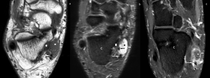
MRIAxial foot MRI, three sequences, showing a synovial cyst with T1 hypointensity and T2 hyperintensity in contact with the flexor retinaculum, the medial plantar nerve and the posterior tibial artery in the tarsal tunnel.
Source : Ormeci et al., International Journal of Surgery Case Reports, 2015, figure 3 · CC BY-NC-ND
🔍 The compression has an identifiable cause in the great majority of cases
Before concluding that a form is idiopathic, look for a mass, previous trauma or a biomechanical abnormality: the purely idiopathic syndrome is rare.
Note: these proportions come from different series and overlap; they do not add up. An impingement mechanism (a mass, cyst, lipoma, valgus, a systemic cause such as diabetes) is found in about 80 % of cases; up to 43 % of patients report an injury (often a sprain); varicosities of the posterior tibial vein are present in 20 % of patients; it remains idiopathic in about 20 % of cases. Sources: Kiel et al., StatPearls 2024 (PMID 30020645); Rodríguez-Merchán et al., EFORT Open Reviews 2021 (PMID 35839088); Sha et al., The Iowa Orthopaedic Journal 2024 (PMID 39811161).
Tarsal tunnel syndrome is a compressive neuropathy (entrapment) of the posterior tibial nerve , or of one of its terminal branches, as it passes through a fibro-osseous tunnel on the medial side of the ankle, deep to the flexor retinaculum 123. The nerve runs there in an inextensible space: any reduction in the volume available, whether from a mass, oedema or a deformity, tends to compress it and to generate symptoms along its territory. It is the lower limb equivalent of the far better known carpal tunnel syndrome 1.
Clinically, this entity is rare and regularly under-diagnosed 431. First described in 1962, it still suffers today from the absence of a definitive clinical or imaging test, which often delays its recognition 5. For the physiotherapist the consequence is twofold: it must be considered in atypical plantar pain, but caution is also needed before accepting it as the explanation, given how uncommon it is and how uncertain its diagnosis.
Key point. Tarsal tunnel syndrome is a compression of the posterior tibial nerve under the flexor retinaculum, behind the medial malleolus. It is the « carpal tunnel of the ankle »: rare, under-diagnosed, with no reference test, and with symptoms that affect above all the plantar surface of the foot 134.
The tarsal tunnel: a constrained fibro-osseous anatomy
The tarsal tunnel is an anatomical passage whose roof is formed by the flexor retinaculum (the laciniate ligament), a fibrous sheet stretched between the medial malleolus and the calcaneus 5. That ceiling closes a space containing, from front to back, the tendons of tibialis posterior, flexor digitorum longus and flexor hallucis longus, together with the posterior tibial neurovascular bundle: the posterior tibial artery, vein and nerve 5. This proximity between tendinous, vascular and nervous structures in a closed compartment explains the nerve's vulnerability: a tenosynovitis of one of the flexors, a venous dilatation or a mass is enough to encroach on its space.
The posterior tibial nerve does not stay a single trunk: it divides into plantar branches whose knowledge governs the topographical interpretation of the symptoms 5 :
- the medial plantar nerve supplies the medial arch, the plantar surface of the first three toes and the two medial lumbricals;
- the lateral plantar nerve supplies the lateral arch, the plantar surface of the 4th and 5th toes, all the interossei and the remaining lumbricals.
Depending on whether the compression sits on the tibial trunk or on one of these branches, the distribution of paraesthesia and pain will vary, hence the importance, on examination, of mapping precisely the territory affected on the plantar surface of the foot 45.
An inextensible tunnel, a nerve that divides there into plantar branches: the whole semiology of the tarsal tunnel is read on the sensory map of the sole of the foot.
A useful analogy, and its limits
Comparing the tarsal tunnel with the carpal tunnel helps both patient and clinician picture the mechanism: a nerve, an osteofibrous tunnel, a compression 1. The analogy is anatomically sound; in both cases a mixed nerve crosses a passage capped by a retinaculum. But it has its limits. At the wrist the entrapment syndrome is extremely common and well codified; at the ankle it remains rare, of unknown exact incidence, and with no diagnostic gold standard 15. In other words, the mechanical reasoning transfers, but the frequency and the diagnostic certainty do not.
Aetiologies: look for the lesion that occupies the space
This is probably the most operational message of the section. Contrary to a widespread idea, the tarsal tunnel is only rarely « idiopathic »: an identifiable impingement mechanism is found in about 80 % of cases, the purely idiopathic form being rare 1. Put another way, the aetiology remains unexplained in only about 20 % of cases 6. The clinical approach must therefore be assertive: actively look for a cause before concluding that there is no lesion.
The causes fall into several families 156 :
| Aetiological family | Examples | Clinical landmark |
|---|---|---|
| Space-occupying lesions (intrinsic) | Ganglion cysts, lipomas, neuromas, varicosities of the posterior tibial vein | Justify imaging (ultrasound / MRI) to look for a compressive mass |
| Biomechanical abnormalities | Valgus or varus hindfoot, pes planovalgus | Chronic tension on the nerve; the target of arch support orthoses |
| Post-traumatic | Ankle sprain, fracture sequelae | Previous trauma reported in up to 43 % of patients 1 |
| Systemic causes | Diabetes (first among them), hypothyroidism, rheumatoid arthritis, obesity | To ask about systematically; they can blur the neurological picture |
| Idiopathic | No cause found | A diagnosis of exclusion (~20 % of cases) |
Two numerical landmarks are worth remembering. First, the post-traumatic origin is frequent : up to 43 % of patients report previous trauma, an ankle sprain in particular 1 , a valuable anchor in a physiotherapist's history-taking. Second, among the intrinsic lesions, the varicosities of the posterior tibial vein are found in 20 % of affected patients 5, which on its own justifies completing the examination with imaging to look for vascular compression.
The systemic causes must not be neglected: diabetes ranks first among the associated conditions, alongside hypothyroidism, rheumatoid arthritis and obesity 5. In a patient with diabetes the difficulty deepens: a distal sensory polyneuropathy can overlap the entrapment picture and complicate attributing the symptoms to the tarsal tunnel alone 6. This systemic background is not trivial for the prognosis; associated obesity and diabetes reduce the chances of a good result from treatment.
Epidemiology: a rare and poorly quantified entity
Recognising the limits of the epidemiological data is part of an honest evidence-based reading. The exact incidence of tarsal tunnel syndrome remains unknown : the condition is relatively rare and often under-diagnosed, so that no reliable prevalence figure can be put forward 1. It is nonetheless known that it affects women more 1. That rarity, combined with the absence of a reference test, should make the physiotherapist cautious: the tarsal tunnel is neither the first nor the most likely explanation for plantar pain, and it should be retained only after the far more common causes have been ruled out.
Key point for practice. When a tarsal tunnel is suspected, the priority is not to assert the diagnosis but to look for the cause : a space-occupying lesion or a mechanical factor is present in ~80 % of cases 1. Ask about previous trauma (a sprain in up to 43 % of cases), request imaging whenever a mass or varicosities are suspected (20 % of patients), and identify the systemic comorbidities, diabetes first. The idiopathic form (~20 %) remains a diagnosis of exclusion 56.
🔍 How is it recognised?
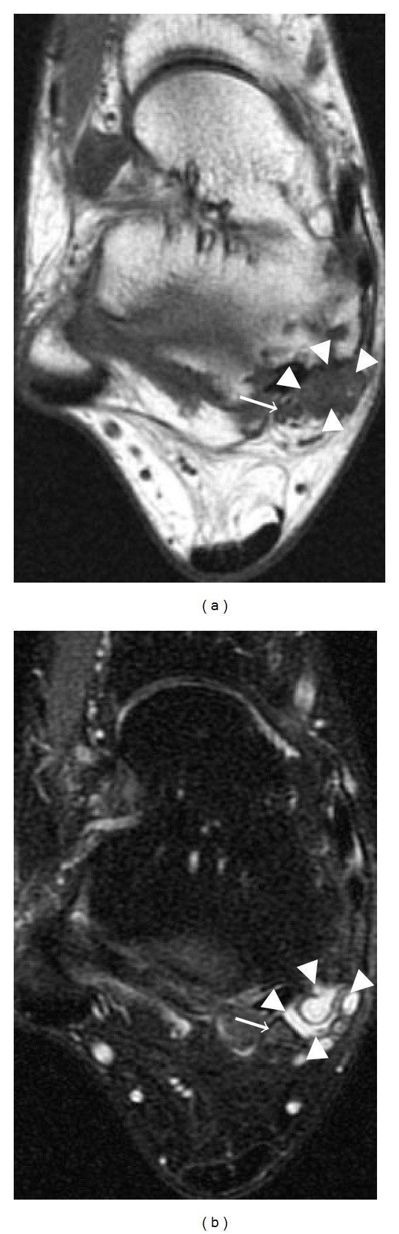
MRIAxial ankle MRI, T1-weighted then T2 fat-saturated, showing a multilobulated synovial cyst in the tarsal tunnel, in contact with the tibial nerve indicated by an arrow.
Source : Dong et al., Radiology Research and Practice, 2012, figure 20 · CC BY
📡 Ultrasound: highly specific, moderately sensitive
Measuring the cross-sectional area of the tibial nerve in the tunnel helps to document the compression and to look for a mass.
With an intra-tunnel cross-sectional area threshold of 15 mm², sensitivity is 74 % for a specificity of 100 %; with an area difference of 5 mm², sensitivity rises to 81 % for a specificity of 100 %. For comparison, MRI shows the point of compression in 82.1 % of cases 11, and electrodiagnosis often remains normal with frequent false negatives 1. Ultrasound source: Fantino et al., Orthopaedics & Traumatology Surgery & Research 2021 (PMID 32682728).
🦶 The dorsiflexion-eversion test reproduces the symptoms
The ankle is passively taken into maximum eversion and dorsiflexion, the toes in dorsiflexion, and the position is held for 5 to 10 seconds: the manoeuvre puts the tibial nerve under tension.
These are rates of symptom reproduction (a provocation manoeuvre), not a formal sensitivity. In the original study (37 operated patients, 50 healthy volunteers, that is 100 feet), no symptom or sign could be induced in the healthy controls. Source: Kinoshita et al., The Journal of Bone and Joint Surgery Am 2001 (PMID 11741063).
Recognising a tarsal tunnel syndrome is an exercise in clinical humility. It is a compressive neuropathy of the posterior tibial nerve, or of one of its branches, as it passes under the flexor retinaculum behind the medial malleolus 13. The condition is rare, regularly under-diagnosed, and its exact incidence remains unknown 1. First described in 1962, this syndrome still suffers from the absence of a truly definitive clinical or imaging test, which delays the diagnosis 5. It is worth writing straight away: no single investigation, taken alone, allows the diagnosis to be made with certainty 6. The reasoning is built by correlating the clinical history, the examination, imaging and electrodiagnosis, and the physiotherapist has every reason to stay cautious before accepting this label for plantar pain 1.
The clinical picture: decoding plantar and heel pain
The presenting complaint is pain or paraesthesia on the plantar surface of the foot and the medial heel. The topography is no accident: the posterior tibial nerve divides into two plantar branches whose sensory territory draws precisely the symptomatic zone. The medial plantar nerve supplies the medial arch and the plantar surface of the first three toes; the lateral plantar nerve supplies the lateral arch and the plantar surface of the 4th and 5th toes 5. A compression of the trunk or of a single branch therefore explains presentations that are sometimes global and sometimes limited to one territory. The symptoms, burning, tingling or numbness, affect the plantar border of the foot and fall within the posterior tibial territory 41.
This picture is in no way specific, and that is the whole difficulty. The differential diagnosis must be conducted rigorously: plantar fasciitis, lumbosacral radiculopathy, S1 in particular, rheumatological disease, metatarsal stress fractures and Morton's neuroma 6. The trap is worse in a patient with diabetes and a distal sensory polyneuropathy, where diffuse nerve involvement can be superimposed on, or mimic, a focal compression, blurring the reading completely 7. And diabetes is precisely among the systemic causes associated with the syndrome, alongside hypothyroidism, rheumatoid arthritis and obesity 5.
Key point. Pain or paraesthesia in the sole and the medial heel, against a background of possible previous trauma 1, should raise the tarsal tunnel. But the picture overlaps that of plantar fasciitis and of S1 radiculopathy: recognition comes from methodically eliminating the differential diagnoses, never from an isolated sign 6.
The stake is not only to assert the diagnosis but to look actively for its cause. A compression mechanism, a space-occupying lesion (varicosities, ganglion cyst, lipoma), a biomechanical abnormality of the hindfoot (valgus or varus), or a systemic condition such as diabetes, is identifiable in about 80 % of cases, the purely idiopathic form being rare 1. That figure has a strong practical consequence: concluding too quickly that a form is idiopathic risks missing a compressive mass that treatment could reach.
The provocation tests: Tinel's sign and dorsiflexion-eversion
Tinel's sign is the historical manoeuvre: percussion behind the medial malleolus reproduces pain or tingling radiating into the posterior tibial territory 1. Its performance is nonetheless modest and very variable, a sensitivity of 25 to 75 % for a specificity of 70 to 90 % 1. The lesson is twofold. First, a negative Tinel does not rule the diagnosis out : with a sensitivity that low, between a quarter and three quarters of affected patients can have a normal test. Second, a positive Tinel has a double value: relatively specific, it points the way, and above all it is a strong predictor of relief after surgical decompression 16.
Two recent series confirm this weakness by another route, and their figures speak more clearly than an interval. A systematic review of the published cases of tarsal tunnel syndrome secondary to an accessory or variant muscle, 25 studies, 39 patients, 47 ankles, reports the frequency of each sign in patients whose diagnosis is established and whose cause is identified: pain 82.9 %, tenderness on palpation 78.7 %, dysaesthesia 57.4 %, swelling 25.5 %, and Tinel's sign only 44.6 % 18. Be careful what that figure is: a frequency in a series of published cases, therefore selected, and not a sensitivity in the diagnostic sense. Its bearing is not diminished for all that: in a population where the compression is proven, the Tinel misses more than one case in two. The prospective Indian study cited below points the same way and simply notes that « Tinel's sign was inconsistent » 17.
It is this lack of reproducibility that motivated the development of the dorsiflexion-eversion test 8. The manoeuvre is standardised: the ankle is passively taken into maximum eversion and dorsiflexion, while all the metatarsophalangeal joints are held in maximum dorsiflexion, and the position is held for 5 to 10 seconds, which puts the tibial nerve under tension in its tunnel. In the original study (37 operated patients, 50 healthy volunteers), the test reproduced numbness in 15 of the 20 feet concerned (75 %), pain in 15 of 17 (88 %) and intensified local tenderness in 42 of the 43 affected feet before surgery (98 %), while no symptom or sign could be induced in the healthy volunteers 8. That complete absence of false positives in healthy subjects makes it a particularly interesting provocation manoeuvre on examination.
| Test | How it is done | Performance | Interpretation |
|---|---|---|---|
| Tinel's sign | Percussion behind the medial malleolus | Sensitivity 25–75 %; specificity 70–90 % 1 | Negative = rules nothing out; positive = points the way and predicts the surgical response |
| Dorsiflexion-eversion | Maximum eversion + dorsiflexion, toes in dorsiflexion, 5–10 s | Local tenderness increased in 42/43 affected feet; 0 induction in healthy subjects 8 | Reproduces or intensifies the symptoms; useful but from a single series |
Electroneuromyography: essential, but fallible
Electrodiagnosis, nerve conduction studies (NCS) and electromyography (EMG), remains the reference investigation for documenting involvement of the tibial nerve at the ankle, but its limits must be known and stated to the patient. Its sensitivity and specificity are suboptimal, and false negatives are frequent: a normal test does not rule the diagnosis out 1. The results are indeed often normal in mild forms or when the symptoms have built up gradually over years, to the point where repeated studies over time may be needed 5.
The AANEM evidence-based review adds a useful nuance: sensory conduction studies are more often abnormal than motor ones, but the real sensitivity and specificity of these tests could not be established, and electrodiagnosis is recommended only at level C evidence for confirming a tibial neuropathy at the ankle 9. Beyond false negatives, electroneuromyography shares another weakness: it cannot predict which cases will respond to surgical decompression, which explains why its role remains debated 3. In practice, electrodiagnosis confirms and documents; it does not settle the matter alone.
The only published sensitivity-specificity pair for the idiopathic form gives the measure of the problem, and it is the specificity that must be remembered. In a prospective Indian study of 26 patients (52 limbs) compared with 45 healthy controls, motor conduction was abnormal in 80 % of symptomatic limbs and also in 66.6 % of asymptomatic limbs of the same patients. The authors conclude that sensitivity is 80 % for a specificity of 33.3 %, and retain the test only as a screening tool 17. In other words: two painless limbs out of three already had an abnormal trace. A positive electrodiagnosis therefore does not make the diagnosis on its own; it accompanies a clinical picture that already convinces. This result comes from a single single-centre series and needs confirmation, but it points the same way as the AANEM level C recommendation.
Imaging: ultrasound and MRI in search of the cause
Since a mechanical cause exists in about 80 % of cases, imaging has a decisive role, less to assert the syndrome than to locate its site and flush out a compressive lesion. Ultrasound documents the increase in the cross-section of the tibial nerve in the tunnel: an intra-tunnel cross-sectional area threshold of 15 mm² gave 74 % sensitivity and 100 % specificity, and a difference in cross-section (ΔCSA) of 5 mm² reached 81 % sensitivity for 100 % specificity 10. Ultrasound, non-irradiating and dynamic, also deserves to be aimed at the search for a mass: varicosities of the posterior tibial vein are found in 20 % of patients 5.
MRI, for its part, serves mainly to locate the point of compression precisely and any space-occupying mass. In one series it showed the point of nerve compression in 82.1 % of cases (23 sides out of 28), but failed to reveal all the detail needed for surgical planning 11. In other words, imaging maps and documents the cause without replacing clinical judgement or the operation itself.
No gold standard: a diagnosis by correlation
This reality must be owned in front of the patient: there is no gold standard. Controversy persists about which diagnostic modalities really contribute, and no gold standard has been established 7. The diagnosis therefore rests on the correlation between the clinical history, imaging, nerve conduction studies and electromyography, each of these elements bringing a piece and none providing certainty 6.
The diagnostic approach, in summary.
- Clinical first: plantar and medial heel pain or paraesthesia, in the posterior tibial territory, worse on weight-bearing; look for previous trauma 1 and rule out plantar fasciitis, S1 radiculopathy and Morton's neuroma 6.
- Provocation tests: Tinel (specific but not very sensitive; a negative rules nothing out) and dorsiflexion-eversion 18.
- Electrodiagnosis: confirms a tibial neuropathy but false negatives are frequent; a normal test does not exclude it 19.
- Imaging: ultrasound (15 mm² threshold) and MRI to locate the compression and look for a mass; a cause is identifiable in ~80 % of cases 10111.
- Synthesis: no gold standard, the diagnosis comes from the coherence of the whole, not from an isolated test 7.
This caution is not an admission of impotence but good practice: in a condition that is rare, under-diagnosed and without a definitive test 5, making the diagnosis means bringing a bundle of concordant arguments together and, whenever possible, identifying the cause, since it is the cause that will govern the prognosis and the choice of treatment.
🎭 The trap of the differential diagnosis
Tarsal tunnel syndrome is a compressive neuropathy of the posterior tibial nerve, or of one of its branches, under the flexor retinaculum behind the medial malleolus, where the nerve crosses the tunnel with the flexor tendons and the posterior tibial pedicle 31. Symptomatically it shows itself as pain, burning and paraesthesia on the plantar surface of the foot. And that is precisely where the diagnosis goes off the rails: this plantar presentation is in no way specific, and half a dozen other pictures mimic it. The condition is described as rare, regularly under-diagnosed, of unknown exact incidence 145. Two symmetrical errors then lie in wait for the clinician: missing a true tarsal tunnel, and invoking it wrongly for any plantar pain. This section sets out the five diagnoses to confront systematically, then the proximal and distal topographical distinction, often neglected.
Why this syndrome is both missed and over-diagnosed
The paradox rests on a simple fact: there is no diagnostic gold standard. The diagnosis rests on the correlation between the clinical history, imaging, nerve conduction studies and electromyography, with no single investigation able to settle it with certainty 6. Controversy persists about which modalities really contribute, and no gold standard has emerged 7. Each clinical tool is fallible: Tinel's sign percussed behind the medial malleolus has only a sensitivity of 25 to 75 % for a specificity of 70 to 90 %, so that a negative test rules nothing out 1. Electrodiagnosis is frequently normal, particularly in mild forms or those building up gradually over years, with frequent false negatives; a normal test does not reject the diagnosis, and electroneuromyography cannot predict which cases will respond to decompression 153.
This lack of an objective anchor pulls in two opposite directions. On one side, the rarity of the entity and its low profile mean it is not considered, delaying the diagnosis: the physiotherapist should think of it in resistant, poorly labelled plantar pain. On the other, the absence of a discriminating test exposes the opposite risk, labelling as « tarsal tunnel » any medial or plantar ankle pain, without having ruled out the competing diagnoses or looked for the compressive cause. For there is almost always a cause: an impingement mechanism is identifiable in about 80 % of cases (a space-occupying mass, a valgus or varus hindfoot, a systemic condition including diabetes), the purely idiopathic form remaining rare 1. Accepting the diagnosis without looking for that cause is to be wrong twice.
Plantar fasciitis: the false twin of the heel
Plantar fasciitis is the first differential diagnosis to confront 6, and the most misleading, because the tarsal tunnel is itself an under-diagnosed cause of heel pain 5. Both share the medial plantar site of the pain. What should point to a nerve origin are the neuropathic features, burning, tingling, radiation into the posterior tibial territory, rather than the mechanical weight-bearing pain of the first morning step typical of fasciitis. A positive Tinel's sign points to the nerve, but its low sensitivity forbids making it a criterion for exclusion 1. In practice, a « plantar fasciitis » that does not respond to the usual treatment and comes with paraesthesia should make the tarsal tunnel hypothesis worth reconsidering.
Polyneuropathy (diabetic) and the double crush trap
This is the most delicate overlap. Diabetes is the first of the systemic causes associated with the tarsal tunnel 51 , so the same patient can have both. The diagnostic difficulty is explicitly increased by the overlap with other lower limb disorders, particularly in people with diabetes and a distal sensory polyneuropathy, where the diffuse nerve involvement can correspond to a « first crush » phenomenon that weakens the nerve upstream 7. Two practical consequences. First, a symmetrical distal polyneuropathy explains bilateral plantar paraesthesia on its own and must not be relabelled a tarsal tunnel. Second, electrodiagnosis, already unreliable in the tarsal tunnel, becomes even harder to interpret against a background of diffuse neuropathy: conduction abnormalities and false negatives add up 1. In a person with diabetes the reasoning must stay probabilistic and caution maximal before attributing plantar pain to a focal compression.
S1 radiculopathy: do not forget the spine
Lumbosacral radiculopathy, S1 in particular, appears explicitly among the differential diagnoses 6. The S1 territory covers the plantar surface and the lateral border of the foot, hence the possible confusion with symptoms of the tibial nerve and its lateral plantar branch. The examination must look for signs of spinal origin: lumbar or buttock pain, downward radiation, signs of neuromeningeal tension on the straight leg raise, an absent Achilles reflex. A positive local provocation test at the tarsal tunnel argues on the contrary for the distal origin: the dorsiflexion-eversion test, done with the ankle passively taken into maximum eversion and dorsiflexion, toes in dorsiflexion, the position held for 5 to 10 seconds, reproduces or intensifies the symptoms by putting the tibial nerve under tension at the tunnel 8. Its original validity is locally solid, no symptom induced in the 50 healthy volunteers, local tenderness intensified in 42 of the 43 affected feet, but it has not been retested on a large scale: to be treated as an argument, not as proof.
Morton's neuroma: distal, but not in the tunnel
Morton's neuroma completes the list 6. It shares the plantar forefoot topography but differs by its intermetatarsal location (classically the 3rd space), its electric pain triggered by walking and by transverse compression of the forefoot. Conversely, tarsal tunnel symptoms travel up towards the medial malleolus and follow the distribution of the plantar branches from the tunnel. The neuroma is also cited as one of the types of space-occupying lesion that can itself compress the nerve 5 : the nuance matters, because a neuroma sitting on the course of the plantar branches is not the same entity as an intermetatarsal Morton's neuroma.
Proximal versus distal tarsal tunnel: an underestimated topography
The proximal and distal distinction shapes both the clinical picture and the differential reasoning. Proximally the compression bears on the trunk of the posterior tibial nerve under the flexor retinaculum, behind the medial malleolus: the symptoms tend to involve the whole plantar aspect. Distally, the involvement bears on the terminal branches, whose distribution is well described 5 : the medial plantar nerve serves the medial arch and the plantar surface of the first three toes; the lateral plantar nerve serves the lateral arch, the plantar surface of the 4th and 5th toes, and the intrinsic muscles. Locating the sensory deficit to one of these two territories points to a distal compression of a branch and, clinically, reduces the risk of confusion; involvement of the lateral plantar alone can, for example, wrongly suggest a lateral border problem of S1 origin, whereas the precise topography brings it back to the tunnel. Imaging takes on its meaning here: ultrasound can document the increased cross-section of the tibial nerve in the tunnel (a 15 mm² threshold: 74 % sensitivity, 100 % specificity), and MRI locates the compression site and any mass (the compression point was seen in 82.1 % of cases in one series) 1011. It is these investigations that attach a plantar pain to a precise focal cause rather than to a diagnostic label.
| Diagnosis | What points to it | What brings it back to the tarsal tunnel | Evidence |
|---|---|---|---|
| Plantar fasciitis | Mechanical heel pain, the first morning step, no neuropathic character | Plantar burning or paraesthesia, failure of the usual treatment, a positive Tinel | A cited differential 6 |
| Diabetic polyneuropathy | Diffuse, symmetrical involvement, a diabetic context, a stocking distribution | Focal compression, a positive local provocation, but a real overlap | Documented overlap, « first crush » 7 |
| S1 radiculopathy | Lumbar and buttock pain, downward radiation, straight leg raise, absent Achilles reflex | Symptoms centred on the medial malleolus, a positive dorsiflexion-eversion | A cited differential 6 |
| Morton's neuroma | Intermetatarsal pain (3rd space), transverse compression of the forefoot | Symptoms travelling up towards the tunnel, the distribution of the plantar branches | A cited differential 6 |
| Distal compression of a branch | A deficit limited to the medial plantar territory or lateral | The precise topography of the branches, a mass documented on imaging | Anatomy of the branches 5 ; imaging 1011 |
Key points
- No test is definitive. The diagnosis rests on the correlation of the clinical picture, imaging and electrodiagnosis, with no gold standard and no investigation that settles it with certainty 67.
- Five mandatory differentials: plantar fasciitis, polyneuropathy (diabetic above all), S1 radiculopathy, Morton's neuroma, rheumatological disease and metatarsal stress fractures 6.
- In a person with diabetes, be doubly cautious: the distal polyneuropathy can simulate or coexist (double crush) and makes electroneuromyography even less interpretable 71.
- Locate the deficit: the territory of the medial plantar nerve (medial arch, first three toes) or the lateral one (lateral arch, 4th and 5th toes) marks a distal branch lesion 5.
- Look for the cause: a compressive mechanism is found in ~80 % of cases; accepting « tarsal tunnel » without imaging to look for a mass is settling for a label 1.
- Do not over-diagnose: the entity is at once rare and under-diagnosed; think of it in resistant atypical plantar pain, but set it aside until the differentials have been lifted 15.
There is no test that makes the diagnosis with certainty: it is the correlation between the history, imaging and electrodiagnosis that decides, and the work of ruling out the diagnoses that resemble it.
In practice on the ground, the differential approach is therefore as much a work of exclusion as of confirmation. One methodically confronts the heel (fasciitis), the spine (S1), the nerve upstream or diffusely (polyneuropathy), the forefoot (Morton) and the systemic or rheumatological causes, before accepting a focal compression of the tibial nerve, and once that is accepted, one looks for its cause rather than stopping at the diagnosis. Given the generally low level of evidence around this condition, that differential rigour is the main defence against both errors: ignoring a true tarsal tunnel, or inventing one.
🎯 What conservative management?
🧑⚕️ Conservative treatment sometimes avoids surgery, but inconsistently
First line before any procedure, but on weak evidence: the results vary a great deal by modality and by series.
Different endpoints: of 218 patients treated first line with an ultrasound-guided injection, 77.5 % (169/218) did not need surgical release; NSAIDs combined with a night immobilisation splint were judged sufficient in only 30.4 % (14/46) of another series. A 6-week physiotherapy programme also improved pain and range in the 28 patients of a small study 14. Low-level evidence. Sources: Atesok et al., Orthopedic Reviews Pavia 2022 (PMID 35769651); Haq et al., Journal of Clinical Orthopaedics and Trauma 2024 (PMID 39101044).
Conservative treatment is the first line in tarsal tunnel syndrome, before surgical decompression is considered. It must nonetheless be said plainly: the level of evidence supporting these interventions is low, heterogeneous and rests almost entirely on small series. The most complete scoping review retained only one level 3 article and 29 level 4 case series, which allows no conclusion either on prognostic factors or on the superiority of any management modality 12. In practice the physiotherapist therefore works in a zone of limited evidence, where clinical reasoning and the search for a mechanical cause take precedence over the blind application of a protocol.
Key point. Conservative management (activity modification, medial arch support orthoses, stretching and strengthening, neural mobilisation, NSAIDs, injections) is offered first line but rests on weak evidence (case series, 28 to 218 patients). Before any symptomatic treatment, the priority is to identify a space-occupying cause or a biomechanical abnormality: a compression mechanism is found in about 80 % of cases 1. Ultrasound-guided injections avoided surgery in 77.5 % of patients in a first-line series 13, but NSAIDs plus a splint alone were enough in only 30 % in another 12.
Look for the cause before treating the symptom
Unlike other tendinopathies or purely functional entrapment syndromes, the tarsal tunnel demands a preliminary step: the search for a space-occupying lesion or a mechanical factor. A compression mechanism can be identified in about 80 % of cases, varicosities of the posterior tibial vein (found in 20 % of patients), ganglion cysts, lipomas, valgus or varus abnormalities of the hindfoot, or systemic conditions such as diabetes, the purely idiopathic form remaining rare 1. That step is not academic: the prognosis depends directly on the aetiology. Patients with an identified cause (a space-occupying mass, previous trauma) respond better to treatment than idiopathic forms, while associated obesity and diabetes reduce the chances of a good result 14.
In concrete terms, a physiotherapist starting conservative management should make sure that imaging (ultrasound, MRI) has ruled out a compressive mass calling for surgery. Rehabilitating indefinitely a nerve compressed by a large ganglion is a programmed failure.
Foot orthoses and control of hindfoot valgus
Among the biomechanical abnormalities associated with the syndrome, hindfoot valgus features prominently as a factor that puts the tibial nerve under tension and compresses it on the medial side of the ankle 1. That is the rationale for foot orthoses and medial arch supports: reducing the collapse of the medial arch and the valgus deviation should limit the load on the nerve in its tunnel. Descriptions of conservative treatment classically combine these orthoses and medial arch supports with corrective wedges 14.
We must nonetheless stay honest about the status of this intervention: it comes from coherent mechanical reasoning rather than from a high-level demonstration of effectiveness. No quality study quantifies the benefit of an orthosis in isolation for this indication; it belongs to a set of conservative measures whose overall effectiveness remains modest. The orthosis is therefore to be offered to a patient with a documented valgus, and reviewed against the reproduction of symptoms rather than against a dogma of morphological correction.
Neural mobilisation (neurodynamics)
Neurodynamic mobilisation of the tibial nerve is the best documented physiotherapy modality, which in this context does not mean strongly documented. A small randomised trial compared, in 28 patients over 6 weeks, conservative treatment (physiotherapy + support orthoses) with and without the addition of neural mobilisation exercises 15. Both groups improved on mobility, strength and pain, with no significant difference between them on those three parameters. The mobilisation group, on the other hand, made additional gains on fine neurological measures: two-point discrimination, light touch and Tinel's sign.
The clinical interpretation deserves nuance. Neural mobilisation did no better on the major functional outcomes of range, strength and pain, but it improved markers of the nerve's sensory function. That is a promising signal, consistent with a mechanism acting on the mobility and sensitivity of the nerve trunk, but small and from a single trial with a small sample. Tibial neurodynamics can reasonably be built into the programme, without overselling it as a decisive treatment.
Manual therapy, stretching and strengthening
The conservative programmes described in the literature combine, beyond orthoses and neurodynamics, stretching and strengthening exercises together with modification of the provocative activities 14. The most recent conservative study cited included only 28 patients, but reported an improvement in pain and joint range in all of them after a 6-week physiotherapy programme 14. This kind of result, encouraging on the face of it, must be read with the caution imposed by a small sample and the absence of a robust control group.
Manual therapy of the foot and ankle complex, joint mobilisation and soft tissue work belong to the same logic of reducing local load and restoring mobility. They have no specific high-level evidence in tarsal tunnel syndrome, but they fit defensibly into overall management aimed at correcting the contributing mechanical factors (hindfoot valgus, flexor tightness, restricted ankle mobility). Here again, the relevant outcome remains the reproduction of the patient's symptoms, not an abstract biomechanical target.
NSAIDs and injections
The pharmacological and interventional side completes the picture. Non-steroidal anti-inflammatory drugs are an integral part of classic conservative treatment, but their yield in isolation is low: in one series, NSAIDs combined with a night immobilisation splint were enough in only 14 patients out of 46, that is 30.4 %, the rest going on to surgery 12. That figure illustrates well how modest simple medical treatment is when a structural cause persists.
Injections, ultrasound-guided in particular, look more promising first line. Of 218 patients initially treated with an ultrasound-guided injection (local anaesthetic + corticosteroid), 169, that is 77.5 %, did not need surgical release of the tarsal tunnel 13. Ultrasound guidance, by securing placement of the product against the nerve in the tunnel, is a strong argument for preferring this route to a blind injection. That finding nonetheless comes from a case series and does not constitute high-level evidence.
Summary of the modalities and their level of evidence
The table below summarises the main conservative modalities, what the physiotherapist can expect from them and the associated level of evidence. None of these interventions has solid support: the colour of the rows conveys a relative gradation within a set that is weak overall.
| Modality | What the physiotherapist offers | Available evidence |
|---|---|---|
| Ultrasound-guided injection (anaesthetic + corticosteroid) | Steer towards the ultrasound-guided procedure rather than a blind one; a first-line option | A series of 218 patients: 77.5 % avoided surgery 13 , a case series |
| Neural mobilisation (neurodynamics) | Add tibial nerve mobilisation exercises to the conservative programme | An RCT of 28 patients: gains on two-point discrimination, light touch and Tinel; no superiority on pain, strength or range 15 |
| Foot orthoses / medial arch support | Correct hindfoot valgus and the collapse of the medial arch | Biomechanical rationale 1 ; no isolated quantification |
| Stretching, strengthening, manual therapy | Reduce local load, restore mobility and modify the provocative activities | A 6-week programme in 28 patients: improvement in pain and range 14 , a small sample |
| NSAIDs + a night splint | Control pain and inflammation, immobilise at night | 14/46 patients sufficiently relieved (30.4 %), the rest operated on 12 |
What the physiotherapist can offer, concretely
In summary, reasoned conservative management can combine: first the confirmation that a surgical cause has been ruled out (imaging) and the identification of the modifiable mechanical factors; then a programme combining valgus control orthoses in the patients concerned, neurodynamic mobilisation of the tibial nerve, stretching and strengthening work and manual therapy of the foot and ankle, modification of the provocative activities, and, depending on the pain, NSAIDs or referral for an ultrasound-guided injection. This programme is offered over several weeks, with regular review of the reproduction of symptoms.
The message to give the patient should stay balanced: conservative treatment is legitimate and often tried before any surgery, several patients gain real benefit from it, but its effectiveness is modest and its evidence base fragile. If well-conducted conservative treatment fails, particularly when a structural cause persists, surgical decompression returns to the discussion, with its own limits of evidence. Being clear-eyed about these uncertainties is an integral part of quality care.
🔪 When surgery? And the physiotherapist's role afterwards
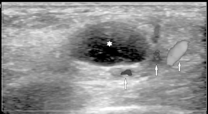
UltrasoundFoot ultrasound: well-circumscribed anechoic synovial cyst, arrows on the posterior tibial artery and vein in direct contact with the cyst in the tarsal tunnel.
Source : Ormeci et al., International Journal of Surgery Case Reports, 2015, figure 2 · CC BY-NC-ND
📋 The detail of a series of 31 operated patients
The fine breakdown of results is a reminder that « a good result » covers varied situations, and that the prognosis depends on the patient's profile.
The best results were in patients with an identified cause, a short delay between symptom onset and surgery, and a positive preoperative Tinel's sign. Source: Reichert et al., Foot and Ankle Surgery 2015 (PMID 25682403).
🔪 After decompression: about three patients in four improved
When conservative treatment fails, surgical release of the tarsal tunnel gives favourable results in most cases, but from series with a low level of evidence.
Pooled results from a scoping review (a single level 3 study and 29 level 4 case series). The reported success rates range very widely from 44 % to 96 % across series. Sources: Haq et al., Journal of Clinical Orthopaedics and Trauma 2024 (PMID 39101044); Kiel et al., StatPearls 2024 (PMID 30020645); Rodríguez-Merchán et al., EFORT Open Reviews 2021 (PMID 35839088).
Tarsal tunnel syndrome is a focal compressive neuropathy of the posterior tibial nerve, or of one of its branches, as it passes under the flexor retinaculum on the medial side of the ankle 3. That retinaculum runs between the medial malleolus and the calcaneus and forms the roof of a fibro-osseous tunnel housing, alongside the nerve, the tendons of tibialis posterior, flexor digitorum longus and flexor hallucis longus, together with the posterior tibial vascular pedicle 5. When conservative treatment fails and a compression is documented, surgical decompression , release of the retinaculum and removal of any mass, becomes the option to discuss. It is a decision rarely taken in a hurry, and never without first having looked for what is compressing the nerve.
Key point. Tarsal tunnel surgery comes only after well-conducted conservative treatment has failed and, ideally, when a compressive cause has been identified. It releases the flexor retinaculum and removes what is crushing the nerve. About three operated patients in four obtain an excellent or good result, but that figure rests on low-level evidence and the published success rates run from 44 % to 96 %. The physiotherapist frames the failure or success of the conservative phase beforehand, then supports sensory and motor recovery after the operation.
Setting the indication: conservative failure and an identified cause
Physiotherapy and conservative treatment remain the first line before any surgery 12. That precondition is not a formality: it shapes both the surgical indication and its prognosis. Conservative care combines activity modification, orthoses and medial arch supports, stretching and strengthening exercises, anti-inflammatory drugs and corticosteroid injections 14. Its effectiveness is real but modest. In one series, anti-inflammatory drugs combined with a night immobilisation splint were enough in only 14 patients out of 46, that is 30.4 %, the rest going on to surgery 12. Conversely, ultrasound-guided injections can avoid the operating theatre: of 218 patients treated first line with an ultrasound-guided injection, 169 (77.5 %) did not need surgical release 13.
The second condition is aetiological. A mechanical cause, a space-occupying mass, tenosynovitis or a hindfoot deformity, is identifiable in about 80 % of cases, the purely idiopathic form being rare 1. Looking for that lesion before concluding is therefore not optional: among the intrinsic causes, varicosities of the posterior tibial vein are found in 20 % of patients, which justifies imaging in search of a compressive mass 5. MRI serves precisely to locate the compression site and any mass; in one series it showed the point of nerve compression in 82.1 % of cases (23 sides out of 28), but failed to reveal all the detail needed for surgical planning 11. Operating on a nerve compressed by an identified ganglion does not mean the same thing as opening a tunnel with no target: surgery repairs all the better when it knows what to remove.
What decompression achieves: results and uncertainty
After conservative treatment fails, surgical decompression of the tarsal tunnel gives an excellent or good result in about 75.3 % of cases, the remaining 24.7 % obtaining a fair or poor result 12. These proportions are consistent with another series of 31 operated patients: 71 % very good or good results, 22 % satisfactory and 7 % poor 16. These are useful orders of magnitude for informing a patient, provided their origin is recalled.
For caution is needed about how solid these figures are. The most recent scoping review retained only one level 3 article and 29 level 4 case series, which does not allow firm conclusions either on prognostic factors or on the superiority of any management 12. That is the structural limit of this whole field: tarsal tunnel syndrome is a rare condition, regularly under-diagnosed, whose clinical evidence for guiding management is limited 4. The most telling consequence lies in the spread of the surgical results: the success rates of decompression range very widely from 44 % to 96 % across series 16. Between those two bounds sit very heterogeneous populations, selection criteria and definitions of success, all reasons not to present any percentage as a promise.
The best results are not decided in theatre but well before: a clear cause, recent symptoms, a young patient, a nerve that still responds.
Who responds well: the prognostic factors
The variability of results is not random; it follows fairly constant lines from one series to another. The best surgical results are obtained in young patients, with a clear aetiology, a positive preoperative Tinel's sign, a short symptom history, an early diagnosis and no previous ankle pathology 6. Tinel's sign percussed at the tarsal tunnel deserves a special place here: a positive test before the operation is a strong predictor of relief after decompression, even though its overall sensitivity remains low and variable, from 25 % to 75 % 1. In other words, it does not serve to make the diagnosis but it helps to predict the response to the procedure.
The favourable and unfavourable sides answer each other. The prognosis depends on the aetiology: patients with an identified cause, a space-occupying mass or previous trauma, obtain better surgical results than idiopathic forms, while associated obesity and diabetes reduce the chances of a good result 14. The prognosis is all the better when a space-occupying lesion, a cyst or a ganglion for example, is identified and then removed; conversely, the idiopathic form, bilateral involvement, nerve fibrosis, a long duration of symptoms, advanced age and comorbidities are associated with poorer results 12. The series of 31 patients confirms that hierarchy: the best results were in patients with an identified cause, a short delay before surgery and a positive preoperative Tinel's sign 16.
| Factor | Direction | Source |
|---|---|---|
| An identified compressive cause (mass, ganglion) removed | Better result | Vij 2022; Haq 2024 |
| A positive preoperative Tinel's sign | A strong predictor of relief | Rodríguez-Merchán 2021; Kiel 2024 |
| A young patient, recent symptoms, an early diagnosis | Better result | Rodríguez-Merchán 2021; Reichert 2015 |
| An idiopathic form, bilateral involvement, nerve fibrosis | Poorer result | Haq 2024 |
| A long duration of symptoms, advanced age, comorbidities | Poorer result | Haq 2024; Vij 2022 |
| Associated obesity and diabetes | Poorer result | Vij 2022 |
The physiotherapist's role after the operation
Decompression lifts the mechanical load on the nerve, but it does not by itself restore function. Sensory and motor recovery, ankle mobility and tolerance to load call for rehabilitation support, the same therapeutic base as conservative treatment, mobilised this time to consolidate the procedure. That base combines restoring joint ranges, strengthening the intrinsic and extrinsic muscles of the foot, managing pain and gradually reintroducing activity 14. The most recent conservative study, although limited to 28 patients, reported an improvement in pain and joint range in all subjects after a six-week physiotherapy programme 14 : a coherent signal, though of low evidential level, in favour of structured management.
The neurodynamic component deserves a mention. Adding neural mobilisation exercises to conservative treatment was tested in a small randomised trial of 28 patients over six weeks: both groups improved in mobility, strength and pain, the mobilisation group additionally gaining on two-point discrimination, light touch and Tinel's sign 15. The effect therefore remains mainly sensory and modest in size, with no demonstrated superiority on range, strength or pain; this is a promising lead, not an established standard. After decompression, these cautious neural glides fit logically into rehabilitation to maintain the excursion of the released nerve, provided healing is respected and the symptom threshold is not crossed.
For practice. Before the operation the physiotherapist runs the conservative treatment which, when it documents failure, validates the surgical indication; they also help to spot a compressive cause to be imaged. After the operation they restore range and strength, manage pain and reintroduce load, and can add gentle neurodynamic mobilisation whose benefit, mainly sensory, remains of low evidential level. In every case the message given to the patient must reflect the uncertainty: about 75 % good results on average, but real rates ranging from 44 % to 96 %, and a prognosis largely dictated by the cause and the earliness of management, not by the operation alone.
🗂️ What do concrete case reports teach us?
Clinical reasoning reads poorly from sensitivity and specificity data alone. To make the approach tangible, sorting plantar pain, ruling out the traps, looking for a space-occupying cause, then running reasoned conservative management, we follow two detailed paths below. They are a reminder that the tarsal tunnel is a compression of the posterior tibial nerve or its branches under the flexor retinaculum, on the medial side of the ankle 3, a rare entity, regularly under-diagnosed, with no definitive test 5.
Read this before starting. The case that follows is published and carries its PubMed identifier: the age, the mechanism and the course are those its authors report. The note markers point to the sources that ground each step of the reasoning.
Published case: a tarsal tunnel that had a cause, and one that had to be looked for
The case. Mix and colleagues report a man aged 17 years referred for an electrodiagnostic assessment for symptoms of tarsal tunnel syndrome, after being stung by a stingray19.
What the electrodiagnosis showed. Moderate axonal involvement, localised to the tarsal tunnel19.
What followed. Surgical exploration removed a stingray barb lodged in the tarsal tunnel. The symptoms were almost entirely resolved at the five-week postoperative review19. The authors conclude that electrodiagnosis, in this patient with delayed recovery, proved a valuable component of management19.
What this case teaches, beyond its exotic nature. The cause here is spectacular, but the reasoning is ordinary and it is the reasoning that counts: an entrapment syndrome that does not recover as expected has a reason, and that reason is often something occupying the space. A mass, a foreign body, varicosities, tenosynovitis, the sequel of a fracture: the tarsal tunnel is a tunnel, and a tunnel fills up. When rehabilitation stalls, the question is not to intensify it but to ask what is inside.
The thread running through the two vignettes , imaginary ones, let us recall, holds in one sentence: the tarsal tunnel is diagnosed by correlating the clinical history, the provoked examination, imaging and, incidentally, electrodiagnosis, no test making the diagnosis with certainty 6. Between Claire's under-diagnosis and the risk of over-diagnosis in Marc, the physiotherapist walks a ridge: the space-occupying cause is looked for systematically 1, conservative treatment is tried first 12, and the uncertainty is stated, because the evidence in this field remains weak.
🧭 How is this applied in practice?
Tarsal tunnel syndrome is a compressive neuropathy of the posterior tibial nerve or its terminal branches, as they pass under the flexor retinaculum on the medial side of the ankle 12. It is a rare condition, regularly under-diagnosed, whose exact incidence remains unknown and for which the clinical evidence remains limited 41. That double characteristic, rarity and the absence of a definitive test, requires a structured approach from the physiotherapist: neither accepting the diagnosis too quickly nor missing it in the patient who truly has it. This section offers a pragmatic algorithm, from clinical reasoning to the moment of referral.
A practical three-step algorithm
No investigation makes the diagnosis with certainty: it rests on the correlation between the clinical history, imaging, nerve conduction studies (NCS) and electromyography (EMG) 6. In practice the reasoning can be structured in three steps.
1. Recognise the picture. Pain, burning, numbness or paraesthesia on the plantar surface of the foot, corresponding to the territory of the tibial nerve and its plantar branches 34. The medial plantar nerve covers the medial arch and the plantar surface of the first three toes, the lateral plantar nerve the lateral arch and the 4th–5th toes: the topography of the symptoms points to the side involved 5. Previous trauma, with up to 43 % of patients reporting an injury, an ankle sprain in particular, strengthens the hypothesis 1.
2. Provoke and localise. Two manoeuvres are useful at the couch. The Tinel's sign (percussion behind the medial malleolus, reproducing pain or tingling in the posterior tibial territory) has modest and variable performance: sensitivity 25 to 75 %, specificity 70 to 90 % 1. The dorsiflexion-eversion test puts the tibial nerve under tension: the ankle is passively taken into maximum eversion and dorsiflexion, all the metatarsophalangeal joints in maximum dorsiflexion, the position held for 5 to 10 seconds 8. In the original study (37 operated patients, 50 healthy volunteers), the manoeuvre reproduced numbness in 15/20 feet (75 %), pain in 15/17 (88 %) and intensified local tenderness in 42/43 affected feet (98 %), while no symptom or sign was induced in the healthy volunteers 8.
3. Correlate and confirm. A negative test does not rule the diagnosis out; a bundle of concordant arguments (history, topography, provocation) supports it. In case of doubt, imaging and electrodiagnosis come in support, but with the limits detailed below.
| Test / investigation | Performance | Practical interpretation |
|---|---|---|
| Dorsiflexion-eversion test 8 | Local tenderness intensified in 42/43 affected feet; no sign in 50 healthy subjects | A reproducible provocation manoeuvre, to be built into every examination |
| Tinel's sign 1 | Sensitivity 25–75 %, specificity 70–90 % | A negative does not exclude; a positive predicts surgical relief |
| Ultrasound 10 | Cross-sectional area threshold 15 mm²: Se 74 %, Sp 100 % | Documents nerve enlargement; useful for looking for a mass |
| MRI 11 | Compression point seen in 23/28 sides (82.1 %) | Locates the site and a mass; insufficient alone for planning |
| Electroneuromyography, NCS/EMG 19 | Suboptimal sensitivity and specificity, frequent false negatives | Normal does not exclude; a level C recommendation |
Do not over-diagnose
The commonest error is to accept the tarsal tunnel for any plantar pain. Yet the condition is rare and often under-diagnosed, and its exact incidence is unknown: that observation should make the physiotherapist cautious before making it the explanation for heel or foot pain 15. First described in 1962, this syndrome suffers from the absence of a definitive clinical or imaging test, and there is no diagnostic gold standard 57.
The reasoning therefore has to pass through the differential diagnosis. The entities to rule out first are plantar fasciitis, lumbosacral radiculopathy (S1 in particular), rheumatological disease, metatarsal stress fractures and Morton's neuroma 6. The difficulty is increased by the overlap with other lower limb disorders, particularly in a patient with diabetes and a distal sensory polyneuropathy, where the diffuse nerve involvement blurs the picture 7.
Faced with plantar pain, the question is not « is this a tarsal tunnel? » but « what else could it be, and have I looked for a cause compressing the nerve? »
Systematically look for a space-occupying cause
This is the pivot of management. An impingement mechanism is identifiable in about 80 % of cases, the purely idiopathic form being rare 1. The aetiology is varied: space-occupying lesions (ganglion cysts, neuromas, lipomas), biomechanical abnormalities (valgus or varus hindfoot), a post-traumatic origin and systemic causes 16. Among the intrinsic lesions, varicosities of the posterior tibial vein are found in 20 % of patients, which on its own justifies imaging in search of a compressive mass 5. The systemic causes to ask about are, first of all, diabetes, but also hypothyroidism, rheumatoid arthritis and obesity 5.
The stake is not only diagnostic, it is prognostic: patients with an identified cause (a space-occupying mass or previous trauma) obtain better results than idiopathic forms, while associated obesity and diabetes reduce the chances of a good result 14. Neglecting the search for a mass risks rehabilitating indefinitely a mechanical compression that calls for surgical removal.
What can be offered in rehabilitation
Conservative treatment is the first line before surgery and combines activity modification, orthoses and medial arch supports, wedges, stretching and strengthening exercises, NSAIDs and corticosteroid injections 146. Its effectiveness remains modest and rests on weak evidence: the overall review is level IV, essentially case series 12.
On the physiotherapy side proper, the data are limited but encouraging. A small randomised trial (28 patients, 6 weeks) tested the addition of neurodynamic mobilisation exercises to conservative treatment: both groups improved (mobility, strength, pain), with no between-group difference on those parameters, but the mobilisation group additionally gained on two-point discrimination, light touch and Tinel's sign 1514. The ultrasound-guided injection (anaesthetic + corticosteroid) is an interesting conservative option: of 218 patients treated first line with an ultrasound-guided injection, 169 (77.5 %) did not need surgical release 13.
When to refer: electroneuromyography, imaging and a surgical opinion
For electroneuromyography. Its role remains controversial and it must be requested in full knowledge of its limits. Electrodiagnosis is often abnormal but its sensitivity and specificity are suboptimal, with frequent false negatives: a normal test does not rule the diagnosis out 1. The results are all the more often normal when the forms are mild or have built up gradually over years, so that repeated studies may be needed 5. Above all, electroneuromyography cannot predict which cases will respond to surgical decompression 3. It confirms a tibial neuropathy at the ankle (a level C recommendation) but does not replace clinical reasoning 9.
For imaging. Ultrasound documents the increased cross-section of the tibial nerve (a 15 mm² threshold: 74 % sensitivity, 100 % specificity; a ΔCSA of 5 mm²: 81 % / 100 %) and MRI locates the compression site and any mass in 82.1 % of cases 1011. Imaging is indicated as soon as a space-occupying lesion is suspected, so in practice widely.
For a surgical opinion. After conservative treatment fails, decompression gives excellent or good results in about 75.3 % of cases, the remaining 24.7 % having a fair or poor result, figures from series with a low level of evidence 12. The reported success rates range very widely from 44 % to 96 % 16. Selection therefore matters: the best results are in young patients, with a clear aetiology, a positive preoperative Tinel's sign, a short symptom history and an early diagnosis, with no previous ankle pathology 6. Conversely, the idiopathic form, bilateral involvement, nerve fibrosis, a long duration, advanced age and comorbidities herald poorer results 12. Referring early the patient whose cause is identified and whose Tinel is positive offers them their best therapeutic window.
Common mistakes to avoid
- Accepting the diagnosis on a single test. Neither Tinel nor electroneuromyography is decisive in isolation; it is the clinical, imaging and electrodiagnostic correlation that counts 6.
- Ruling the diagnosis out on a negative test. A negative Tinel, a normal electroneuromyography or a weak provocation do not exclude the condition 15.
- Forgetting to look for a mass. In 80 % of cases a cause is identifiable, and its presence changes the prognosis: not imaging means missing a removable ganglion or varicosity 114.
- Neglecting the differential. Plantar fasciitis, S1 radiculopathy, Morton's neuroma and a stress fracture mimic the picture 6.
- Ignoring the comorbidities. Diabetes, hypothyroidism, rheumatoid arthritis and obesity take part in the genesis and weigh on the prognosis 514.
- Referring too late. A long duration degrades the surgical results 12.
Key points
- Posterior tibial nerve compression under the flexor retinaculum, medial side of the ankle; plantar symptoms 31.
- Rare and under-diagnosed : do not make it the default diagnosis for any plantar pain; rule out fasciitis, S1 radiculopathy, Morton's neuroma and a stress fracture 16.
- Look for a space-occupying cause : identifiable in ~80 % of cases; varicosities in 20 %; image when in doubt 15.
- Imperfect clinical tests : dorsiflexion-eversion useful and reproducible, Tinel modest; no normal test excludes the diagnosis 81.
- Conservative first (weak evidence): orthoses, exercise, neurodynamic mobilisation, ultrasound-guided injection (77.5 % without surgery) 121513.
- Refer : electroneuromyography in support despite its limits (it neither excludes nor predicts); a surgical opinion after conservative failure, ~75 % good results, better with a clear cause, a positive Tinel and early management 3126.
Bibliography
Every reference checked individually on PubMed (clickable PMID). 18 sources. Click a superscript note in the text: the bibliography opens and highlights the source.
- Kiel J (2024). StatPearls [Internet], StatPearls Publishing. PMID 30020645.
- Doneddu PE (2017). Neurological Sciences. PMID 28664501. doi:10.1007/s10072-017-3039-x.
- Ahmad M (2012). Foot and Ankle Surgery. PMID 22857954. doi:10.1016/j.fas.2011.10.007.
- McSweeney SC (2015). The Foot (Edinburgh). PMID 26546070. doi:10.1016/j.foot.2015.08.008.
- Sha I I (2024). The Iowa Orthopaedic Journal. PMID 39811161.
- Rodríguez-Merchán EC (2021). EFORT Open Reviews. PMID 35839088. doi:10.1302/2058-5241.6.210031.
- Boers N (2026). JBJS Reviews. PMID 41662474. doi:10.2106/JBJS.RVW.25.00222.
- Kinoshita M (2001). The Journal of Bone and Joint Surgery. American Volume. PMID 11741063. doi:10.2106/00004623-200112000-00011.
- Patel AT (2005). Muscle & Nerve. PMID 16003732. doi:10.1002/mus.20393.
- Fantino O (2021). Orthopaedics & Traumatology, Surgery & Research. PMID 32682728. doi:10.1016/j.otsr.2020.02.021.
- Kim K (2022). Neurologia medico-chirurgica. PMID 36184477. doi:10.2176/jns-nmc.2022-0118.
- Haq II (2024). Journal of Clinical Orthopaedics and Trauma. PMID 39101044. doi:10.1016/j.jcot.2024.102489.
- Atesok K (2022). Orthopedic Reviews (Pavia). PMID 35769651. doi:10.52965/001c.35455.
- Vij N (2022). Orthopedic Reviews. PMID 36072502. doi:10.52965/001c.37539.
- Kavlak Y (2011). Journal of Manipulative and Physiological Therapeutics. PMID 21875518. doi:10.1016/j.jmpt.2011.05.017.
- Reichert P (2015). Foot and Ankle Surgery. PMID 25682403. doi:10.1016/j.fas.2014.08.013.
- Sodani A (2018). Annals of Indian Academy of Neurology. PMID 29720796. doi:10.4103/aian.AIAN_320_17.
- Yammine K (2022). Surgical and Radiologic Anatomy. PMID 35353216. doi:10.1007/s00276-022-02932-9.
- Mix FM, Sucher BM, Brooks JE (2021). Stingray-Induced Tarsal Tunnel Syndrome. Cureus. PMID 34513381. doi:10.7759/cureus.16761.
❓ Frequently asked questions
What is tarsal tunnel syndrome?
It is a focal compressive neuropathy (entrapment) of the posterior tibial nerve, or of one of its branches, as it passes through the tarsal tunnel under the flexor retinaculum, on the medial side of the ankle behind the medial malleolus. It is described as the lower limb equivalent of carpal tunnel syndrome. The symptoms affect the plantar border of the foot. It is a rare condition whose exact incidence remains unknown, regularly under-diagnosed, and for which the clinical evidence guiding management remains limited 143.
What are the causes?
The aetiology is very varied. A mechanical cause (an impingement mechanism) is identifiable in about 80 % of cases: space-occupying lesions (ganglion cysts, neuromas, lipomas), varicosities of the posterior tibial vein found in 20 % of patients, biomechanical abnormalities such as a valgus hindfoot, previous trauma, with up to 43 % of patients reporting an injury, an ankle sprain in particular. Systemic causes are associated: diabetes first among them, but also hypothyroidism, rheumatoid arthritis and obesity. In about 20 % of cases no cause is found, and the form is then called idiopathic 156.
How is the diagnosis made?
The diagnosis rests above all on the clinical picture, because there is no gold standard test and the clinical diagnosis lacks objectivity and reproducibility. Tinel's sign percussed behind the medial malleolus has a low and variable sensitivity of 25 to 75 % for a specificity of 70 to 90 %: a negative test therefore does not rule the diagnosis out. The dorsiflexion-eversion test serves as a reproducible provocation manoeuvre. The final diagnosis rests on the correlation between the clinical history, imaging (ultrasound, MRI) and electrodiagnosis (nerve conduction studies and EMG) 186.
What does the dorsiflexion-eversion test involve?
It is a clinical provocation manoeuvre that puts the tibial nerve under tension: the ankle is passively taken into maximum eversion and dorsiflexion while all the metatarsophalangeal joints are held in maximum dorsiflexion, and the position is held for 5 to 10 seconds. In the original study of 37 operated patients and 50 healthy volunteers, the manoeuvre reproduced numbness in 15 feet out of 20 (75 %), pain in 15 out of 17 (88 %) and intensified local tenderness in 42 of the 43 affected feet (98 %); conversely, no symptom or sign could be induced in the healthy volunteers 8.
Is electromyography (EMG) reliable for confirming the diagnosis?
Its contribution is limited and debated. Electrodiagnosis (EMG and nerve conduction studies) is often abnormal, but its sensitivity and specificity are suboptimal and false negatives are frequent: the results are often normal, particularly in mild forms or when symptoms build up gradually over years, so that repeated studies may be needed. The test is also unable to predict which cases will respond to surgical decompression. A normal electrodiagnosis therefore does not rule the diagnosis out, and the diagnosis remains above all clinical 153.
How is tarsal tunnel syndrome treated?
Conservative treatment comes first: activity modification, orthoses and medial arch supports, wedges, stretching and strengthening exercises, NSAIDs and corticosteroid injections; the level of evidence remains low. Ultrasound-guided injection is an effective option: of 218 patients treated first line, 169 (77.5 %) did not need surgery. After conservative treatment fails, surgical decompression gives an excellent or good result in about 75.3 % of cases, but the reported success rates vary widely from 44 % to 96 %. A positive preoperative Tinel's sign, an identified cause, young age and a short history are good predictors of relief 1413121.
Also worth reading in the review

