















In brief
Plantar fasciitis, more accurately called plantar fasciopathy, is the leading cause of heel pain in adults: it is a degenerative rather than inflammatory process affecting the plantar fascia at the heel. It presents as sharp pain on the first steps in the morning, reproduced by palpating the medial calcaneal tubercle; the main risk factors are a high BMI, reduced ankle dorsiflexion and prolonged standing. First-line management combines education, fascia-specific stretching and orthoses, with high-load strengthening superior to stretching at three months. Lifetime prevalence is about 1 person in 10.
Clinical synthesis based on the JOSPT 2023 Clinical Practice Guideline (Koc TC Jr et al.), the BJSM 2016-2023 meta-analyses (van Leeuwen, Whittaker, Hamstra-Wright) and the FIX-Heel RCT (Riel 2023).
Clinical summary
- In practice, plantar fasciopathy (a more accurate term than “fasciitis”) is the leading cause of heel pain in adults. Histological studies (Lemont 2003) confirm that it is a degenerative and not an inflammatory process : disorganisation of the collagen fibres, myxoid degeneration, neovascularisation.
- Three risk factors carry a strong level of evidence : a high BMI (van Leeuwen 2016: RR ≈ 2.5 where BMI > 30), reduced ankle dorsiflexion and prolonged occupational standing. Runners and military personnel are at-risk subgroups (Hamstra-Wright 2021).
- The course is generally self-limiting : 80 % of patients recover at 12 months with conservative treatment (Trojian 2019). One third nonetheless develop pain that persists beyond 12 months, which is why early, structured management matters.
- Diagnosis is clinical according to the JOSPT CPG 2023 (Koc et al.) : pain on the first steps in the morning, palpation reproducing the pain at the medial calcaneal tubercle. The Windlass test has high specificity but low sensitivity.
- Imaging is not recommended routinely, but reserved for atypical cases, red flags, or failure after 3-6 months of well-conducted conservative treatment. A fascia thickness > 4 mm on ultrasound is compatible with the diagnosis but correlates poorly with pain.
- Differential diagnoses to rule out systematically: S1 radiculopathy, tarsal tunnel syndrome, calcaneal stress fracture, fat pad atrophy, Baxter's nerve entrapment, and, rare but serious, soft-tissue tumours.
- First-line treatment (Grade A, Koc 2023): patient education on load management, fascia-specific stretching (DiGiovanni 2006), prefabricated or custom orthoses (Whittaker 2018), anti-pronation taping in the short term.
- Beyond that, high-load strengthening (heavy slow resistance) is superior to stretching alone at 3 months (Rathleff 2015, BJSM 2019). Protocol: single-leg heel raises on a rolled towel under the toes, 3 × 8 repetitions ⇒ 3 × 12 repetitions, 3 times a week.
- For refractory chronic cases (> 6 months): extracorporeal shockwave therapy (ESWT) (Grade A), corticosteroid injection (short-term effect only, with a risk of rupture and fat pad atrophy ⇒ use sparingly). FIX-Heel trial (Riel 2023, BJSM): adding the injection to strengthening does no better than education plus orthosis alone at 12 weeks.
- Return to sport: functional criteria before the calendar (walking 30-60 min without pain, 10 pain-free single-leg hops). The pain rule: do not exceed 3/10 during the exercise, with no persistence the following morning.
- Red flags (Finucane 2020): intense night pain, progressive neurological signs, a history of cancer, fever, suspected fracture (a fall, BSI). Yellow flags (Cotchett 2016): catastrophising, kinesiophobia ⇒ a biopsychosocial approach.
- Measure outcomes with validated PROMs (FAAM, FFI, FHSQ) and aim for the MCID. Evidence-based practice demands continuing education and access to resources, but remains constrained by time and by patients' expectations of passive modalities.
Contents
- What are the fundamentals to know about plantar fasciitis?
- How do you assess and diagnose plantar fasciitis with certainty?
- Which treatment strategies are the most effective for plantar fasciitis?
- How do you secure lasting recovery and prevent recurrence of plantar fasciitis?
- What is specific to plantar fasciopathy in runners and in at-risk subgroups?
- What do real clinical cases teach us about plantar fasciitis?
- How do you apply these recommendations concretely in your practice?
What are the fundamentals to know about plantar fasciitis?
How is this condition defined, who does it affect and what are the risk factors?
Plantar fasciopathy is heel pain arising from the plantar fascia at its insertion on the medial calcaneal tubercle. The pain is typically sharp on the first step in the morning or after a prolonged period of rest, then eases after a few minutes of walking before returning at the end of the day.¹ ² The lifetime prevalence is estimated at 1 person in 10 in the American Family Physician 2019 review.¹ It accounts for about 15 % of the foot conditions presenting in primary care.¹ ² The peak age falls between 40 and 60 years, with a bimodal distribution: overweight middle-aged sedentary people, and runners of all ages.²📊 Risk factors for plantar fasciopathy: levels of evidence
van Leeuwen 2016 (BJSM) and Hamstra-Wright 2021 (Sports Health) meta-analyses: pooled odds ratios (point estimate + 95 % CI)
Three factors carry a strong level of evidence: high BMI, reduced dorsiflexion, prolonged standing. The radiographic calcaneal spur is no longer considered a significant risk factor: it is present in about 20 % of the asymptomatic population (van Leeuwen 2016).
Certain subgroups are particularly exposed: runners (peaking in distance runners, Hamstra-Wright 2021),⁹ military personnel (uneven ground, load carriage), workers on hard floors (checkout staff, construction workers, care staff).¹ ⁴ These subgroups are covered in the dedicated chapter further on.
What happens in the body, and how does plantar fasciitis evolve naturally?
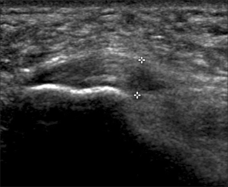
UltrasoundLongitudinal ultrasound of the heel: markedly thickened plantar fascia, measured between the callipers, suggestive of plantar fasciitis.
Source : Park et al., Annals of Rehabilitation Medicine, 2014, figure 2 · CC BY-NC
The natural course is broadly favourable but slow. The classic estimates (Trojian 2019; Koc CPG 2023) indicate complete resolution in about 80 % of patients at 12 months under conservative treatment.¹ ⁵ However, one third of cases progress to pain persisting beyond a year, which is why early, structured management matters.¹ ⁵
Key points
- The term plantar fasciopathy is more precise than “fasciitis”: this is a degenerative condition, not an inflammatory one (Lemont 2003).
- The three risk factors with strong evidence are: a high BMI (van Leeuwen 2016), limited dorsiflexion and prolonged standing.
- A favourable but slow course: 80 % resolution at 12 months, but 1 patient in 3 may develop pain persisting beyond 1 year.
- The radiographic calcaneal spur is an adaptive sign, not the cause of the pain: 20 % of asymptomatic people have one.
Bibliography, chapter 1
- Trojian T, Tucker AK. Plantar Fasciitis. Am Fam Physician. 2019;99(12):744-750. PMID 31194492
- Buchanan BK, Sina RE, Kushner D. Plantar Fasciitis. StatPearls [Internet]. Treasure Island (FL): StatPearls Publishing; 2024. NBK431073
- Lemont H, Ammirati KM, Usen N. Plantar fasciitis: a degenerative process (fasciosis) without inflammation. J Am Podiatr Med Assoc. 2003;93(3):234-237. PMID 12756315
- Martin RL, Davenport TE, Reischl SF, et al. Heel pain-plantar fasciitis: revision 2014. J Orthop Sports Phys Ther. 2014;44(11):A1-A33. PMID 25361863
- Koc TC Jr, Bise CG, Neville C, Carreira D, Martin RL, McDonough CM. Heel Pain - Plantar Fasciitis: Revision 2023. Clinical Practice Guidelines. J Orthop Sports Phys Ther. 2023;53(12):CPG1-CPG39. DOI 10.2519/jospt.2023.0303
- Latt LD, Jaffe DE, Tang Y, Taljanovic MS. Evaluation and Treatment of Chronic Plantar Fasciitis. Foot Ankle Orthop. 2020;5(1):2473011419896763. DOI 10.1177/2473011419896763
- van Leeuwen KDB, Rogers J, Winzenberg T, van Middelkoop M. Higher body mass index is associated with plantar fasciopathy/'plantar fasciitis': systematic review and meta-analysis of various clinical and imaging risk factors. Br J Sports Med. 2016;50(16):972-981. PMID 26644427
- Hamstra-Wright KL, Huxel Bliven KC, Bay RC, Aydemir B. Risk Factors for Plantar Fasciitis in Physically Active Individuals: A Systematic Review and Meta-analysis. Sports Health. 2021;13(3):296-303. PMID 33530860
- Cotchett M, Munteanu SE, Landorf KB. Depression, Anxiety, and Stress in People With and Without Plantar Heel Pain. Foot Ankle Int. 2016;37(8):816-821. PMID 27137796
How do you assess and diagnose plantar fasciitis with certainty?
Diagnosing plantar fasciopathy is essentially clinical according to the JOSPT 2023 Clinical Practice Guideline (Koc TC Jr et al.).¹ ² Imaging is not recommended routinely: it is reserved for atypical presentations, suspected red flags, or failure after 3 to 6 months of well-conducted conservative treatment.¹ ³
Which questions should you ask to understand the patient and their history?
The history seeks to recognise the characteristic clinical pattern and to identify modifiable risk factors:
- Pain on the first steps : the most discriminating sign. Sharp, stabbing pain in the morning or after any prolonged rest (sitting through a film, a long car journey). The pain fades within a few minutes, then can return at the end of the day.¹ ²
- Location : the medial calcaneal tubercle (anteromedial heel). Diffuse or night pain should raise another aetiology.¹ ³
- Triggering factors : an abrupt increase in activity (running, walking, hiking), a change of footwear, weight gain, a new standing job.⁴ ⁵
- Risk factors : BMI, occupation, sport practised (running above all), a history of heel pain on the other side.⁵ ⁶
- Psychosocial factors : Cotchett 2016 shows a significant association between plantar fasciopathy and catastrophising / kinesiophobia, to be screened with the TSK-11 or the PCS in chronic cases.⁷
Which clinical tests should you perform, and which other conditions should you rule out?
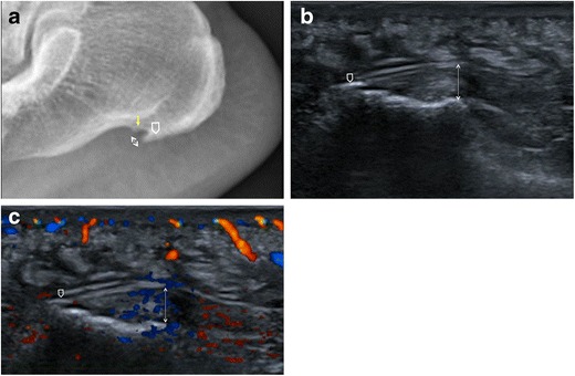
UltrasoundLateral foot: X-ray showing an inferior calcaneal spur (a), ultrasound revealing a thickened, hypoechoic plantar fascia (b), with hypervascularity on colour Doppler (c).
Source : Draghi et al., Insights into Imaging, 2017, figure 4 · CC BY
The physical examination aims to reproduce the patient's pain and to exclude the differential diagnoses. No single test is sufficient; it is the convergence of the picture that makes the diagnosis.²
- Palpation of the medial calcaneal tubercle : the most sensitive test. Immediate reproduction of sharp pain on digital pressure.¹ ²
- Windlass test : passive dorsiflexion of the toes (the great toe in particular) with the patient weight-bearing. Positive if it reproduces the heel pain. High specificity but low sensitivity : a negative test does not exclude the diagnosis.¹ ²
- Assessment of ankle dorsiflexion : range < 10° with the knee extended = a risk factor and a treatment target.¹ ⁴ ⁵
- Neural tension tests (SLR / slump) : to rule out S1 radiculopathy.³ ⁸
- Retromalleolar Tinel test : to rule out tarsal tunnel syndrome (compression of the posterior tibial nerve).³ ⁸
🔀 Diagnostic algorithm for plantar heel pain
Adapted from the JOSPT CPG 2023 (Koc et al.) and the Finucane 2020 red-flag framework
Imaging (ultrasound, MRI) does not change the initial management. The sensitivity of ultrasound for measuring fascia thickness (> 4 mm) is about 80 %, but its correlation with pain is weak.¹ ³
Critical differential diagnoses to rule out
- S1 radiculopathy : pain radiating from the buttock to the sole, motor deficit (plantar flexion), absent Achilles reflex, positive SLR.³ ⁸
- Tarsal tunnel syndrome : burning or tingling in the sole, positive medial retromalleolar Tinel, sometimes at night.³ ⁸
- Calcaneal stress fracture : a history of abruptly increased activity, diffuse pain, positive lateral compression (“squeeze”) test. Think of a BSI in a runner with hypogonadism or energy deficiency.³
- Plantar fat pad atrophy : an older patient, central plantar heel pain, a soft surface that cushions less well. A clinical diagnosis.¹
- Baxter's nerve entrapment (1st branch of the lateral plantar nerve): pain mimicking fasciopathy, palpation more lateral, possible weakness of abduction of the 5th toe.³
- Rare but not to be missed : soft-tissue tumours, osteomyelitis, seronegative arthritis (spondyloarthritis where heel pain is bilateral in a young adult).¹ ³
Should patients with plantar fasciopathy be classified, and what are the benefits?
The JOSPT CPG 2023 (Koc) incorporates the ICF classification (International Classification of Functioning) and acknowledges that patients are not homogeneous. Without formal validation of therapeutic subtypes, several clinical profiles emerge:⁴ ⁵
- The “mechanical overload” profile : an athlete, a suddenly increased load, limited dorsiflexion. Treatment target: load management + HSR strengthening.
- The “weight” profile : a high BMI, prolonged standing, a flattened biomechanics. Target: orthoses + weight loss + non-impact aerobic activity.⁵ ⁶
- The “chronic pain” profile : pain > 6 months, kinesiophobia, catastrophising. Target: therapeutic education + a biopsychosocial approach + ESWT if refractory.⁷
Key points
- Diagnosis is clinical (JOSPT CPG 2023, Koc): pain on the first steps + reproducible palpation of the medial tubercle.
- The Windlass test has high specificity but low sensitivity: a negative test does not exclude the diagnosis.
- Imaging is not recommended routinely ; it is reserved for atypical cases or failure at 3-6 months.
- Always rule out: S1 radiculopathy, tarsal tunnel, calcaneal stress fracture, and Baxter's nerve entrapment.
- The yellow flags (catastrophising, kinesiophobia, depression, Cotchett 2016) should be screened for in chronic cases.
Bibliography, chapter 2
- Koc TC Jr, Bise CG, Neville C, Carreira D, Martin RL, McDonough CM. Heel Pain - Plantar Fasciitis: Revision 2023. Clinical Practice Guidelines. J Orthop Sports Phys Ther. 2023;53(12):CPG1-CPG39. DOI 10.2519/jospt.2023.0303
- Martin RL, Davenport TE, Reischl SF, et al. Heel pain-plantar fasciitis: revision 2014. J Orthop Sports Phys Ther. 2014;44(11):A1-A33. PMID 25361863
- Buchanan BK, Sina RE, Kushner D. Plantar Fasciitis. StatPearls [Internet]. 2024. NBK431073
- Latt LD, Jaffe DE, Tang Y, Taljanovic MS. Evaluation and Treatment of Chronic Plantar Fasciitis. Foot Ankle Orthop. 2020;5(1):2473011419896763. DOI 10.1177/2473011419896763
- Trojian T, Tucker AK. Plantar Fasciitis. Am Fam Physician. 2019;99(12):744-750. PMID 31194492
- van Leeuwen KDB, Rogers J, Winzenberg T, van Middelkoop M. Higher BMI is associated with plantar fasciopathy: SR/MA. Br J Sports Med. 2016;50(16):972-981. PMID 26644427
- Cotchett M, Munteanu SE, Landorf KB. Depression, Anxiety, and Stress in People With and Without Plantar Heel Pain. Foot Ankle Int. 2016;37(8):816-821. PMID 27137796
- Finucane LM, Downie A, Mercer C, et al. International Framework for Red Flags for Potential Serious Spinal Pathologies. J Orthop Sports Phys Ther. 2020;50(7):350-372. PMID 32438853
Which treatment strategies are the most effective for plantar fasciitis?
The JOSPT CPG 2023 (Koc TC Jr et al.) is the current international reference.¹ It carries a major update of the 2014 CPG (Martin) and ranks the interventions using the GRADE system.
Where do you start? What is the recommended hierarchy of interventions?
The Grade A recommendations of the 2023 CPG are:¹
- Patient education : explaining the condition (degenerative, not inflammatory), a favourable but slow prognosis, and the principle of optimal load management.¹ ²
- Plantar fascia-specific stretching (the DiGiovanni 2006 protocol): preferable to stretching the triceps surae alone. To be done 3 times a day, 10 repetitions of 10 seconds, above all on getting up.²
- Anti-pronation taping : a well-documented short-term analgesic effect (3 weeks).¹
- Foot orthoses (prefabricated or custom): moderate evidence of efficacy at 2-12 weeks (Whittaker 2018, meta-analysis of 19 RCTs, 1660 participants).³ No superiority of custom over prefabricated orthoses has been demonstrated.³ ⁴
- Manual therapy plus exercise (Grade B): a positive effect on ankle mobility and on pain in the short term.¹
🏗️ Treatment pyramid for plantar fasciopathy (JOSPT CPG 2023)
Ranked by frequency of use and level of evidence: base = universal interventions; apex = interventions targeted at refractory cases
Universal · Grade A · indispensable for everyone
Grade A · DiGiovanni 2006 foot-specific protocol
Superior to stretching at 3 months · Rathleff 2015 RCT
Grade A short/medium term · Whittaker 2018 meta-analysis
Chronic cases > 6 months · Grade A · 3-5 sessions
Reserved · Risk of rupture and atrophy · FIX-Heel 2023: no added value over exercise alone
The base of the pyramid (education + optimal load + exercise) covers the great majority of patients. The interventions at the apex (ESWT, injection, surgery) remain reserved for well-selected refractory chronic cases.
What is the place of exercise, and is there a superior approach?
Rathleff's randomised controlled trial (2015, Scand J Med Sci Sports) was a turning point.⁵ 48 patients with fasciopathy confirmed on ultrasound were randomised to two arms: fascia-specific stretching (DiGiovanni) or high-load strengthening (HSR). At 3 months, the HSR group had an FFI score 29 points lower (a clinically and statistically significant superiority). At 12 months the two groups converged, so HSR delivers a faster recovery without additional long-term benefit.⁵
The HSR protocol, Rathleff 2015:
- Position: standing single-leg heel raise, toes in dorsiflexion over a rolled towel (to put the fascia under tension through the windlass effect).
- Slow tempo: 3 seconds concentric phase (up), 2 seconds isometric at the top, 3 seconds eccentric (down).
- Progression: weeks 1-2 = 3 × 12 repetitions at 12RM. Weeks 3-4 = 4 × 10 repetitions at 10RM (wearing a weighted backpack). Weeks 5-8 = 5 × 8 repetitions at 8RM. Beyond that: maintain 3 sessions a week.
- Frequency: every other day. Acceptable pain during the exercise ≤ 3-5/10.⁵ ⁶
The FIX-Heel RCT (Riel 2023, BJSM, n = 180) compared advice plus a heel cup ± exercise ± corticosteroid injection. At 12 weeks the three arms showed similar improvement: neither exercise (on a background of education) nor the addition of an injection outperformed education alone on the primary outcome.⁷ A nuanced reading: education is powerful; exercise brings a clinical benefit but a moderate one; corticosteroids bring no added value and expose the patient to adverse effects.
Manual therapies, shockwave, injections: how effective are they really?
| Intervention | Effect on pain | Level of evidence (GRADE) | Indication |
|---|---|---|---|
| Education + load management | +++ | High (Grade A) | All patients, the base |
| Fascia stretching (DiGiovanni) | ++ | High (Grade A) | First line |
| HSR strengthening (Rathleff) | +++ | Moderate (Grade A) | Faster recovery at 3 months |
| Prefabricated / custom orthoses | ++ | Moderate | Short/medium term (2-12 weeks) |
| Anti-pronation taping | ++ | Moderate | Short-term effect (3 weeks) |
| Manual therapy as an adjunct | + | Moderate (Grade B) | Limited ankle mobility |
| ESWT (shockwave) | ++ | Moderate-High (Grade A) | Chronic cases > 6 months |
| Dry needling | + | Low | Exploratory adjunct |
| Corticosteroid injection | ++ short term only | Low (risks) | Conservative failure, use sparingly |
| PRP (platelet-rich plasma) | + | Low (heterogeneous) | Refractory cases, not routinely recommended |
| Ultrasound / LLLT (laser) | ± (highly variable) | Very low | Not recommended first line |
| Surgery (partial release) | ++ (but risky) | Low | Strict failure of 12 months of conservative treatment |
ESWT is the best-validated second-line option for chronic cases of more than 6 months.¹ Focused or radial shockwave (3 to 5 weekly sessions) shows significant superiority over placebo in several recent meta-analyses.¹
Corticosteroid injection gives marked relief at 1-4 weeks, but the effect has gone by 3-6 months.⁷ The risk of fascia rupture and of fat pad atrophy calls for sparing use and limits repetition (a maximum of 1 to 2 injections).¹ The FIX-Heel trial 2023 confirmed the absence of added value at 12 weeks over exercise plus education.⁷
Beyond the physical: how do you educate the patient and address psychological factors?
Therapeutic education is the first recommendation of the 2023 CPG.¹ It should cover:
- Reconceptualisation : fasciopathy is a degenerative fasciosis, not acute inflammation (Lemont 2003).⁸ ⇒ Move away from the “rest + NSAIDs” logic.
- Prognosis : 80 % recovery at 12 months, but slowly, so prepare the patient for a horizon of 3 to 6 months.¹
- Load management : the principle of an “optimal zone” of stimulation (neither too much nor too little). The progressive load concept borrowed from Gabbett 2016.
- Demystifying pain : pain does not equal active tissue damage. Keep moving below the 3/10 threshold.⁹
- Addressing beliefs : “I have a spur, it needs removing” (false), “complete rest is needed” (false), “injections will cure it” (a limited effect).
In chronic cases with catastrophising or kinesiophobia (Cotchett 2016), a strengthened biopsychosocial approach is needed, possibly including CBT or a referral.⁹
Key points
- First line (Grade A): education + fascia-specific stretching + orthosis + taping.
- The high-load strengthening (HSR) protocol of Rathleff 2015 delivers faster recovery at 3 months than stretching alone.
- Note that custom orthoses are no better than prefabricated ones (Whittaker 2018), so prefer prefabricated models on cost.
- ESWT is validated (Grade A) for chronic cases > 6 months where conservative treatment has failed.
- By contrast, corticosteroid injections have only a short-term effect and carry a risk of rupture and atrophy, so use them sparingly. FIX-Heel (Riel 2023) shows no added value over exercise plus education.
- Passive modalities (ultrasound, laser) have limited efficacy and must not replace an active approach.
Bibliography, chapter 3
- Koc TC Jr, Bise CG, Neville C, Carreira D, Martin RL, McDonough CM. Heel Pain - Plantar Fasciitis: Revision 2023. J Orthop Sports Phys Ther. 2023;53(12):CPG1-CPG39. DOI 10.2519/jospt.2023.0303
- DiGiovanni BF, Nawoczenski DA, Malay DP, et al. Plantar fascia-specific stretching exercise improves outcomes in patients with chronic plantar fasciitis. Prospective clinical trial with two-year follow-up. J Bone Joint Surg Am. 2006;88(8):1775-1781. PMID 16882901
- Whittaker GA, Munteanu SE, Menz HB, Tan JM, Rabusin CL, Landorf KB. Foot orthoses for plantar heel pain: a systematic review and meta-analysis. Br J Sports Med. 2018;52(5):322-328. PMID 28935689
- Martin RL, Davenport TE, Reischl SF, et al. Heel pain-plantar fasciitis: revision 2014. J Orthop Sports Phys Ther. 2014;44(11):A1-A33. PMID 25361863
- Rathleff MS, Mølgaard CM, Fredberg U, et al. High-load strength training improves outcome in patients with plantar fasciitis: A randomized controlled trial with 12-month follow-up. Scand J Med Sci Sports. 2015;25(3):e292-e300. PMID 25145882
- Trojian T, Tucker AK. Plantar Fasciitis. Am Fam Physician. 2019;99(12):744-750. PMID 31194492
- Riel H, Jensen MB, Olesen JL, Vicenzino B, Rathleff MS. Does a corticosteroid injection plus exercise or exercise alone add to the effect of patient advice and a heel cup for patients with plantar fasciopathy? A randomised clinical trial (FIX-Heel). Br J Sports Med. 2023;57(18):1180-1186. PMID 37414460
- Lemont H, Ammirati KM, Usen N. Plantar fasciitis: a degenerative process (fasciosis) without inflammation. J Am Podiatr Med Assoc. 2003;93(3):234-237. PMID 12756315
- Cotchett M, Munteanu SE, Landorf KB. Depression, Anxiety, and Stress in People With and Without Plantar Heel Pain. Foot Ankle Int. 2016;37(8):816-821. PMID 27137796
How do you secure lasting recovery and prevent recurrence of plantar fasciitis?
The recurrence rate of plantar fasciopathy remains poorly quantified in the literature, but clinical experience and the JOSPT CPG 2023 stress that prevention rests on three pillars: patient self-management, a maintained exercise programme, and progressive load management on returning to activity.¹
How do you make the patient an active participant in their recovery through self-management?
Self-management rests on four main components:
- Understanding load management : fasciopathy is a response to a load that exceeds the current capacity of the fascia. The patient learns to modulate their activities to stay below the irritation threshold, while progressively increasing tissue tolerance.² ³
- Home exercises : a programme combining specific stretching (DiGiovanni 2006) and high-load strengthening (Rathleff 2015) is superior to stretching alone. Adherence is the major predictor of success.⁴ ⁵
- Self-monitoring with the 3/10 rule : pain must not exceed 3 out of 10 during the exercise, and any pain provoked must settle quickly without persisting into the next morning.¹ ⁶
- Choice of footwear and use of orthoses : shoes with impact absorption and arch support. Orthoses are a temporary tool that allows activity to continue, not a permanent solution.⁷
When and how should a safe return to sport and to activity be planned?
The return to sport should be based on functional criteria, not on a calendar.⁶ ⁸
Prerequisites before resuming running or impact sport:
- Walking 30-60 minutes without heel pain
- No pain on the first steps in the morning
- 10 pain-free single-leg hops on the affected foot
- Plantar flexor strength symmetrical or > 90 % of the healthy side
- Functional ankle dorsiflexion (≥ 10° with the knee extended)
A typical return-to-running programme :
- Weeks 1-2: intervals of 1 min running / 2 min walking × 5 (3 sessions a week)
- Weeks 3-4: 2 min running / 1 min walking × 5
- Weeks 5-6: 5 min continuous running / 1 min walking × 3
- Weeks 7-8: 10-15 min continuous × 2-3
- Beyond that: a progressive return to the usual volume, +10 % a week at most
Monitoring : pain ≤ 3/10 during the session, no worsening at 24 h. If not, reduce the load or add a rest day.⁶
Warning signals during the return to activity
- Pain on the first steps in the morning returning after several weeks of remission
- Pain > 5/10 during or after exertion
- Pain persisting > 24 h after a session
- Increased tenderness on palpation of the medial tubercle
- The appearance of radiating pain or paraesthesia (consider a differential diagnosis)
Key points
- Self-management is the cornerstone: education, home exercises, choice of footwear, pain monitoring.
- Combining stretching + HSR strengthening is more effective than stretching alone (Rathleff 2015).
- The return to sport is guided by functional criteria and not by the calendar: walking 30-60 min pain-free, single-leg hops, symmetrical strength.
- The pain rule: ≤ 3/10 during exertion, with no worsening at 24 h. Otherwise, reduce the load.
- Orthoses and appropriate footwear are temporary tools and do not replace an active strengthening approach.
Bibliography, chapter 4
- Koc TC Jr, Bise CG, Neville C, Carreira D, Martin RL, McDonough CM. Heel Pain - Plantar Fasciitis: Revision 2023. J Orthop Sports Phys Ther. 2023;53(12):CPG1-CPG39. DOI 10.2519/jospt.2023.0303
- Martin RL, Davenport TE, Reischl SF, et al. Heel pain-plantar fasciitis: revision 2014. J Orthop Sports Phys Ther. 2014;44(11):A1-A33. PMID 25361863
- Trojian T, Tucker AK. Plantar Fasciitis. Am Fam Physician. 2019;99(12):744-750. PMID 31194492
- DiGiovanni BF, Nawoczenski DA, Malay DP, et al. Plantar fascia-specific stretching exercise improves outcomes in patients with chronic plantar fasciitis. J Bone Joint Surg Am. 2006;88(8):1775-1781. PMID 16882901
- Rathleff MS, Mølgaard CM, Fredberg U, et al. High-load strength training improves outcome in patients with plantar fasciitis: RCT 12-month follow-up. Scand J Med Sci Sports. 2015;25(3):e292-e300. PMID 25145882
- Riel H, Jensen MB, Olesen JL, Vicenzino B, Rathleff MS. FIX-Heel: Corticosteroid injection plus exercise vs exercise alone in plantar fasciopathy. Br J Sports Med. 2023;57(18):1180-1186. PMID 37414460
- Whittaker GA, Munteanu SE, Menz HB, Tan JM, Rabusin CL, Landorf KB. Foot orthoses for plantar heel pain: SR/MA. Br J Sports Med. 2018;52(5):322-328. PMID 28935689
- Hamstra-Wright KL, Huxel Bliven KC, Bay RC, Aydemir B. Risk Factors for Plantar Fasciitis in Physically Active Individuals: SR/MA. Sports Health. 2021;13(3):296-303. PMID 33530860
What is specific to plantar fasciopathy in runners and in at-risk subgroups?
Plantar fasciopathy is a heterogeneous condition. Three subgroups deserve a differentiated approach: the runner, the patient with a high BMI, and the worker who stands for long periods. Each shares the same tissue target (the plantar fascia) but with distinct biomechanical drivers.
The runner: biomechanical risk factors and load management
The meta-analysis by Hamstra-Wright 2021 (Sports Health, 13 pooled studies) is the reference for risk factors specific to physically active individuals.¹ The variables significantly associated with fasciopathy in runners are:
- Reduced ankle dorsiflexion (< 10° with the knee extended): a major biomechanical factor, with less absorption during the stance phase, increased traction on the fascia.¹ ²
- A high BMI in the recreational runner (BMI > 27 in runners, not only in sedentary people).¹
- A recent history of lower-limb injury : deconditioning and persistent imbalance.¹
- Poor load management : an increase in weekly mileage > 10 %, a rapid transition to minimalist shoes, or the abrupt addition of interval or hill work.² ³
- Hard surfaces (concrete, asphalt) predominantly, above all in worn shoes (> 800 km).
🏃 Acute:Chronic Workload Ratio (ACWR) in runners: the Gabbett 2016 concept
The “sweet spot” of optimal load sits between 0.8 and 1.3. Above 1.5, the risk of injury (including fasciopathy) rises significantly.
A runner coming back after fasciopathy should stay in the 0.8-1.3 zone through the first 8 weeks of the return. The typical error: jumping abruptly from 0.5 (deconditioning) to 1.8 (“I feel better, I'm going all in”).
Obesity and prolonged standing: weight as the main driver
The meta-analysis by van Leeuwen 2016 (BJSM, n > 5000 patients) remains the reference on the role of BMI.⁴ A BMI > 30 multiplies the risk of fasciopathy by about 2.5. In the overweight sedentary patient who stands for long periods, the dominant mechanism is not “sporting overload” but the cumulative static load on a poorly vascularised, degenerate fascia.
The clinical implications differ accordingly:
- The runner benefits above all from a temporary reduction in volume, from HSR strengthening, and from a progressive return to load.
- The overweight sedentary patient benefits from a multimodal approach including weight loss (cardiovascular and locomotor effects), non-impact aerobic activity (cycling, swimming, progressive walking), and an orthosis to cushion the static load.⁴ ⁵
- The worker who stands benefits particularly from anti-pronation taping, from orthoses, and from micro-breaks in standing (5 min seated each hour).⁵ ⁶
Clinical stratification: a practical decision tool
| Profile | Characteristics | Treatment targets | Yellow flags |
|---|---|---|---|
| The sporting runner | 20-50 years, normal BMI, recent overload, limited dorsiflexion | Load management (ACWR), HSR, stretching, gait analysis | Kinesiophobia about resuming |
| The overweight sedentary patient | 40-65 years, BMI > 28, frequently bilateral pain | Weight loss, orthosis, non-impact aerobic work, education | Catastrophising, mistaken beliefs |
| The standing worker | Checkout staff, care staff, construction worker, teacher | Short-term taping, orthosis, micro-breaks, occupational footwear | Conflict between work and treatment |
| The refractory chronic patient | Pain > 6 months despite 1st line treatment, central sensitisation | ESWT, a biopsychosocial approach, CBT, prolonged follow-up | Depression, anxiety, social withdrawal |
Red flags specific to runners
- Pain that does not follow the “first steps” pattern (intense at night, rising continuously with exertion) ⇒ consider a calcaneal stress fracture (BSI). Urgent imaging, above all where there is a triad or REDs (energy deficiency).
- An amenorrhoeic female runner with heel pain: think calcaneal BSI, energy assessment (LEAF-Q questionnaire), calcium and vitamin D intake.
- Bilateral heel pain in a young man < 40 years with low back pain ⇒ consider ankylosing spondyloarthritis (plantar enthesitis).
- Plantar paraesthesia, weakness of plantar flexion ⇒ tarsal tunnel syndrome or S1 radiculopathy.
- Pain reproduced by lateral compression of the calcaneus (squeeze test) ⇒ a stress fracture is likely.
Key points
- Three subgroups: the runner, the patient with a high BMI, the worker who stands , with different biomechanical drivers and treatments adapted to each.
- Runner: ACWR (Gabbett 2016), optimal zone 0.8-1.3. A progressive return, +10 %/week at most.
- BMI > 30: RR ≈ 2.5 (van Leeuwen 2016). Include weight loss in the treatment plan.
- The major red flag in runners: calcaneal stress fracture, above all where there is a triad or REDs.
- Stratifying the patient at the outset makes it possible to personalise treatment and to avoid the “one protocol fits all” trap.
Bibliography, chapter 5
- Hamstra-Wright KL, Huxel Bliven KC, Bay RC, Aydemir B. Risk Factors for Plantar Fasciitis in Physically Active Individuals: A Systematic Review and Meta-analysis. Sports Health. 2021;13(3):296-303. PMID 33530860
- Koc TC Jr, Bise CG, Neville C, Carreira D, Martin RL, McDonough CM. Heel Pain - Plantar Fasciitis: Revision 2023. J Orthop Sports Phys Ther. 2023;53(12):CPG1-CPG39. DOI 10.2519/jospt.2023.0303
- Martin RL, Davenport TE, Reischl SF, et al. Heel pain-plantar fasciitis: revision 2014. J Orthop Sports Phys Ther. 2014;44(11):A1-A33. PMID 25361863
- van Leeuwen KDB, Rogers J, Winzenberg T, van Middelkoop M. Higher BMI is associated with plantar fasciopathy: SR/MA. Br J Sports Med. 2016;50(16):972-981. PMID 26644427
- Trojian T, Tucker AK. Plantar Fasciitis. Am Fam Physician. 2019;99(12):744-750. PMID 31194492
- Latt LD, Jaffe DE, Tang Y, Taljanovic MS. Evaluation and Treatment of Chronic Plantar Fasciitis. Foot Ankle Orthop. 2020;5(1):2473011419896763. DOI 10.1177/2473011419896763
- Cotchett M, Munteanu SE, Landorf KB. Depression, Anxiety, and Stress in People With and Without Plantar Heel Pain. Foot Ankle Int. 2016;37(8):816-821. PMID 27137796
What do real clinical cases teach us about plantar fasciitis?
Published clinical cases illustrate how the recommendations apply in practice and flag the diagnostic traps. We present three representative situations, referring to the case series published in the peer-reviewed literature.
Analysis of a “classic” case: from assessment to resolution
The typical profile is a person of 40-55 years with unilateral morning heel pain for 4 to 8 weeks, following an increase in activity (walking, resuming running) or a change of footwear.¹ ² Structured management following the JOSPT CPG 2023 (Koc) comprises:
- Initial assessment : clinical examination (palpation of the medial tubercle, Windlass), assessment of dorsiflexion (often < 10° with the knee extended), BMI and activity review, screening for red and yellow flags.
- A first-line multimodal plan :
- Education: the degenerative (not inflammatory) nature, the prognosis, the load principle
- Fascia-specific stretching (DiGiovanni) × 3 a day
- Gastrocnemius stretching × 2 a day
- A prefabricated orthosis worn daily
- Anti-pronation taping for the first 2-3 weeks
- Phase 2 (weeks 3-4) : adding HSR strengthening (Rathleff) 3 × a week
- Phase 3 (weeks 6-12) : progressive withdrawal of the taping and the orthosis, progression of the strengthening, a progressive return to sport with the 3/10 rule
- Discharge criteria : walking 30-60 min without pain, FAAM-ADL improved by ≥ 8 points (MCID), a return to the original activity
The expected course in this typical case: significant improvement at 6-12 weeks, complete resolution at 3-6 months, discharge at 4-6 months depending on functional demands.¹ ²
The diagnostic challenge: when plantar fasciitis mimics another condition
The case reports published on PubMed/PMC document several situations where a presumed “plantar fasciitis” turns out to be something else. The critical differential diagnoses always to consider:³ ⁴
- S1 radiculopathy : pain starting in the buttock, radiating down the leg to the heel, absent Achilles reflex, positive SLR. The heel pain is not reproduced by local palpation but by the neurodynamic tests.³
- Baxter's nerve compression (1st branch of the lateral plantar nerve): pain similar to fasciopathy, but palpation more lateral (between abductor hallucis and quadratus plantae), possible weakness of abduction of the 5th toe, with ultrasound sometimes showing atrophy of abductor digiti minimi.³ ⁴
- Tarsal tunnel syndrome : burning pain, plantar paraesthesia, positive retromalleolar Tinel, sometimes worse at night.³
- Calcaneal stress fracture : a history of abrupt loading (military service, ultra-trail), diffuse heel pain, positive squeeze test. In the amenorrhoeic female runner: think BSI in the context of a triad or REDs.
- Rare but critical cases : soft-tissue tumours (sarcomas), seronegative arthritis (spondyloarthritis, above all where bilateral in a young man), osteomyelitis (immunosuppressed or diabetic patients).¹ ³
The practical rule: any heel pain that does not respond to 3-6 months of well-conducted conservative treatment, or that carries an atypical signal (night pain, neurological signs, fever, deterioration in general health, a history of cancer), warrants a diagnostic reassessment and targeted imaging.¹ ³
A complex case
The refractory chronic profile (pain > 6 months despite well-conducted first-line treatment) remains a challenge. The individualised second-line options are:⁵ ⁶
- ESWT (3-5 sessions) : the most solid evidence for chronic cases. To be considered ahead of injections.¹ ⁵
- Corticosteroid injection : a short-term effect, to be limited to 1-2 spaced injections. Document informed consent about the risk of rupture and atrophy.⁶
- PRP (platelet-rich plasma) : an alternative under evaluation, with heterogeneous evidence and an off-label indication.
- A strengthened biopsychosocial approach : where catastrophising, kinesiophobia or depression are high (Cotchett 2016) ⇒ CBT, psychological intervention.
- Surgery (partial release) : considered only after 12 months of strictly conducted conservative treatment has failed. Adverse effects are documented (arch instability, residual pain).¹
Key points
- A “classic” case resolves in 3-6 months with a multimodal approach: education + stretching + orthosis + HSR.
- The differential diagnoses always to rule out: S1 radiculopathy, tarsal tunnel, calcaneal stress fracture, Baxter's nerve entrapment, and inflammatory or tumoral conditions.
- Any case refractory after 3-6 months of well-conducted treatment warrants a diagnostic reassessment and targeted imaging.
- ESWT is the best-validated second-line option, ahead of injections or surgery.
- Case reports illustrate how the recommendations apply, but their publication bias calls for a critical reading: an isolated success does not prove superiority.
Bibliography, chapter 6
- Koc TC Jr, Bise CG, Neville C, Carreira D, Martin RL, McDonough CM. Heel Pain - Plantar Fasciitis: Revision 2023. J Orthop Sports Phys Ther. 2023;53(12):CPG1-CPG39. DOI 10.2519/jospt.2023.0303
- Martin RL, Davenport TE, Reischl SF, et al. Heel pain-plantar fasciitis: revision 2014. J Orthop Sports Phys Ther. 2014;44(11):A1-A33. PMID 25361863
- Buchanan BK, Sina RE, Kushner D. Plantar Fasciitis. StatPearls [Internet]. 2024. NBK431073
- Latt LD, Jaffe DE, Tang Y, Taljanovic MS. Evaluation and Treatment of Chronic Plantar Fasciitis. Foot Ankle Orthop. 2020;5(1):2473011419896763. DOI 10.1177/2473011419896763
- Riel H, Jensen MB, Olesen JL, Vicenzino B, Rathleff MS. FIX-Heel: Corticosteroid injection plus exercise vs exercise alone for plantar fasciopathy. Br J Sports Med. 2023;57(18):1180-1186. PMID 37414460
- Rathleff MS, Mølgaard CM, Fredberg U, et al. High-load strength training improves outcome in patients with plantar fasciitis: RCT 12-month follow-up. Scand J Med Sci Sports. 2015;25(3):e292-e300. PMID 25145882
How do you apply these recommendations concretely in your practice?
When and to which other health professionals should you refer?
The autonomy of the physiotherapist in direct access comes with a responsibility to identify the situations that go beyond their scope.¹
Referral to a physician or the emergency department where red flags are present (Finucane 2020):
- Recent trauma, suspected fracture (positive calcaneal squeeze test)
- Intense, non-mechanical night pain that rest does not relieve
- Fever, deterioration in general health, unexplained weight loss
- A history of cancer, immunosuppression
- Progressive neurological signs (motor, sensory or sphincter deficit)
- Bilateral heel pain + low back pain in a young man (spondyloarthritis)
- An amenorrhoeic or hypogonadal runner with heel pain (calcaneal BSI)
Referral to a podiatrist or orthotist : where there is marked pronated loading, structural deformity, failure of a prefabricated orthosis, or a need for a custom orthosis for a specific activity (elite sport).²
Referral to an orthopaedic surgeon : only after 12 months of well-conducted conservative treatment has failed, and with the diagnosis confirmed on imaging. Surgical release remains marginal and demands an informed discussion of the risks.¹
Referral to a psychologist or chronic pain specialist : where yellow flags predominate (TSK-11 > 37, PCS > 30, clinically significant depression). Cotchett 2016 confirms the association of catastrophising and kinesiophobia with chronic plantar fasciopathy.³ ⁴
How do you measure outcomes and overcome barriers to implementation?
The PROMs recommended by the JOSPT CPG 2023 (Koc) are:¹
- FAAM (Foot and Ankle Ability Measure) : 21 ADL items + 8 Sports items. MCID about 8 points (ADL) and 9 points (Sports). The most widely validated for fasciopathy.
- FFI (Foot Function Index) : 23 items, 3 subscores (pain, disability, activity limitation). Often used in research.
- FHSQ (Foot Health Status Questionnaire) : 13 items, including the foot-related quality-of-life dimension.
- NPRS / VAS : pain on the first steps in the morning and overall pain at the end of the day, a simple daily measure.
Recommended clinical follow-up: baseline → 4 weeks → 12 weeks → 6 months. Aim to exceed the MCID at 12 weeks.
📚 GRADE pyramid: the hierarchy of evidence for plantar fasciopathy
5 levels of evidence (Oxford CEBM 1a-5), steering the clinical decision by the quality of the available data
Whittaker 2018 (orthoses), van Leeuwen 2016 (BMI), Hamstra-Wright 2021 (runners)
Rathleff 2015 (HSR), DiGiovanni 2006 (stretching), Riel 2023 FIX-Heel
Koc 2023 JOSPT CPG, Martin 2014 CPG, Finucane 2020 red-flag framework
Cotchett 2016 (catastrophising), Latt 2020 (narrative review)
Histological studies (Lemont 2003), case reports, clinical opinion
Plantar fasciopathy benefits from high-level evidence (1a, 1b) for education, stretching, HSR strengthening and orthoses. The place of injections and surgery rests on evidence of a lower level, or uncertain evidence.
Documented barriers to implementation :⁵
- Lack of time in consultation (patient education = 15-20 min)
- Clinicians' beliefs and habits (a preference for passive modalities)
- Patients' expectations (“a massage”, “an injection”)
- Difficulty accessing resources and continuing education
Practical levers :
- A written patient handout (load management + exercises + the 3/10 rule)
- A video of the exercises sent by secure messaging
- A mobile app for tracking pain and adherence
- A review at 4 weeks with PROMs to make progress objective
- An explicit discussion of expectations at the start of care (“the pain will take 3-6 months to disappear completely”)
Key points
- Refer to a physician where there are red flags (Finucane 2020): trauma, night pain, fever, neurological signs, a history of cancer.
- Refer to a psychologist or pain specialist where yellow flags predominate (TSK-11 > 37, high PCS, depression).
- Measure outcomes with validated PROMs : FAAM (preferred), FFI or FHSQ. Aim for the MCID at 12 weeks.
- Clinical follow-up: baseline → 4 weeks → 12 weeks → 6 months.
- Overcome the barriers to implementation with written handouts, exercise videos, tracking apps and an explicit discussion of expectations.
Bibliography, chapter 7
- Koc TC Jr, Bise CG, Neville C, Carreira D, Martin RL, McDonough CM. Heel Pain - Plantar Fasciitis: Revision 2023. Clinical Practice Guidelines. J Orthop Sports Phys Ther. 2023;53(12):CPG1-CPG39. DOI 10.2519/jospt.2023.0303
- Whittaker GA, Munteanu SE, Menz HB, Tan JM, Rabusin CL, Landorf KB. Foot orthoses for plantar heel pain: SR/MA. Br J Sports Med. 2018;52(5):322-328. PMID 28935689
- Finucane LM, Downie A, Mercer C, et al. International Framework for Red Flags for Potential Serious Spinal Pathologies. J Orthop Sports Phys Ther. 2020;50(7):350-372. PMID 32438853
- Cotchett M, Munteanu SE, Landorf KB. Depression, Anxiety, and Stress in People With and Without Plantar Heel Pain. Foot Ankle Int. 2016;37(8):816-821. PMID 27137796
- Martin RL, Davenport TE, Reischl SF, et al. Heel pain-plantar fasciitis: revision 2014. J Orthop Sports Phys Ther. 2014;44(11):A1-A33. PMID 25361863
And after this article?
This article is part of a collection of evidence-based clinical syntheses. A question, a comment, a correction to suggest? Contact us directly through the WhatsApp button at the bottom right of the screen.

