

















In brief
Achilles tendinopathy is a degenerative overload condition (tendinosis) that is not primarily inflammatory, affecting the mid-portion of the tendon (2 to 7 cm above the calcaneus) or its insertion, two presentations whose management differs. The diagnosis is clinical: pain localised on palpation, morning stiffness, Arc sign and Royal London Hospital test; imaging is not required as a first-line investigation. Treatment combines patient education with progressive load management, the exercise protocols (Alfredson eccentric, HSR, isometric) having equivalent effects. Point prevalence is around 6% in athletes, and diabetes is the strongest risk factor (OR 7.22).
Clinical synthesis based on the most recent meta-analyses and international consensus statements: Dutch CPG 2021, van der Vlist 2021 living NMA, ICON 2023 COS-AT, 2024 JOSPT revision, Cochrane / SR 2022-2025.
Clinical synthesis
- Achilles tendinopathy is a degenerative overload condition ("tendinosis"), not primarily inflammatory. It comes in a mid-portion form (2-7 cm above the calcaneus) and an insertional form, whose management differs.
- Point prevalence ≈ 6% in athletes, 17% in gymnastics (Wang 2022). Incidence per season: 7.4% in marathon runners (Lagas 2020). The peak falls between 35 and 50 years of age.
- Key risk factors: diabetes (OR 7.22), hypercholesterolaemia, limited dorsiflexion, previous tendinopathy, fluoroquinolone use, high BMI, poorly managed training load.
- The continuum model of Cook-Purdam (reactive → dysrepair → degenerative) remains the dominant teaching framework, but it stays conceptual: it guides load modulation more than it formally classifies.
- The diagnosis is clinical : pain localised on palpation, morning stiffness, Arc sign and Royal London Hospital test. Imaging is not required as a first-line investigation (poor correlation between imaging and symptoms).
- The VISA-A is the reference PROM tool. It is built into the COS-AT 2024 (ICON 2023, de Vos), with pain, function and participation as the core outcomes.
- The patient education and progressive load management are the universal basis of treatment (Dutch CPG 2021, JOSPT 2018 revised 2024).
- The exercise protocols (Alfredson eccentric, HSR Beyer 2015, Silbernagel combined, isometric for pain relief) have equivalent effects in the NMA (van der Vlist 2021). The key is individualisation and progression, not the choice of protocol.
- The shockwave therapy (ESWT) is a worthwhile adjunct for chronic refractory cases after 12 weeks of exercise, above all for the insertional form (Stania 2024 SR/MA).
- The PRP injection is not superior to sham (Kearney 2021 JAMA, n=240). Corticosteroid injections are to be avoided (risk of rupture).
- The pain-monitoring model (Silbernagel 2007) allows sport to continue with pain ≤ 5/10 that does not worsen within 24 hours. It is the pivotal approach for self-management.
- The return to sport is based on objective functional criteria: symmetry ≥ 90% on heel-rise endurance, hop test, VISA-A close to normal (Silbernagel & Crossley 2015).
- Hanlon 2021 identified 3 clinical subgroups (activity-, psychosocial-, structure-dominant) with distinct trajectories at 6 months, an argument for individualised stratification.
- The red flags (acute rupture, stress fracture, infection, tumour process) call for medical referral. The psychological yellow flags (kinesiophobia, catastrophising, Mallows 2017) are major predictors of failure.
- Critical differential diagnoses: partial rupture, paratenonitis, Haglund deformity, retrocalcaneal bursitis, calcaneal stress fracture, S1 radiculopathy, tarsal tunnel syndrome.
- Medication history : look for recent exposure to fluoroquinolones (ciprofloxacin, levofloxacin, ofloxacin), and to corticosteroid therapy given systemically or locally. Both raise the risk of tendinopathy and rupture, especially after the age of 60, in weight-bearing tendons and when the two are combined.
Contents
- What are the essentials to know about Achilles tendinopathy?
- How can Achilles tendinopathy be assessed and diagnosed with confidence?
- Which treatment strategies are most effective for Achilles tendinopathy?
- How can lasting recovery be secured and recurrence of Achilles tendinopathy prevented?
- What do concrete clinical cases teach us about Achilles tendinopathy?
- How do you apply these recommendations concretely in your practice?
What are the essentials to know about Achilles tendinopathy?
How is this condition defined, who does it affect and what are the risk factors?
The nomenclature recommended by the Dutch multidisciplinary guideline 2021 and the 2024 JOSPT revision distinguishes two major clinical entities, whose management differs:³,⁴- Tendinopathy of the tendon body (mid-portion) : pain localised 2-7 cm above the calcaneal insertion, tender on palpation, often associated with fusiform thickening.
- Insertional tendinopathy : pain at the bony attachment of the tendon on the calcaneus, frequently associated with an enthesopathy, a Haglund deformity or calcaneal exostoses.⁵
📊 Prevalence of Achilles tendinopathy by sport (Wang 2022, SR/MA)
Point prevalence, 95% confidence intervals
Source: Wang Y, Zhou H, Nie Z, et al. Sports Med Health Sci. 2022;4(3):152-159. PMID 36090915.
- Previous personal history of tendinopathy of the lower limb, or of fracture: the strongest risk factor identified by the Lagas 2019 cohort.¹⁰
- Diabetes : the De Luca 2025 meta-analysis (J Exp Orthop, 53 studies) finds an OR of 7.22 (95% CI 2.61-19.97) for Achilles tendinopathy in people with diabetes, linked to non-enzymatic glycation of collagen and to microangiopathy.¹¹
- Fluoroquinolone use (notably ofloxacin) and corticosteroids: dose-dependent risk of tendinopathy and of rupture.⁹
- Reduced dorsiflexion range of the ankle: Rabin 2014, in a cohort of 1,405 military recruits, finds an HR of 3.49 for participants with limited dorsiflexion.
- High BMI / obesity : repeated mechanical overload, and a probable systemic inflammatory role of adiposity.
- Training errors : an abrupt increase in weekly volume (> 30% from one week to the next), a sudden change of surface or of footwear.
- Hypercholesterolaemia and arterial hypertension: systemic metabolic factors associated with altered tendon structure.
- Reduced plantar flexor strength, in particular of the soleus (the main load bearer during running).⁴
- The age > 40 years : progressive alteration of the extracellular matrix and of tendon repair capacity.
⚖️ Hierarchy of risk factors (odds ratios / hazard ratios)
Synthesis of the strongest predictors identified in the 2019-2025 literature
Sources: De Luca 2025 (diabetes), van der Vlist 2019 (SR synthesis), Rabin 2014 (dorsiflexion), Lagas 2019 (history).
What happens inside the tendon and how does it evolve naturally?
The most widely used conceptual framework for understanding tendon pathology remains the Cook & Purdam 2009 continuum (PMID 18812414), taken up and nuanced by the same authors in 2016. It describes three states of tendon structure that can coexist and move in one direction or the other, but that guide load modulation:¹²,¹³- Reactive tendinopathy : a proliferative, non-inflammatory response to acute overload (an abrupt change in training volume). The tendon thickens transiently through an increase in water and proteoglycan content in order to withstand the stress. A reversible state, managed with a reduction in load and isometric holds for pain relief.
- Tendon dysrepair : if the overload persists, the matrix becomes disorganised, cellularity increases and angiogenic factors appear (neovessels). A partially reversible state: progressive loading (HSR, eccentric) stimulates remodelling.
- Degenerative tendinopathy : areas of cell necrosis, severe matrix disorganisation, dense neovascularisation. Often confined to one zone and surrounded by still-healthy tissue. The structural changes are barely reversible; the therapeutic aim is to optimise the function of the remaining healthy tissue so that it compensates for the degenerated zone, not to "cure" that zone.¹³
🔄 The Cook-Purdam continuum: 3 tendon states
A key model for adapting loading to the clinical stage
Source: Cook JL, Purdam CR. Br J Sports Med. 2009;43(6):409-416. PMID 18812414. Revised in Cook 2016 BJSM 50(19):1187-1191.
Key points
- Achilles tendinopathy is a tendinosis (a degenerative, non-inflammatory condition), split into two clinical entities: the tendon body (2-7 cm above the insertion) and the insertion (at the calcaneus), each managed differently.
- Prevalence : 6% in athletes, 17% in gymnastics, and 7.4% incidence per season in marathon runners. Peak in adults aged 35-50.
- Ranked risk factors : diabetes (OR 7.22, the strongest), limited dorsiflexion (HR 3.49), previous tendinopathy, fluoroquinolones, high BMI, training errors.
- The Cook-Purdam continuum (reactive → dysrepair → degenerative) remains the reference teaching framework for guiding load modulation. The three states can coexist.
- Unfavourable natural course without intervention: one third of runners develop persistent symptoms at one year (Lagas 2020). A structured conservative approach is required from the first episode onwards.
Bibliography
- Maffulli N, Wong J, Almekinders LC. Types and epidemiology of tendinopathy. Clin Sports Med. 2003;22(4):675-692. PMID 14560540.
- Silbernagel KG, Hanlon S, Vicenzino B. Current Clinical Concepts: Conservative Management of Achilles Tendinopathy. J Athl Train. 2020;55(5):438-447. PMID 32267723.
- de Vos RJ, van der Vlist AC, Zwerver J, et al. Dutch multidisciplinary guideline on Achilles tendinopathy. Br J Sports Med. 2021;55(20):1125-1134. PMID 34187784.
- Martin RL, Chimenti R, Cuddeford T, et al. Achilles Pain, Stiffness, and Muscle Power Deficits: Midportion Achilles Tendinopathy Revision 2018. J Orthop Sports Phys Ther. 2018;48(5):A1-A38. PMID 29712543.
- Chimenti RL, Cychosz CC, Hall MM, Phisitkul P. Current Concepts Review Update: Insertional Achilles Tendinopathy. Foot Ankle Int. 2017;38(10):1160-1169. PMID 28789557.
- Wang Y, Zhou H, Nie Z, et al. Prevalence of Achilles tendinopathy in physical exercise: A systematic review and meta-analysis. Sports Med Health Sci. 2022;4(3):152-159. PMID 36090915.
- de Jonge S, van den Berg C, de Vos RJ, et al. Incidence of midportion Achilles tendinopathy in the general population. Br J Sports Med. 2011;45(13):1026-1028. PMID 21926076.
- Lagas IF, Tol JL, Weir A, et al. How many runners with new-onset Achilles tendinopathy develop persisting symptoms? A large prospective cohort study. Scand J Med Sci Sports. 2020;30(10):1939-1948. PMID 32615645.
- van der Vlist AC, Breda SJ, Oei EHG, Verhaar JAN, de Vos RJ. Clinical risk factors for Achilles tendinopathy: a systematic review. Br J Sports Med. 2019;53(21):1352-1361. PMID 30718234.
- Lagas IF, Fokkema T, Verhaar JAN, et al. Incidence of Achilles tendinopathy and associated risk factors in recreational runners: A large prospective cohort study. J Sci Med Sport. 2020;23(5):448-452. PMID 31892510.
- De Luca A, Trecca EMC, et al. The interplay between metabolic disorders and tendinopathies: Systematic review and meta-analysis. J Exp Orthop. 2025;12:e70429. doi:10.1002/jeo2.70429.
- Cook JL, Purdam CR. Is tendon pathology a continuum? A pathology model to explain the clinical presentation of load-induced tendinopathy. Br J Sports Med. 2009;43(6):409-416. PMID 18812414.
- Cook JL, Rio E, Purdam CR, Docking SI. Revisiting the continuum model of tendon pathology: what is its merit in clinical practice and research? Br J Sports Med. 2016;50(19):1187-1191. PMID 27127294.
- Magnussen RA, Dunn WR, Thomson AB. Nonoperative treatment of midportion Achilles tendinopathy: a systematic review. Clin J Sport Med. 2009;19(1):54-64. PMID 19124985.
How can Achilles tendinopathy be assessed and diagnosed with confidence?
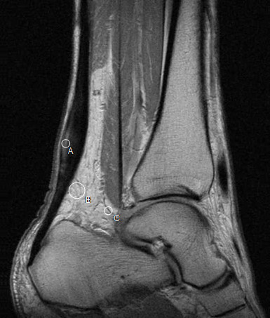
MRISagittal MRI of the Achilles tendon showing fusiform thickening of the mid-portion with intratendinous high signal, in a patient with chronic tendinopathy.
Source : Gärdin et al., BMC medical imaging, 2013, figure 1 · CC BY
Which questions should you ask to understand the patient and their history?
The history opens the whole reasoning process. 📝 It must characterise the pain, identify actionable risk factors and point straight away towards the mid-portion or the insertional form, which determines the choice of exercise protocol.³- Precise location of the pain : ask the patient to point with one finger. Pain 2-7 cm above the calcaneus → tendon body. Pain at the insertion on the calcaneus → insertional tendinopathy. Diffuse or radiating pain → reconsider the diagnosis (S1 radiculopathy, tarsal tunnel, stress fracture).
- Mode of onset : insidious and gradual over several weeks (typical of an overload tendinopathy) versus acute with a sensation of being "whipped" or of "snapping" (suspected tendon rupture – a red flag, see chapter 6).
- Characteristic time course of the pain :
- Morning pain and stiffness on rising, easing after a few steps, an almost pathognomonic sign.⁴
- Warm-up pain that eases during exercise then returns afterwards (and the following morning).
- Constant or night pain → reconsider (suspected inflammatory, infectious or tumour process).
- Recent changes in load : an increase in weekly volume (the classic +10%/week rule not respected), a change of surface (from track to road, from tartan to grass), new shoes (reduced drop), a return after a break, a change of programme.⁵
- Previous personal history of tendinopathy (Achilles or patellar), the strongest risk factor.⁵
- Metabolic factors : age > 40 years, BMI, known or suspected diabetes, hypercholesterolaemia, hypertension, recent fluoroquinolone use (ofloxacin, ciprofloxacin), corticosteroids.⁵,⁶
- Psychosocial profile : fear of movement, catastrophising, beliefs about pain ("my tendon is going to snap"), level of self-efficacy, all major predictors of prognosis (Mallows 2017).⁷
- Baseline VISA-A : 8 questions (0-100, 100 = asymptomatic), the reference PROM tool recommended by the COS-AT 2024 (de Vos ICON 2023). It allows severity to be quantified objectively and progress to be tracked.⁸
🚩 Red flags to screen for from the history onwards
- An audible "snap" plus an inability to rise onto the toes → Achilles tendon rupture (positive Thompson test). An orthopaedic emergency.
- Fever + hot swelling + exquisite tenderness → cellulitis, necrotising fasciitis, septic ankle arthritis → emergency.
- Focal bone pain + night pain + inability to hop on one leg → suspected calcaneal stress fracture → MRI/bone scan.
- A palpable mass, progressive enlargement, night sweats, weight loss → oncological work-up (soft-tissue tumour, rare but to be considered).
- Shooting pain, paraesthesia, foot-drop weakness → S1 radiculopathy, tarsal tunnel syndrome, cauda equina syndrome → neurological examination + lumbar MRI.
Which clinical tests should be performed and which other conditions must be ruled out?
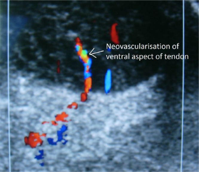
UltrasoundTransverse colour Doppler ultrasound of the Achilles tendon showing neovascularisation on the ventral aspect of the tendon, a characteristic sign of tendinopathy.
Source : Pearce et al., EFORT Open Reviews, 2016, figure 2 · CC BY-NC
- Arc sign : the area of maximal thickening is identified, then the patient is asked to move the ankle into flexion and extension. If the thickening moves with the tendon (upwards in plantar flexion, downwards in dorsiflexion) → an intratendinous lesion. If it stays fixed → suspected paratenonitis (inflammation of the peritendinous sheath). High specificity, moderate sensitivity.⁹
- Royal London Hospital test : the patient lies prone with the foot over the edge of the couch. The clinician palpates the most painful point, then passively takes the ankle into maximal dorsiflexion. If the pain decreases or disappears in that position → the test is positive (an intratendinous lesion, the tendon put under tension away from the point of pressure). Good inter-examiner reliability.⁹
- Single-leg heel-rise test : rising onto the toes of one foot. Reproduction of pain, asymmetry of height or of endurance versus the uninvolved side (< 90%) → significant functional impairment. The endurance test (the number of consecutive heel raises to fatigue) is a key indicator for return to sport.²,⁴
- Single-leg hop test : for the high-level athlete. Asymmetry > 10% versus the uninvolved side → functional deficit.²
| Clinical test | What it assesses | Performance | Level of evidence |
|---|---|---|---|
| Localised palpation | Reproduction of pain over the tendon | Sensitive (routine practice) | High |
| Arc sign | Distinguishes an intratendinous lesion from paratenonitis | High specificity, moderate sensitivity | Moderate |
| Royal London Hospital test | Confirms an intratendinous lesion | Good inter-examiner reliability | Moderate |
| Heel-rise endurance test | Strength and endurance of the triceps surae | Functionally predictive (RTS) | High |
| VISA-A (PROM) | Overall severity and follow-up (COS-AT 2024) | Excellent validity and responsiveness | High |
| Thompson test | Screening for complete rupture | Sensitivity and specificity ≈ 95% | High |
| Imaging (ultrasound/MRI) | Anatomical work-up as a second-line investigation | Poor correlation between symptoms and imaging | Low for primary diagnosis |
- Suspected partial or complete rupture (Doppler ultrasound or MRI).
- Failure of at least 3 months of well-conducted conservative treatment → re-evaluate the diagnosis.
- Suspected alternative diagnosis (stress fracture, tumour process, nerve involvement).
- A surgical request, as part of a preoperative work-up.
- Partial or complete rupture of the Achilles tendon : an audible snap, acute pain, a positive Thompson test, loss of plantar flexion.
- Isolated paratenonitis : inflammation of the peritendinous sheath, crepitus on movement, fixed thickening (negative Arc sign).
- Retrocalcaneal or pre-Achilles bursitis : posterior pain, above all in forced dorsiflexion, often associated with a Haglund deformity.
- Calcaneal stress fracture : focal bone pain, inability to hop on one leg, a runner in a context of overload.
- Tenosynovitis of flexor hallucis longus or of tibialis posterior: medial pain, sometimes mistaken for it.
- S1 radiculopathy : referred pain following the dermatome, paraesthesia, possible motor deficit, positive straight leg raise.
- Tarsal tunnel syndrome : compression of the posterior tibial nerve, plantar paraesthesia, positive Tinel sign at the medial ankle.
- Subtalar or tibiotalar osteoarthritis, systemic rheumatological disease (spondyloarthritis, rheumatoid arthritis with tendon involvement).
Should patients be classified, and what are the benefits?
Yes, but with nuance. Several classification systems coexist:- Topographical (mid-portion vs insertional) : the most clinically useful, because it directly determines the exercise modalities (avoiding forced dorsiflexion for the insertional form).⁵
- Cook-Purdam continuum (reactive/dysrepair/degenerative): educational, it guides load modulation but remains conceptual.¹⁰
- Hanlon 2021 clinical subgroups (JOSPT, n = 145, latent class analysis): three profiles, activity-dominant (a healthy athlete, acute presentation, a good response to the standard exercise protocol), psychosocial-dominant (kinesiophobia, catastrophising, chronic pain, comorbidities, calling for a biopsychosocial approach), structure-dominant (a middle-aged woman, chronic tendinopathy with marked structural changes, slower progress). The 6-month follow-up (Hanlon 2023) confirms that recovery trajectories differ significantly between subgroups, a strong argument for individualised therapeutic stratification.¹¹,¹²
Key points
- The diagnosis of Achilles tendinopathy is essentially clinical. Imaging is not recommended as a first-line investigation (Dutch CPG 2021, JOSPT 2024).
- Diagnostic triad : localised pain + morning stiffness + reproduction on palpation of the tendon. Confirmed by the Arc sign and Royal London Hospital test.
- The VISA-A (8 questions, 0-100) is the reference PROM tool recommended by the COS-AT 2024 (ICON 2023, de Vos).
- Always distinguish the body (2-7 cm above the calcaneus) from the insertion : management differs (avoid forced dorsiflexion for the insertional form).
- Red flags to screen for: rupture (Thompson test), stress fracture, infection, tumour process, S1 radiculopathy.
- Hanlon 2021 identified 3 clinical subgroups (activity / psychosocial / structure-dominant) with distinct trajectories, an argument for individualised stratification.
Bibliography
- de Vos RJ, van der Vlist AC, Zwerver J, et al. Dutch multidisciplinary guideline on Achilles tendinopathy. Br J Sports Med. 2021;55(20):1125-1134. PMID 34187784.
- Martin RL, Chimenti R, Cuddeford T, et al. Achilles Pain, Stiffness, and Muscle Power Deficits: Midportion Achilles Tendinopathy Revision 2018. J Orthop Sports Phys Ther. 2018;48(5):A1-A38. PMID 29712543.
- Silbernagel KG, Hanlon S, Vicenzino B. Current Clinical Concepts: Conservative Management of Achilles Tendinopathy. J Athl Train. 2020;55(5):438-447. PMID 32267723.
- Chimenti RL, Neville C, Houck J, Cuddeford T, Carreira D, Martin RL. Achilles Pain, Stiffness, and Muscle Power Deficits: Midportion Achilles Tendinopathy Revision 2024. J Orthop Sports Phys Ther. 2024;54(12):CPG1-CPG32. PMID 39611662.
- Chimenti RL, Cychosz CC, Hall MM, Phisitkul P. Current Concepts Review Update: Insertional Achilles Tendinopathy. Foot Ankle Int. 2017;38(10):1160-1169. PMID 28789557.
- van der Vlist AC, Breda SJ, Oei EHG, Verhaar JAN, de Vos RJ. Clinical risk factors for Achilles tendinopathy: a systematic review. Br J Sports Med. 2019;53(21):1352-1361. PMID 30718234.
- Mallows A, Debenham J, Walker T, Littlewood C. Association of psychological variables and outcome in tendinopathy: a systematic review. Br J Sports Med. 2017;51(9):743-748. PMID 27852585.
- de Vos RJ, van der Vlist AC, Winters M, et al. ICON 2023: International Scientific Tendinopathy Symposium Consensus - the core outcome set for Achilles tendinopathy (COS-AT) using a systematic review and a Delphi study. Br J Sports Med. 2024;58(20):1175-1186. PMID 39271248.
- Maffulli N, Kenward MG, Testa V, Capasso G, Regine R, King JB. Clinical diagnosis of Achilles tendinopathy with tendinosis. Clin J Sport Med. 2003;13(1):11-15. PMID 12544158.
- Cook JL, Purdam CR. Is tendon pathology a continuum? Br J Sports Med. 2009;43(6):409-416. PMID 18812414.
- Hanlon SL, Pohlig RT, Silbernagel KG. Beyond the Diagnosis: Using Patient Characteristics and Domains of Tendon Health to Identify Latent Subgroups of Achilles Tendinopathy. J Orthop Sports Phys Ther. 2021;51(9):440-448. PMID 34074130.
- Hanlon SL, Pohlig RT, Silbernagel KG. Differences in Recovery of Tendon Health Explained by Midportion Achilles Tendinopathy Subgroups: A 6-Month Follow-up. J Orthop Sports Phys Ther. 2023;53(4):200-208. PMID 36688719.
Which treatment strategies are most effective for Achilles tendinopathy?
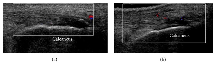
UltrasoundColour Doppler ultrasound of the Achilles tendon insertion on the calcaneus before treatment and four weeks later, showing the appearance of neovascularisation.
Source : Cheng et al., BioMed Research International, 2016, figure 3 · CC BY
Where do you start? What is the recommended hierarchy of interventions?
The Dutch CPG 2021 and the JOSPT 2024 are aligned: two pillars that are essential before any other intervention.³,⁴- Patient education and load modulation : explain the degenerative nature of the tendinopathy, dismantle the myth of "inflammatory tendinitis" requiring rest, present the pain-monitoring model (Silbernagel 2007), set realistic expectations (recovery = 3-12 months, not linear), and give the patient ownership of their load management.⁵
- A progressive exercise programme loading the triceps surae and the tendon: the cornerstone of treatment, high level of evidence (GRADE high).¹,⁶
🔺 Treatment pyramid for Achilles tendinopathy
From the most invasive (top) to the most universal (base): a hierarchy of intent
⚠️ Critical reading: the percentages are expert clinical estimates illustrating the hierarchy of therapeutic intent according to the guidelines (Dutch CPG 2021, JOSPT 2018/2024), not drawn from an epidemiological study. The hierarchy itself is solidly established by the SRs/MAs cited.
What is the place of exercise, and is there a superior approach?
🏋️ Exercise is the best documented active treatment, with a level of evidence rated high (Dutch CPG 2021, JOSPT 2024).¹,³,⁴ The debate of the past thirty years has turned on the best protocol. The van der Vlist 2021 living NMA (BJSM, 29 RCTs covering 42 compared treatments) has settled it: no exercise protocol is clinically superior to another at 12 weeks, nor in the long term.² Four protocols have solid data and remain the reference:- Alfredson 1998 protocol (pure eccentric) : 3 sets × 15 repetitions, twice a day, seven days a week, for 12 weeks. Eccentric heel drop off the edge of a step, with the knee straight (gastrocnemius) and bent (soleus). Load progressively increased with a weighted rucksack. The "founding" protocol, validated in 15 recreational athletes, all of whom returned to sport.⁸
- Heavy Slow Resistance (HSR), Beyer 2015 : 3 sets × 6 to 15 RM performed slowly (3 sec concentric phase + 3 sec eccentric phase), 3 times a week, 12 weeks. External load (machine or rucksack) increased progressively from 15 RM to 6 RM. Equivalent to eccentric loading at 12 and 52 weeks, with better adherence (92% vs 78%) because the weekly volume is lower (3 x 15 vs 2 x 7 x 45 repetitions with Alfredson), a key argument in everyday practice.⁹
- Silbernagel combined protocol (concentric + eccentric) : 4 progressive phases over 12 weeks, incorporating bilateral then unilateral heel rises, concentric-eccentric work and jumping. It tolerates pain ≤ 5/10 during exercise (the pain-monitoring model). The Habets 2021 randomised trial (Orthopaedic Journal of Sports Medicine, n = 40 recreational athletes) shows results equivalent to Alfredson at 1 year on the VISA-A.¹⁰
- Isometric holds for pain relief (Rio) : useful for the acute reactive phase or for managing pain during the competitive season. A short-term analgesic effect, they do not replace a structured exercise programme.
📌 International consensus 2025 on exercise parameters (Demangeot et al., BJSM) – A modified Delphi study conducted with 24 international experts in Achilles tendinopathy (including Alfredson, Silbernagel, Cook, Chimenti, Malliaras and Rio) reached consensus on the 4 parameters with a major influence on rehabilitation: contraction intensity, total time under tension, the number of repetitions and sets, and the type of contraction.¹⁶ For the tendon body, it is contraction intensity that is judged the most decisive; for the insertion, it is dorsiflexion range. The optimal target for tendon adaptation lies between 70 and 90% of 1RM. This consensus, the most recent to date, pragmatically confirms the keystone of treatment: what counts is not the name of the protocol, but the dose applied to the tendon.¹⁶
⚖️ Comparison of exercise protocols (van der Vlist 2021 NMA)
Effect vs sham / wait-and-see on the VISA-A at 12 weeks
Illustrative values based on the van der Vlist 2021 NMA (PMID 32522732). The differences between HSR / ECC / Silbernagel are not statistically significant and the choice is made according to patient preference, expected adherence and the equipment available.
Adjunct therapies: what are shockwave therapy, PRP and manual therapy worth?
💡 All adjunct modalities must be considered alongside an exercise programme, never as a substitute for one.¹ Extracorporeal shockwave therapy (ESWT) : the Stania 2024 meta-analysis (Explore, 12 RCTs of monotherapy) shows a positive effect on pain and on the VISA-A, particularly for insertional tendinopathy. The effect is more marked when ESWT is combined with an exercise programme.¹¹ Practical recommendation: reserve it for chronic cases (> 3 months of symptoms) that have already been on an exercise programme for at least 12 weeks without sufficient improvement. Moderate to high level of evidence, above all for the insertional form. PRP (Platelet-Rich Plasma) : the large Kearney 2021 study (JAMA, n = 240, a multicentre English RCT with 6-month follow-up) has settled the matter for good: a single PRP injection is not superior to a sham injection for chronic mid-portion Achilles tendinopathy, on any of the composite measures (pain, function, activity).⁷ PRP is not recommended in routine practice (high level of evidence against its use). Intratendinous or peritendinous corticosteroids : contraindicated. A documented increased risk of tendon rupture.¹ Manual therapy (deep transverse friction, ankle joint mobilisations): evidence is limited specifically for Achilles tendinopathy. It may be justified to improve a restricted range (talocrural) that would prevent correct loading, but it must not form the core of treatment.¹ Other modalities (low-level laser, ultrasound, taping, heel-lift insoles, intratendinous sclerosant): insufficient or contradictory evidence. Insoles with a heel lift (15-20 mm) may be useful in the very short term to reduce the load in dorsiflexion in a very painful acute phase, particularly for insertional tendinopathy. To be removed quickly so as to avoid a harmful adaptation.⁴| Modality | Main indication | Level of evidence | Expected effect |
|---|---|---|---|
| Education + load modulation | All patients | High | ↗ adherence, ↘ recurrence, ↘ fear of movement |
| Progressive exercise (HSR / ECC / Silbernagel) | All patients, 12+ weeks | High | ↘ pain (-15 to -25 pts VISA-A), ↗ function |
| Isometric holds for pain relief | Reactive phase, in-season | Moderate | Short-term pain relief |
| ESWT (insertional or refractory) | Adjunct for chronic cases | Moderate to high | ↘ pain, above all for the insertional form (Stania 2024) |
| Manual therapy (ankle) | Associated talocrural restriction | Low | Adjunct if mobility is limited |
| Heel-lift insole (15-20 mm) | Acute insertional phase | Low | Transient relief, to be removed quickly |
| Intratendinous PRP | – | Not superior to sham (Kearney 2021) | Not recommended in routine practice |
| Intra/peritendinous corticosteroids | – | Risk of rupture | ⚠️ To be avoided |
| Ultrasound / laser / TENS | Symptomatic adjunct | Low | Insufficient evidence |
| Surgery (tenosynovectomy, debridement) | Failure at ≥ 6 months of conservative care | Exceptional resort | No superiority over well-conducted ECC |
Beyond the physical: how can psychological factors be addressed?
🧠 Psychological factors are a major determinant of chronicity, and are frequently under-treated. The Mallows 2017 systematic review (BJSM) shows that kinesiophobia (fear of movement), catastrophising (a negative, amplified interpretation of pain) and low self-efficacy are independent predictors of a poor prognosis at 12 months.¹² Patient education must be active and proactive on four points:- Reframe the pain : tolerable pain (≤ 5/10 VAS) during the exercises, which does not worsen during the effort and which returns to baseline within 24 hours, is acceptable and even productive for adapting the tendon (Silbernagel 2007 model).¹³
- Dismantle the myths : "the tendon is not going to snap because I am doing heel rises", "complete rest makes things worse rather than better", "an abnormal scan does not mean the pain is serious".
- Set realistic expectations : recovery = 3 to 12 months depending on the profile, not linear, with fluctuations to be expected. This information prevents discouragement when a bad day follows a week of improvement.
- Self-efficacy : give the patient concrete tools (a diary, a daily VAS score, simple criteria for adjusting load) so that they become an agent of their own recovery rather than a passive recipient.¹⁴
Critique and controversy
The exercise protocol war belongs to the past: the van der Vlist 2021 NMA has definitively established their equivalence.² Yet many publications, courses and commercial brands continue to claim the superiority of one protocol over another, which owes more to marketing than to evidence. More problematic still, the injection industry (PRP, sclerosant, stem cells) continues to grow despite solid and concordant scientific data against its effectiveness.⁷ The clinician must take care not to give in to commercial pressure, or to patient expectations of unvalidated "modern treatments". Finally, the mid-portion versus insertional dichotomy, although clinically relevant, remains imperfectly studied: the majority of RCTs include patients with mid-portion tendinopathy, and the evidence is weaker for the insertional form. The JOSPT 2024 explicitly acknowledges this knowledge gap.⁴Key points
- ✅ First line: education + load modulation + a progressive exercise programme over 12+ weeks. High level of evidence.
- ✅ No exercise protocol is superior (van der Vlist 2021 NMA): choose HSR, Alfredson or Silbernagel according to preference and feasibility. HSR often has better adherence (a reduced weekly volume).
- 📌 International consensus 2025 (Demangeot, BJSM) : what counts is the dose (intensity 70-90% of 1RM, time under tension, repetitions, type of contraction), not the name of the protocol.
- ✅ Shockwave therapy (ESWT) : a worthwhile adjunct for chronic refractory cases, particularly for insertional tendinopathy. Always alongside exercise.
- ❌ PRP not superior to sham (Kearney 2021 JAMA, n = 240). Not for routine use.
- ❌ Intra/peritendinous corticosteroids contraindicated (risk of rupture).
- ✅ Systematically address psychosocial factors : kinesiophobia and catastrophising are major predictors of failure (Mallows 2017). Pain-monitoring model ≤ 5/10.
Bibliography
- de Vos RJ, van der Vlist AC, Zwerver J, et al. Dutch multidisciplinary guideline on Achilles tendinopathy. Br J Sports Med. 2021;55(20):1125-1134. PMID 34187784.
- van der Vlist AC, Winters M, Weir A, et al. Which treatment is most effective for patients with Achilles tendinopathy? A living systematic review with network meta-analysis of 29 randomised controlled trials. Br J Sports Med. 2021;55(5):249-256. PMID 32522732.
- Martin RL, Chimenti R, Cuddeford T, et al. Achilles Pain, Stiffness, and Muscle Power Deficits: Midportion Achilles Tendinopathy Revision 2018. J Orthop Sports Phys Ther. 2018;48(5):A1-A38. PMID 29712543.
- Chimenti RL, Neville C, Houck J, Cuddeford T, Carreira D, Martin RL. Midportion Achilles Tendinopathy Revision 2024. J Orthop Sports Phys Ther. 2024;54(12):CPG1-CPG32. PMID 39611662.
- Silbernagel KG, Hanlon S, Vicenzino B. Current Clinical Concepts: Conservative Management of Achilles Tendinopathy. J Athl Train. 2020;55(5):438-447. PMID 32267723.
- Murphy MC, Travers MJ, Chivers P, et al. Efficacy of heavy eccentric calf training for treating mid-portion Achilles tendinopathy: a systematic review and meta-analysis. Br J Sports Med. 2019;53(17):1070-1077. PMID 30636702.
- Kearney RS, Ji C, Warwick J, et al. Effect of Platelet-Rich Plasma Injection vs Sham Injection on Tendon Dysfunction in Patients With Chronic Midportion Achilles Tendinopathy: A Randomized Clinical Trial. JAMA. 2021;326(2):137-144. PMID 34255009.
- Alfredson H, Pietila T, Jonsson P, Lorentzon R. Heavy-load eccentric calf muscle training for the treatment of chronic Achilles tendinosis. Am J Sports Med. 1998;26(3):360-366. PMID 9617396.
- Beyer R, Kongsgaard M, Hougs Kjaer B, Ohlenschlaeger T, Kjaer M, Magnusson SP. Heavy Slow Resistance Versus Eccentric Training as Treatment for Achilles Tendinopathy: A Randomized Controlled Trial. Am J Sports Med. 2015;43(7):1704-1711. PMID 26018970.
- Habets B, van Cingel REH, Backx FJG, van Elten HJ, Zuithoff P, Huisstede BMA. No Difference in Clinical Effects When Comparing Alfredson Eccentric and Silbernagel Combined Concentric-Eccentric Loading in Achilles Tendinopathy: A Randomized Controlled Trial. Orthop J Sports Med. 2021;9(10):23259671211031254. PMC 8554573.
- Stania M, Juras G, Chmielewska D, et al. The Efficacy of Extracorporeal Shock Wave Therapy as a Monotherapy for Achilles Tendinopathy: A Systematic Review and Meta-Analysis. Explore (NY). 2024;20(4):488-499. PMID 38205224.
- Mallows A, Debenham J, Walker T, Littlewood C. Association of psychological variables and outcome in tendinopathy: a systematic review. Br J Sports Med. 2017;51(9):743-748. PMID 27852585.
- Silbernagel KG, Thomeé R, Eriksson BI, Karlsson J. Continued sports activity, using a pain-monitoring model, during rehabilitation in patients with Achilles tendinopathy: a randomized controlled study. Am J Sports Med. 2007;35(6):897-906. PMID 17307888.
- Chimenti RL, Frey-Law LA, Sluka KA. A Mechanism-Based Approach to Physical Therapist Management of Pain. Phys Ther. 2018;98(5):302-314. PMID 29669091.
- Hanlon SL, Pohlig RT, Silbernagel KG. Beyond the Diagnosis: Using Patient Characteristics and Domains of Tendon Health to Identify Latent Subgroups of Achilles Tendinopathy. J Orthop Sports Phys Ther. 2021;51(9):440-448. PMID 34074130.
- Demangeot Y, O'Neill S, Degache F, et al. Exercise parameters to consider for Achilles tendinopathy: a modified Delphi study with international experts. Br J Sports Med. 2025;59(19):1337-1349. doi:10.1136/bjsports-2025-110183.
How can lasting recovery be secured and recurrence of Achilles tendinopathy prevented?
How can the patient be made an active participant through the pain-monitoring model?
🎯 The Silbernagel 2007 pain-monitoring model transformed management by showing, in a randomised trial (n = 38), that continuing sport during rehabilitation is not only safe but also effective, provided a simple rule is followed:²- Pain tolerable (≤ 5/10 VAS) during the effort.
- Pain that does not worsen from session to session.
- Pain that returns to baseline within 24 hours after the effort.
- Morning stiffness that does not worsen week after week.
🚦 The Silbernagel pain-monitoring model
A simple rule for allowing, adjusting or stopping activity
Adapted from Silbernagel KG et al. Am J Sports Med. 2007;35(6):897-906 (PMID 17307888) and from the JOSPT RTS programme 2015.
- Weekly diary : running volume (km), intensity, pain VAS on waking and after exercise, morning stiffness. It makes trends objective and allows decisions to be taken free of emotional bias.³
- Monthly VISA-A : a 0-100 score to make progress objective. A 10-point improvement is clinically significant (MCID).⁴
- Keeping up the strengthening programme in the long term: at least 2-3 sessions of loaded heel rises a week, even after complete resolution of symptoms, to maintain tendon capacity.⁵
- Regulating the external load : the maximum +10%/week rule on weekly volume (km, number of sessions), a Strava-type app for objective tracking, a season plan including 1 deload week every 3-4 weeks.
When and how should a safe return to sport be planned?
🏃 Return to sport (RTS) must never be based on elapsed time alone, or on the absence of pain. The Silbernagel & Crossley 2015 RTS programme (JOSPT) proposes an approach based on objective functional criteria, which has become the international reference.⁵ Minimum criteria to meet before returning to running / jumping :- No pain on the single-leg heel rise (10 consecutive repetitions).
- Single-leg heel-rise endurance symmetry > 90% versus the uninvolved side (number of repetitions to fatigue).
- Heel-rise height symmetry (maximum height) > 90% versus the uninvolved side.
- Hop tests (single-leg hop, triple hop) with asymmetry < 10%.
- VISA-A > 80/100 or a clinically significant improvement ≥ 20 points.
- Tolerance of bilateral then unilateral jumping without reproduction of pain.
- Phase 1: rehabilitation and strengthening (weeks 0-4): load modulation, isometric holds for pain relief, basic HSR or ECC. No running.
- Phase 2: muscle capacity and basic plyometrics (weeks 4-8): progressive HSR/ECC, loaded single-leg heel rises, bilateral then unilateral mini-hops.
- Phase 3: gradual return to running (weeks 8-12): alternating walking and running (start 30 sec running / 90 sec walking × 8, progressing over 4 weeks). Soft surface (track, tartan). Short distance (1-3 km).
- Phase 4: increasing volume and intensity (weeks 12-20): maximum +10%/week increase, return to sport-specific training (speed, hills, uneven ground progressively).
- Phase 5: return to competition : competitive reintegration while keeping up the strengthening programme and pain monitoring.
Critique and controversy
The threshold of "5/10 VAS" in the Silbernagel model is a clinical heuristic that is widely adopted, but its exact cut-off has never been formally validated on a large scale. It may be too permissive for some patients (catastrophisers, those with kinesiophobia) and too restrictive for others (elite athletes used to tolerating pain). Personalisation remains necessary according to psychological profile and level of activity. The return-to-sport criteria (symmetry ≥ 90%, VISA-A > 80) are logical, but their predictive value for freedom from recurrence in the long term remains only moderately established. Most studies measure the return-to-sport rate, not the recurrence rate at 2-3 years. Finally, in everyday practice these criteria take time, equipment (a mat for heel rises, a stopwatch) and specific training: the risk is that they remain theoretical and that the clinician allows return to sport on looser criteria (absence of pain at rest, which is far from sufficient).Key points
- The Silbernagel 2007 pain-monitoring model is the pivotal tool for self-management: pain ≤ 5/10 VAS, no worsening, back to baseline within 24 hours → green zone, carry on.
- Concrete tools for the patient: a diary, a monthly VISA-A, keeping up the HSR/ECC programme in the long term.
- The return to sport must be based on objective functional criteria (Silbernagel & Crossley 2015): heel-rise endurance and height symmetry > 90%, hop test with < 10% asymmetry, VISA-A > 80.
- Programming in 5 progressive phases over 12-20 weeks, with the maximum +10%/week rule.
- A realistic trajectory: perceived improvement at 4-6 weeks, a peak at 12 weeks, return to competition between 4 and 12 months. One third of patients remain symptomatic at 1 year (Lagas 2020) – keeping up the programme is essential.
Bibliography
- Lagas IF, Tol JL, Weir A, et al. How many runners with new-onset Achilles tendinopathy develop persisting symptoms? A large prospective cohort study. Scand J Med Sci Sports. 2020;30(10):1939-1948. PMID 32615645.
- Silbernagel KG, Thomeé R, Eriksson BI, Karlsson J. Continued sports activity, using a pain-monitoring model, during rehabilitation in patients with Achilles tendinopathy: a randomized controlled study. Am J Sports Med. 2007;35(6):897-906. PMID 17307888.
- Silbernagel KG, Hanlon S, Vicenzino B. Current Clinical Concepts: Conservative Management of Achilles Tendinopathy. J Athl Train. 2020;55(5):438-447. PMID 32267723.
- de Vos RJ, van der Vlist AC, Winters M, et al. ICON 2023: International Scientific Tendinopathy Symposium Consensus - Core Outcome Set for Achilles tendinopathy (COS-AT). Br J Sports Med. 2024;58(20):1175-1186. PMID 39271248.
- Silbernagel KG, Crossley KM. A Proposed Return-to-Sport Program for Patients With Midportion Achilles Tendinopathy: Rationale and Implementation. J Orthop Sports Phys Ther. 2015;45(11):876-886. doi:10.2519/jospt.2015.5885.
- Murphy M, Travers M, Gibson W, et al. Rate of improvement of pain and function in mid-portion Achilles tendinopathy with loading protocols: A systematic review and longitudinal meta-analysis. Sports Med. 2018;48(8):1875-1891. PMID 29766442.
- Habets B, van Cingel REH, Backx FJG, et al. No Difference in Clinical Effects When Comparing Alfredson Eccentric and Silbernagel Combined Concentric-Eccentric Loading in Achilles Tendinopathy: A RCT. Orthop J Sports Med. 2021;9(10):23259671211031254. PMC 8554573.
- Mallows A, Debenham J, Walker T, Littlewood C. Association of psychological variables and outcome in tendinopathy: a systematic review. Br J Sports Med. 2017;51(9):743-748. PMID 27852585.
- Martin RL, Chimenti R, Cuddeford T, et al. Midportion Achilles Tendinopathy Revision 2018. J Orthop Sports Phys Ther. 2018;48(5):A1-A38. PMID 29712543.
- de Vos RJ, van der Vlist AC, Zwerver J, et al. Dutch multidisciplinary guideline on Achilles tendinopathy. Br J Sports Med. 2021;55(20):1125-1134. PMID 34187784.
What do concrete clinical cases teach us about Achilles tendinopathy?
Analysis of a published refractory case: the Omodani 2024 report
The case reported by Omodani T (2024, Cureus – PMC10975086) illustrates the complexity of a refractory Achilles tendinopathy and the value of a multimodal approach combining conventional therapies with advanced adjuncts.¹ The patient was a 23-year-old middle-distance track athlete, with bilateral Achilles tendinopathy present for 4 years, refractory to standard treatment. Previous treatments included: standard rehabilitation (eccentric exercises), orthotic insoles, corticosteroid injections, and extracorporeal shockwave therapy (ESWT) – with no lasting improvement. The patient had had to stop competing. The diagnosis, confirmed on ultrasound, showed marked thickening with dense neovascularisation, the signature of a chronic degenerative tendinopathy. The multimodal approach that finally succeeded combined:- Transarterial embolisation targeting the pathological neovessels.
- ESWT repeated with optimised parameters.
- Prolotherapy given intratendinously.
- A programme of progressive strengthening structured in HSR style.
🧩 Components of the refractory protocol, Omodani 2024 case
A 23-year-old middle-distance athlete, bilateral tendinopathy of 4 years, previous treatments failed
Source: Omodani T. Cureus. 2024;16(2):e55030. PMC 10975086. The levels of evidence come from the SRs/MAs cited in the article.
The diagnostic challenge: when Achilles pain hides something else
Retromalleolar and tibiotarsal pain is not always tendinous in origin. ⚠️ Several conditions can mimic an Achilles tendinopathy, sometimes with serious consequences if the diagnosis is delayed:- 🔍 Partial or complete rupture of the Achilles tendon – the most critical cause not to miss. The Thompson test (squeezing the calf with the patient prone and the foot over the edge of the couch → absence of plantar flexion = complete rupture) has a sensitivity and specificity > 95%. Clinical suspicion is high if there was an audible snap, intense acute pain, or an inability to rise onto the toes. Ultrasound or MRI to confirm.² Acute rupture is an injury with loss of continuity, not an advanced stage of tendinopathy: its management is the opposite of what is described here. It is the subject of a dedicated article, Achilles tendon rupture.
- 🔍 Isolated paratenonitis – inflammation of the peritendinous sheath, distinguished by a negative Arc sign (fixed thickening that does not move with the tendon during movement). Crepitus on palpation during flexion and extension. Management differs (anti-inflammatories more effective, a shorter period of relative rest).⁹
- 🔍 Calcaneal stress fracture – to be considered in a runner in overload with focal bone pain, pain on percussion of the calcaneus, and pain on hopping on one foot (which is impossible). MRI or bone scan to confirm. A delayed diagnosis leads to complete fracture.
- 🔍 Haglund deformity + retrocalcaneal bursitis – a posterosuperior bony prominence of the calcaneus, rubbing against the heel counter of the shoe, with an associated bursitis. Often confused with insertional tendinopathy (the two can coexist). A lateral radiograph of the calcaneus is enough.
- 🔍 S1 radiculopathy – pain referred in the S1 territory (posterior aspect of the leg, heel), possibly with low back pain, plantar paraesthesia, a positive straight leg raise, and a reduced Achilles reflex or reduced plantar flexor strength. Lumbar MRI as a work-up.
- 🔍 Tarsal tunnel syndrome – compression of the posterior tibial nerve behind the medial malleolus. Plantar paraesthesia, a positive Tinel sign at the medial ankle. Electromyography to confirm.
- 🔍 Tibialis posterior tendinopathy – medial pain, loss of the medial arch, a positive "too many toes" sign (seen from behind the standing patient, too many toes are visible on the affected side). It must be told apart carefully, as the treatment is specific.
Study of a complex case: insertion, diabetes and clinical subgroups
Complexity increases when several factors combine. Three situations deserve particular attention: 1. Insertional tendinopathy complicated by a Haglund deformity – particularly common in middle-aged women wearing shoes with a rigid posterosuperior counter. Management is specific: avoid forced dorsiflexion which worsens compression between the tendon and the calcaneus. Adapt the exercise programme by avoiding the drop off a step (stay on flat ground, or with a slight rise). A temporary heel lift is useful in the short term.³ ESWT gives particularly good results in this insertional form.⁴ 2. The patient with diabetes and Achilles tendinopathy – the De Luca 2025 meta-analysis established an OR of 7.22 for diabetes as a risk factor.⁵ The mechanisms include non-enzymatic glycation of collagen (formation of AGEs), microangiopathy reducing the blood supply to the tendon, and probably a systemic inflammatory component. Specific features of management: slower recovery than average (a longer wait), an increased risk of rupture (corticosteroids absolutely to be avoided), and close collaboration with the diabetes specialist to optimise glycaemic control (HbA1c < 7%).⁶ 3. The "psychosocial-dominant" subgroup of Hanlon 2021 – a patient with high kinesiophobia, catastrophising, painful comorbidities (chronic low back pain, fibromyalgia), a low VISA-A but modest imaging findings. An exercise programme alone is not enough. Management calls for a biopsychosocial approach: pain education, cognitive behavioural therapy, management of kinesiophobia, and sometimes referral to a psychologist specialising in chronic pain. The 6-month follow-up (Hanlon 2023) shows that this subgroup has a significantly slower recovery trajectory than the others.⁷,⁸Critique and controversy: where do case reports sit in the hierarchy of evidence?
🧠 Case reports are fundamental for generating hypotheses and flagging rare presentations, but their level of evidence is the lowest of the whole scientific hierarchy. A single case illustrates a possibility, not a generality. It cannot control for placebo, regression to the mean or spontaneous recovery.⁹ Publication bias is massive (failures are rarely published).📐 Hierarchy of scientific evidence: where does each type of study belong?
Strength of evidence decreasing from the top (meta-analyses) towards the bottom (isolated cases)
A simplified GRADE / Oxford CEBM hierarchy. The length of the coloured bar on the right illustrates the relative strength of evidence. Practical implication: where a seductive case report and a meta-analysis diverge, the decision must follow the meta-analysis. Case reports remain valuable for generating hypotheses, flagging rare presentations, or illustrating a line of clinical reasoning.
⭐ Key points
- The Omodani 2024 case (Cureus) illustrates that a multimodal approach (embolisation + ESWT + prolotherapy + HSR) can be attempted in refractory cases after standard treatment has failed, but does not demonstrate its effectiveness (n = 1).
- Critical differential diagnoses to know: rupture (Thompson test), paratenonitis (negative Arc sign), calcaneal stress fracture, Haglund deformity, S1 radiculopathy, tarsal tunnel syndrome, tibialis posterior tendinopathy.
- The insertional tendinopathy complicated by Haglund requires a specific approach: avoid forced dorsiflexion, a temporary heel lift, ESWT particularly effective.
- The presence of diabetes multiplies the risk by 7.2 (De Luca 2025) and calls for specific management: slower recovery, avoid corticosteroids (risk of rupture), collaboration with the diabetes specialist (HbA1c < 7%).
- The 3 clinical subgroups of Hanlon 2021 (activity-, psychosocial-, structure-dominant) have distinct trajectories at 6 months, an argument for individualised stratification.
- ⚠️ Level of evidence : a case report = level 5 (the lowest). Where there is divergence, follow the meta-analyses (level 1a), not the isolated case. Cases illustrate, they do not demonstrate.
Bibliography
- Omodani T. Multifaceted Treatment Using Advanced Modalities for Refractory Achilles Tendinopathy: A Case Report. Cureus. 2024;16(2):e55030. PMC 10975086.
- Reiman M, Burgi C, Strube E, et al. The utility of clinical measures for the diagnosis of achilles tendon injuries: a systematic review with meta-analysis. J Athl Train. 2014;49(6):820-829. PMID 25243736.
- Chimenti RL, Cychosz CC, Hall MM, Phisitkul P. Current Concepts Review Update: Insertional Achilles Tendinopathy. Foot Ankle Int. 2017;38(10):1160-1169. PMID 28789557.
- Stania M, Juras G, Chmielewska D, et al. The Efficacy of Extracorporeal Shock Wave Therapy as a Monotherapy for Achilles Tendinopathy: A Systematic Review and Meta-Analysis. Explore (NY). 2024;20(4):488-499. PMID 38205224.
- De Luca A, Trecca EMC, et al. The interplay between metabolic disorders and tendinopathies: Systematic review and meta-analysis. J Exp Orthop. 2025;12:e70429. doi:10.1002/jeo2.70429.
- Cannata F, Vadala G, Ambrosio L, et al. The impact of type 2 diabetes on the development of tendinopathy. Diabetes Metab Res Rev. 2021;37(8):e3417. PMID 33156563.
- Kearney RS, Ji C, Warwick J, et al. Effect of Platelet-Rich Plasma Injection vs Sham Injection on Tendon Dysfunction in Patients With Chronic Midportion Achilles Tendinopathy: A Randomized Clinical Trial. JAMA. 2021;326(2):137-144. PMID 34255009.
- Hanlon SL, Pohlig RT, Silbernagel KG. Beyond the Diagnosis: Using Patient Characteristics and Domains of Tendon Health to Identify Latent Subgroups of Achilles Tendinopathy. J Orthop Sports Phys Ther. 2021;51(9):440-448. PMID 34074130.
- Nissen T, Wynn R. The clinical case report: a review of its merits and limitations. BMC Res Notes. 2014;7:264. PMID 24758689.
- Chimenti RL, Neville C, Houck J, Cuddeford T, Carreira D, Martin RL. Midportion Achilles Tendinopathy Revision 2024. J Orthop Sports Phys Ther. 2024;54(12):CPG1-CPG32. PMID 39611662.
- Hanlon SL, Pohlig RT, Silbernagel KG. Differences in Recovery of Tendon Health Explained by Midportion Achilles Tendinopathy Subgroups: A 6-Month Follow-up. J Orthop Sports Phys Ther. 2023;53(4):200-208. PMID 36688719.
- de Vos RJ, van der Vlist AC, Zwerver J, et al. Dutch multidisciplinary guideline on Achilles tendinopathy. Br J Sports Med. 2021;55(20):1125-1134. PMID 34187784.
- Beyer R, Kongsgaard M, Hougs Kjaer B, et al. Heavy Slow Resistance Versus Eccentric Training as Treatment for Achilles Tendinopathy: A RCT. Am J Sports Med. 2015;43(7):1704-1711. PMID 26018970.
- van der Vlist AC, Winters M, Weir A, et al. Which treatment is most effective for patients with Achilles tendinopathy? A living systematic review with network meta-analysis. Br J Sports Med. 2021;55(5):249-256. PMID 32522732.
- Lagas IF, Tol JL, Weir A, et al. How many runners with new-onset Achilles tendinopathy develop persisting symptoms? Scand J Med Sci Sports. 2020;30(10):1939-1948. PMID 32615645.
How do you apply these recommendations concretely in your practice?
When and to which other health professionals should patients be referred?
The first non-negotiable step in triage is screening for red flags. The clinician must have a low threshold for referral: sensitivity takes precedence over specificity when serious conditions are at stake.🚩 Red flags specific to the Achilles region
- Achilles tendon rupture : an audible or felt snap + an inability to rise onto the toes + positive Thompson test (squeezing the calf with the feet over the edge of the couch → absence of plantar flexion). An orthopaedic emergency.
- Calcaneal stress fracture : focal bone pain, pain on percussion, inability to hop on one leg. Above all in the runner in overload, the soldier, and the amenorrhoeic athlete (REDs).
- Infection (cellulitis, necrotising fasciitis, septic arthritis) : fever + hot swelling + exquisite tenderness + rapidly spreading erythema. An emergency.
- Tumour process (rare but to be considered): a palpable mass appearing gradually, night pain, night sweats, unexplained weight loss, deterioration in general condition.
- Chronic compartment syndrome : calf pain on exercise, confirmed by intracompartmental pressure measurement.
- S1 radiculopathy / cauda equina syndrome : radicular pain (S1 territory), paraesthesia, motor deficit, sphincter disturbance, saddle anaesthesia.
- Systemic rheumatological disease : spondyloarthritis (HLA-B27, morning axial stiffness, uveitis), rheumatoid arthritis (multi-joint involvement), gout (an acute attack).
- Recent fluoroquinolone treatment + acute tendon pain: an increased risk of rupture, alert the prescribing doctor.
⚠️ Any red flag → prompt medical referral (general practitioner, emergency department, sports physician, orthopaedic surgeon) before, or alongside, physiotherapy management.
- General practitioner / sports physician : metabolic work-up (HbA1c, cholesterol, uric acid), medication review (fluoroquinolones, corticosteroids), overall follow-up.
- Endocrinologist / diabetes specialist : for patients with diabetes (OR 7.22, De Luca 2025).²
- Orthopaedic surgeon / foot surgeon : failure at ≥ 6 months of structured conservative treatment, a severe symptomatic Haglund deformity, a suspected complex intratendinous lesion.
- Podiatrist / pedorthist : biomechanical analysis of running, corrective insoles if needed (excessive pronation, and so on), footwear advice.
- Coach / strength and conditioning coach : reviewing and restructuring the training programme to manage load over the long term.
How do you measure outcomes and overcome barriers to implementation?
📊 Measuring outcomes is the sine qua non of quality practice. The COS-AT 2024 (Core Outcome Set for Achilles Tendinopathy), from the ICON 2023 consensus (de Vos, BJSM, PMID 39271248), standardised the outcomes to be measured in any management of, or research on, Achilles tendinopathy.³ Core outcomes of the COS-AT 2024 :- Pain : numerical rating scale (NRS) 0-10.
- Function : VISA-A (Victorian Institute of Sport Assessment - Achilles), 8 questions, score 0-100. MCID (Minimal Clinically Important Difference) ≈ 10 points.
- Participation : return to sporting activity, level of practice versus before the injury.
- Quality of life : EQ-5D or similar.
- Global impression of change reported by the patient (PGIC).
- Digital tools : a tablet or smartphone app that the patient fills in in the waiting room, automatic scoring, graphical display of progress.
- Continuing education : practical workshops on using PROMs and on shared decision-making.
- Clinical champions : opinion leaders in the practice or the clinic who spread the use of EBP.
- Integration into the routine : VISA-A at every follow-up consultation (every 4-6 weeks), not only at the start and end of care.
- Shared decision-making : present the options to the patient (HSR vs ECC vs Silbernagel), discuss the advantages and drawbacks, decide together according to their preferences and their timetable. It significantly increases adherence and satisfaction.
Critique and controversies: beyond the guidelines
The red flag paradox still holds: no sign taken in isolation has a high positive predictive value. It is the convergence of evidence plus probabilistic clinical reasoning that guides referral, not a checklist. The risk is twofold: over-diagnosing (sending every painful tendon for MRI) or under-diagnosing (missing a rupture by putting the pain down to a simple tendinopathy). The "knowing-doing" gap is particularly striking in Achilles tendinopathy: the van der Vlist 2021 NMA established the equivalence of exercise protocols 5 years ago, the rejection of PRP by Kearney 2021 has been known for 4 years, and yet everyday practice sometimes continues to impose a single protocol or to prescribe costly PRP injections without indication. Moving to genuine EBP requires more than disseminating information: it requires a rethink of economic incentives and of continuing education. Finally, the standardisation versus personalisation tension is central. The COS-AT 2024 is essential for research, but the clinician must know how to navigate between standardised tools (which allow comparability) and individualisation (which answers the patient's real needs). The Hanlon 2021 subgroups are one way of reconciling these two demands.⁴Key points
- Referral is crucial for safety : red flags (rupture, fracture, infection, tumour process, radiculopathy) → prompt medical referral.
- Psychosocial yellow flags (kinesiophobia, catastrophising, Mallows 2017) → collaboration with a psychologist, a biopsychosocial approach.
- Measure outcomes with the COS-AT 2024 (de Vos ICON 2023): pain (NRS), function (VISA-A), participation, quality of life.
- The VISA-A at every follow-up (every 4-6 weeks), MCID = 10 points.
- For cases still refractory after 3-6 months: reconsider the diagnosis (imaging), discuss with a sports physician, consider ESWT, and possibly a surgeon after failure of 6 months of structured conservative treatment.
- The shared decision-making (presenting HSR vs ECC vs Silbernagel, discussing according to preferences) increases adherence and satisfaction.
Bibliography
- Mallows A, Debenham J, Walker T, Littlewood C. Association of psychological variables and outcome in tendinopathy: a systematic review. Br J Sports Med. 2017;51(9):743-748. PMID 27852585.
- De Luca A, Trecca EMC, et al. The interplay between metabolic disorders and tendinopathies: Systematic review and meta-analysis. J Exp Orthop. 2025;12:e70429. doi:10.1002/jeo2.70429.
- de Vos RJ, van der Vlist AC, Winters M, et al. ICON 2023: International Scientific Tendinopathy Symposium Consensus - Core Outcome Set for Achilles tendinopathy (COS-AT) using a systematic review and a Delphi study. Br J Sports Med. 2024;58(20):1175-1186. PMID 39271248.
- Hanlon SL, Pohlig RT, Silbernagel KG. Beyond the Diagnosis: Using Patient Characteristics and Domains of Tendon Health to Identify Latent Subgroups of Achilles Tendinopathy. J Orthop Sports Phys Ther. 2021;51(9):440-448. PMID 34074130.
- de Vos RJ, van der Vlist AC, Zwerver J, et al. Dutch multidisciplinary guideline on Achilles tendinopathy. Br J Sports Med. 2021;55(20):1125-1134. PMID 34187784.
- Chimenti RL, Neville C, Houck J, Cuddeford T, Carreira D, Martin RL. Midportion Achilles Tendinopathy Revision 2024. J Orthop Sports Phys Ther. 2024;54(12):CPG1-CPG32. PMID 39611662.
- van der Vlist AC, Winters M, Weir A, et al. Which treatment is most effective for patients with Achilles tendinopathy? A living systematic review with network meta-analysis. Br J Sports Med. 2021;55(5):249-256. PMID 32522732.
- Kearney RS, Ji C, Warwick J, et al. Effect of PRP Injection vs Sham Injection on Tendon Dysfunction in Patients With Chronic Midportion Achilles Tendinopathy: A RCT. JAMA. 2021;326(2):137-144. PMID 34255009.
- Silbernagel KG, Crossley KM. A Proposed Return-to-Sport Program for Patients With Midportion Achilles Tendinopathy: Rationale and Implementation. J Orthop Sports Phys Ther. 2015;45(11):876-886. doi:10.2519/jospt.2015.5885.
- Stania M, Juras G, Chmielewska D, et al. The Efficacy of ESWT as a Monotherapy for Achilles Tendinopathy: A SR/MA. Explore (NY). 2024;20(4):488-499. PMID 38205224.
- O'Neill S, Watson PJ, Barry S. A Delphi Study of Risk Factors for Achilles Tendinopathy - Opinions of World Tendon Experts. Int J Sports Phys Ther. 2016;11(5):684-697. PMID 27757281.
- Silbernagel KG, Hanlon S, Vicenzino B. Current Clinical Concepts: Conservative Management of Achilles Tendinopathy. J Athl Train. 2020;55(5):438-447. PMID 32267723.
- Lagas IF, Tol JL, Weir A, et al. How many runners with new-onset Achilles tendinopathy develop persisting symptoms? Scand J Med Sci Sports. 2020;30(10):1939-1948. PMID 32615645.
And after this read?
This article is part of a collection of evidence-based clinical syntheses. A question, a comment, a correction to suggest? Contact us directly through the WhatsApp button at the bottom right of the screen.

