

















In brief
This article deals with the acute injury: a loss of tendon continuity, occurring in a single second, in a tendon that was giving no trouble the day before. It is not the same disease as Achilles tendinopathy, which is a condition of progressive overload (pain that builds over weeks, a tendon that stays continuous, treatment by increasing load). Nothing in the management is alike: where tendinopathy is treated by loading, rupture begins with immobilisation in equinus and turns on protecting a healing scar. Confusing the two leads to loading a ruptured tendon, or to immobilising a tendon that needs to be worked. For Achilles pain of gradual onset, with no sudden snap or abrupt loss of function, it is the other article that should be read.
Clinical synthesis on acute Achilles tendon rupture. Three things decide this patient's outcome, and this article takes them in order: the delay before anyone makes the diagnosis, because he is still walking and that wrongly reassures everybody; the choice between surgery and functional treatment, where the state of the evidence has shifted enough to make the question less settled than it is usually presented; and the residual plantarflexion strength deficit, which persists for years, can be measured, and shows up on no satisfaction questionnaire.
Clinical summary
- An injury, not wear and tear. The presentation is stereotyped: a push-off or a sprint start, the sensation of a violent blow behind the ankle (many patients turn round to see who has struck them), sometimes an audible snap, and immediate loss of function. The tendon loses its continuity; this is not a painful tendon but a tendon cut in two.
- The patient is still walking, and that is the central trap. The toe flexors, tibialis posterior and the peroneals produce enough plantarflexion to allow walking on the flat. The compensation is not a hypothesis: at fourteen years of follow-up, flexor hallucis longus on the ruptured side is 5 % larger than on the healthy side, a hypertrophy measured on MRI.
- Only one test really carries the diagnosis. The calf squeeze (Thompson-Simmonds) reaches a sensitivity of 0.96 and a specificity of 0.93. Loss of resisted plantarflexion, on the other hand, proves nothing: it can be preserved in a completely ruptured tendon. The practical rule fits on one line: a rupture is never ruled out on the grounds that the patient can push through the foot.
- The great debate has changed in nature. Surgery more than halves the risk of re-rupture in general meta-analyses, but that advantage disappears statistically when both arms receive early functional rehabilitation with protected weight-bearing. The price of surgery, by contrast, does not disappear: complications multiplied by 2.8, including infection and nerve injury.
- The protocol counts for more than the procedure. Across 43 randomised trials, postoperative re-rupture ranges from 1.36 % (accelerated rehabilitation without immobilisation) to 5.95 % (early immobilisation with active mobility) depending on the protocol chosen. The gap between protocols is of the same order as the gap between operating and not operating.
- Rehabilitation is steered by criteria, not by a calendar. Time frames serve as a safety rail, not as a green light: it is the ability to perform ten single-leg heel-rises, to walk without a limp and to rise onto the toes that opens the next stage, seven criteria, from which calf circumference has been explicitly excluded by expert consensus.
- The strength deficit is the expected sequela, not the exception. It persists at seven years, no longer improves after two years, and is not correlated with patient satisfaction: in one series where the median ATRS reached 97 out of 100, only 41 % of the operated patients had recovered 90 % symmetry on the heel-rise test.
What are the fundamentals to know about Achilles tendon rupture?
Who ruptures the tendon, in what circumstances, and why the number of these injuries has been rising for sixty years. This chapter also sets out the anatomical mechanism that explains everything else in the article: the reason a patient whose tendon is cut in two walks out of the consultation on both legs.
The Achilles tendon is the thickest and strongest in the human body. It transmits to the calcaneus the force of the three heads of the triceps surae, the two gastrocnemii and the soleus, and withstands, during running, loads of several times body weight. Its rupture is the commonest tendon rupture of the lower limb.40
An injury on push-off, not a wearing-out that gives way
The story is almost always the same. A man of forty takes up sport again on the Saturday, sets off in a sprint or jumps at badminton, and feels a violent blow in the lower third of the calf. Many turn round to look for the opponent who has just struck them, a detail of the history so constant that it alone carries a strong presumption. Immediate loss of function follows, often an inability to run, and walking that becomes possible again within a few minutes.
That is exactly what separates this presentation from Achilles tendinopathy. Tendinopathy builds over weeks, gives warning through morning stiffness, warms up with exercise, and the tendon stays continuous. Rupture, by contrast, assumes no warning: the injury can happen in a tendon that was silent until then, and that is precisely what makes it unpredictable. The two entities share a tendon and nothing else.
How many ruptures, and in whom?
A 2026 meta-analysis brought together twenty-eight population studies covering more than 630 million individuals and 568,000 ruptures, from 1950 to 2022. The pooled worldwide incidence comes to 15.7 per 100,000 person-years, but that average masks a clear trend: it rose from 6.1 in 1979 to 31.1 per 100,000 in 2021, that is a mean increase of 2.7 % a year (95% CI 2.0-3.3).1
The same analysis puts the male-to-female ratio at 3.18 (95% CI 2.50-4.04) and places the peak incidence in men between 30 and 49 years, at 42.6 per 100,000; in women the peak comes later, between 40 and 49 years, at 17.2. About 68 % of ruptures are sport-related.1
The Swedish national registry, which covers 53,688 ruptures between 2002 and 2021, confirms this and adds detail: incidence there rose from 28.8 to 41.7 per 100,000 (+45 %), including a 21 % acceleration over the last five years alone. Over the same period, the incidence of operated ruptures has fallen from 13.4 to 6.0 per 100,000, and the mean delay between injury and surgery has gone from 0.6 to 5.1 days.2 In other words: more and more patients rupture the tendon, and fewer and fewer are operated on. That is the population landing in rehabilitation, and it is changing in nature.
The most recent American data, from the emergency department surveillance system over 2019-2024 (an estimated 31,549 ruptures), give 86.3 % men, a peak between 20 and 39 years, and identify basketball as the leading sport involved in men.3 The age difference from the European series comes largely from recruitment, emergency department against national registry, and it is a reminder that none of these series describes "the" typical patient: they describe the patient of their own care pathway.
Why the patient is still walking: the compensation, measured
This is the point of functional anatomy that governs the whole of the next chapter. The triceps surae is not the only plantarflexor of the ankle. Flexor hallucis longus, flexor digitorum longus, tibialis posterior and the peroneals all pass behind the malleolus and take part in plantarflexion. Their lever arm is far shorter than that of the Achilles tendon, and they are incapable of lifting the body onto the toes of a single foot, but they are amply enough to produce flat walking on level ground.
This compensation is not an explanation in principle: it can be measured. A prospective MRI study with a mean follow-up of fourteen years, in 52 operated patients, finds on the ruptured side a soleus reduced by 13 %, a medial gastrocnemius by 13 % and a lateral one by 11 %, and, against the trend, a flexor hallucis longus 5 % larger than on the healthy side (p = 0.002), that is to say a compensatory hypertrophy.8 An electromyographic study one year after a rupture treated without surgery confirms the mechanism: differences in the length of the medial gastrocnemius and soleus subtendons alone explain 48 % of the variance in the electrical activity of flexor hallucis longus.9 The more tension the triceps has lost, the harder the hallux works.
What weakens the tendon before the injury
The best documented iatrogenic factor is the fluoroquinolone class, but the most recent systematic review shows that they are not all equivalent. Across twelve studies and 439,299 patients, the risk of Achilles tendinopathy or rupture reaches 1.40 % with ofloxacin, against 0.17 % with levofloxacin as with ciprofloxacin (p < 0.0001 for each comparison) and 0.31 % for the other molecules.10 The useful piece of history is therefore not "have you taken an antibiotic?" but which one.
Key points, chapter 1
- Incidence has been rising by 2.7 % a year for sixty years, and the proportion of patients operated on is falling: rehabilitation receives a growing flow of non-operated tendons.
- A man aged 30 to 49 years, in a push-off sport: that is the dominant profile, but 32 % of ruptures are not sport-related.
- Preserved walking is explained by accessory flexors whose compensatory hypertrophy is still measurable at fourteen years. It has no value for ruling out the diagnosis.
- This article and the one on tendinopathy do not describe two stages of the same disease: their treatments are opposed in their very principle.
Why is the diagnosis delayed, and how do you avoid missing it?
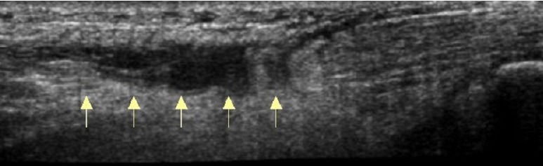
UltrasoundSagittal ultrasound of the Achilles tendon: rupture of the middle third of the tendon, arrows beneath the discontinuity.
Source : Belmoubarik et al., The Pan African medical journal, 2015, figure 2 · CC BY
One rupture in nine reaches treatment more than two weeks late. This chapter breaks down where that delay comes from (the answer is not the one usually taught), then gives the real diagnostic values of each test, and says which of them deserve to be trusted.
Where the delay really comes from: measurement against classical teaching
It is commonly read that Achilles tendon rupture is "frequently missed at the first consultation". The most solid figure available today tells a different, and more usable, story.
A Gothenburg team went back over the 958 ruptures treated at Sahlgrenska University Hospital between 2015 and 2020, and isolated those managed more than fourteen days late. They account for 102 patients, that is 11 % of the series. Seventy-five of them were studied in detail, and when the cause of the delay is sought, the first culprit is not the doctor: 52 patients, reported as 84 % in the publication, had delayed seeking care. Medical diagnostic errors, for their part, concerned 10 patients (1 % of the whole series) at the time of the injury, and 28 (3 %) at later consultations. The authors explicitly conclude that these delays were due first of all to "patient's delay" rather than to a diagnostic error, "relatively rare".4
Two practical consequences, and they do not point the way one might think.
The first: medical error exists but it is in the minority, and above all it occurs more at later consultations than at the time of the injury (three times more, in this series). The patient whose rupture is missed is generally not the one who arrives in the emergency department limping on the day itself; it is the one who comes back three weeks later with a "calf that is not recovering", taken for a healing muscle injury. That is precisely the physiotherapist's consultation.
The second: if 84 % of the delay comes from patients who do not seek care, it is because the presentation is not frightening enough. And it is not frightening enough because the patient walks. The compensation described in the previous chapter has its effect upstream of the health system: it convinces the injured person that this is a muscle tear.
The chronic patient is not a late acute patient
In the same series, chronic ruptures involved patients who were older, with a higher BMI and more comorbidities than acute ruptures. And their outcomes deteriorate markedly: in these patients managed late, the median ATRS reaches 77 after surgery against 34 without surgery, and self-rated recovery 85 % against 40 %.4 In other words, the surgery-versus-functional debate in chapter 3 concerns ruptures that are acute. Once that delay has passed, the balance of arguments changes.
The clinical tests, and what they are really worth
The reference on this point remains a thirteen-year prospective study of 174 patients with confirmed complete rupture and 28 patients suspected but intact, a rare design, since it supplies both true positives and true negatives.5 A systematic review with meta-analysis has since pooled the available data and confirms the ranking.6
| Test | Manoeuvre | Sensitivity | Specificity | Verdict |
|---|---|---|---|---|
| Calf squeeze Thompson-Simmonds |
Patient prone, feet over the edge of the couch. Squeeze the muscle belly of the calf. The foot should move into plantarflexion. | 0.96 | 0.93 | The reference test with the best sensitivity and the best specificity of all the tests studied |
| Matles resting angle |
Prone, active knee flexion to 90°. The healthy foot stays in slight plantarflexion; the injured foot falls into neutral or dorsiflexion. | 0.88 | 0.85 | Solid and the second test to do, always comparing sides |
| Palpation of the gap | Palpate along the tendon looking for a depression. | 0.73 (0.81 under anaesthesia) |
0.89 | Insufficient on its own because the haematoma fills the defect within a few hours, and a tendon can be ruptured with no palpable gap |
| Copeland blood pressure cuff |
Cuff around the calf inflated with the foot in plantarflexion, then moved into dorsiflexion: the pressure should rise if the tendon is continuous. | 0.80 | — | Little used since the equipment is superfluous when the calf squeeze is enough |
| O'Brien needle test |
Needle inserted into the tendon, ankle moved: the needle tilts if the tendon is continuous. | 0.80 | — | Invasive and of no interest in physiotherapy practice |
| Active or resisted plantarflexion | Ask the patient to push through the foot, against resistance or not. | No published value appears in none of the validation series |
Never to be used to rule out because the accessory flexors produce it in a completely ruptured tendon | |
Sensitivities and specificities: Maffulli 1998, 174 confirmed ruptures and 28 controls (PMID 9548122). Concordant pooled values: Reiman 2014 (PMID 25243736), negative likelihood ratio 0.04 and positive likelihood ratio 13.71 for the calf squeeze.
The rule that protects against the false negative
The most useful finding of the 1998 study is not an isolated sensitivity, it is a sentence in the conclusion: whatever tests were performed, at least two of them were positive in every patient in the series.5 No single test catches a rupture on its own; a combination of two does.
The 2014 meta-analysis puts the same caution differently: most Achilles clinical tests have a diagnostic capacity greater than their screening capacity.6 Translated into practice: a positive test points strongly, a negative test reassures poorly. Hence the course of action, which comes down to two manoeuvres and one decision.
What place for imaging?
A systematic review of 56 studies examined the question. Ultrasound shows sensitivities from 79.6 to 100 % depending on the series, with widely scattered specificities, and its conclusion is explicit: rely first on the clinical examination, and reserve imaging for excluding other injuries or for providing additional information.7 Ultrasound beats MRI for this purpose.
That said, in 2026 imaging acquired an indication it did not have before: guiding the treatment decision, by measuring the overlap of the tendon ends. That is the principle of the Danish algorithm presented in chapter 4, and it is a different use from positive diagnosis.
Red flags in acute posterior leg pain
- A calf that is unilaterally warm, tense and painful on passive dorsiflexion, with no clear trauma. Deep vein thrombosis is the diagnosis not to be missed in the other direction: it is common around this condition, since 42 % of patients operated on for an Achilles rupture had one on systematic Doppler ultrasound screening.11
- A wound over the tendon. An open rupture belongs in theatre, not in the consulting room.
- Bilateral rupture, or rupture occurring without adequate trauma. Look for fluoroquinolone use, corticosteroid therapy, systemic inflammatory or metabolic disease: one published case revealed lupus.12
- A patient already operated on, with sudden pain during rehabilitation. Any re-rupture must be referred back: it is the complication that the whole of the next chapter seeks to avoid.
- "A calf that is not recovering" three weeks after an injury. That is the typical presentation of the missed rupture: repeat both tests, whatever the initial diagnosis was.
The main differential: medial gastrocnemius injury
The other major cause of acute posterior leg pain is muscle injury at the myotendinous junction of the medial gastrocnemius, known as tennis leg. The mechanism is close (sudden knee extension with the foot in dorsiflexion), the pain sits higher and more medially, the calf squeeze stays normal, and recovery takes 6 to 12 weeks with conservative treatment.13 This entity is mentioned here only as a differential: it is covered in its own right in calf muscle injury.
| Achilles tendon rupture | Medial gastrocnemius injury | Achilles tendinopathy | |
|---|---|---|---|
| Onset | Sudden, dated to the second | Sudden, dated to the second | Gradual, over weeks |
| Site of the pain | Lower third, over the tendon | Mid and medial part of the calf | Body of the tendon or its insertion |
| Calf squeeze | No plantarflexion | Normal | Normal |
| Resting angle (Matles) | Lost on the injured side | Preserved | Preserved |
| Single-leg heel-rise | Impossible | Painful, often possible | Painful, possible |
| Treatment in principle | Protection, then supervised return to loading | Conservative, 6 to 12 weeks | Progressively increasing load |
| Where to read on | This article | Dedicated article | Dedicated article |
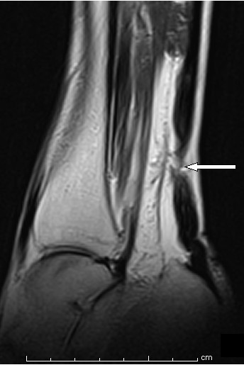
MRISagittal ankle MRI: rupture of the Achilles tendon, arrow on the area of fibre discontinuity.
Source : Khanzada et al., Journal of medical case reports, 2011, figure 1 · CC BY
Key points, chapter 2
- Eleven per cent of ruptures arrive more than fourteen days late, and that delay comes first from the patient who does not seek care, not from the doctor who gets it wrong.
- When diagnostic error does occur, it happens three times more often at later consultations than at the time of the injury. That is the physiotherapist's place in the chain.
- Two tests are enough: calf squeeze (Se 0.96 / Sp 0.93) and Matles (Se 0.88 / Sp 0.85), always comparing sides. At least two tests were positive in every patient in the reference series.
- Preserved resisted plantarflexion, possible walking and the absence of a palpable gap are all compatible with a complete rupture. None of the three rules out the diagnosis.
- Imaging does not make the diagnosis: it rules out other injuries and, more recently, guides the treatment choice.
Surgery or functional treatment: what does the evidence really say?
This is the question that has structured the whole literature on this condition for forty years. It has changed in nature without being closed. This chapter sets out what the evidence establishes, what it does not settle, and why two equally solid sources can appear to contradict each other.
The historical argument, and what shifted it
Surgery established itself on a single, solid argument: it reduces the risk of re-rupture. The 2010 Cochrane review, over twelve trials and 844 participants, established this unambiguously (relative risk of re-rupture 0.41, 95% CI 0.21-0.77, in favour of open surgery) while documenting the price to be paid: a risk of infection multiplied by nearly five (RR 4.89).14
The reference meta-analysis published in the BMJ in 2019 revisited the question across 29 studies, ten randomised trials and nineteen observational studies, 15,862 patients in all. It confirms both sides of the trade-off:
- Re-rupture: 2.3 % after surgery against 3.9 % without (risk difference 1.6 %; RR 0.43, 95% CI 0.31-0.60; p < 0.001).
- Complications: 4.9 % after surgery against 1.6 % without (RR 2.76, 95% CI 1.84-4.13; p < 0.001), most of the gap being due to infection, present in 2.8 % of operated patients.15
The result that shifted the debate lies in a subgroup analysis within the same publication. In the studies where both arms received accelerated functional rehabilitation with early mobility, the re-rupture difference was no longer significant: RR 0.60, 95% CI 0.26-1.37, p = 0.23, with zero heterogeneity (I² = 0 %).15
Two earlier randomised trials had prepared this result. Willits in 2010, in 144 patients where both arms followed an accelerated protocol with early weight-bearing and mobility, finds only 2 re-ruptures in the operated group against 3 in the non-operated group, with no clinically relevant difference in strength, range of motion or calf circumference, but thirteen complications on the surgical side against six.16 Olsson in 2013, in 100 patients, counts no re-rupture after stable repair with accelerated rehabilitation against five without surgery, a gap that does not reach the significance threshold (p = 0.06), at the price of six superficial infections.17
The contradiction that has to be faced
If the matter were settled, the largest recent randomised trial would have confirmed it. It says something else.
The Norwegian trial published in the New England Journal of Medicine in 2022 randomised 554 patients into three arms (non-operative treatment, open repair, minimally invasive surgery) with 526 analysed at twelve months. On the primary outcome it finds no difference: an ATRS change of −17.0 without surgery, −16.0 after open repair and −14.7 after minimally invasive surgery (p = 0.57), with equivalent physical performance and reported function. But on re-ruptures the gap is considerable: 6.2 % without surgery against 0.6 % in each of the two operated arms. And the surgical cost turns up where nobody was looking for it: nine nerve injuries with minimally invasive surgery (5.2 % of patients) against five with open repair (2.8 %) and one without surgery (0.6 %).18
How can a meta-analytic subgroup that abolishes the difference be held together with a trial of 554 patients that finds it tenfold? Three elements of an answer, which have to be given without resolving them artificially:
- Re-rupture is a rare event: confidence intervals are wide and trials are fragile. A reverse fragility index analysis, covering nine randomised trials, 713 patients and 46 re-ruptures, measured how solid the "neutrality" of the trials that find no difference really is. Their median re-rupture rates were 4.00 % on the operated side and 10.00 % on the non-operated side, a gap that, despite its apparent size, did not reach significance for want of numbers.19 "No significant difference" does not mean "no difference".
- The protocols compared are not the same from one trial to another and the following chapter shows that the protocol counts as much as the procedure.
- Function, for its part, does not differ. This is the point on which every source converges, including those that find a re-rupture gap: ATRS, strength, range of motion and activity level are comparable between arms at one year.1820
The most recent meta-analysis, over fourteen randomised trials and 1,628 participants, rules the same way as the BMJ on risk, and refines the cost side: re-rupture reduced with minimally invasive surgery (RR 0.28; CI 0.11-0.74) as with open surgery (RR 0.30; CI 0.19-0.50), but a significant excess of complications only for open surgery (RR 3.03; CI 1.75-5.26), the minimally invasive approach not differing from non-operative treatment (RR 2.40; CI 0.52-10.98). No functional difference, and a faster return to work after minimally invasive surgery.20
The comparison table, with levels of evidence
| Criterion | Surgery | Functional treatment | What the evidence says | Level |
|---|---|---|---|---|
| Re-rupture | 2.3 % (0.6% in the NEJM) | 3.9 % (6.2% in the NEJM) | A consistent advantage to surgery, RR 0.43. But the difference is not significant when both arms have early functional rehabilitation (RR 0.60; 0.26-1.37). | Moderate |
| Overall complications | 4.9 % | 1.6 % | A clear advantage to functional treatment, RR 2.76 (1.84-4.13). The excess is significant for open surgery (RR 3.03), not for the minimally invasive approach. | Moderate |
| Infection | 2.8 % | Almost nil | The main contributor to the complications gap. Cochrane found an RR of 4.89. | Moderate |
| Nerve injury | 2.8% (open) 5.2% (minimally invasive) |
0.6 % | The minimally invasive approach reverses the trade-off: less infection, more sural nerve. Data from a three-arm randomised trial. | Moderate |
| Function at 12 months ATRS, strength, range of motion |
No difference demonstrated | All the sources converge, including those that find a re-rupture gap. Three-arm trial: p = 0.57. | High | |
| Return to work | Faster (minimally invasive) | Reference | The only isolated functional advantage of minimally invasive surgery in the 2025 meta-analysis. | Low |
| Deep vein thrombosis | A shared risk, tied to immobilisation | 2.67% on average in the network meta-analysis; up to 42% on systematic Doppler ultrasound screening in operated patients. | Moderate | |
| Chronic rupture > 14 days |
Clearly superior | Poor results | Median ATRS 77 against 34; self-rated recovery 85% against 40%. The balance of the acute debate does not apply here. | Low cohort |
Levels of evidence: an editorial appraisal applying GRADE logic to the sources cited (meta-analyses of randomised trials downgraded for imprecision on rare events, non-randomised cohorts for chronic rupture). This is not a published GRADE assessment. Sources: Ochen 2019 (PMID 30617123), Myhrvold 2022 (PMID 35417636), Xu 2025 (PMID 41243574), Khan 2010 (PMID 20824836), Wu 2019 (PMID 30781966), Aufwerber 2024 (PMID 38796725), Nilsson 2026 (PMID 42067876).
What the network meta-analysis adds: the procedure-protocol pairing
A Bayesian network meta-analysis covering 29 randomised trials and 2,060 patients compared not procedures but combinations of procedure × rehabilitation protocol. The mean rate of major complications, all strategies taken together, comes to 9.13 %, of which 5 % re-ruptures, 1.50 % deep infections and 2.67 % deep vein thromboses. Two lessons:
- The worst combination is not "not operating", it is "not operating and immobilising": non-operative treatment combined with early immobilisation is significantly associated with the highest risk of major complications.
- The best combination pairs minimally invasive surgery with accelerated rehabilitation, with a 79.7 % probability of being the optimal strategy for minimising major complications.21
This result shifts the question put to the physiotherapist. It is not "should this patient have been operated on?", a decision beyond their reach, but "whatever procedure was chosen, is the loading protocol the one that goes with it?". A non-operated tendon immobilised for six weeks accumulates the drawbacks of both strategies.
Key points, chapter 3
- Surgery more than halves the risk of re-rupture (RR 0.43) and multiplies complications by nearly three (RR 2.76). It is a trade-off, not a superiority.
- With early functional rehabilitation in both arms, the re-rupture gap is no longer statistically significant (RR 0.60; 0.26-1.37).
- The largest recent randomised trial nevertheless finds 6.2 % against 0.6 %. The contradiction is not resolved: re-rupture is a rare event, and the trials lack the power to settle it.
- On function at one year, by contrast, all the sources converge: no difference.
- The minimally invasive approach trades infection for sural nerve injury (5.2 % against 2.8 % in open surgery).
- The most harmful combination is immobilised non-operative treatment. The protocol is not an execution detail of the strategic choice: it is part of it.
How do you choose the strategy with this particular patient?
Since the evidence names no winner, the decision is taken patient by patient. This chapter sets out what tips the balance one way or the other, and the most accomplished attempt to make that choice reproducible rather than a matter of doctrine.
Getting out of the default choice: the individualised algorithm
The Danish CARTA trial, published in the British Journal of Sports Medicine in 2026, framed the question differently. Rather than setting "everybody operated on" against "nobody operated on", it randomised 300 patients into three arms: non-operative treatment by default, operative treatment by default, or an individualised decision on an ultrasound criterion (surgery indicated if ultrasound showed less than 25 % overlap of the tendon ends or an elongation of at least 7 %).
The primary outcome, a heel-rise work test at twelve months, does not differ between the three arms. But among the secondary outcomes, the individualised arm reduces the re-rupture rate by 73 % compared with systematic non-operative treatment from 11 % (CI 6-19) to 3 % (CI 1-8), p = 0.03, improves the ATRS by 8 points (CI 1-15) and the resting angle by 2° (CI 1-4). Against the systematic operative arm there is no difference, except that 36 % fewer patients were operated on (64 % operated on in the individualised arm).22
One nuance not to be glossed over: the reliability of the ultrasound measurement of the gap between the fragments is good overall, but becomes uncertain in the region of a 5 mm threshold.23 An algorithm that rests on an imaging measurement is no more solid than that measurement.
What tips the balance, in practice
| Factor | Points towards surgery | Points towards functional treatment |
|---|---|---|
| Time since the injury | Beyond 14 days (the results of non-surgical treatment collapse: ATRS 34 against 77) | Immediate management, within the first few days |
| Gap between the tendon ends | Overlap < 25% or elongation ≥ 7% on ultrasound, ankle in equinus | Tendon ends that meet in plantarflexion |
| Activity level | High-level pivoting or push-off sport, an argument of preference, not of functional evidence | Recreational or occupational activity without push-off |
| Background | Healthy background, non-smoker, good skin condition | Diabetes, smoking, arterial disease, corticosteroid therapy, immunosuppression (the infection risk, already 2.8%, is higher in these patients) |
| Ability to follow the protocol | A patient who will not be able to hold to a demanding supervised loading protocol | An adherent patient, access to regular rehabilitation, a brace available |
| Risk tolerance | A patient for whom a re-rupture would be catastrophic (a professional athlete, a self-employed worker who cannot take time off) | A patient for whom an infection or a nerve injury would weigh more heavily than a re-rupture |
| What does not decide the matter | Function at one year, strength, range of motion, the rate of return to sport. These outcomes are equivalent and must not serve as an argument either way. | |
The clinical practice guidelines published in 2026 by a committee of forty orthopaedic surgeons, in answer to nine clinical questions, conclude no differently: surgery is "a reliable option", while the indications, the timing and the techniques remain the subject of unsettled controversy, and the emphasis is placed on the overall operative strategy rather than on the isolated procedure.24
What the physiotherapist brings to this decision
The choice belongs to the surgeon and the patient, but three elements come from the physiotherapist and carry weight: the patient's real ability to follow a supervised loading protocol (does he have regular access to the practice? can he manage a brace?), his true functional demands (often different from the ones he announces in the consultation on the day of the injury) and the level of apprehension, which is known to shape recovery and to be commoner in patients treated without surgery (59 % against 48 %).25 These three elements are gathered in ten minutes and appear in no operation note.
What phased rehabilitation after an Achilles tendon rupture?
The previous chapter showed that the loading protocol counts as much as the choice to operate. This one sets it out in detail: what the trials actually varied, what that changed, and why time frames should serve as a safety rail rather than as a green light.
The protocol counts as much as the procedure
A meta-analysis of 43 randomised trials and 2,553 operated patients classified the postoperative protocols and measured re-rupture for each. The overall incidence comes to 3.15 % (95% CI 2.26-4.17), but the spread between protocols is considerable:
The very title of this publication is a warning: mobilisation immediately after repair may increase the incidence of re-rupture.26 "Early" and "immediate" are not synonyms, and that nuance is exactly the one rehabilitation has to hold.
What is established, and what is not
The mode of immobilisation changes nothing. The UKSTAR trial randomised 540 patients treated without surgery in 39 British hospitals between a plaster cast and a functional brace worn for eight weeks: no difference in ATRS at nine months (74.4 against 72.8; adjusted difference −1.38, CI −4.9 to 2.1; p = 0.44), and none in re-rupture rate.27
The date of return to weight-bearing, in the non-operated patient, changes nothing either. Nine randomised trials and 1,046 participants find no difference between early weight-bearing (within four weeks) and late, whether on re-rupture (RR 0.75; CI 0.49-1.16), the ATRS, return to previous sport or time off work.28 A second meta-analysis on accelerated rehabilitation in non-operative treatment concludes the same way, with no significant gap on the ATRS, re-rupture, return to sport or return to work, while recommending it all the same, for the absence of any demonstrated cost.29
An uncomfortable conclusion, but it is the one the data give
In the non-operated patient, the literature does not demonstrate the superiority of early weight-bearing: it demonstrates its safety. The argument in its favour is therefore not "it speeds up healing" but "it costs nothing and gives the patient back his mobility, his independence and his work sooner". That is a sufficient argument, but it has to be presented for what it is. In the patient who has been operated on, by contrast, the ranking of protocols does show a gradient, and strictly immediate mobilisation sits on the wrong side of it.
The timeline: five phases, and what allows you to move to the next
The points to watch in each phase
Phase 1: thrombosis is the real risk of this period
The figure is striking and deserves to be known: in a cohort of 181 operated patients drawn from two randomised trials, with systematic Doppler ultrasound screening at two and six weeks, 76 patients (42 %) had a deep vein thrombosis. And it is not an event without sequel: at three years, those patients still had a heel-rise work symmetry index of 68 % against 78 % (p = 0.027), and thrombosis emerged as an independent risk factor for a poor functional outcome, alongside age and calf wasting.11 The network meta-analysis, for its part, finds a mean incidence of 2.67 %, the gap between the two figures coming from systematic screening against clinical detection, and it says above all how many thromboses go unnoticed.21
Phase 2: the angle, not the pain, sets the limit
The healing tendon does not hurt when it is stretched too far, and that is what makes this phase treacherous.47 The limit is mechanical and is set on range of motion, not on what the patient feels. The consequence of overstepping it is expressed not in pain but in tendon elongation (chapter 7 shows that it is paid for in strength, years later).
Phase 3: the step that half the patients do not clear
A useful marker for calibrating expectations: out of 81 patients treated with early active rehabilitation, with or without surgery, 40 (49 %) were unable to perform a single heel-rise on one leg at twelve weeks. Those who managed it were younger, more often men, less symptomatic and more active.30 A patient who cannot do it at three months is not failing: he is in the expected half. Saying so avoids installing an anxiety that does have a measured cost.
The modalities, and what they are worth
Two comments on the last two rows, because they run counter to widespread practice. Electrical stimulation has been tested against placebo in a double-blind randomised trial of 40 operated patients, with MRI and calf circumference at 2, 6 and 12 weeks: no significant difference, only a trend towards preserved muscle volume.31 Work under blood flow restriction, often put forward as the obvious answer to a phase in which loading is forbidden, has been the subject of a feasibility series of 18 non-operated patients: adherence was good (88 %) and pain minimal, but three adverse events occurred: two re-ruptures after the programme and one deep vein thrombosis, which led the authors themselves to write that efficacy and safety warrant further research.32 The randomised trial meant to settle the matter is under way.33
Key points, chapter 5
- Between the best and the worst postoperative protocol, re-rupture ranges from 1.36 % to 5.95 %, a gap comparable to the one separating surgery from functional treatment.
- "Early" is not "immediate": strictly immediate mobilisation after repair is associated with more re-ruptures.
- Cast or brace, early or late weight-bearing in the non-operated patient: no difference in outcome. The choice is made on comfort, independence and cost.
- Deep vein thrombosis affects 42 % of operated patients on systematic screening and still weighs on function three years later.
- A single-leg heel-rise is impossible in half the patients at twelve weeks. That is the norm, not a failure.
- Electrical stimulation: no demonstrated effect. Blood flow restriction: a safety signal not to be ignored until the ongoing trial has reported.
How do you decide on return to running, and then on return to sport?
In this condition the question "when?" long received an answer in weeks. An international consensus published in 2026 replaced it with a list of criteria, and explicitly discarded one that many practices still measure.
The seven return-to-running criteria
Thirty-five international Achilles tendon experts took part in a modified three-round Delphi study, with a 75 % agreement threshold, on sixteen candidate criteria. Eight reached consensus: seven to be included, and one to be excluded.34
| Criterion retained | How to check it in the clinic |
|---|---|
| No pain in everyday life | Targeted questioning on walking, stairs and prolonged standing |
| No pain during and after sessions | The "after" counts just as much: waking in pain the day after a heavy session |
| Walking without a limp | Observation over a sufficient distance, barefoot and shod |
| Ability to walk on tiptoe | Over a few metres, without support |
| Ten single-leg heel-rises | The most discriminating criterion: full range, with no compensation from the opposite limb |
| Good single-leg balance | Stable single-leg stance, eyes open then closed |
| The patient feels psychologically ready | A question asked directly: see below why it is not decorative |
| Calf circumference symmetry | Explicitly EXCLUDED by consensus. Calf wasting persists for years without preventing a return to activity; requiring it would amount to delaying everybody |
The authors themselves state the limit of their work: these criteria, arising from expert agreement, require prospective validation before they can serve as a decision rule.34 They are worth having as a shared framework, not as an approved test.
A composite tool for what follows: the Ankle-GO
For return to sport proper, a composite score combining four functional tests and two questionnaires, the Ankle-GO, initially validated after lateral ankle sprain, has been the subject of a prospective multicentre validation study in 50 patients operated on for an Achilles rupture, assessed at six and nine months and compared with 30 uninjured controls.35 It is, to date, the best studied instrument for objectifying that particular decision.
What return to sport actually delivers
A systematic review of fifteen studies of professional athletes in American football, basketball, baseball and football provides the clearest markers: 76 % return to professional competition, with a mean delay of eleven months. The corollary: nearly a quarter never return at all. Those who do see their efficiency ratings and their sport-specific statistics decline compared with uninjured controls, and the return rate is significantly lower than after an anterior cruciate ligament rupture, a meniscal injury or an ankle fracture in the same leagues.36
These figures concern professionals and do not transfer as they stand to the patient in the clinic, and the authors themselves put estimated recovery in the general population at around six months. They serve above all to reframe an expectation: this injury costs more, in sporting career terms, than the injuries it is spontaneously compared with.
On the treatment choice, by contrast, the rate of return to sport decides nothing: a dedicated meta-analysis finds no difference between surgery and conservative treatment, nor between early and late rehabilitation after open repair. The only gap measured concerns the timing: early rehabilitation after open repair saves 4.19 weeks on average (p = 0.002).37 In other words: the decision to operate must not be taken on a promise of return to sport.
Fear of re-injury is not a detail of comfort
A cohort of 550 patients followed from one to six years after their rupture gives the measure of the phenomenon: more than half report a fear of re-injury, and those patients have a median ATRS lower by 15 points than the others (p < 0.001). The fear is commoner in patients treated without surgery: 59 % against 48 % (p = 0.024).25 A gap of 15 points on a 100-point scale is of the same order as the one the trials try to obtain by changing surgical technique.
The link had already been established earlier in the pathway: at twelve weeks, kinesiophobia is negatively correlated with every patient-reported outcome and with the level of physical activity.30 That is what justifies the seventh consensus criterion, "the patient feels ready", standing on the same footing as the other six.
Key points, chapter 6
- Seven criteria carry consensus for allowing running, among them ten single-leg heel-rises and the sense of psychological readiness. They are still awaiting prospective validation.
- Calf circumference symmetry has been explicitly excluded by the experts: do not make it a gate.
- In the professional, 76 % return, in eleven months on average, with a measurable drop in performance, a harsher prognosis than for the ACL.
- The rate of return to sport does not separate surgery from functional treatment: it must not serve as an argument in that choice.
- More than one patient in two fears re-injury, and that is worth 15 points of ATRS. Treating it is part of the treatment.
What becomes of plantarflexion strength in the long term?
This is the sequela nobody warns the patient about, because it does not hurt. It is constant, lasting, measurable, and completely uncorrelated with his satisfaction, which is why it goes unnoticed on both sides.
The deficit persists, and stops improving
Two studies from the same Swedish randomised cohort trace the trajectory. At two years, the 81 patients show major functional deficits on the injured side whatever the treatment, with only minor improvements between one and two years, even though their mean ATRS reaches 89 to 90 out of 100. The authors draw an operational conclusion from this: it is in the first year that the final outcome is decided.38
At seven years, 66 of these subjects were reassessed: the deficits persist on every calf performance test (p between < 0.001 and 0.012), and above all, no significant progress occurred after the two-year follow-up, with the exception of heel-rise height, which rose from 10.8 to 11.5 cm.39
The most recent British series confirms and quantifies the final state: 60 participants seen again on average 6.8 years after their rupture have a cross-sectional area of the injured tendon 62 % greater, with four times more disorganised fibrillar structure than on the healthy side (28.7 mm² against 7.3 mm²; p < 0.001), a maximum isometric strength 18 % lower and heel-rise test work 40 % lower.40 The tendon heals by becoming bigger and worse.
Why this deficit: elongation, and its limits as an explanation
The dominant hypothesis is mechanical: a healed tendon that is longer places the triceps at an unfavourable length and makes it lose torque. It rests on a solid correlation, since in the fourteen-year MRI series the difference in tendon length between the two sides is substantially correlated with the strength deficit (ρ = 0.51; p < 0.001), as well as with atrophy of the medial gastrocnemius (ρ = 0.46) and of the soleus (ρ = 0.42), for a mean elongation of 12 mm, that is 6 %.8
A 2026 study nevertheless qualifies this explanation. Twenty-nine non-operated patients, classified as high or low functioning according to their heel-rise work index, were compared on bilateral MRI: elongation of the free tendon exists in both groups (3.59 cm in the good recoverers against 5.19 cm in the poor ones, p = 0.053, right on the threshold). What really separated the two groups was age, thirteen years apart, the tendon cross-section, and the state of the soleus.41 Elongation contributes, but it does not explain everything: the soleus and age count at least as much.
An early predictor, available in the clinic
One finding deserves to be known by every physiotherapist who follows these patients. In 35 patients treated without surgery, the symmetry of the ankle resting angle measured at two months predicts plantarflexion strength symmetry at six and at twelve months (β = 2.530, CI 1.041-4.018, adjusted R² 0.416, p = 0.002 at six months; β = 1.659, CI 0.330-2.988, p = 0.016 at twelve months).42 It is the same measurement as the Matles test in chapter 2, taken again at two months as a prognostic indicator. It costs nothing and is documented in thirty seconds.
The trap: the satisfied patient who has not recovered
This is the most counter-intuitive result in this chapter. A series of 76 men operated on, seen again at a median of 41 months after open repair, shows a median ATRS of 97 out of 100 and an AOFAS score of 98 (figures that are almost perfect). The same sample, tested on the single-leg heel-rise, gives a median symmetry index of 87.5 %, and only 31 patients (40.8 %) reach the 90 % threshold. Above all: no correlation between the symmetry index and the ATRS (rho = 0.190; p = 0.100) or the AOFAS score (rho = 0.218; p = 0.058).43
The practical consequence is direct: if you do not test, you do not see. A satisfied patient, free of pain, back to his activities, can still carry a 40 % deficit of plantarflexion work. That deficit is not trivial, since it alters the biomechanics of walking and running one year after the rupture44 and it will not be made up if the first two years are allowed to slip by.
Key points, chapter 7
- The plantarflexion strength deficit is constant: −18 % of isometric strength and −40 % of work at nearly seven years.
- It stops improving after two years. Everything that has to be gained has to be gained before then.
- Tendon elongation (12 mm on average) contributes to it and correlates with the deficit (ρ = 0.51), but it is found in patients who recover well too.
- Resting angle symmetry at two months predicts strength symmetry at six and twelve months: it is a clinic measurement, free of charge, to be recorded.
- Satisfaction questionnaires do not detect this deficit: an ATRS of 97 out of 100 and a symmetry index of 87.5 % coexist without correlation. You have to test.
What do concrete clinical cases teach us?
Three published and indexed case reports, chosen because each of them contradicts a received idea from the previous page. No reconstructed or composite case is used here: every patient described corresponds to a publication cited with its identifier.
Case 1: a diagnosis at four weeks, and yet a good result without surgery
A 19-year-old female athlete feels calf pain in the middle of sport. The complete Achilles tendon rupture is not diagnosed until four weeks after the injury. Ultrasound then shows a discontinuity with a gap of 2.0 cm that persists even in maximum plantarflexion which is precisely the situation that, by the criteria of the Danish algorithm in chapter 4, would point towards surgery.
The team nevertheless chooses non-operative treatment: immobilisation in plantarflexion, then gradual recovery of dorsiflexion up to the tenth week. At one year, MRI shows a tendon that has become continuous again, the patient is pain-free and has returned to high-level sport with performances equivalent to those before.45
What this case shifts
It is a reminder that a diagnostic delay is not a sentence, and that a residual gap does not forbid healing, in a young female patient. That is the caveat to keep: the data in chapter 7 show that thirteen years of age difference separated the good from the poor recoverers in a comparable series. An isolated case does not license transferring this approach to the 55-year-old diabetic patient seen at four weeks.
Case 2: the neglected rupture on an inflammatory background
A man of 69 years, with rheumatoid arthritis, presents with an Achilles tendon rupture that is neglected. Two difficulties add up: a large tendon gap, and tendon quality degraded by the inflammatory disease, which rules out using his own tendons as a graft. Reconstruction is carried out with an Achilles tendon allograft with a calcaneal bone block, with a clinical and functional result reported as excellent.46
This case illustrates what the surgical side of chronic rupture actually looks like: repair gives way to reconstruction, with material that does not belong to the patient. That is the underlying price of the 11 % of ruptures managed late, and the most concrete argument for insisting to patients that a calf that "is not recovering" must be re-examined.
Case 3: when the rupture is the symptom of something else
A woman of 32 years is injured playing basketball and arrives in the emergency department with an Achilles tendon rupture, repaired surgically. The course is abnormal: delayed skin healing requiring a skin graft, then, two months later, a second rupture at a different level of the tendon, requiring a tendon transfer. It is only at this stage that the rheumatology department makes the diagnosis of systemic lupus erythematosus, of which the rupture was the presenting manifestation.12
Three warning signals were adding up and could have been read earlier: a woman of 32 years (an atypical profile when the female peak lies between 40 and 49 years and men account for nearly 80 % of ruptures), a disproportionate delay in wound healing, and a recurrence at a different site. None of these elements is specific; their sum is.
Where these cases sit in the hierarchy of evidence
It has to be said clearly: an isolated case report occupies the bottom rank of the hierarchy of evidence and founds no recommendation. Its value lies elsewhere, since it documents what a meta-analysis cannot show: the concrete shape of an exception, the chain of decisions, what a failure looks like.
The three cases above therefore do not prove that chronic ruptures should be treated without surgery in young people (case 1), nor that rheumatoid arthritis demands an allograft (case 2), nor that every rupture in a young woman should prompt a search for lupus (case 3). They indicate that these situations exist, which is enough to justify keeping them in mind, and nothing more.
How do you apply all this concretely in practice?
What this article changes in an ordinary consultation, what to measure and record, and when to refer back.
The four actions that change something
| When | Action | What it prevents |
|---|---|---|
| In any acute posterior leg pain, even seen late | Calf squeeze and Matles test, comparing sides. Always both; never one alone; never resisted plantarflexion as an argument. | The rupture missed at a later consultation, three times commoner than the initial error |
| Immobilisation phase | Question and examine looking for venous thrombosis at every session: the opposite calf compared, new pain, swelling. | A DVT that, undetected, still weighs on function three years later |
| At two months | Measure and record the resting angle on both sides. Thirty seconds, patient prone, knees flexed. | Losing the only early predictor available of strength symmetry at six and twelve months |
| At three months, six months, one year | Test the single-leg heel-rise and calculate the symmetry index (injured side / healthy side × 100), in number of repetitions and in height. | Concluding from a satisfaction questionnaire, which does not detect a 40% deficit |
What to tell the patient, and when
- From the first session: that rehabilitation is counted in months, not weeks, and that the useful window closes at around two years. This is not discouraging, it is mobilising, since it justifies the effort asked for at the moment when it pays off most.
- Around the twelfth week: that a heel-rise on one leg is impossible in half the patients at this stage. Saying so before the test avoids installing an anxiety known to cost 15 points of ATRS.
- Before return to running: the seven criteria, set out as a checklist to go through together. The seventh, "do you feel ready?", is asked straight out, and a negative answer is treated instead of being worked around.
- On the calf that stays thinner: that the wasting is expected, that it persists for years, and that it has been explicitly discarded from the return criteria. Many patients cling to it as an indicator of failure.
When to refer back without waiting
- Sudden pain during rehabilitation with loss of function: repeat both tests, suspect a re-rupture, refer back the same day.
- A warm, tense, painful calf: suspected deep vein thrombosis.
- Delayed healing, discharge, wound breakdown: infection concerns 2.8 % of operated patients and is not monitored in the clinic.
- A patient referred for a "calf muscle injury" in whom the calf squeeze is abnormal: it is not for the physiotherapist to settle it, but it is for him to do the test and report it.
- A resting angle that deteriorates between two measurements: progressive tendon elongation during rehabilitation warrants an opinion, before the loss becomes permanent.
Frequently asked questions
Can a patient who is still walking have a complete rupture?
Yes, and it is the commonest situation. The toe flexors, tibialis posterior and the peroneals produce enough plantarflexion to walk on the flat. This compensation is still measurable fourteen years after the injury, in the form of hypertrophy of flexor hallucis longus.8 Preserved walking rules nothing out; a single-leg heel-rise, by contrast, is impossible.
Should an athlete be operated on?
The evidence does not require it. Function at one year, strength, range of motion and the rate of return to sport do not differ between strategies.1837 What surgery brings is a reduction in the risk of re-rupture, an argument that weighs more heavily in a patient for whom a second rupture would be professionally catastrophic. It is a decision about risk tolerance, not about expected performance.
How long before I can run again?
The question is badly framed: the international consensus has replaced the time frame with seven criteria, among them ten single-leg heel-rises and walking without a limp.34 As an order of magnitude, these criteria are rarely met before the fourth month, but a patient who meets them at four months is readier than a patient who does not meet them at six.
Will the calf regain its size?
Most often no, and that prevents neither a return to activity nor a good outcome. Wasting of the triceps surae persists, 11 to 13 % less volume at fourteen years,8 and circumference symmetry has been explicitly excluded from the return-to-running criteria.34
Is a partial rupture treated like a complete one?
No, and it is a frequent source of confusion. The diagnostic values quoted in this article were established on ruptures that were complete: the calf squeeze can stay normal in a partial rupture. That presentation is closer to acute tendinopathy, and calls for its own assessment.
What is the risk of rupturing the other tendon?
This article cites no data allowing that risk to be quantified, so no figure should be offered. What is documented, on the other hand, is that bilateral ruptures, or ruptures occurring without adequate trauma, should prompt a search for a systemic cause: fluoroquinolone use, corticosteroid therapy, systemic inflammatory disease.1012
Does this article apply to Achilles pain that has been present for months?
No. Pain of gradual onset, with no dated injury and a continuous tendon, falls under Achilles tendinopathy, whose treatment rests on progressively increasing load, the opposite of the protection principle described here.
References
Forty-seven references, each checked against the PubMed metadata (NCBI E-utilities) on 15 August 2026: authors, title, journal, year, volume, pagination and DOI. The PMID links point to PubMed, the DOI links to doi.org. The two clinical cases in chapter 8, along with the lupus case cited in chapter 2, are indexed publications cited with their identifier: no reconstructed or composite case has been used.
Epidemiology and natural history
- Kotsifaki R, Malliaras P, Byron C et al. Incidence, Temporal Trends, and Surgical Shift of Achilles Tendon Rupture: A Systematic Review and Meta-analysis. Sports Med 2026;56(6):1467-1487.PMID 41933260 ·DOI 10.1007/s40279-026-02397-5
- Svedman S, Marcano A, Ackermann PW et al. Acute Achilles tendon ruptures between 2002-2021: sustained increased incidence, surgical decline and prolonged delay to surgery-a nationwide study of 53,688 ruptures in Sweden. BMJ Open Sport Exerc Med 2024;10(3):e001960.PMID 39040046 ·DOI 10.1136/bmjsem-2024-001960
- Burke OC Jr, Milano ME, Onor GI Jr et al. Epidemiology of Achilles tendon ruptures in the United States: 2019 to 2024. Phys Sportsmed 2026.PMID 42506584 ·DOI 10.1080/00913847.2026.2710601
Diagnostic delay and the values of the clinical tests
- Nilsson N, Larsson E, Dyrehag E et al. Incidence, reason for treatment delay and patient-reported outcome of patients affected by a chronic Achilles tendon rupture in a Swedish population. BMC Musculoskelet Disord 2026;27(1).PMID 42067876 ·DOI 10.1186/s12891-026-09890-y
- Maffulli N. The clinical diagnosis of subcutaneous tear of the Achilles tendon. A prospective study in 174 patients. Am J Sports Med 1998;26(2):266-70.PMID 9548122 ·DOI 10.1177/03635465980260021801
- Reiman M, Burgi C, Strube E et al. The utility of clinical measures for the diagnosis of achilles tendon injuries: a systematic review with meta-analysis. J Athl Train 2014;49(6):820-9.PMID 25243736 ·DOI 10.4085/1062-6050-49.3.36
- Dams OC, Reininga IHF, Gielen JL et al. Imaging modalities in the diagnosis and monitoring of Achilles tendon ruptures: A systematic review. Injury 2017;48(11):2383-2399.PMID 28943056 ·DOI 10.1016/j.injury.2017.09.013
Functional anatomy and compensation
- Heikkinen J, Lantto I, Piilonen J et al. Tendon Length, Calf Muscle Atrophy, and Strength Deficit After Acute Achilles Tendon Rupture: Long-Term Follow-up of Patients in a Previous Study. J Bone Joint Surg Am 2017;99(18):1509-1515.PMID 28926379 ·DOI 10.2106/JBJS.16.01491
- Khair RM, Stenroth L, Cronin NJ et al. Exploration of muscle-tendon biomechanics one year after Achilles tendon rupture and the compensatory role of flexor hallucis longus. J Biomech 2023;152:111586.PMID 37080080 ·DOI 10.1016/j.jbiomech.2023.111586
- Sangiorgio A, Sirone M, Adravanti FM et al. Achilles tendon complications of fluoroquinolone treatment: a molecule-stratified systematic review and meta-analysis. EFORT Open Rev 2024;9(7):581-588.PMID 38949172 ·DOI 10.1530/EOR-23-0181
- Aufwerber S, Svedman S, Silbernagel KG et al. Long-term patient outcome is affected by deep venous thrombosis after Achilles tendon rupture repair. Knee Surg Sports Traumatol Arthrosc 2024;32(8):2184-2193.PMID 38796725 ·DOI 10.1002/ksa.12240
- Jiménez-Yarza M, Jiménez-Puga M, Ramírez-Jasso J et al. Recurrent Achilles Tendon Rupture in Multiple Sites as a Primary Manifestation of Systemic Lupus Erythematosus in a 32-Year-Old Patient: A Case Report. Cureus 2024;16(5):e61231.PMID 38939241 ·DOI 10.7759/cureus.61231
- Schwach M, Gaulin B, Vermorel PH et al. Tennis leg: Diagnosis and management - A state-of-the-art review. Foot (Edinb) 2026;68:102267.PMID 42258915 ·DOI 10.1016/j.foot.2026.102267
Surgery against functional treatment
- Khan RJ, Carey Smith RL. Surgical interventions for treating acute Achilles tendon ruptures. Cochrane Database Syst Rev 2010.PMID 20824836 ·DOI 10.1002/14651858.CD003674.pub4
- Ochen Y, Beks RB, van Heijl M et al. Operative treatment versus nonoperative treatment of Achilles tendon ruptures: systematic review and meta-analysis. BMJ 2019;364:k5120.PMID 30617123 ·DOI 10.1136/bmj.k5120
- Willits K, Amendola A, Bryant D et al. Operative versus nonoperative treatment of acute Achilles tendon ruptures: a multicenter randomized trial using accelerated functional rehabilitation. J Bone Joint Surg Am 2010;92(17):2767-75.PMID 21037028 ·DOI 10.2106/JBJS.I.01401
- Olsson N, Silbernagel KG, Eriksson BI et al. Stable surgical repair with accelerated rehabilitation versus nonsurgical treatment for acute Achilles tendon ruptures: a randomized controlled study. Am J Sports Med 2013;41(12):2867-76.PMID 24013347 ·DOI 10.1177/0363546513503282
- Myhrvold SB, Brouwer EF, Andresen TKM et al. Nonoperative or Surgical Treatment of Acute Achilles' Tendon Rupture. N Engl J Med 2022;386(15):1409-1420.PMID 35417636 ·DOI 10.1056/NEJMoa2108447
- Bragg JT, Ruelos VCB, McIntyre JA et al. Reverse Fragility Index Comparing Rates of Rerupture After Open Achilles Tendon Repair Versus Early Functional Rehabilitation: A Systematic Review of Randomized Controlled Trials. Am J Sports Med 2024;52(4):1116-1121.PMID 37306060 ·DOI 10.1177/03635465231178831
- Xu S, Xiao J, Li Y et al. Operative versus nonoperative management of acute Achilles tendon rupture: a systematic review and meta-analysis of clinical outcomes from randomized controlled trials. Ann Med 2025;57(1):2537349.PMID 41243574 ·DOI 10.1080/07853890.2025.2537349
- Wu Y, Mu Y, Yin L et al. Complications in the Management of Acute Achilles Tendon Rupture: A Systematic Review and Network Meta-analysis of 2060 Patients. Am J Sports Med 2019;47(9):2251-2260.PMID 30781966 ·DOI 10.1177/0363546518824601
- Toft M, Hansen MS, Vestergaard JD et al. Randomised three-armed trial investigation of the Copenhagen Achilles tendon Rupture Treatment Algorithm (CARTA) for individualised treatment of acute Achilles tendon rupture. Br J Sports Med 2026;60(12):848-855.PMID 42082321 ·DOI 10.1136/bjsports-2025-110210
- Mocanu D, Bokwa-Dąbrowska K, Larsson E et al. Ultrasound gap measurement after acute Achilles rupture is reliable overall but uncertain near a 5-mm decision threshold. Skeletal Radiol 2026;55(9):2301-2313.PMID 42103998 ·DOI 10.1007/s00256-026-05243-x
- Feng SM, Maffulli N, Oliva F et al. Evidence-based clinical practice guidelines on the surgical management of acute Achilles tendon rupture. Br Med Bull 2026;157(1).PMID 41528729 ·DOI 10.1093/bmb/ldaf026
Rehabilitation, weight-bearing and modalities
- Larsson E, LeGreves A, Brorsson A et al. Fear of reinjury after acute Achilles tendon rupture is related to poorer recovery and lower physical activity postinjury. J Exp Orthop 2024;11(4):e70077.PMID 39493002 ·DOI 10.1002/jeo2.70077
- Wang R, Huang L, Jiang S et al. Immediate mobilization after repair of Achilles tendon rupture may increase the incidence of re-rupture: a systematic review and meta-analysis of randomized controlled trials. Int J Surg 2024;110(6):3888-3899.PMID 38477123 ·DOI 10.1097/JS9.0000000000001305
- Costa ML, Achten J, Marian IR et al. Plaster cast versus functional brace for non-surgical treatment of Achilles tendon rupture (UKSTAR): a multicentre randomised controlled trial and economic evaluation. Lancet 2020;395(10222):441-448.PMID 32035553 ·DOI 10.1016/S0140-6736(19)32942-3
- Ghaddaf AA, Alomari MS, Alsharef JF et al. Early versus late weightbearing in conservative management of acute achilles tendon rupture: A systematic review and meta-analysis of randomized controlled trials. Injury 2022;53(4):1543-1551.PMID 35115168 ·DOI 10.1016/j.injury.2022.01.028
- Coopmans L, Amaya Aliaga J, Metsemakers WJ et al. Accelerated Rehabilitation in Non-operative Management of Acute Achilles Tendon Ruptures: A Systematic Review and Meta-analysis. J Foot Ankle Surg 2022;61(1):157-162.PMID 34400090 ·DOI 10.1053/j.jfas.2021.07.007
- Olsson N, Karlsson J, Eriksson BI et al. Ability to perform a single heel-rise is significantly related to patient-reported outcome after Achilles tendon rupture. Scand J Med Sci Sports 2014;24(1):152-8.PMID 22716232 ·DOI 10.1111/j.1600-0838.2012.01497.x
- Hyer CF, Berlet G, Philbin T et al. Does Functional Neuromuscular Electrical Stimulation (NMES) Influence Calf Atrophy Following Achilles Tendon Surgery? Prospective Double-Blind Randomized Controlled Trial on the Use of Immediate Postoperative Electrical Muscle Stimulation to Preserve Muscle Function and Volume. J Foot Ankle Surg 2021;60(4):683-688.PMID 33736944 ·DOI 10.1053/j.jfas.2020.12.005
- Bentzen A, Jørgensen SL, Birch S et al. Feasibility of Blood Flow Restriction Exercise in Adults with a Non-surgically Treated Achilles Tendon Rupture; a Case Series. Int J Exerc Sci 2024;17(3):140-153.PMID 38665686 ·DOI 10.70252/QGAF3184
- Bentzen A, Gundtoft PH, Silbernagel KG et al. The effectiveness of low-load Blood flow restriction Exercise in patients with an acute Achilles tendon rupture treated Non-surgically (BEAN): Protocol for a randomized controlled trial. Foot (Edinb) 2024;61:102133.PMID 39260068 ·DOI 10.1016/j.foot.2024.102133
Return to running, return to sport
- Gaspar M, Maffulli N, Memain G et al. Criteria for Return to Running After Surgical Repair of Acute Achilles Tendon Rupture: A Modified Delphi Consensus Study. Sports Med 2026.PMID 42503592 ·DOI 10.1007/s40279-026-02506-4
- Lopes R, Freiha K, Carmont MR et al. Validation of a Composite Outcome Score for Assessing Return to Sports After Achilles Tendon Repair. Am J Sports Med 2025;53(7):1707-1715.PMID 40263952 ·DOI 10.1177/03635465251333142
- Johns W, Walley KC, Seedat R et al. Career Outlook and Performance of Professional Athletes After Achilles Tendon Rupture: A Systematic Review. Foot Ankle Int 2021;42(4):495-509.PMID 33218267 ·DOI 10.1177/1071100720969633
- Bak BM, Seow D, Teo YZE et al. Return to Play and Functional Outcomes Following Treatment of Acute Achilles Tendon Ruptures: A Systematic Review and Meta-Analysis. J Foot Ankle Surg 2024;63(3):420-429.PMID 38296023 ·DOI 10.1053/j.jfas.2023.12.008
Residual deficit and long-term prognosis
- Olsson N, Nilsson-Helander K, Karlsson J et al. Major functional deficits persist 2 years after acute Achilles tendon rupture. Knee Surg Sports Traumatol Arthrosc 2011;19(8):1385-93.PMID 21533539 ·DOI 10.1007/s00167-011-1511-3
- Brorsson A, Grävare Silbernagel K, Olsson N et al. Calf Muscle Performance Deficits Remain 7 Years After an Achilles Tendon Rupture. Am J Sports Med 2018;46(2):470-477.PMID 29068725 ·DOI 10.1177/0363546517737055
- Briggs-Price S, Mangwani J, Kilcran A et al. Structure and Function of the Achilles Tendon and Plantarflexors 1 Year Following Achilles Tendon Rupture in the United Kingdom: A Cross-Sectional Study. J Foot Ankle Res 2026;19(1):e70134.PMID 41673242 ·DOI 10.1002/jfa2.70134
- Szaro P, Meunier A, Bokwa-Dabrowska K et al. Tendon elongation in the free tendon is evident in patients with and without persistent muscle weakness following an Achilles tendon rupture. Knee Surg Sports Traumatol Arthrosc 2026;34(7):2623-2638.PMID 42159201 ·DOI 10.1002/ksa.70445
- Sukanen M, Khair RM, Reito A et al. Early Predictors of Recovery From Nonoperatively Treated Achilles Tendon Rupture: 1 Year Follow-Up Study. Scand J Med Sci Sports 2024;34(7):e14700.PMID 39010659 ·DOI 10.1111/sms.14700
- Dogruoz F, Askin A, Ergun A et al. Discordance Between Patient-Reported Outcomes and Heel-Rise Limb Symmetry After Surgical Repair of Acute Achilles Tendon Rupture. J Clin Med 2026;15(12).PMID 42355836 ·DOI 10.3390/jcm15124669
- Huseth K, Harðarson GR, Aagaard P et al. Side-to-side differences in neuromuscular function and associated joint mechanics in the lower limbs and trunk during walking and running one year after acute unilateral Achilles tendon rupture. J ISAKOS 2026.PMID 42556514 ·DOI 10.1016/j.jisako.2026.101198
Published clinical cases
- Schwartz PE, Schwartz MH, Georgiadis AG. Delayed Diagnosis of Complete Achilles Tendon Rupture in a Teenage Athlete: A Case Report of Nonoperative Treatment. JBJS Case Connect 2024;14(1).PMID 38306444 ·DOI 10.2106/JBJS.CC.23.00624
- Moreira FP, Sousa A, Machado S. Neglected Achilles tendon rupture associated with rheumatoid arthritis: a case report and a brief review of the literature. BMJ Case Rep 2021;14(1).PMID 33504535 ·DOI 10.1136/bcr-2020-239477
Functional rehabilitation protocol
- Orji C, Goudie P, Mason L. Protocol for Functional Rehabilitation of Achilles Tendon Ruptures. Foot Ankle Clin 2026;31(3):363-379.PMID 42538069 ·DOI 10.1016/j.fcl.2025.12.004
For the progressive overload form of the same tendon (pain that sets in without an injury, a continuous tendon, treatment by increasing load), the dedicated article is Achilles tendinopathy. For the whole range of ankle and foot conditions, see the lower limb hub.

