














On-siteDPCFIFPL

Lower limb
Myo-collagenous calf injuries and Achilles tendon rupture: from clinic-based rehabilitation to patient self-management
Damien Rech
Go further
Calf injury: ruling out deep vein thrombosis and Achilles rupture, locating gastrocnemius or soleus, and returning to sport on criteria. Sources checked.
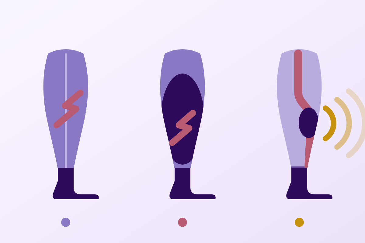
Facing a painful, swollen calf, first rule out deep vein thrombosis: one patient in ten diagnosed with “muscle injury” actually had an isolated DVT.
9.9%of patients referred for tennis leg actually had an isolated thrombosis
What follows covers each of these points in detail, with sources. It is there if you need it.
This topic is taught in a course: Myo-collagenous calf injuries and Achilles tendon rupture: from clinic-based rehabilitation to patient self-management, on-site with Damien Rech.
1 / 14
13 article chapters · 53 min in total
Rule out DVT◔ 8 min
A painful, swollen calf can have several causes: clinical presentation is not enough.
Rule out Achilles◔ 5 min
Unlike thrombosis, Achilles rupture is screened with excellent clinical tests.
Anatomy◔ 3 min
The gastrocnemius crosses the knee, the soleus does not: that governs the whole reasoning.
Locating◔ 4 min
Contrary to the tennis leg image, the soleus accounts for 84.6 % of calf injuries.
Classifying◔ 5 min
It is not the muscle that limits return: it is the connective tissue that transmits the force.
Risk factors◔ 4 min
The only two solid risk factors are age and a previous history of the same injury.
Loading◔ 2 min
The triceps surae is the muscle you cannot unload without unloading walking.
Rehabilitating◔ 6 min
The therapeutic literature on the calf is poor, and the experts say so themselves.
Strengthening◔ 4 min
Strengthening the calf with the knee extended only neglects the muscle most often injured.
Return to sport◔ 4 min
20 % of reinjuries occur before full recovery: returning too soon costs dear.
Case reports◔ 3 min
Three published cases each illustrate a different error. None is reconstructed.
In practice◔ 2 min
A triceps surae injury nearly always has its movement and its second: their absence is a red flag.
Questions◔ 3 min
“Tennis leg” classically denotes injury of the medial head of gastrocnemius, not the whole calf.
Train on this
Lower limb
Damien Rech
Next comes practice: the course that teaches this topic, with Damien Rech.
Our courses on this topic may be covered by DPC or FIFPL, the French continuing-education funding schemes, depending on your profession and situation.
Sports trauma · Lower limb
A patient arrives with a calf that has been painful since yesterday, swollen, and that they cannot roll through when walking. Before laying hands on it, two questions must have been answered: is it really muscle, and is the Achilles tendon intact? Both answers are obtained in the clinic, in a few minutes, and they decide everything else.
of patients referred with a clinical diagnosis of “tennis leg” in fact had an isolated deep vein thrombosis, with no muscle injury at all
Delgado 2002, ultrasound series of 141 patients (PMID 12091669)
of calf injuries involve the soleus in elite Australian footballers, and not the gastrocnemius, contrary to the classic picture of tennis leg
Green 2020, 184 injuries from the AFL registry (PMID 31494970)
mean time to return when the connective tissue is disrupted, against 8 days when it is intact: a sixfold difference within the same condition
Prakash 2018, 100 consecutive patients on MRI (PMID 29074478)
What this chapter gives: the short version, the one you reread in the corridor before going into the room. The rest of the article details each of these lines and gives the source of every figure.
Calf muscle injury is a common and deceptive presentation. Common: the medial head of gastrocnemius is the third most injured muscle in the elite athlete, after biceps femoris and rectus femoris (Bright 2017, PMID 28661826). Deceptive: behind an identical clinical picture (sudden pain in the back of the leg, swelling, limping), hide at least four entities, two of which are emergencies.
The approach comes down to three steps, in this order, and the order is not negotiable.
First step, rule out deep vein thrombosis. In the reference ultrasound series of 141 patients referred with a clinical diagnosis of tennis leg, 14 of them (9.9 %) had no muscle injury at all: they had an isolated DVT (Delgado 2002, PMID 12091669). One patient in ten. That is why massage, vigorous mobilisation or pneumatic compression applied to an uninvestigated calf are not neutral acts.
Second step, rule out Achilles tendon rupture. The calf squeeze test, known as the Thompson or Simmonds test, has a sensitivity of 0.96 and a specificity of 0.93 in a prospective series of 174 ruptures and 28 unconfirmed suspicions (Maffulli 1998, PMID 9548122). It costs fifteen seconds.
Only then, third step, locate the muscle injury. Medial gastrocnemius at the myotendinous junction, soleus, plantaris: the picture, the timelines and the reinjury risk differ markedly. The soleus dominates heavily in volume: 84.6 % of calf injuries in the elite Australian football registry, and accounts for 91.4 % of reinjuries (Green 2020, PMID 31494970).
The clinical question is not “what grade?” but “is it really muscle?”. A grade can be corrected along the way; a massaged thrombosis cannot.
On prognosis, one landmark is worth more than any table of timelines: it is the state of the connective tissue, not the volume of muscle oedema, that predicts how long the patient will be unavailable. Across 100 consecutive patients, the mean time to return went from 8 days when the connective interfaces were intact to 48 days when they were disrupted (Prakash 2018, PMID 29074478).
Key points
The danger of this presentation lies not in the muscle injury, which nearly always heals well, but in the two diagnoses it mimics: deep vein thrombosis and Achilles tendon rupture. Ruling them out takes less than five minutes and is done entirely through the history and the examination. Only then does the site of the injury, soleus or gastrocnemius, guide the timelines and the reinjury risk.
This article covers injury of the triceps surae. For injury from a direct blow, whose pathophysiology and complications are different, see muscle contusion (dead leg) and its complications. For chronic tendon distress in the same muscle-tendon complex, see Achilles tendinopathy.
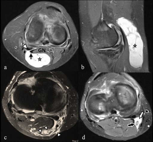
MRIMRI of the knee and calf, four views: large multiseptated popliteal cyst extending towards the calf, to be distinguished from a muscle tear in the differential diagnosis of tennis leg.
Source : Perdikakis et al., Insights into Imaging, 2013, figure 1 · CC BY-NC
This is the chapter that involves patient safety, and that is why it comes first rather than in the middle. A painful, swollen calf looks like a painful, swollen calf, whatever the cause.
Clinical experience builds a mental picture of tennis leg: the tennis or badminton player in their forties, the sensation of being kicked or hit by a stone in the muscle belly, sometimes an audible snap, the immediate inability to push off the forefoot. That picture is accurate, and that is precisely what makes it dangerous: it is so characteristic that it discourages looking for anything else.
The reference series on this point is old but has not been refuted. Delgado and colleagues reviewed the ultrasound scans of 141 patients referred for imaging with a clinical diagnosis of tennis leg already made. In other words, patients whom a clinician had already examined and labelled. This is what ultrasound found.
What ultrasound really finds in 141 patients labelled “tennis leg”
Retrospective series, consensus reading by two radiologists, Radiology 2002
Source: Delgado GJ, Chung CB, Lektrakul N, et al. Radiology 2002;224(1):112-9 (PMID 12091669). The five categories are those reported by the authors and cover the whole series: 94 + 30 + 2 + 1 + 14 = 141 patients.
Two results from this series deserve to be kept separate.
The first is the safety figure: an isolated DVT in 14 patients out of 141, that is roughly one patient in ten, in subjects already examined by a clinician who had concluded to a muscle injury. That is not a statistical curiosity, it is a measured clinical error rate.
The second corrects a stubborn belief. Tennis leg was long taught as a rupture of the plantaris. In this series, the plantaris is at fault in only 2 cases out of 141, that is 1.4 %. The muscle really involved is the medial head of gastrocnemius, in two thirds of cases.
DVT and calf muscle injury share their cardinal signs: calf pain, increased volume, local heat, pain on passive dorsiflexion, discomfort on weight-bearing. They also share part of their background: recent immobilisation, prolonged travel, age. And they can coexist: Slawski reported a case of ipsilateral thrombophlebitis occurring on an already established tennis leg, recalling in passing that the reverse error, taking a tennis leg for thrombophlebitis, had led to unnecessary anticoagulation and to bleeding complications (Slawski 1994, PMID 8027900).
This point matters so as not to swing to the opposite excess. The aim is not to send every painful calf to the vascular emergency department: it is to know how to recognise the situations that cannot be settled in the clinic, and not to treat before they are settled.
Red flags: stop the session and refer
Faced with acute calf pain, each of these features means no massage, no forceful mobilisation, no compression, and referral for a medical opinion the same day.
A single one of these is enough. They do not add up towards a threshold.
The Wells score is the usual landmark for pre-test probability of DVT. It is useful to the physiotherapist for structuring the history, but you have to know what it does not do. In 1,135 inpatients with suspected DVT, the area under the ROC curve for the score was only 0.60, and 5.9 % of patients classed as low probability nevertheless had a proximal DVT: the authors conclude that the score “is not sufficient to rule out DVT or to guide decisions” in that setting (Silveira 2015, PMID 25985219).
The practical consequence is clear and it is liberating: the physiotherapist's role is not to calculate a probability in order to decide whether to treat. It is to spot the doubt and pass it on. The decision to image, to anticoagulate or to monitor belongs to the doctor, and it is itself debated: for symptomatic isolated distal DVT in the low-risk patient, therapeutic anticoagulation has not been shown superior to placebo for reducing thromboembolic events, at the price of increased bleeding risk (Righini & Robert-Ebadi 2024, PMID 38546285). If the question divides haemostasis specialists, it is not settled on a treatment couch.
The five entities that make up most acute calf pain, with what separates them.
| Entity | What suggests it | What rules it out | What to do |
|---|---|---|---|
| Injury of the medial head of gastrocnemius “tennis leg” |
A precise moment, knee extended and ankle dorsiflexed (lunge, sprint start). Pain localised at the myotendinous junction, upper-middle third, medial side. Dependent bruising at 48-72 h. | Negative calf squeeze test, resting equinus preserved, no thrombogenic background, pain reproduced by resisted contraction. | Rehabilitation. Ultrasound useful if the extent is in doubt. |
| Soleus injury | More insidious onset, often on a recent training load. More diffuse, deeper pain, harder to localise. Discomfort greater with the knee flexed. | The same exclusion criteria. The gradual character is in itself reassuring with regard to tendon rupture. | Rehabilitation, with longer timelines to announce. |
| Deep vein thrombosis | No clear mechanism. Progressive swelling, heat, superficial venous dilatation, constant pain. Thrombogenic background. | Nothing rules it out clinically with certainty. Duplex ultrasound decides, not the examination. | Do not massage, do not compress. Medical opinion the same day; emergency if breathless or with chest pain. |
| Achilles tendon rupture | The sensation of a violent blow above the heel, sometimes a snap. Inability to rise onto the toes in single-leg stance. | Negative calf squeeze test and resting equinus preserved and no palpable gap. | Prompt surgical opinion. Immobilisation in equinus meanwhile, no weight-bearing in dorsiflexion. |
| Ruptured popliteal cyst pseudo-thrombophlebitis |
A history of swelling in the popliteal fossa, knee joint disease (osteoarthritis, arthritis). Pain running down from the popliteal fossa into the calf. | Cannot be told from DVT clinically: it is ultrasound that shows the anechoic pocket behind the muscles. | Medical opinion. See Baker's cyst. |
| Medial tibial stress syndrome and exertional leg pain | Load-related pain of gradual onset, posteromedial border of the tibia, in a runner. No acute event. | The chronic character and the absence of an injury moment are usually enough. | See medial tibial stress syndrome in the runner. |
A remark on the ruptured popliteal cyst: it is rare but it is not negligible in a population presenting with a swollen leg. Across 106 patients referred to vascular surgery for a swollen leg, DVT accounted for 44.8 % of causes and a ruptured Baker's cyst for 2.4 % (Sato 2001, PMID 11381504). The lesson of that series is above all that fewer than one swollen leg in two is a thrombosis : the doubt must be investigated, not turned into certainty in one direction or the other.
The order of questions faced with an acute calf
Three gates to pass before treating: each one is done in the clinic, with no imaging
A synthesis of the sources cited in this chapter: Delgado 2002 (PMID 12091669) for the frequency of the entities, Maffulli 1998 (PMID 9548122) and Garras 2012 (PMID 22538958) for examination of the Achilles tendon, Silveira 2015 (PMID 25985219) for the limits of the Wells score: the diagram is a practical ordering, not a prospectively validated algorithm.
Key points
One patient in ten referred for tennis leg has no muscle injury but a deep vein thrombosis. No clinical sign formally rules out a DVT, and the Wells score itself does not rule it out. The physiotherapist's role is therefore to spot the doubt, suspend local treatment, and refer, not to decide. The order is: red flags, then Achilles tendon, then only the muscle.
Achilles rupture is the other major differential, and it is the easier of the two to rule out: unlike thrombosis, here there are clinical tests whose diagnostic value has been measured, and it is excellent.
The patient who ruptures their Achilles tendon very often describes the same thing as the patient who tears their medial gastrocnemius: a violent blow from behind, a snap, the sense of something having been thrown at their leg. The difference is not in the story, it is in the examination.
Knowing the background helps to calibrate vigilance. A recent meta-analysis of 28 population studies, covering more than 630 million people and 568,000 ruptures, puts the worldwide incidence at 15.7 per 100,000 person-years, rising by a mean 2.7 % a year (95 % CI 2.0-3.3) over six decades. Men are affected 3.18 times more often than women (95 % CI 2.50-4.04), with a peak in men aged 30 to 49 at 42.6 per 100,000 person-years. About 68 % of ruptures are sport-related (Kotsifaki 2026, PMID 41933260).
The four values come from the same source: Kotsifaki R, Malliaras P, Byron C, et al. Sports Med 2026;56(6):1467-87 (PMID 41933260), meta-analysis of 28 population studies, 1950-2022.
In other words, the occasionally sporting man in his forties, exactly the tennis leg profile, is also the Achilles rupture profile. The two populations overlap. That is why the test is not done “if in doubt”: it is done routinely.
Patient prone, feet over the end of the couch, ankles free. The examiner grasps the calf muscle belly and squeezes it transversely. If the muscle-tendon complex is continuous, the squeeze deforms the muscle, pulls on the tendon and produces visible plantarflexion. If the tendon is ruptured, the chain is broken: the foot does not move.
Its performance was measured in a prospective series run over thirteen years, comparing 174 patients with confirmed complete subcutaneous rupture and 28 patients suspected but without rupture (Maffulli 1998, PMID 9548122).
Diagnostic value of the clinical tests for Achilles tendon rupture
174 confirmed complete ruptures and 28 unconfirmed suspicions, prospective series over 13 years
Source: Maffulli N. Am J Sports Med 1998;26(2):266-70 (PMID 9548122). The sensitivity of palpation rises from 0.73 in the awake patient to 0.81 under anaesthesia, which illustrates the part played by muscle guarding in the difficulty of the examination.
Two practical lessons come out of those figures.
First, palpation alone is not enough. It is the least sensitive test, and it is nevertheless the commonest reflex. A tendon that feels continuous under the fingers is not necessarily so: the haematoma fills the gap, muscle guarding blurs the contour.
Second, and this is the point Maffulli himself stresses, in the 174 genuinely ruptured patients, at least two tests were positive. The clinical strategy that follows is not to choose the best test, but to perform two or three routinely.
The combination of signs has been evaluated directly. In 66 patients operated on for an acute rupture confirmed intraoperatively, the triad abnormal Thompson test + loss of resting tension + palpable gap was present in all of them, that is a sensitivity of 100 %. In the same study, MRI read 60 complete ruptures, 4 partial ones and 2 inconclusive examinations: it was therefore less accurate than clinical examination, while delaying management by several days (Garras 2012, PMID 22538958).
Three clinical signs together identified 100 % of the ruptures confirmed in theatre. MRI, for its part, missed six out of sixty-six. Faced with suspected Achilles rupture, examination is not the preliminary to imaging: it is the reference standard.
What not to do when a rupture is suspected
A useful point to avoid a classic false negative: a ruptured Achilles tendon does not abolish all active plantarflexion. The toe flexors, tibialis posterior, the peroneals and, when it is intact, the plantaris still produce movement. A patient who “can move the foot” does not therefore have a continuous tendon. This trap is documented right into the case literature (Rohilla 2013, PMID 23345486).
Key points
The calf squeeze test has a sensitivity of 0.96 and a specificity of 0.93; it is never performed alone, because in every ruptured patient at least two tests were positive. The triad of an abnormal Thompson test, loss of resting tension and a palpable gap identified 100 % of confirmed ruptures, better than MRI. Faced with a suspicion: non-weight-bearing, equinus, surgical opinion, never stretching or weight-bearing in dorsiflexion.
Three anatomical facts explain on their own where the injuries sit, their mechanism and how to rehabilitate them. The rest is descriptive detail that clinical practice does not need.
First fact: the gastrocnemius crosses the knee, the soleus does not. The two heads of gastrocnemius arise above the femoral condyles: they are biarticular, and their tension depends simultaneously on the position of the knee and that of the ankle. The soleus arises below the knee, from the tibia and fibula: it is monoarticular, and only the ankle concerns it. That difference governs the injury mechanism, the clinical test and the strengthening exercise.
Second fact: the injury occurs at the interface, not in the muscle belly. The medial head of gastrocnemius ends on a broad aponeurosis that glides against that of the soleus. It is that transition zone, both the stiffest and the most loaded, that gives way. About two thirds of calf injuries sit at the fascial junction between the medial head of gastrocnemius and the soleus (Bright 2017, PMID 28661826).
Third fact: the plantaris exists, but it explains almost nothing. That small vestigial muscle, absent in part of the population, long carried the blame for tennis leg. Delgado's ultrasound series implicated it in 2 cases out of 141, that is 1.4 % (Delgado 2002, PMID 12091669).
The three injury sites in the triceps surae
Schematic posterior view of the right leg, superimposed planes
Original diagram. Proportions deliberately simplified. Sources of the data cited: Bright JM, Fields KB, Draper R. Sports Health 2017;9(4):352-5 (PMID 28661826) for the fascial junction; Delgado GJ, et al. Radiology 2002;224(1):112-9 (PMID 12091669) for the share of the plantaris.
The most dangerous position for the medial head of gastrocnemius combines knee extension and ankle dorsiflexion : the two joints it crosses lengthen it at the same time. That is the forward lunge of the tennis or badminton player, the uphill sprint start, the missed step off a kerb. The muscle is then loaded eccentrically at maximal length, exactly the condition that produces stretch injuries.
For the soleus, the knee protects nothing. It works in every knee position, which explains two things: its far greater cumulative loading during running, and the fact that its injuries appear more readily on a recent training load than on a single movement. It is an overload injury more than an accident injury.
Key points
The gastrocnemius crosses the knee, the soleus does not. That difference alone explains why tennis leg occurs with the knee extended on a precise movement, why the soleus suffers from accumulated load, why the test is done with the knee flexed for one and extended for the other, and why strengthening must include both positions. The injury sits at the aponeurotic interface, not in the middle of the muscle belly.
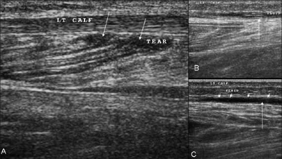
UltrasoundUltrasound of the calf, longitudinal view: hypoechoic area corresponding to a tear of the medial head of the gastrocnemius, arrows on the tear site, a typical case of tennis leg.
Source : Shah et al., The Indian Journal of Radiology & Imaging, 2010, figure 6 · CC BY
Yes, but not in the way it is often put. The difference in timeline between soleus and gastrocnemius exists in the data; it does not reach statistical significance. What does differ clearly is the clinical profile and the reinjury risk.
The Australian Football League soft tissue injury registry is the best source available on this question, because it prospectively follows a homogeneous population with routine MRI. Across 184 calf injuries collected between 2014 and 2017 in 16 clubs (Green 2020, PMID 31494970) :
That last point is the most operational. The soleus may not significantly lengthen the time to first return, but it massively dominates relapses. The issue with the soleus is therefore not “how long”, it is “will it hold”.
The soleus carries 84.6 % of injuries and 91.4 % of reinjuries. Treating a calf as a “tennis leg” by default means getting the muscle wrong nine times out of ten in the field athlete.
The gastrocnemius data have been refined recently. Across 82 MRI-confirmed gastrocnemius injuries between 2014 and 2023 in the same registry, the medial head accounts for 78 % of cases, and the median times to recovery are 3 days to walk without pain, 14 days to run at more than 90 % of maximal speed, 14 days to return to full training and 19 days to return to competition. More than one injury in six was a reinjury, and 79 % of those reinjuries occurred within six months (Gray 2025, PMID 40985375).
| Criterion | Medial head of gastrocnemius | Soleus | Plantaris |
|---|---|---|---|
| Share of injuries | 78 % of gastrocnemius injuries (Gray 2025) ; 66.7 % of the pictures referred for tennis leg (Delgado 2002) | 84.6 % of calf injuries in the elite Australian footballer (Green 2020) | 1.4 % (Delgado 2002) |
| Mode of onset | Abrupt, an identifiable moment, often with a perceived snap | Gradual or semi-abrupt, often on a recent increase in load | Abrupt, a picture close to tennis leg |
| Pain | Localised, medial myotendinous junction, upper-middle third | More diffuse, deeper, hard to point to | Posterolateral or high medial, poorly systematised |
| Position that provokes it | Knee extended, ankle dorsiflexed | Knee flexed: the position that relaxes the gastrocnemius | Variable |
| Reported timelines | Medians: 3 d painless walking, 14 d running > 90 %, 19 d return to match play (Gray 2025) | Mean 25.4 ± 16.2 d (Green 2020) ; difference from the gastrocnemius not significant (P = 0.097) | Generally favourable, data limited to case reports |
| Reinjury | More than one injury in six is a reinjury; 79 % occur within 6 months (Gray 2025) | 91.4 % of calf reinjuries (Green 2020) | Not documented |
| Main trap | Returning too early because walking is painless from day 3 onwards | Underestimating diffuse, poorly localised pain and taking it for simple soreness | Being labelled DVT on ultrasound (Rohilla 2013) |
The manoeuvre follows directly from the biarticular arrangement. You test resisted plantarflexion in two successive positions:
Discriminating manoeuvre
Knee extended: gastrocnemius and soleus work together. Pain here points to nothing.
Knee flexed to 90°: the gastrocnemius is slackened by knee flexion, the soleus provides most of the force. Pain that persists or worsens in that position points to the soleus; pain that disappears points to the gastrocnemius.
The same reasoning applies to stretching: knee extended, the tension falls on the gastrocnemius; knee flexed, it isolates the soleus.
A limitation to know: this discrimination is a common-sense anatomical argument, widely taught, but its diagnostic value has not been measured against imaging in a dedicated study. It points the way, it does not prove.
That reservation deserves to be explicit rather than hidden. A large part of calf semiology rests on coherent anatomical reasoning whose sensitivity nobody has published. Saying so does not weaken the approach: it avoids announcing to the patient a certainty you do not have.
Key points
In the field athlete, the soleus carries most calf injuries and nearly all reinjuries. The difference in time to return between soleus and gastrocnemius is not statistically significant; what is significant is the 12-day surcharge of injuries occurring while running. Clinical discrimination is done knee flexed against knee extended: solid anatomical reasoning whose diagnostic value has nevertheless not been measured.
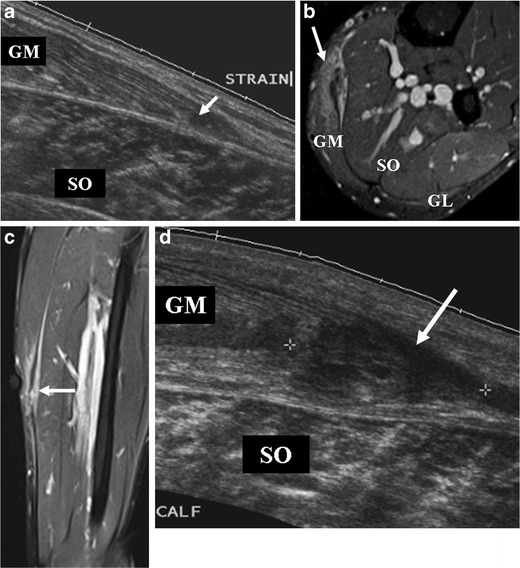
UltrasoundUltrasound then follow-up MRI of the calf: grade 2 tear of the medial head of the gastrocnemius, hypoechoic area evolving into feathery hyperintensity.
Source : Hayashi et al., Insights into Imaging, 2012, figure 3 · CC BY
Three systems coexist, and the clinician does not need to memorise them all. They need to understand what these systems measure, and what they all measure, in the end, is the same thing: involvement of the connective tissue.
For a long time, muscle injuries were graded on the extent of fibre involvement: grade 1 strain, grade 2 partial tear, grade 3 complete rupture. That classification never predicted timelines well, for a reason we understand better today: it is not the muscle that limits return, it is the connective tissue that transmits its force.
The study that established this point most clearly retrospectively assessed 100 consecutive patients referred with suspected calf injury, that is 114 injuries on MRI. The authors graded each injury from 0 to 3 by the involvement of the connective structures and their interfaces, then related that grade to the actual time to return. Connective involvement was present in 63 of the 100 patients, and connective tissue failure, grade 3, in 18 (Prakash 2018, PMID 29074478).
The time to return follows connective tissue involvement, not the volume of oedema
100 consecutive patients, 114 calf injuries on MRI, mean times to return to sport
Source: Prakash A, Entwisle T, Schneider M, et al. Br J Sports Med 2018;52(14):929-33 (PMID 29074478). Difference between grades significant at p < 0.001. These values are group means and do not constitute an individual prognosis.
A sixfold difference within the same condition, on a criterion that clinical examination cannot see. It is the strongest argument in favour of imaging when a prognosis has to be announced, to an athlete at the end of a season, to a patient whose job requires standing.
The same mechanism is found in a different population. Across 149 calf injuries in the Australian registry, players with severe aponeurotic involvement took 31.3 ± 12.6 days to return, against 19.4 ± 10.8 days in its absence (P = 0.003) (Green 2020, PMID 33030961).
| System | What it grades | Validation on the calf | Practical use |
|---|---|---|---|
| Munich consensus Mueller-Wohlfahrt 2013 |
Four types: 1 and 2 functional disorders with no visible fibre rupture, 3 partial tears, 4 subtotal or total tears and tendon avulsions | Correlated with time to return across 20 soleus injuries in professional footballers, Spearman's ρ = 0.982 (p < 0.0001) (Pezzotta 2017) | A common vocabulary. Note: level of evidence V, expert opinion |
| BAMIC British Athletics Muscle Injury Classification |
Grades 0 to 4 crossed with a suffix a, b or c according to whether the involvement is myofascial, myotendinous or intratendinous | Correlated with time to return in the same series, ρ = 0.886 (p < 0.0001) (Pezzotta 2017) | The most used in sports medicine; the suffix carries most of the prognosis |
| Pedret ultrasound classification | Five types specific to the medial head of gastrocnemius, by involvement of the gastrocnemius aponeurosis and of the free aponeurosis | 115 subjects (64 athletes, 51 workers), significant relationship with time to return, P < 0.001 (Pedret 2020) | Usable in the clinic with an ultrasound machine; identifies the forms with a long prognosis |
The Pedret classification deserves a further word, because it is designed specifically for tennis leg. The five types are: type 1 myo-aponeurotic; type 2A involvement of the gastrocnemius aponeurosis over less than 50 % of its width; type 2B over more than 50 %; type 3 involvement of the free, tendinous aponeurosis; type 4 mixed. The longest timelines are associated with involvement of the free aponeurosis. Two ultrasound signs accompany the forms with a poor prognosis: the presence of an intermuscular haematoma and asynchronous movement between gastrocnemius and soleus during ankle flexion-extension: signs that appear when more than 50 % of the aponeurotic width is involved (Pedret 2020, PMID 32854168).
The honest answer is: most often, no, but when you do image, you know what to look for.
Ultrasound is the first-line investigation. It is available, dynamic, it shows the inter-aponeurotic fluid, and above all it sees deep vein thrombosis, which settles both questions at once. A review of the use of ultrasound in calf injuries concludes that the great majority of injuries can be diagnosed and managed with no further imaging, and that repeated ultrasound allows healing to be followed (Bright 2017, PMID 28661826).
MRI brings the fine connective grading. It is justified when a precise prognosis has a consequence (selection, contract, long sick leave) or when the course does not follow. In the Pezzotta series, the extent of the oedema emerged as an independent prognostic factor for time to return in two multivariable regression models (Pezzotta 2017, PMID 28770309).
Initial imaging predicts the time to return. It does not predict reinjury. Across 149 injuries followed up, the initial MRI data were associated with no relapse risk: it was the clinical data that were.
That result, from the same Australian cohort, deserves to be taken seriously (Green 2020, PMID 33030961). It means that a fine follow-up MRI does not reassure about the risk of relapse, and that an image that is still imperfect in a patient whose functional criteria are met is not necessarily a reason to delay. The return decision is taken on function, not on the image.
Key points
What grades a calf injury is the involvement of the connective tissue: from 8 days to return when it is intact to 48 days when it is disrupted. Ultrasound is enough in the great majority of cases and settles the thrombosis question at the same time. MRI refines the prognosis but does not predict reinjury: the relapse factors are clinical.
Few risk factors survive systematic review, and those that do are uncomfortably simple: age, and having already had the same injury. Neither is modifiable.
The reference systematic review retained 10 studies assessing risk factors for calf injury, representing 5,397 athletes and 518 calf or leg injuries, in football, Australian football, rugby union, basketball and triathlon. Its synthesis by level of evidence gives (Green & Pizzari 2017, PMID 28259848) :
What predicts a calf injury, and what predicts nothing
Synthesis by best level of evidence, 10 studies, 5,397 athletes, 518 injuries
Source: Green B, Pizzari T. Br J Sports Med 2017;51(16):1189-94 (PMID 28259848). The authors themselves stress the scarcity of data and the tendency of the included studies to high bias.
Two comments are needed on that figure.
The first concerns the absence of association with sex, height and weight. That is not evidence of no effect: it is the absence of evidence of an effect, in a literature the authors themselves describe as scarce and biased. It would be excessive to conclude that these variables do not matter.
The second is more useful in the consultation. Since the two solid factors are not modifiable, all prevention plays out on what is not in this list : load management, calf capacity, the quality of the initial rehabilitation. That is exactly what the 20 international experts interviewed about their practices report: there is no universal preventive programme for the calf, and strategies are individualised to the athlete's characteristics and the demands of their sport (Green 2022, PMID 35032233).
This is where the calf differs clearly from other muscle injuries, and where the published figures need to be read carefully, because they depend entirely on the definition of “reinjury” adopted.
An analysis of 563 calf injuries in 359 players over ten years measured that definitional effect precisely (Green 2025, PMID 40919406) :
The four values come from the same source: Green B, Schache AG, Pizzari T. BMJ Open Sport Exerc Med 2025;11(3):e002865 (PMID 40919406). Reinjuries cost at least 2,153 cumulative days of unavailability over the decade, that is at least 35.6 days on average per reinjury.
The figure to remember is the third. One subsequent injury in five occurs in a player who had not finished recovering, and it then costs 46.7 days on average, more than double an index injury. It is the quantified demonstration that returning too early does not save time: it costs time.
The clinical factors associated with reinjury have been identified separately. For an early reinjury, within two months: older age (aHR 1.3; P = 0.001) and a previous ankle injury (aHR 3.9; P = 0.032). For a reinjury within two seasons: age (aHR 1.1; P = 0.013) and above all a previous calf injury, with an adjusted hazard ratio of 6.7 (P = 0.002) (Green 2020, PMID 33030961). Consistency with the European registry is good: across 22,942 injuries in the UEFA cohort, calf injury is among the six diagnoses for which reinjury significantly lengthens the absence compared with the index injury (Ekstrand 2020, PMID 31182429).
Warning signals during follow-up
These features, during rehabilitation, should prompt you to start the examination again from scratch rather than continue the protocol.
Key points
The two solid risk factors, age and previous calf injury, are not modifiable, which shifts the whole issue onto load management and the quality of the return. One subsequent injury in five occurs before full recovery and then costs 46.7 days instead of about twenty. A previous calf injury multiplies the reinjury risk over two seasons by 6.7.
Once the two emergencies are ruled out, the question becomes ordinary and is handled with the principles common to any muscle injury. This chapter therefore stays deliberately short: the pathophysiology of muscle healing and the general principles of return to loading are covered elsewhere on the site.
For the biology of muscle repair, the inflammatory, repair and remodelling phases, and the general logic of progressive reloading after injury, see muscle contusion (dead leg) and its complications, which covers the direct-blow side, and proximal hamstring tendinopathy for the progressive loading side of a thigh muscle-tendon complex. What follows covers only what is specific to the triceps surae.
The calf's specificity at this stage lies in a simple mechanical constraint: it is the muscle you cannot unload without unloading walking. Unlike a hamstring or an adductor, the triceps surae works at every step, eccentrically at heel strike and concentrically at push-off. Loading is therefore not an exercise to be added to the programme: it is the patient's basic activity, and it is that which has to be dosed.
Dose the walking rather than forbid it
The criterion is the limp, not the pain. A patient who walks rolling through the step, with no avoidance, with mild and stable pain, is in a good loading regime. A patient who limps keeps their calf in inner range, no longer loads the forefoot, and loses dorsiflexion while they wait.
Crutches serve to remove the limp, not to remove weight-bearing. They are given when rolling through the step is impossible, and withdrawn as soon as it becomes possible again, often within a few days.
A temporary heel lift of 5 to 10 mm, bilateral so as not to unbalance the pelvis, shortens the painful lever arm and often allows a normal gait pattern to be recovered sooner. It is withdrawn progressively.
What is not justified: strict immobilisation, early stretching into forced dorsiflexion, and deep transverse friction on a recent injury in which thrombosis has not been ruled out.
The most useful time landmark comes from the Australian registry: the median to walk without pain after a medial gastrocnemius injury is 3 days (interquartile range 3 days) (Gray 2025, PMID 40985375). A patient still unable to walk normally at the end of the first week is off the usual trajectory: that is a reason to re-examine, not to wait.
Walking without pain on the third day does not mean the injury has healed. It means the injury allows walking. The gap between those two sentences explains most early reinjuries.
A warning at the outset: the treatment literature specific to the calf is thin. There is no randomised controlled trial of triceps surae injury rehabilitation comparable with those that exist for the hamstrings. This chapter says so rather than hiding it behind firm recommendations.
That gap is not an opinion: it is the observation that opens the qualitative study of 20 international expert clinicians, which begins by noting “the scarcity of research to guide clinicians facing these injuries” (Green 2022, PMID 35032233). That study is, paradoxically, the best source of practice available, but it is structured expert opinion, not experimental evidence.
Analysis of the interviews brings out several converging points. Calf injuries are considered different from other muscle injuries and treated as such. Rigorous clinical work establishes the diagnosis; what gives the most reliable prognosis, on the other hand, is not the initial imaging but monitoring of calf capacity and its response to load exposure. Rehabilitation is structured in six phases, guided by principles aimed at optimising performance on return to play while avoiding reinjury.
Dosing the walking, heel lift if needed, partial unloading only if rolling through the step is impossible. Early sub-painful isometrics in plantarflexion, knee extended then flexed. Maintaining the rest of the chain: hip, knee, trunk, and cardiovascular work without impact.
Recovery of dorsiflexion with the knee extended and with the knee flexed: both are measured, because they do not progress at the same rate. Active mobilisation, progressive loading into range rather than prolonged passive stretching.
The core of the treatment. Double-leg then single-leg heel raises, knee extended and knee flexed, aiming first for repetition volume, then for load. This is where reinjury prevention is decided.
Moving to fast regimes: jumping on the spot, skipping, repeated foot contacts, then single-leg plyometrics. The triceps surae works like a spring; slow endurance capacity says nothing about tolerance of elastic regimes.
Progress speed before volume, because it is speed that loads the calf in the injury regime. AFL registry landmark: a median of 14 days to run at more than 90 % of maximal speed after a gastrocnemius injury (Gray 2025).
Sport-specific movements, changes of direction, accelerations and decelerations, then partial and full training. Acceleration was the commonest injury mechanism during running in the Gray series: it must be exposed in training before it is exposed in a match.
Six-phase structure after Green B, McClelland JA, Semciw AI, et al. Sports Med Open 2022;8(1):10 (PMID 35032233), qualitative study of 20 expert clinicians. The quantified landmarks are those of Gray C, et al. J Orthop Sports Phys Ther 2025;55(10):681-8 (PMID 40985375). The exit criteria proposed are a practical arrangement, not validated as such.
The table below applies GRADE logic to the modalities used on the calf. The reader will note that no line reaches the “high” level. That is a faithful reflection of the state of the literature on this site, and not stylistic caution.
| Modality | Level | What the evidence says exactly | In practice |
|---|---|---|---|
| Progressive plantarflexion loading (heel raises, knee extended and flexed) | MODERATE | Consensus of the 20 international experts on the central role of calf capacity and its response to load (Green 2022). Established biomechanical rationale for soleus selectivity (Kovács 2024). No RCT specific to the calf. | The mainstay of treatment. Both knee positions, without exception. |
| Return guided by functional criteria rather than by a fixed timeline | MODERATE | Explicit recommendation of the state-of-the-art review: return must be guided by functional recovery and not by the disappearance of symptoms alone (Schwach 2026). Consistent with the 46.7 days of early relapses (Green 2025). | Set the criteria in writing from the first session. |
| Dosing the walking and returning to a normal gait pattern | MODERATE | A median of 3 days to painless walking in a cohort of 82 gastrocnemius injuries (Gray 2025) ; normal walking is a documented milestone, not a theoretical objective. | Crutches to remove the limp, never beyond that. |
| Progressing running speed before volume | LOW | Injuries occurring while running take ~12 days longer (P = 0.001) (Green 2020) and acceleration is the dominant mechanism (Gray 2025) : running is the critical loading factor. The optimal sequence of reintroduction, however, has not been tested. | Expose acceleration in training before the match. |
| First-line conservative treatment (relative rest, ice, compression, elevation) | LOW | A consistent practice across reviews and case series, recovery in 6 to 12 weeks (Schwach 2026). No randomised comparison of the components with each other. | Reasonable and risk-free, provided DVT has been ruled out before any compression. |
| Follow-up ultrasound to monitor healing | LOW | Level 4 clinical review: serial ultrasound allows healing to be followed and may help with return decisions (Bright 2017). No effect on outcomes has been demonstrated. | Useful if the course is surprising; not routine. |
| Prolonged passive stretching in the acute phase | VERY LOW | Cited in the narrative reviews among the rehabilitation measures (Schwach 2026), with no comparative data. The biological rationale, tensioning a healing junction, invites caution in the early phase. | Prefer loading into range over sustained passive stretching. |
| Deep transverse friction on the injured area | NONE | No effectiveness study in triceps surae injury. And a documented risk if DVT has not been ruled out (Delgado 2002 ; Slawski 1994). | Not recommended in the acute phase. Never without having ruled out thrombosis. |
| A standardised preventive programme for the calf | NONE | The 20 experts conclude that a universal preventive programme for the calf probably does not exist, and favour individualised strategies (Green 2022). | Individualise to the sport and to the patient's history. |
Key points
No modality reaches a high level of evidence in calf injury: the specific treatment literature is thin, and that is a fact to state rather than to get round. What approaches consensus comes down to two lines: load the triceps surae progressively in both knee positions, and decide on return using functional criteria. Transverse friction in the acute phase has no evidence of effectiveness and raises a safety problem as long as thrombosis has not been ruled out.
This is the most operational chapter of the article, and the one where the commonest error is also the easiest to correct: strengthening the calf only with the knee extended means leaving out the muscle that carries most injuries and nearly all reinjuries.
The argument is mechanical and it is measurable. When the knee flexes, the gastrocnemius, biarticular, loses length and therefore force-producing capacity. The soleus, monoarticular, is unaffected. The result is that knee flexion shifts the load of the complex towards the soleus.
That shift has been quantified. In nine physically active men, maximal isometric plantarflexion torque went from 154.4 ± 37.8 N·m with the knee extended to 93.1 ± 22.3 N·m with the knee flexed. And above all, the correlation analysis shows that knee position changes the relationship between muscle size and torque produced for the gastrocnemius but not for the soleus : only soleus volume and total triceps surae volume remained correlated with torque produced with the knee flexed (Kovács 2024, PMID 39577118).
What knee flexion changes in the work of the triceps surae
Maximal isometric plantarflexion torque, 9 physically active men, dynamometer
Source: Kovács B, Yaodong G, Kóbor I, et al. J Biomech 2024;177:112436 (PMID 39577118). Limited sample (n = 9 physically active men): the absolute values are not norms, it is the direction of the effect that is usable.
The reservation about the sample deserves stating: nine subjects, all active men. The absolute values are not meant to serve as an individual reference. The direction of the effect, on the other hand, is consistent with the anatomy and with the whole literature on triceps surae architecture, and it is that direction that governs practice.
A calf strengthening programme done exclusively with the knee extended neglects the muscle that carries 84.6 % of injuries and 91.4 % of reinjuries. The knee-flexed position is not a complementary exercise: it is the only one that loads the soleus selectively.
The logic is constant: tolerance first, then volume, then load, then speed. Each stage is done in both knee positions, and you progress in the two independently: it is common for a patient to tolerate load with the knee extended well before tolerating it with the knee flexed, or the reverse depending on the muscle involved.
Static push into plantarflexion against fixed resistance, at sub-painful intensity. Seated with the knee flexed to 90° first, the position least demanding for the gastrocnemius myotendinous junction, then standing with the knee extended.
Rising onto the toes on both feet, on flat ground then on a step to gain eccentric range. Two compulsory variants:
Moving to single-leg doubles the load: it is the real start of strengthening. Always both knee positions. The objective is to match the sound side in number of repetitions and in height of rise, not to reach an absolute figure.
Adding load (weighted vest, barbell, press) once symmetry is reached at body weight. This is where knee-flexed work becomes most discriminating: the soleus supports high loads, and a residual deficit shows up there when it is invisible with the knee extended.
Jumping on the spot, skipping, bouncing contacts, then single-leg jumps and plyometrics. The triceps surae works like a spring in running: good capacity in a slow regime guarantees nothing about tolerance of the elastic regime, which must be tested separately.
The commonest dosing error
The calf takes volumes that few other muscles take: at every running stride it absorbs several times body weight, hundreds of times per kilometre. A programme of 3 sets of 10 heel raises, three times a week, amounts to nothing on that scale.
The useful landmark is not the load of an isolated exercise but total capacity, in repetition volume and in tolerance of repetition. That is precisely what the expert clinicians monitor: calf capacity and its response to load exposure, rather than a one-off strength value (Green 2022, PMID 35032233).
Key points
Strengthening the triceps surae is always done in two knee positions, and you progress in the two separately. Knee extended loads the whole complex; knee flexed isolates the soleus, the only muscle whose size-torque relationship stays unchanged by knee flexion. In a muscle that absorbs several times body weight at every stride, the target is volume and repeatability, not the load of an isolated set.
The question is asked back to front in most consultations: “how long?” rather than “on what conditions?”. The reinjury data give a quantified reason to reverse the question.
The most useful reminder is this: in a cohort of 563 calf injuries followed over ten years, 20 % of subsequent injuries occurred before full recovery, and those relapses cost 46.7 ± 25.6 days to return, against about twenty days for an index injury (Green 2025, PMID 40919406). Returning a week too early does not gain a week: it costs three.
The state-of-the-art review on tennis leg puts the same thing in terms of principle: recovery usually occurs in 6 to 12 weeks, and return to activity must be guided by functional recovery, not by the disappearance of symptoms alone (Schwach 2026, PMID 42258915).
The heel rise endurance test is the best tool the physiotherapist has for objectifying calf capacity, because it is reproducible, needs no equipment, and has just been given normative values in a large population.
An international study of 500 subjects with no history of Achilles tendinopathy or recent immobilisation gives medians of 25 repetitions on the dominant side and 24 on the non-dominant side, for total work of 1,374 and 1,325 J, cumulative vertical displacement of 192 and 186 cm, and a peak height of 9.3 and 9.6 cm. The authors note lower performance with female sex, higher BMI and lower physical activity level (Visser 2025, PMID 40020545).
Normative values of the single-leg heel raise test
500 subjects with no history of Achilles tendinopathy, validated Calf Raise application, medians
Source: Visser TSS, Neill SO, Hébert-Losier K, et al. Braz J Phys Ther 2025;29(3):101188 (PMID 40020545). Medians (50th percentile) across 500 subjects, 55 % women, 88 % physically active. Female sex, high BMI and low activity level are associated with lower values: the general medians do not replace comparison with the sound side.
The most operational result of that study is not the median of 25 repetitions, it is the absence of a significant difference between dominant and non-dominant side. It validates the practice of side-to-side comparison: in a patient with a unilaterally injured calf, the sound side is an acceptable reference, which avoids having to correct for sex, BMI and activity level.
| Domain | Criterion | How to measure it | Evidence status |
|---|---|---|---|
| Pain | Absent on walking, running, single-leg jumping and on palpation of the injured area | History and comparative palpation | A consensus principle (Green 2022) |
| Range | Symmetrical dorsiflexion with the knee extended and with the knee flexed | Wall test, toe-to-wall distance, both positions measured separately | Anatomical rationale, not validated as a return criterion |
| Endurance capacity | Single-leg heel raises symmetrical with the sound side, in number and in height | Bilateral heel rise endurance test; norms available (Visser 2025) | Normative values established in 500 subjects; the absence of dominant/non-dominant asymmetry validates side-to-side comparison |
| Knee-flexed capacity | Symmetry also in the seated position, knee at 90° | Loaded seated heel raises, side-to-side comparison | Biomechanical rationale (Kovács 2024) ; no published threshold |
| Elastic regime | Symmetrical repeated single-leg jumps, with no pain or apprehension | Drop jump, timed repeated jumps, side-to-side comparison | Functional rationale; no validated threshold on the calf |
| Running | Maximal speed reached without pain, accelerations and decelerations exposed | Progress speed before volume; median landmark of 14 days for > 90 % of maximal speed (Gray 2025) | Cohort of 82 injuries; a descriptive landmark, not a target to reach |
| Training load | Return to full training before return to competition, with exposure to accelerations | Load monitoring; acceleration was the dominant injury mechanism (Gray 2025) | Descriptive; the causal link is not established |
| Previous injury | In a patient already injured in the same calf, require full symmetry and not the absence of complaint | History of previous injury, all the criteria above met | Reinjury aHR at 2 seasons = 6.7 with a previous injury (Green 2020) |
Why it makes sense to reason in criteria rather than in timelines
A mean timeline is a property of a population: 19 days for a gastrocnemius injury, 25 for a soleus. A patient is not a population, and the published intervals are wide: 25.4 ± 16.2 days means that many patients fall outside the mean.
Above all, the data show that the cost of error is asymmetrical. Returning late costs the days of waiting. Returning too early costs 46.7 days on average. When an error costs twice as much in one direction as in the other, the rational decision is to err on the cautious side.
Key points
Return is decided on criteria, not on a calendar: no pain, symmetrical dorsiflexion in both knee positions, symmetrical single-leg heel raises knee extended and flexed, tolerance of the elastic regime, maximal running speed reached. The sound side is a valid reference, the normative study having found no difference between dominant and non-dominant. In a patient already injured on the same side, you require full symmetry, not merely the absence of complaint.
Three published cases, chosen because each illustrates a different error and because all three are genuine referenced case reports. None is reconstructed or composite.
A 51-year-old man, playing tennis, feels as though he has been kicked in the back of the calf, with an audible snap and severe pain. Ultrasound finds fluid between the muscle planes and a hypoechoic structure; the radiology registrar concludes to a deep vein thrombosis. MRI corrects the diagnosis: rupture of the plantaris, with the fluid and the muscle stump visible between gastrocnemius and soleus. The patient is treated conservatively (rest, ice, compression, elevation), with clear resolution of the pain and the swelling (Rohilla S, Jain N, Yadav R. BMJ Case Rep 2013;2013:bcr-2012-007840, PMID 23345486).
What this case teaches. The error goes the other way too: here it is a muscle injury that was taken for a thrombosis. The authors note in passing a second trap in the same picture: an intact plantaris can still produce plantarflexion when the Achilles tendon is ruptured, which gives “a misleading picture”. The practical corollary is that of chapter 3: never conclude that the tendon is intact from the persistence of active movement alone.
A 35-year-old man, sudden intense calf pain after a minor injury with the leg extended. The diagnosis of tennis leg, with a partial gastrocnemius tear, is made on clinical examination and ultrasound. The authors explicitly stress that ultrasound served to distinguish it from a deep vein thrombosis. Conservative management: rest, ice, compression, elevation, analgesia, physiotherapy. Normal muscle function at the four-month review (Adhikari S, Devkota S, Lamichhane S, et al. SAGE Open Med Case Rep 2024;12:2050313X241272690, PMID 39430721).
What this case teaches. Two things, one reassuring and one demanding. The reassuring one: the prognosis of conservatively treated tennis leg is good, and surgery is reserved for severe forms only. The demanding one: follow-up ran to four months. The time to return to sport published in the cohorts, about twenty days in the elite athlete supervised daily, does not describe the trajectory of the clinic patient. The injury mechanism was moreover “minor”, which is a reminder that the intensity of the trauma does not predict that of the injury.
A patient with an established and obvious tennis leg subsequently develops thrombophlebitis in the same limb. The author, who reports this case as the first of its kind published, recalls in the same article that the earlier literature documented mainly the reverse error: tennis legs taken for thrombophlebitis, treated with unnecessary anticoagulation, with bleeding complications as a result (Slawski DP. J Orthop Trauma 1994;8(3):263-4, PMID 8027900).
What this case teaches. It is the most important of the three, and the one that defeats the most natural reasoning. Having established a muscle injury does not protect against a thrombosis: the two can coexist, and the relative immobilisation that follows the injury is itself a thrombogenic factor. Screening for red flags is not done once, at the first session; it is done again at every session, particularly if the swelling increases again or if the pain changes in character.
The lesson common to the three cases
A diagnosis made is not a diagnosis settled. The first case shows a muscle injury labelled thrombosis, the third a thrombosis occurring on a genuine muscle injury. In both directions, the error comes from the same cause: having considered the question closed.
The operational summary, in the order of the consultation.
Report template
Mechanism: movement, position of the knee and ankle, whether it was audible, ability to continue the activity.
Red flags: the list gone through, with an explicit “absent” or the detail of what prompted the referral.
Achilles tendon: the two tests performed and their result, plus comparison of the resting equinus. Write which ones, not “examination normal”.
Site: muscle identified and the reasoning (pain with knee extended against knee flexed, palpation).
Baseline measurements: bilateral circumference, dorsiflexion in both positions, single-leg heel raises on the sound side.
Return criteria: set in writing from the first session and communicated to the patient.
Not exactly. “Tennis leg” classically denotes injury of the medial head of gastrocnemius at its myo-aponeurotic junction. It is one form of calf injury, but not the only one: in the field athlete, the soleus is at fault in 84.6 % of cases (Green 2020, PMID 31494970). Using “tennis leg” as a synonym for “calf injury” leads to neglecting the muscle most often involved.
That idea, long taught, does not stand up to the data. Across 141 patients referred with a clinical diagnosis of tennis leg, the plantaris was at fault in only 2 cases, that is 1.4 %; the medial head of gastrocnemius was in 66.7 % (Delgado 2002, PMID 12091669). In the same series, an isolated deep vein thrombosis was seven times commoner than plantaris involvement.
You cannot be sure clinically, and that is precisely the message of this chapter. No examination sign rules out a deep vein thrombosis, and the Wells score itself is not enough: in a cohort of 1,135 inpatients, 5.9 % of those classed as low probability had a proximal DVT (Silveira 2015, PMID 25985219). The physiotherapist's role is to spot the doubt, suspend all local treatment and refer for duplex ultrasound, not to decide.
The cohort landmarks, in elite athletes supervised daily, give medians of 19 days for a gastrocnemius injury (Gray 2025, PMID 40985375) and a mean of 25.4 ± 16.2 days for the soleus (Green 2020, PMID 31494970). The reviews on tennis leg suggest rather 6 to 12 weeks in the general population (Schwach 2026, PMID 42258915). These figures diverge because they describe different populations and different return criteria. That is one more reason to decide on functional criteria and not on a calendar.
Because it is what transmits the muscle's force to the tendon. Across 100 consecutive patients, the mean time to return ran from 8 days when the connective interfaces were intact to 48 days when they were disrupted, that is a sixfold difference within the same condition (Prakash 2018, PMID 29074478). The extent of muscle oedema, for its part, is a poorer predictor.
No, but it has two advantages that often make it useful first line: it locates the injury and, above all, it sees deep vein thrombosis, which settles the safety question at the same time. The great majority of calf injuries can be diagnosed and managed with no further imaging (Bright 2017, PMID 28661826). MRI is reserved for situations where a precise prognosis has a consequence, or where the course does not follow.
Because it is the only position that loads the soleus selectively. The gastrocnemius crosses the knee: when it flexes, the muscle shortens and loses force-producing capacity. Maximal isometric plantarflexion torque goes from 154.4 to 93.1 N·m between knee extended and knee flexed, and the relationship between muscle volume and torque produced stays unchanged for the soleus while it changes for the gastrocnemius (Kovács 2024, PMID 39577118).
Yes, in how demanding the exit criteria are rather than in the content of the exercises. A previous calf injury multiplies the reinjury risk over the following two seasons by 6.7 (Green 2020, PMID 33030961), and along with age it is one of only two solidly established risk factors (Green & Pizzari 2017, PMID 28259848). In those patients, you require measured full symmetry, not merely the absence of complaint.
Yes, and it is documented: a case of ipsilateral thrombophlebitis occurring on an already established tennis leg has been reported (Slawski 1994, PMID 8027900). That is why red flag screening is repeated at every session, particularly if the swelling increases again after a phase of resolution.
No. The toe flexors, tibialis posterior, the peroneals and the plantaris still produce plantarflexion when the Achilles tendon is ruptured. It is a classic trap, flagged in the case literature (Rohilla 2013, PMID 23345486). Only the specific tests decide, and at least two are needed.
Every reference was verified on PubMed, and the content of each abstract read to ensure that it does establish what is attributed to it in the text. The identifiers are clickable.
Behind this article
An author who explains, a reviewer who checks.
How we write and check our content

Physiotherapist · co-founder of Physio Learning
Marked for life by his first four-hour lecture without a single image, he took a master’s in instructional design so that it would never happen to anyone again. He hunts down publication bias and unreadable slides with the same intransigence.

Head of scientific content
Physiotherapist specialising in neuro-musculoskeletal practice and holder of a master’s in public health. He checks the methodological rigour of every article: primary sources, levels of evidence, no exceptions.
