
















On-siteDPCFIFPL

Sports physiotherapy
From injury to return to play: structure every stage of reconditioning
Maxence Ponthus
Go further
Grade the dead leg on active knee flexion at 12 to 24 hours: minor above 90 degrees, severe below 45. Myositis ossificans hit 9% of West Point cadets.
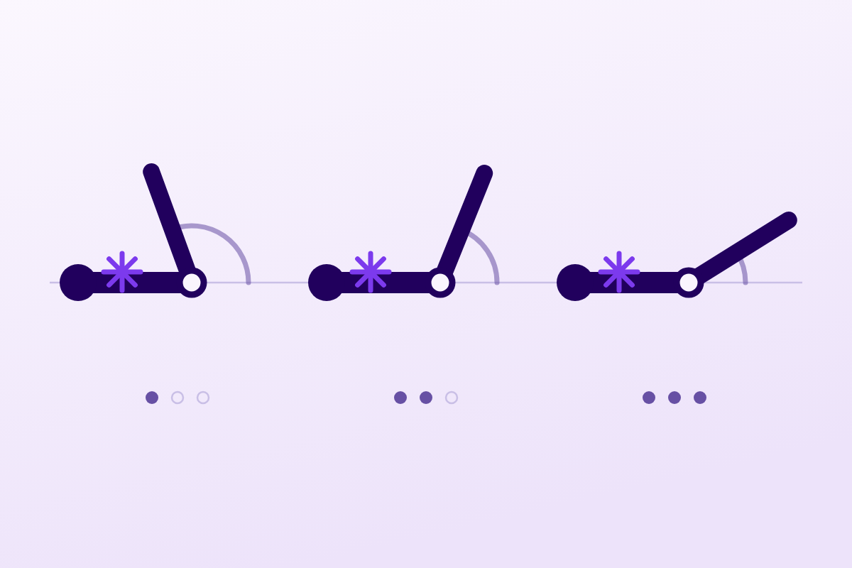
The severity of a dead leg is read from active knee flexion, not pain or bruising, and the first move is to put the knee into flexion.
120°of knee flexion regained before a safe return to sport
What follows covers each of these points in detail, with sources. It is there if you need it.
This topic is taught in a course: From injury to return to play: structure every stage of reconditioning, on-site with Maxence Ponthus.
1 / 15
14 article chapters · 64 min in total
Understanding◔ 4 min
A dead leg comes from an impact: an opponent's knee, helmet or shoulder crushes the muscle against the femur.
Frequency◔ 4 min
Contusion accounts for 12% of thigh injuries in the pro footballer, and almost always occurs in a match.
Grading◔ 6 min
The severity of a dead leg is read from active knee flexion measured twelve to twenty-four hours after the impact.
First hours◔ 6 min
The only move that changes what follows: put the knee into flexion, and leave it there.
Rehabilitation◔ 5 min
Dead-leg rehabilitation rests almost entirely on pathophysiological reasoning and case series.
Myositis◔ 5 min
Despite its name, it is not inflammation: it is a benign, most often self-limiting heterotopic ossification.
Recognising it◔ 5 min
After initial improvement, a plateau or regression of flexion between the 2nd and 6th week should raise alarm.
Treating myositis◔ 5 min
The best-documented answer: do nothing specific, continue rehabilitation and monitor with ultrasound.
Compartments◔ 4 min
It is the rarest complication, but the only one that can cost a limb.
Return to sport◔ 4 min
The determining factor for a safe return to play is recovered knee flexion, not a fixed timeframe.
Figures◔ 5 min
Several percentages circulate without anyone tracing them back to their real origin in the literature.
Case studies◔ 3 min
Six published cases map the real timetable of complications, which pooled series erase.
In practice◔ 3 min
Worsening within 24 hours is not part of the normal course of a contusion.
Questions◔ 5 min
How long a dead leg lasts depends on its severity and how quickly it's treated, and any average is misleading.
Train on this
Sports physiotherapy
Maxence Ponthus
Next comes practice: the course that teaches this topic, with Maxence Ponthus.
Our courses on this topic may be covered by DPC or FIFPL, the French continuing-education funding schemes, depending on your profession and situation.
A knee that no longer bends after a blow to the thigh: the dead leg is the most ordinary of contact injuries, and the only one that can end in ossification inside the muscle or in emergency fasciotomy. Here is how to grade it, what to do in the first 24 hours, and what myositis ossificans really hinges on.
Review article. Every figure quoted carries its source at the point where it is written. Claims that circulate without an identifiable primary source are flagged as such rather than repeated: a whole chapter is devoted to them.
Three figures to place the dead leg
Primary data: prospective UEFA cohort 2001-2013 and West Point series 1991
Sources: Ueblacker P, Müller-Wohlfahrt HW, Ekstrand J. Br J Sports Med 2015;49(22):1461-5 (PMID 25755277) for the first two; Ryan JB et al. Am J Sports Med 1991;19(3):299-304 (PMID 1867338) for the third.
Contents
What the available literature says, in ten points. Each one is taken up and sourced in the chapters that follow.
Who this article is for
For physiotherapists who see contact-sport athletes, in the clinic as much as pitchside. It covers the quadriceps dead leg and its three complications, myositis ossificans, compartment syndrome and residual stiffness, in a single text, because none of the three can be understood without the injury that produces it.
The distinction between direct and indirect injury is not a nicety of nomenclature. It changes the mechanism, the site of the lesion, the prognosis and the risk of complication. An article that treats the two together gets it wrong twice.
A strain arises from an eccentric contraction: the muscle contracts while it lengthens, and the myotendinous junction gives way. A dead leg arises from an impact: an opponent's knee, helmet or shoulder crushes the muscle belly against the femur, which acts as an anvil. The lesion is therefore deep, centred on the point of impact, and it readily involves the muscle plane closest to the bone.
The reference review on muscle trauma, published in 2023 in Nature Reviews Disease Primers, sets out this dichotomy from the start: indirect injuries (strains, ruptures) occur "without the influence of direct external trauma" and result from muscle activation during lengthening, whereas direct injuries (contusions, lacerations) result from an external impact12. The Munich consensus, which has structured the terminology of muscle injuries in sport since 2013, moreover deals with contusion outside its main axis: its four types (overuse, neuromuscular, partial tear, subtotal tear or avulsion) describe the indirect side, and contusion is named separately13.
The management of muscle injuries has shifted in five years: from rest, immobilisation and initial (over)protection towards early activation and progressive loading. That is the central message of the Nature Reviews Disease Primers 2023 review, and it is exactly what the 24-hour flexion protocol was anticipating back in 1991.
The sequence was described in the review by Beiner and Jokl: microscopic rupture and injury to muscle cells, macroscopic defects in the muscle belly, infiltrating bleeding, inflammation. The authors sum up repair in a formula that is useful in the clinic, it is a race between remodelling and scar formation14. Everything that helps remodelling win (early pain-free mobilisation, progressive loading) works in the right direction; everything that feeds the haematoma or increases local inflammation works the other way.
Two forms of haematoma coexist and they do not behave alike. The intermuscular haematoma spreads between the fascial planes, migrates with gravity, colours the skin within a few days, and resolves quickly. The intramuscular haematoma stays enclosed within the fascial sheath of the muscle: it raises local pressure, restricts the excursion of the muscle, and it is the one that causes trouble. The prospective study by Thorsson et al. measured this difference on ultrasound: lesions appearing as a circumscribed anechoic, hypoechoic or mixed collection normalised more slowly and took longer to heal completely (p = 0.001) than lesions with a diffuse hyperechoic appearance5.
The quadriceps occupies the anterior compartment of the thigh: rectus femoris superficially, vastus medialis and vastus lateralis on either side, vastus intermedius flattened against the femoral shaft. A direct anterior impact therefore crushes the deep plane preferentially, which the surgical data confirm: in the series by Rööser et al. on eight thigh compartment syndromes, the haematoma lay in vastus intermedius in four cases and in rectus femoris in three8.
For ossification, the most recent systematic review gives a slightly different result. Across 89 published patients, Stammer et al. find the thigh to be the most frequent site and the quadriceps the most affected muscle, but they name vastus lateralis as the head most often involved15. The radiological series by Saad et al. (68 cases over 13 years in a tertiary orthopaedic centre) points the same way on localisation: 73% of cases in the lower limb, mostly in the quadriceps16. In other words: the thigh and the quadriceps, yes, without argument; the exact head, the literature does not settle.
Four markers on direct thigh injury in professional football
UEFA Elite League cohort, 30 clubs, 1,981 players, seasons 2001-2002 to 2012-2013
Single source for all four values: Ueblacker P, Müller-Wohlfahrt HW, Ekstrand J. Br J Sports Med 2015;49(22):1461-5 (PMID 25755277). Comparison of direct and indirect incidence significant at p < 0.01.
Key points
The dead leg is a deep crush injury, whose prognosis depends on the volume and the location of the intramuscular haematoma, not on the painful surface area. Statistically it heals faster than a strain. Its danger lies entirely in its two complications: ossification of the muscle and, exceptionally, raised compartment pressure.
Simple question, partial answer. There is one solid prospective dataset, but it deals with European male professional football, and nothing says it transfers to amateur rugby or to Saturday handball.
The study by Ueblacker, Müller-Wohlfahrt and Ekstrand, published in 2015 in the British Journal of Sports Medicine, remains the reference: 30 European clubs, 1,981 players followed prospectively from 2001 to 2013, with recording of individual exposure and of every time-loss injury. It counts 2,287 thigh muscle injuries, that is 25% of all injuries; 2,003 were usable, of which 88% were indirect and 12% direct1.
The detail is more instructive than the percentage. The incidence of indirect injuries is eight times higher: 1.48 per 1,000 hours of exposure against 0.19 (p < 0.01). But the gap closes when you look at the cost: indirect injuries account for 19% of total absence time, direct ones for 1%, and the mean absence is 18.5 days against 7 (p < 0.001). Finally, 76% of direct injuries occur in matches, against 60% of indirect ones, and a foul is involved in 42% of direct injuries, against 2% of indirect ones1.
Direct versus indirect thigh injury: four comparisons
Each row has its own scale; the values are written at the end of the bar
Source: Ueblacker P, Müller-Wohlfahrt HW, Ekstrand J. Epidemiological and clinical outcome comparison of indirect versus direct anterior and posterior thigh muscle injuries in male elite football players. Br J Sports Med 2015;49(22):1461-5 (PMID 25755277). Each of the four rows has its own scale: they cannot be compared with one another.
Professional basketball. A study published in February 2026 in BMC Sports Science, Medicine and Rehabilitation characterised quadriceps injuries in the NBA over the 2015 to 2020 seasons: 116 injuries in 89 players, and contusion is the most frequent type. Most of the players missed fewer than ten games. The authors observe a significant fall in player efficiency rating one year after the injury (16.5 before, 15.8 after, p = 0.015), which is no longer significant at two years (16.8, p = 0.166)17. That is a performance signal, not incidence data: the database used is public and self-declared.
Rugby league. The work of Alonso, Hekeik and Adams followed 100 injured rugby league players in an Australian professional club. This is not an incidence study either, it is a prognostic study, and one of the best we have on this subject, but it does attest in passing that a single club can gather a hundred dead legs18.
American military academies. Jackson and Feagin counted 65 quadriceps injuries in a single school year in cadets at West Point19. Ryan et al. counted 117 over three years in the same institution2. Aronen et al. gathered 47 cases at the Naval Academy, but over eighteen years3. These populations are young, male, exposed daily to compulsory contact sports, and followed by a single department: that is what makes these series valuable, and it is also what forbids generalising from them.
What we did not find
No incidence data for thigh contusion in the general sporting population. The searches carried out for this article identified no study giving a rate per 1,000 hours of exposure in rugby, handball, hockey or amateur sport, at any level. The figures circulating about these disciplines come from injury registers that group together "contusions" at all sites, which is not the same thing. We prefer to write that than to transpose the 0.19 per 1,000 hours of professional football to sports where contact is more frequent and the equipment different.
Injury registers count only time-loss injuries. Yet Ueblacker points out that 76% of direct injuries occur in matches, and that the player often carries on playing: the minor dead leg, the one that leaves more than 90° of flexion, never crosses the reporting threshold. The practical consequence is that the 12% in the UEFA cohort is a floor, not an estimate. This point matters for what follows: the clinic physiotherapist mostly sees the dead legs that were not treated on the day, and those are precisely the ones Ryan showed to become complicated more often2.
Key points
A single large prospective cohort, on a single sport and a single level. It establishes that contusion accounts for 12% of thigh muscle injuries in the professional footballer, costs 7 days on average, and almost always occurs in a match. Outside that setting, the true frequency is not known.
With a goniometer and a watch. The severity of a quadriceps contusion is read from active knee flexion measured twelve to twenty-four hours after the impact, not from pain, not from the volume of the thigh, not from the visible haematoma.
In 1973, Jackson and Feagin published in the Journal of Bone and Joint Surgery the study that still structures management today: 65 quadriceps injuries in cadets, over one school year, classified into three grades. Their central finding is that the degree of flexion limitation matches the severity of the injury, and so does the time lost19.
The clinical descriptors of the three grades, as Kary's review reports them, attach a functional picture to each range: normal walking and localised tenderness for the minor form above 90°; antalgic limp and a swollen, tender muscle mass between 45 and 90°; marked limp and a clearly swollen mass below 45°20.
Ryan et al. took up these thresholds in 1991, specifying when the measurement is taken, 12 to 24 hours after the injury , and it is that detail which makes the grading reproducible2. Measuring on the pitch, in the minute that follows, measures only the shock; measuring at day 3 already measures the treatment.
| Grade | Active knee flexion | Clinical picture | Disability, Jackson & Feagin 1973 | Disability, Ryan 1991 |
|---|---|---|---|---|
| Minor | > 90° | Normal walking, tenderness localised to the point of impact | Not reported separately | 13 days on average |
| Moderate | 45–90° | Antalgic limp, swollen and tender muscle mass | 33 to 95 days | 19 days on average |
| Severe | < 45° | Marked limp, clearly swollen and very tender mass | 28 to 180 days | 21 days on average |
Breakdown of the 1973 series: 47 minor injuries, 7 moderate, 11 severe, that is 65 in total19. 1991 series: 117 quadriceps contusions over three years2.
A moderate injury kept a cadet out for 33 to 95 days in 1973 and 19 days on average in 1991. Ryan himself explains where the gap comes from: his protocol takes up that of Jackson and Feagin with two major modifications, resting the injured limb in flexion rather than in extension, and favouring early flexion exercises rather than extension exercises2. Two positioning decisions, a factor of three to five on time lost.
The same injury, three protocols, times lost on entirely different scales
Solid bar from zero: reported mean. Floating bar: observed range, with no published mean.
Sources: Jackson DW, Feagin JA. J Bone Joint Surg Am 1973;55(1):95-105 (PMID 4691666); Ryan JB et al. Am J Sports Med 1991;19(3):299-304 (PMID 1867338); Aronen JG et al. Clin J Sport Med 2006;16(5):383-7 (PMID 17016112). None of these three studies is randomised and none compared the protocols directly.
The only work that has sought to predict recovery time is that of Alonso, Hekeik and Adams, published in 2000 in the Australian Journal of Physiotherapy. Six tests were assessed in 100 rugby league players, all treated with the same standardised programme of cryokinetics and adapted training. Inter-rater reliability of the measurements is good to excellent (0.66 to 1.00). Above all, a multiple regression shows that 64% of the variance in the time to return to full training is explained by five variables18:
Four of these five variables are measured with a goniometer, a tape measure and two questions. The fifth, firmness, is the only subjective element, and it is precisely the one whose reliability the study demonstrated. Note too what variable 4 means: "he finished the match" is not reassuring, it is prognostic data.
Not straight away. Kary's review is explicit: imaging is "typically not necessary" to diagnose a contusion, and becomes useful when the patient presents late, with no known mechanism, or when the course does not follow20. The practical guide to the anterior thigh in footballers, published in 2022 by a group of European club doctors, places ultrasound and MRI on the same level of usefulness for defining the extent, MRI having the advantage of identifying a tendon retraction that would call for surgery21.
Two situations change matters and justify imaging:
Grading a dead leg
Select the active flexion range measured 12 to 24 hours after the impact, then tick the risk factors present. Nothing is sent or stored: everything happens in your browser.
Factors associated with myositis ossificans (Ryan 1991)
Select a range above.
The grade and the order of magnitude of the disability will appear here.
Red flags at the initial assessment
Key points
Three grades, a single instrument: active knee flexion at 12-24 hours. Above 90°, minor; between 45 and 90°, moderate; below 45°, severe. Add the difference in suprapatellar circumference and the question "did you finish the match?": you then hold four of the five variables that explain two thirds of the time to return.
This is the only moment when a simple decision visibly changes what follows. It comes down to one sentence: put the knee in flexion, and leave it there.
The series by Aronen et al., published in 2006 in Clinical Journal of Sport Medicine, is a prospective case series of 47 midshipmen at the United States Naval Academy, recruited between 1987 and 2005 on two strict criteria: declared inability to carry on with the activity at the moment of impact, and inability to perform a pain-free isometric quadriceps contraction with the knee extended together with a straight leg raise. The protocol, instituted within ten minutes of the injury, comes down to three actions3:
The mean time to return to unrestricted sporting activity was 3.5 days, with a range of 2 to 5 days. Of the first 23 cases X-rayed at 3 and 6 months, only one developed myositis ossificans3.
This result has to be read for what it is: a descriptive case series, without a control group, in a young and closely supervised military population, with an intervention within ten minutes that no private practice can reproduce. The authors themselves put their conclusion cautiously: holding at 120° appears to shorten the time to return by comparison with the figures published in other studies. It is not a direct comparison.
The idea nonetheless remains consistent with the pathophysiology and with Ryan's result. A quadriceps held under tension by knee flexion offers less room to the haematoma; a quadriceps rested in extension offers it more, and then demands that the lost range be won back. That is exactly the modification Ryan introduced between 1973 and 1991, with the effect we have seen2.
Decision tree for the first hours
The red rail is read at every step, never just once
Compiled from: Aronen JG et al. Clin J Sport Med 2006 (PMID 17016112) for the 120° and the 24 h; Ryan JB et al. Am J Sports Med 1991 (PMID 1867338) for the grading and the durations; Kary JM. Curr Rev Musculoskelet Med 2010 (PMID 21063497) for the active phase and return criteria; Kanlic EM et al. Patient Saf Surg 2010 (PMID 20723263) for the compartment triad.
It is the action everyone performs and no one has validated. Thorsson et al. tested it in 1997 in 40 athletes with a contusion or strain of the thigh or calf, followed to complete healing by range measurement, creatine kinase assay and ultrasound. Nineteen injuries received a maximal compression bandage applied in under five minutes (mean: two minutes); twenty-one were treated with rest and elevation alone5.
Result: no significant difference, nor in the time to complete subjective recovery, nor in the ultrasound size of the lesion, nor in the time to normalisation of the image. The study was not randomised and its numbers are modest, but it is the only one to have asked the question in real pitchside conditions, and its answer is negative.
Two secondary results from this work are worth knowing. First, contusions heal faster than strains: 19 ± 9 days against 26 ± 22 days (p = 0.02), which matches the 7 against 18.5 days of the UEFA cohort fifteen years later. Second, creatine kinase values and loss of range were not correlated with the severity of the trauma, whereas range showed a weak correlation with the ultrasound size of the haematoma (r = 0.42; p < 0.01)5. In other words: do not request CK, and remember that range measures the lesion imperfectly, it is the least bad indicator available, not a perfect one.
The systematic review by Bleakley, McDonough and MacAuley gathered 22 randomised trials on cryotherapy in acute soft-tissue injury. The mean PEDro score is 3.4 out of 10. The authors conclude that there is marginal evidence in favour of combining ice with exercise, little evidence that adding ice to compression changes anything, and above all they note that few studies have dealt with closed injuries and that none establishes an optimal mode or duration22.
There is therefore no ice protocol justified by the data. What remains defensible is modest: apply cold for as long as it relieves, do not let it delay getting into flexion, and do not credit it with an effect on the haematoma that nothing demonstrates.
What not to do in the first 48 hours
The question is an old one and poorly settled. Kary reports that non-steroidal anti-inflammatory drugs, and indometacin in particular for at least seven days, have been proposed to prevent myositis ossificans after severe quadriceps contusion20. That is an inherited practice, not a practice demonstrated in this indication.
The most recent meta-analysis on indometacin, published in 2024, covers 665 patients across randomised trials: it concludes that there is a significant reduction in Brooker grade I-II heterotopic ossification, but not in grades III-IV, with significantly more frequent gastrointestinal adverse effects23. The crucial point is that these trials deal with heterotopic ossification after surgery or joint trauma , hip replacement, acetabular fracture, elbow trauma, and not with sports muscle contusions. Transposing the result would be an extrapolation, and it has to be named as such.
In practice, prescribing is a matter for the doctor, and the reasonable position is this: there is no trial demonstrating that an anti-inflammatory prevents myositis ossificans after a dead leg. The Nature Reviews Disease Primers review sums up the general situation of drug treatments in muscle injuries: numerous, often proposed, and backed by very limited evidence of efficacy12.
Key points
A single intervention in the first hours is supported by a published series: the knee flexed to 120° for 24 hours. Immediate compression has been tested and showed nothing. Ice rests on a low-quality literature. Anti-inflammatories have never been assessed in this precise indication. What remains is positioning, and that is already a great deal.
The honest answer comes down to one sentence: rehabilitation of the dead leg rests almost entirely on pathophysiological reasoning and case series. This chapter shows that modality by modality, rather than hiding it behind a list of exercises.
The structure described by Kary is simple and has not been challenged since20:
The two criteria for moving on are therefore ranges and not time frames: 120° of pain-free active flexion to enter the active phase, then 120° with the hip in extension plus functional tests without limitation for return20. That is good news for the physiotherapist: the patient is his own stopwatch.
The 120° threshold appears three times in this condition: it is the immobilisation angle for the first 24 hours, it is the threshold for entering the active phase, and it is the return criterion. It also appears by implication among the five factors associated with myositis ossificans, since flexion below 120° is one of them.
The Nature Reviews Disease Primers review of 2023 formulates the paradigm shift of the last five years: management of muscle injuries has moved from rest, immobilisation and initial overprotection towards early activation and progressive loading12. That review deals with muscle trauma as a whole, contusion included, and its message aligns with the logic of Ryan's protocol: early flexion rather than protective extension.
But be careful not to over-interpret. "Early activation" does not mean "early stretching", and certainly not "early forced stretching": on a contusion, aggressive stretching is exactly the action Kary says must never go beyond discomfort20, and it is a plausible candidate, though not a demonstrated one, for the formation of an ossification.
How this table was built
There is no published GRADE assessment of the treatment modalities for thigh contusion. The classification below is an appraisal made for this article, following the usual GRADE criteria: the starting level is high for a body of randomised trials and low for a body of observational studies, and it is then downgraded for risk of bias, indirectness of the population or the intervention, imprecision and small numbers. Each row indicates the study or studies it rests on, so that the reader can redo the reasoning and contest it.
| Modality | Certainty | What the data show | Basis |
|---|---|---|---|
| Knee flexion at 120° for 24 h | Very low | Mean return at 3.5 d (2-5) in a series of 47 cases, with no control group and no direct comparison | Aronen 2006, prospective case series3 |
| Rest and exercises in flexion rather than in extension | Low | 117 contusions treated this way: 13, 19 and 21 d of disability against 33-95 and 28-180 d under the extension protocol | Ryan 1991 compared with Jackson & Feagin 1973, two non-concurrent series2,19 |
| Early active mobilisation and progressive loading | Low | Consensus principle for the management of muscle injuries, stated as the paradigm shift of the last five years | Edouard 2023, non-systematic reference review12 |
| Immediate maximal compression (< 5 min) | Low | No effect on the time to recovery, the ultrasound size of the lesion or its normalisation | Thorsson 1997, non-randomised controlled trial, 40 athletes5 |
| Cryotherapy | Very low | 22 trials, mean PEDro 3.4/10; few studies on closed injury; neither mode nor optimal duration established | Bleakley 2004, systematic review of randomised trials22 |
| Gentle stretching to the point of discomfort, never to pain | Very low | Good practice recommendation, with no comparative study in this indication | Kary 2010, narrative review20 |
| Pulsed red and blue photobiomodulation | Low | Peak torque and power higher than placebo at day 4 on induced contusion in 46 healthy subjects | Wells 2024, single-blind randomised trial24 |
| Platelet-rich plasma | Very low | No trial on contusion. The available meta-analyses deal with strains, with certainty judged low to moderate by their authors | Sheth 2018; Nakagawa 2026, meta-analyses outside the indication25,26 |
| Anti-inflammatories to prevent myositis | Very low | Efficacy demonstrated on post-surgical heterotopic ossification of grade I-II only, never after muscle contusion | Wen 2024, meta-analysis of randomised trials outside the indication23 |
| Focused shockwaves on established myositis | Very low | 24 athletes: functional improvement, only partial reduction of the ossification, 87.5% return at 3 months. No controls | Buselli 2010, case series27 |
| Watchful waiting in established myositis | Low | 19 cases followed by ultrasound: 89.5% at their previous level at 6 months, 100% at 12 months, despite the ossification persisting | Simon 2016, consecutive retrospective series6 |
| Protective shell on return | Very low | Recommended to reduce recurrence, with no comparative data | Kary 2010, narrative review20 |
| Massage and deep friction in the acute phase | No data | No study identified. The pathophysiological reasoning is against it | – |
| Therapeutic ultrasound | No data | No study identified in this indication | – |
Distribution of the modalities by level of certainty
The top two tiers are empty. That is not a misreading.
Appraisal made for this article following GRADE criteria; no published GRADE assessment exists on this subject. The sources for each row appear in the table above.
Key points
Nothing we do for a dead leg reaches high or moderate certainty. That does not license doing nothing: positioning in flexion, early active mobilisation and progressive loading are supported by solid reasoning and by the best available data. It does forbid selling a protocol as validated, and it invites us to spend the session on what really decides the outcome: range, load, and monitoring for the two complications.
It is the complication that gives the dead leg its clinical interest. Bone forms where there is none: inside the bruised muscle. The name is misleading, there is neither myositis in the inflammatory sense nor systemic disease, but a benign and most often self-limiting heterotopic ossification.
The clearest definition is that of the EFORT review: "a benign bone formation in an extraskeletal location", whose most frequent subtype, post-traumatic, typically occurs in the young man after trauma or a sporting injury28. The 2025 French-language review published in the Revue Médicale Suisse describes it as a benign heterotopic ossification of the young athlete, occurring most often after muscle trauma, and evolving through three phases with distinct clinical and radiological features29.
The practical consequence is twofold. First, it is not an inflammatory disease: anti-inflammatories have no demonstrated indication once the ossification is established. Second, it is a process that matures , and maturation changes everything that follows, from radiological diagnosis to the indication for surgery.
Three figures circulate and they have to be told apart, because they do not count the same thing.
Frequency of myositis ossificans after quadriceps contusion
Four different denominators: read each bar with its own
Sources: Aronen JG et al. Clin J Sport Med 2006 (PMID 17016112), radiographs at 3 and 6 months in the first 23 enrolled; Ryan JB et al. Am J Sports Med 1991 (PMID 1867338); Jackson DW, Feagin JA. J Bone Joint Surg Am 1973 (PMID 4691666), which reports 13 cases of myositis ossificans and places them among the moderate and severe patients (18 patients). The denominators, the populations and the radiographic screening methods differ: these four percentages cannot be compared term for term.
A fourth value is worth quoting because it is the one the reviews quote: Kary reports, drawing on Beiner and Jokl as well as on Ryan, an incidence "between 9 and 17%" after contusion20. That is the range a peer-reviewed review states, not "9 to 20%".
The lesson of the fourth bar
Announcing that "one dead leg in ten is complicated by myositis ossificans" is an average of no clinical interest. The useful message is that myositis ossificans is a complication of moderate and severe injuries. A dead leg that leaves more than 90° of flexion at the twenty-fourth hour does not call for the same words, or the same monitoring, as a dead leg at 40°.
The West Point series is the only one to have looked for associated factors in a homogeneous population of quadriceps contusions. Ryan et al. identify five2:
| Factor | What it implies in practice |
|---|---|
| Knee flexion below 120° | The same threshold as the protocol and the return criterion. Flexion that does not cross 120° is a signal, not merely a nuisance. |
| Injury sustained in American football | A marker of impact energy and of repeated blows to the same area, in the particular context of this military cohort. |
| Previous quadriceps injury | A strong argument against premature return to play: a muscle already injured that takes another blow starts with a handicap. |
| Treatment delay of more than 3 days | This is the factor over which the physiotherapist has the most influence, and it is also one of Alonso's five prognostic variables18. |
| Ipsilateral knee effusion | A sign of trauma more extensive than the anterior compartment alone; it makes re-examining the knee compulsory. |
Why there is no odds ratio chart here
The usual format for this section is a risk-factor chart in odds ratios or relative risks. It cannot be produced: no study has published an effect measure for these five factors. Displaying them as bars of different lengths would amount to inventing a hierarchy the literature does not establish.
Three recent counts give the demographic picture, and they agree on the essentials.
These three series do not measure an incidence: they describe cases that were published or imaged, which selects atypical forms and forms that posed a diagnostic problem. Their value lies elsewhere: they say where to look, in whom, and above all what to beware of, confusion with a tumour.
Key points
Myositis ossificans is a benign heterotopic ossification of the bruised muscle, which affects the young man and the thigh first of all. Its frequency after a dead leg is of the order of 9 to 17% according to the reviews, but it is concentrated in moderate and severe injuries. Five factors are associated with it in the only series that has looked for them, two of which depend directly on management: the delay before treatment and the residual flexion.
Diagnosis is a problem of timing before it is a problem of imaging. Too early, the image looks like a tumour; at the right moment, it becomes characteristic. Knowing how to wait is a medical act here.
The scenario is stereotyped: a moderate or severe dead leg, initial improvement, then a plateau or a regression of flexion between the second and the sixth week, with a hard, tender, sometimes warm mass palpable in the body of the quadriceps. Pain no longer follows the expected curve and range no longer progresses despite correct rehabilitation.
Two published cases illustrate this delay. In a 27-year-old professional footballer, myositis of vastus intermedius was suspected four weeks after a severe contusion, on the basis of progressive pain and loss of range resistant to conservative treatment31. In a 20-year-old semi-professional rugby player, the diagnosis came nine weeks after a severe quadriceps contusion, on the same picture32. In a third case, a young footballer was diagnosed six weeks after an injury to the right thigh, but it was a strain and not a contusion, which is a reminder that myositis ossificans is not the preserve of direct impact33.
The imaging review published in 2026 in Muscles describes three phases with precise time markers: an early phase from 0 to 4 weeks, itself divided into an acute inflammatory stage from 0 to 1 week and a subacute stage from 1 to 4 weeks, an intermediate phase from 4 to 8 weeks, and a mature phase beyond 8 weeks34.
What each investigation shows, phase by phase
Time markers counted from the initial trauma
Source: Gullì C, Ferrara G, Ferravante E, et al. Revisiting Myositis Ossificans: A Comprehensive Stage-by-Stage Imaging Review. Muscles 2026;5(2):27 (PMID 42029569). Ultrasound follow-up reference: Simon T et al. Joint Bone Spine 2016 (PMID 26934992) and Benatar C, Schwitzguebel A. Rev Med Suisse 2025 (PMID 40671393).
The signature of myositis ossificans is its concentric organisation, described as a zonal phenomenon in three layers: at the centre, immature non-ossified fibroblastic tissue mixed with necrotic debris; in the intermediate zone, osteoid and immature bone; at the periphery, well-organised mature lamellar bone. Maturation therefore progresses from the centre towards the periphery, and a well-defined peripheral cortex becomes visible in 6 to 8 weeks34.
That is exactly the opposite of an extraskeletal osteosarcoma, whose calcification is centrifugal, from the centre towards the periphery, with disorganised peripheral calcification. Three other differences help: the osteosarcoma shows infiltrating margins with soft-tissue invasion, perilesional oedema that persists or increases whereas it gradually decreases in myositis, and central or diffuse heterogeneous enhancement where myositis enhances as a peripheral ring34.
The risk of error is not theoretical. The EFORT review on post-traumatic myositis ossificans is titled, bluntly, "a benign lesion that simulates malignant bone and soft tissue tumours", and points out that in the early phase both imaging and histology can be non-characteristic28. In paediatrics, a neoplasm was the diagnosis initially suspected in 21.7% of the 60 published cases30.
What should take you outside the traumatic framework
In all these cases the decision no longer belongs to the physiotherapist: it belongs to the doctor, and the imaging must be reread by a radiologist alert to the differential diagnosis.
Ultrasound is the most suitable follow-up tool, and this is a point on which the recent French-language literature and the Brest series agree. Simon et al. based their diagnosis on two ultrasound criteria in the context of recent muscle trauma: the presence of intramuscular ossification or calcification on axial and longitudinal B-mode views, and hyperactivity on power Doppler around that ossification6. The review in the Revue Médicale Suisse likewise concludes that the three phases "make ultrasound follow-up particularly useful"29.
Key points
Suspect it when flexion plateaus or regresses between the second and the sixth week after a moderate or severe dead leg. Do not conclude from an image taken too early: it is in the early phase that myositis most resembles a tumour. Wait 6 to 8 weeks for the peripheral cortex to appear, then follow up with ultrasound. Step outside the framework if the trauma is missing, if the mass is still growing, or if the site is unusual.
The best-documented answer is the one that costs least: do nothing specific, carry on with rehabilitation, and monitor with ultrasound. The published outcome is better than the apparent severity suggests.
Simon, Guillodo, Madouas and Saraux followed, between 2006 and 2012, the cases of myositis ossificans diagnosed in a sports medicine practice and a rheumatology department. Of 22 cases, 19 were of traumatic origin on a recent muscle injury and were included, with clinical and treatment data collected at inclusion, at 6 months and at 1 year6.
The result is clear: 89.5% of the patients had resumed light physical activity at 3 months, and all of them at 10 months; 89.5% had regained their previous level at 6 months, and all of them at 12 months. The conclusion of the authors deserves to be quoted in spirit: watchful waiting and the persistence of the ossification do not appear to be unfavourable factors for return to sport at the previous level, under ultrasound monitoring6.
Return to sport after traumatic myositis ossificans
19 cases followed by ultrasound, without specific treatment. Only published values are shown: no intermediate point is interpolated.
Source: Simon T, Guillodo Y, Madouas G, Saraux A. Myositis ossificans traumatica (circumscripta) and return to sport: a retrospective series of 19 cases. Joint Bone Spine 2016;83(4):416-20 (PMID 26934992). Single-centre series without a control group: these percentages describe a natural course under monitoring, they do not demonstrate the superiority of watchful waiting over another strategy.
The review published in July 2025 in the Revue Médicale Suisse sums up management in four points: prevention rests on early management of muscle injuries and haematomas; treatment remains conservative, centred on physiotherapy; focused shockwaves may be added to it; surgery is considered only in case of persistent discomfort beyond six months29.
This six-month threshold is not arbitrary. It corresponds to the moment when, according to the 2026 imaging review, the calcified mass shrinks and separates more readily from the underlying bone, consolidation being accompanied by a gradual improvement in symptoms and function34. To operate before that is to resect an immature lesion in a muscle that is still inflamed.
It is the only active modality for which a series exists. Buselli et al. treated 24 athletes with three sessions of electrohydraulic shockwaves combined with a rehabilitation programme. The result is instructive in two ways: radiography showed only a partial reduction of the ossification, but all the patients showed functional improvement immediately after the therapy; at two months range was normal and without weakness, and 87.5% had resumed their regular sporting activity at three months27.
There is no control group. Yet we have just seen that, with no treatment at all, 89.5% of the Brest patients had resumed light activity at 3 months. The two series do not measure exactly the same thing, but the overlap of the figures invites caution: nothing today allows the favourable course we observe anyway to be attributed to the shockwaves.
A single case points the same way, only faster: a 20-year-old rugby player, treated with three sessions of shockwaves over two weeks combined with an unsupervised exercise programme, recovered pain and range within two weeks and resumed specific activity at four weeks32. One case demonstrates nothing, but it documents that a short protocol is feasible.
A case published in 2025 in the Journal of Sport Rehabilitation proposes the opposite of the six-month rule. A 27-year-old professional footballer, symptomatic one month after a severe contusion and refractory to conservative treatment, underwent an early surgical excision of the myositis of vastus intermedius. He was asymptomatic two months after the operation and returned to competition three months after the initial injury31.
This case deserves to be known and must not become a rule. It concerns a professional athlete whose calendar is not that of a private-practice patient, it is unique, and it stands squarely against the consensus described above. The reasonable reading is that there are situations in which waiting six months is not sustainable, and that those situations call for a specialist surgical decision, not for a default choice.
A Japanese case published in September 2025 in Cureus describes ultrasound-guided manual therapy for limitation of knee flexion secondary to a quadriceps contusion with heterotopic ossification35. It is a single case, and it founds no recommendation. It is quoted here for two reasons: it shows that the question of residual stiffness remains open in 2025, and it illustrates the use of ultrasound no longer merely as a follow-up tool but as a guide to the procedure.
The message to give the patient
Myositis ossificans is frightening because it carries a heavy name and because it can be seen on an X-ray. The available data say something else: of 19 athletes followed without specific treatment, all had regained their previous level at twelve months, and nine out of ten as early as six months, with the ossification still visible. The persistence of the image is not the persistence of the disability. That is probably the most useful piece of information a physiotherapist can pass on at this stage.
Key points
Conservative treatment first, rehabilitation continued, ultrasound monitoring. Focused shockwaves possible, with no evidence of a benefit of their own. Surgery reserved for discomfort persisting beyond six months, except in a particular situation decided in a specialist setting. And a prognosis to announce: nine athletes out of ten regain their level at six months.
It is the rare complication, and the only one that can cost a limb. It deserves a chapter of its own not because it is frequent, but because the physiotherapist is sometimes the first, and sometimes the only person, to see the patient again within twenty-four hours.
Rarity is attested by the literature itself. In 1990, Klasson and Vander Schilden noted that only eight cases of thigh compartment syndrome after isolated closed trauma without fracture had been reported to that date, and added two more, both due to an intramuscular haematoma after a low-energy closed impact and treated by emergency fasciotomy7.
More broadly, the series by Kanlic et al. places thigh compartment syndrome at less than 0.3% of trauma patients: 23 patients and 26 compartments decompressed in eight years across two level 1 centres each admitting more than 2,000 trauma cases a year9.
Three figures on diagnostic delay
Major trauma population, to be read as a warning, not as the prognosis of a dead leg
Source: Kanlic EM, Pinski SE, Verwiebe EG, Saller J, Smith WR. Acute morbidity and complications of thigh compartment syndrome: a report of 26 cases. Patient Saf Surg 2010;4(1):13 (PMID 20723263).
Rööser, Bengtson and Hägglund reported in 1991 eight patients who developed an anterior compartment syndrome after contusion or muscle rupture of the anterior thigh. All had raised pressure in the quadriceps, between 41 and 80 mmHg. At fasciotomy, the haematoma lay in vastus intermedius in four cases, in rectus femoris in three, and a single case showed only oedema of the anterior compartment. Relief was immediate in all of them, and all had normal function after about four weeks. The authors recommend broad indications for fasciotomy and evacuation of the haematoma8.
Two recent cases show that the situation still occurs, and how fast it evolves:
These two observations share a point that is crucial in practice: the picture did not declare itself on the pitch, it declared itself the next day. That is precisely the moment when the athlete consults his physiotherapist rather than the match doctor.
What should stop the session and prompt an emergency referral
A preserved peripheral pulse rules nothing out: compartment pressure becomes pathological well before the main arterial circulation is compromised. The diagnosis rests on pressure measurement, which is not a clinic investigation: the expected action is immediate referral, not monitoring.
Two ideas, and they do not point the same way, which is precisely the problem.
The first: it is rare. Ten published cases of compartment syndrome after isolated closed trauma without fracture at the time of Klasson's review7, less than 0.3% of trauma patients in Kanlic9. A physiotherapist can work a whole career without seeing one.
The second: when it does occur, delay decides everything. A third of Kanlic's patients already had ischaemic muscle at the time of opening, and half of them were amputated9. Conversely, the eight patients of Rööser, operated on broad indications, all recovered normal function within four weeks8.
The course of action that follows from these two facts is not anxious monitoring: it is a low alert threshold and immediate referral when it is crossed. A dead leg that hurts more and more, in a thigh that is hardening, is not a dead leg doing badly, it is a surgical hypothesis until proved otherwise.
Key points
Rare to the point of being published case by case, but devastating when recognised late. The most consistent sign is a thigh tight as a block, together with increasing disproportionate pain and pain on passive stretch. The two sports cases published in 2024 declared themselves the day after the impact, not on the day itself.
On criteria, not on a time frame. It is the one point on which the literature of this condition has been stable for thirty years, and it is also the one that best protects the patient against recurrence.
The most direct formulation dates from 1992, from the pen of Aronen and Chronister: the determining factor for sending the patient back to play safely is whether he has recovered 120° or more of knee flexion4. Kary specifies the examination and adds to it: the athlete must be pain-free, reach 120° of flexion with the hip in extension, and complete all the field functional tests without limitation20.
The qualifier "with the hip in extension" is not decorative. Rectus femoris is biarticular: measuring knee flexion with the hip flexed slackens it and overestimates the range available in the sporting movement, which combines precisely hip extension and knee flexion, the running stride, the plant, the kick.
Kary adds a third condition, often forgotten in the clinic: a thigh guard is recommended before return to sport, to reduce recurrence20. That is consistent with Ryan's data, where previous quadriceps injury is one of the five factors associated with myositis ossificans2.
The systematic review by Haws et al., published in 2017 in the Journal of ISAKOS, is the best available survey. It searched PubMed and SPORTDiscus to compare non-surgical and surgical treatment of thigh haematomas in athletes, taking time to return to sport as the primary outcome. Seven studies met the inclusion criteria, all of level of evidence IV; six dealt with non-operative treatment, only one with a surgical procedure, and that one reported no time to return. The mean time to return to play after conservative treatment comes out at 29.3 days, with a range of 2 to 180 days. The authors conclude that it is unknown whether operative treatment would allow an earlier return, and that this figure should serve as a benchmark for expectation when treating conservatively10.
That range of 2 to 180 days is the real lesson. It does not reflect the imprecision of a measurement: it reflects the fact that "thigh contusion" covers both a minor injury treated within the minute and a severe injury neglected for three days. Announcing a time frame to a patient without having graded the injury therefore means announcing a number drawn from a range of one to ninety.
Two series allow us to go beyond the time frame alone.
In the UEFA cohort, direct thigh injuries account for 1% of the total absence time of the squads, against 19% for indirect injuries1. At club level, the dead leg is therefore a minor organisational problem, which probably explains the scant attention research gives it.
At player level, the 2026 NBA study qualifies that picture. Across 116 quadriceps injuries in 89 players, of which contusion is the most frequent type, most missed fewer than ten games. But the measured efficiency of the player fell significantly one year after the injury, player efficiency rating of 16.5 before against 15.8 after, p = 0.015, returning to baseline at two years (16.8, p = 0.166). Older age was associated with a relative risk of a fall in usage rate of 0.83 (95% CI: 0.76-0.90) and in player efficiency rating of 0.75 (95% CI: 0.64-0.88) at one year17.
This result has to be read with its limits: it rests on a public database, it does not distinguish contusions from strains in the performance analysis, and a fall in efficiency can have many causes other than an injury. It does nevertheless suggest that "return" and "return to level" are not synonymous, and that the second season counts as much as the first.
The narrative review published in 2025 in the British Journal of Radiology covers the whole spectrum of quadriceps injury in the athlete: strains, tears, avulsions, contusions, injuries from degloving, exercise-related signal abnormalities, and points out that knowing these appearances is aimed at ensuring a safe and rapid return, at limiting the risk of recurrence and of long-term sequelae11. None of the recent reviews identified for this article proposes a return criterion that would replace the 1992 one. That is information about the state of the field as much as about the condition.
Do not validate a return if
Key points
Three conditions, none of them a time frame: pain-free, 120° of flexion with the hip in extension, complete functional tests. Plus a thigh guard on return to contact. The published mean time is 29.3 days, but the range runs from 2 to 180 days: it is usable only after grading.
This chapter does not exist in the other articles on the dead leg, and that is precisely why it is here. Several percentages are repeated from page to page without anyone going back to their origin. We have done so, line by line, and the result is not what you would expect.
| Common claim | Verdict | What the search for the source gives |
|---|---|---|
| "Myositis ossificans complicates 9 to 20% of contusions" | Partly traceable | Both bounds exist but they do not refer to the same object. The 9% comes from Ryan 1991 (117 quadriceps contusions)2. The 20% corresponds to the 13 cases out of 65 injuries in Jackson and Feagin 197319 , yet those 13 cases are placed by the authors among the moderate and severe patients, who number 18. A peer-reviewed review moreover writes "between 9 and 17%", not 20%20. The risk is not uniform across the three grades. |
| "60 to 75% of cases of myositis ossificans are of traumatic origin" | Not traceable | No primary study identified. The range is repeated from review to review without an original reference. The most recent systematic review contradicts it: across 89 published patients, Stammer et al. find trauma in 52.81% of cases and explicitly note that this is lower than what had been reported previously15. |
| "Vastus intermedius is the muscle of myositis ossificans" | Contested | The thigh and the quadriceps are established15,16. The exact head is not: the same systematic review names vastus lateralis as the most often affected15, whereas the surgical series on compartment syndrome found the haematoma mainly in vastus intermedius8. Two different populations, two answers. |
| "The Jackson and Feagin classification rests on the 90° and 120° thresholds" | False | The three grades rest on 90° and 45° of active flexion20. The 120° is another threshold, which serves three purposes: the immobilisation angle for the first 24 hours3, entry into the active phase20 and the return criterion4. Confusion between the two scales is common. |
| "A thigh contusion heals in 2 to 3 weeks" | Misleading | The published mean is 29.3 days, with a range of 2 to 180 days10. Depending on the protocol and the severity, the series give 3.5 days3, 13 to 21 days2, or 28 to 180 days19. The time frame is not a property of the injury. |
| "Anti-inflammatories prevent myositis ossificans" | Not demonstrated here | Long proposed, indometacin included, for at least seven days after severe contusion20. But the existing randomised trials deal with post-surgical heterotopic ossification: efficacy on Brooker grades I-II only, more frequent gastrointestinal effects23. No trial after a sports muscle contusion. |
| "Creatine kinase levels reflect the severity of the contusion" | Refuted | Thorsson et al. measured CK serially: neither the CK values nor the loss of range were correlated with the severity of the trauma, whereas range showed a weak correlation with the ultrasound size of the haematoma (r = 0.42; p < 0.01)5. |
| "Immediate compression reduces the haematoma" | Refuted | A maximal compression bandage applied in under five minutes reduced neither the ultrasound size of the lesion, nor the time to normalisation, nor the time to healing5. |
| "You must never massage a contusion" | No data | No study identified, either way. The caution rests on pathophysiological reasoning, not feeding a haematoma whose volume governs the prognosis, and not on evidence. It has to be presented as such. |
Because research has left it. A bibliometric analysis published in 2024 in EFORT Open Reviews sifted through 1,280 articles on myositis ossificans published between 1993 and 2022. Its conclusion on research themes is unambiguous: the hot topics have moved from traumatic myositis ossificans and its clinical management towards the genetic aetiology, pathogenesis and treatment of fibrodysplasia ossificans progressiva38.
In other words, scientific energy has shifted from the athlete's complication to a rare genetic disease. Post-traumatic myositis ossificans has been left with the data it had, that of the 1970s to 1990s, and the figures of that era have been copied ever since.
The subject is not dead for all that, and it would be wrong to write that it is. Here is what the last three years have produced, and what it is worth:
The finding, put honestly
Over the period 2023-2026, no randomised trial has dealt with the treatment of a thigh contusion occurring in a real-life situation, no practice guideline has been published, and no systematic review has updated the one from 2017. What has progressed is imaging and description; what has not moved is treatment. An article that presented management of the dead leg as a consolidated field would be wrong: management is stable because it has not been re-evaluated, not because it has been validated.
Key points
Two very widespread figures do not survive verification: the "9 to 20%" range masks a risk concentrated in moderate and severe injuries, and the "60 to 75% of traumatic myositis" has no primary source, the best available estimate being 52.8%. Creatine kinase and immediate compression have been measured, and showed nothing. When a claim has no source, it has to be said to the patient as well as to the colleague.
Six published observations, all identifiable by their PMID. None has been invented or recomposed for the sake of demonstration. Taken together, they map the real timetable of the complications, the one that pooled series erase.
| Patient | Interval | Complication | Management | Outcome |
|---|---|---|---|---|
| Man, 19, football37 | 24 hours | Thigh compartment syndrome on a large intramuscular haematoma | Pressure measurement, fasciotomy, shoelace closure | Excellent functional outcome |
| Man, 17, American football36 | The next day | Thigh compartment syndrome | Fasciotomy then rehabilitation | Return to sport at 4 months |
| Man, 27, professional footballer31 | 4 weeks | Myositis ossificans of vastus intermedius, refractory to conservative care | Early surgical excision then rehabilitation | Asymptomatic at 2 months post-operatively; back to match play at 3 months from the injury |
| Young footballer33 | 6 weeks | Myositis ossificans of the quadriceps, after a strain, not a contusion | Radiography and ultrasound to confirm then to monitor | Clinical improvement documented by follow-up imaging |
| Man, 20, semi-professional rugby32 | 9 weeks | Myositis ossificans of the anterior thigh after severe contusion | 3 sessions of shockwaves over 2 weeks + unsupervised exercises | Pain and range improved in 2 weeks; specific activity at 4 weeks |
| Adult, quadriceps contusion35 | Late | Limitation of knee flexion on heterotopic ossification | Ultrasound-guided manual therapy | Isolated case reported in 2025; founds no recommendation |
The two observations from 2024 share the same chronology: an impact during the match, then a worsening in the twenty-four hours that follow. Correia's patient presents to the emergency department 24 hours after the trauma, O'Toole's presents the following morning with intractable pain36,37. That window is exactly the one of the first physiotherapy appointment after a weekend injury.
The consequence is direct: at the first contact with a patient who took a dead leg the day before, the question to ask is not "what range?" but first of all "is it better or worse than yesterday?". A contusion that worsens within 24 hours has no physiological reason to do so.
Four weeks for the professional footballer31, six for the young player in the Portuguese case33, nine for the rugby player32. That window matches precisely the intermediate and mature phases described by the 2026 imaging review34, and it explains an observation often made in practice: the patient we thought was cured comes back for advice after having stopped his sessions.
A follow-up appointment scheduled at six weeks after a moderate or severe dead leg is therefore nothing excessive, even if the patient is well at three weeks.
Compare the two most contrasting cases. The 20-year-old rugby player was treated with shockwaves and unsupervised exercises, with a return to specific activity in four weeks32. The 27-year-old professional footballer was operated on one month after the injury, against the consensus that recommends waiting six months, with a return to match play at three months31,29.
Neither of them establishes a rule. What they show together is that the decision depends on functional tolerance and on the patient's calendar more than on the image, and that meets the conclusion of the Brest series, where the persistence of the ossification did not prevent a return to the previous level6.
Why no fabricated case in this article
The invented clinical narrative, "a 24-year-old rugby player, severe dead leg, healed in six weeks", is the classic weak point of review articles: it is plausible, illustrative, and false. The six observations above are published cases, each identifiable by its PMID in the bibliography. The reader can read them and check what they are made to say.
Key points
Two windows to remember. Twenty-four to forty-eight hours: that of compartment syndrome, and it falls at the time of the first appointment. Four to nine weeks: that of myositis ossificans, and it often falls after the sessions have ended. Scheduling a review at six weeks solves the second; asking the question "better or worse than yesterday?" solves the first.
This chapter contains nothing new: it reorders what precedes in the order in which the patient arrives.
Five lines are enough
| Phase | Entry criterion | Content | What is avoided |
|---|---|---|---|
| Protection 0–24 h |
Contusion confirmed, no sign of compartment syndrome | Knee in flexion, ideally 120° if the brace and the pain allow it3. Cold for as long as it relieves. Partial offloading if walking is painful. | Prolonged extension. Massage. Stretching. Heat. |
| Active | Pain-free active flexion ≥ 120°20 | Pain-free active range repeated through the day, gentle stretching taken to the point of discomfort, isometric quadriceps work, then concentric then progressive eccentric work20. | Ballistic stretching. Stretching taken to the point of pain20. |
| Functional | Full range, symmetrical strength on isometric testing | Progressive return to fitness, sport-specific movements, changes of direction, then protected contact. | Return to contact without a protective shell20. |
| Return | Pain-free + 120° with hip in extension + field tests without limitation4,20 | Return to play with a thigh guard. Review appointment scheduled at six weeks from the injury. | Validating on a time frame rather than on the three criteria. |
Three articles on the site extend this one to neighbouring injuries of the athlete's lower limb: stress fracture in the athlete, which shares with the dead leg the difficulty of differential diagnosis when thigh or leg pain does not settle; proximal hamstring tendinopathy, the posterior and indirect side of thigh injury; and patellar tendinopathy, for the rest of the knee extensor chain.
Key points
One question, two measurements, one grade, one date. "Better or worse than yesterday?", active knee flexion and suprapatellar circumference, the grade recorded, and a review appointment scheduled at six weeks. Everything else follows.
The questions patients ask, and the answers the literature really allows.
It depends on severity and on how quickly treatment starts, to a degree that makes any average answer misleading. The reference systematic review reports 29.3 days on average for conservative treatment, with a range running from 2 to 180 days10. In professional football, the mean absence after a direct thigh injury is 7 days1. With immobilisation at 120° of flexion started within ten minutes, a series of 47 athletes reports a return in 3.5 days on average3. Conversely, under a rest-in-extension protocol, the severe cases in the 1973 series ran up to 180 days19.
By measuring active knee flexion 12 to 24 hours after the impact. Above 90°, the injury is minor; between 45 and 90°, it is moderate; below 45°, it is severe2,20. Neither the pain felt nor the size of the visible haematoma replaces this measurement, and creatine kinase levels have not proved to be correlated with the severity of the trauma5.
It will do no harm, but the evidence is thin. The reference systematic review on cryotherapy in acute soft-tissue injury retained 22 randomised trials, with a mean methodological quality score of 3.4 out of 10, few studies dealing with closed injuries, and no data establishing an optimal mode or duration22. What matters in the first hours is not the cold: it is positioning the knee in flexion3.
It is the reflex action, and it has been tested. In a prospective study of 40 athletes, a maximal compression bandage applied in under five minutes reduced neither the ultrasound size of the haematoma, nor the time to normalisation of the image, nor the time to complete healing, compared with rest and elevation alone5. The study was not randomised and its numbers were modest, but it is the only one to have asked the question in field conditions.
No study for or against was identified. The usual caution rests on pathophysiological reasoning: the prognosis of a dead leg depends on the volume of the intramuscular haematoma5,14, and firm massage over the point of impact in the acute phase has no demonstrated benefit to set against that theoretical risk. In practice, the session is better spent on pain-free active range of motion, whose benefit is consistent with the available data.
It is a benign heterotopic ossification: bone forms inside the bruised muscle. The name is misleading, it is neither muscle inflammation nor a systemic disease28,29. The prognosis is good: in a French series of 19 traumatic cases followed by ultrasound and without specific treatment, 89.5% of the athletes had regained their previous level at 6 months and all of them at 12 months, despite the ossification persisting on imaging6.
Not straight away: imaging is generally not needed to diagnose a contusion20. It becomes useful if flexion stalls or regresses between the second and the sixth week, or if a hard mass becomes palpable. Watch the timing: radiographic signs can appear as early as the third week but frequently lag behind the clinical picture20, and it is in the early phase that the image most resembles a tumour28,34. An image taken too soon worries without concluding.
Three conditions must be met, and none of them is a time frame: being pain-free, reaching 120° of knee flexion with the hip in extension, and completing the field functional tests without limitation4,20. A thigh guard is recommended on return in order to limit recurrence20. Carrying on playing after the impact is not trivial: it is one of the five variables that predict the time to full training18.
In the founding 1973 series, despite periods of disability running up to 180 days and many cases of myositis ossificans, no patient was left with permanent disability19. The sequelae reported are mainly functional and transient: stiffness, strength deficit, discomfort at full flexion. A 2026 study of NBA players nevertheless observes a significant fall in measured efficiency one year after a quadriceps injury, back to baseline at two years17.
Three levers have support in the data. The wearing of a thigh guard, recommended on return after a first injury20. The application of the laws of the game: a foul is involved in 42% of direct thigh injuries in professional football, against 2% of indirect injuries1. And the early management of muscle injuries and haematomas, which is the basis of myositis ossificans prevention according to the 2025 French-language review29 , something the "treatment delay of more than 3 days" factor of Ryan confirms by implication2.
That has not been demonstrated in this indication. Anti-inflammatories, and indometacin in particular, have long been proposed after severe contusion20, but the available randomised trials deal with heterotopic ossification after surgery or joint trauma , hip, acetabulum, elbow. In that setting, the most recent meta-analysis concludes that efficacy is limited to Brooker grades I-II, with significantly more frequent gastrointestinal adverse effects23. No trial has assessed this prevention after a sports muscle contusion. Prescribing is a matter for the doctor.
Rarely, and not before six months. The 2025 French-language review reserves surgery for cases of persistent discomfort beyond six months29, which corresponds to the point when the calcified mass shrinks and separates more readily from the bone34. A case published in 2025 reports a successful early excision at one month in a professional footballer refractory to conservative treatment, with a return to match play at three months31 , but it is a single observation, in a professional setting, and it does not overturn the rule.
Thirty-eight references, all verified on PubMed or CrossRef at the time of writing. The identifiers are clickable. Where a piece of data comes from a study whose population differs from the subject of this article, post-surgical heterotopic ossification, hamstring strain, high-energy trauma, the difference is flagged in the text at the point where the figure is quoted.
What this article does not contain
No incidence figure for thigh contusion in the general sporting population: it was not found. No clinical case composed for illustration: the six observations in the dedicated chapter are published and identifiable. No odds ratio for the myositis ossificans risk factors: the only study that identified them publishes none. And no repetition of the "60 to 75% of myositis of traumatic origin" range, for want of a primary source.
Behind this article
An author who explains, a reviewer who checks.
How we write and check our content

Physiotherapist · co-founder of Physio Learning
Marked for life by his first four-hour lecture without a single image, he took a master’s in instructional design so that it would never happen to anyone again. He hunts down publication bias and unreadable slides with the same intransigence.

Head of scientific content
Physiotherapist specialising in neuro-musculoskeletal practice and holder of a master’s in public health. He checks the methodological rigour of every article: primary sources, levels of evidence, no exceptions.
