















In brief
Proximal hamstring tendinopathy is an overload disorder of the common hamstring enthesis (semitendinosus, semimembranosus, biceps femoris) at the ischial tuberosity. The typical picture combines deep, well-localised buttock pain, worsened by prolonged sitting, acceleration when running, sprinting and deep lunges. Diagnosis is essentially clinical. First-line management rests on education and load management (complete rest being harmful), then on progressive exercise (isometrics, heavy slow resistance, plyometrics, return to running). Around 9% of recreational runners report proximal buttock symptoms.
Clinical synthesis based on the Goom narrative pivot (JOSPT 2016), the Cook-Purdam tendon continuum model (BJSM 2009-2016), the systematic reviews of interventions (Nasser IJSPT 2021, Dizon Sports 2023), the ICON 2019 consensus (Scott BJSM 2020) and the ESWT data (Korakakis BJSM 2018).
Clinical synthesis
- The proximal hamstring tendinopathy (PHT) is an overload disorder of the common enthesis (semitendinosus, semimembranosus, long head of biceps femoris) at the ischial tuberosity (Goom 2016 JOSPT).
- Typical clinical picture: deep, well-localised buttock pain, worsened by prolonged sitting, acceleration when running, sprinting, lunges and hill climbing. Onset is most often insidious, sometimes following an abrupt rise in training load.
- Population affected: runners (long distance, sprinters, track and field), sports involving changes of direction (football, hockey), dancers, but also a sedentary population exposed to prolonged compressive sitting (Lempainen 2015 MLT J).
- Mechanism: a combination of tensile loads (elastic energy storage) and compressive loads on the tendon against the ischial tuberosity in hip flexion (Cook & Purdam 2012 BJSM).
- Pathophysiology: failed healing response that is non-inflammatory, with progressive collagen disorganisation, described by the continuum model of Cook-Purdam: reactive (reversible) → dysrepair → degenerative (largely irreversible) (Cook 2009, Cook 2016 BJSM).
- Diagnosis is essentially clinical. Triad: pain localised to the ischial tuberosity + aggravation on sitting > 30 min + reproduction on provocation tests (Puranen-Orava, Bent-Knee Stretch, Modified BKS). Imaging (MRI / ultrasound) is not required first line.
- Essential differential diagnosis: acute proximal avulsion (traumatic mechanism, extensive bruising, strength deficit), deep gluteal syndrome (sciatic compression with paraesthesia), S1 radiculopathy, hip disorders (FAI, osteoarthritis), proximal gluteal tendinopathy, ischiogluteal bursitis.
- First-line management: education + load management (modify the sitting position, avoid provocative stretching early on). Complete rest is harmful.
- Progressive exercise is the best supported intervention (Nasser 2021 IJSPT, Dizon 2023 Sports). Classic sequence: phase 1 isometrics (analgesia) → phase 2 heavy slow isotonic strengthening (HSR) → phase 3 energy storage and release (plyometrics) → phase 4 graded return to running (Goom 2016).
- The extracorporeal shockwave therapy (ESWT) is the best supported adjunct for chronic forms resistant to rehabilitation (Korakakis 2018 BJSM, Cacchio 2011 AJSM).
- The corticosteroid injections may relieve symptoms in the short term but are not recommended routinely (lack of long-term evidence, increased risk of rupture). PRP has not shown superiority over placebo in recent meta-analyses.
- Return to sport must be driven by functional criteria : no pain on palpation, tolerance of heavy HSR exercise, strength symmetry, plyometric tolerance, and not by a calendar (Macdonald 2019 BJSM, BAMIC).
- Monitoring uses the 24-hour rule of Silbernagel: pain acceptable at ≤ 4/10 during exercise AND not worse the following day.
- Measure progress with the VISA-H (Cacchio 2014 BJSM, French validation Causeret 2019), the sit-to-pain duration, and an isometric strength test at 30/60/90° of hip flexion.
- Red flags (major traumatic avulsion, severe night pain, weight loss, history of cancer) call for urgent surgical referral / imaging.
Contents
- What are the fundamentals to know about proximal hamstring tendinopathy?
- How can PHT be assessed and diagnosed with confidence?
- Which treatment strategies are the most effective for PHT?
- How do you secure lasting recovery and prevent flares?
- What do real clinical cases teach us about PHT?
- How do you apply these recommendations concretely in your practice?
What are the fundamentals to know about proximal hamstring tendinopathy?
Proximal hamstring tendinopathy (PHT, also known in the literature as proximal hamstring tendinopathy or high hamstring tendinopathy) is an enthesis disorder affecting the common insertion of the three hamstring muscles (semitendinosus, semimembranosus and long head of biceps femoris) at the ischial tuberosity.¹ The characteristic clinical picture combines deep, well-localised pain at the gluteal fold, worsened by prolonged sitting, acceleration when running, sprinting and movements combining hip flexion with knee extension (deep lunges, hill climbing).¹,²
How is this condition defined, who does it affect and what are the risk factors?
PHT is regarded as an enthesis tendinopathy or insertional tendinopathy, distinct from mid-substance tendinopathies (Achilles, mid-portion patellar). This distinction is crucial because insertions are exposed to a particular combination of tensile loads (when the hamstrings are placed under tension) and compressive loads (compression of the tendon against bone in hip flexion), which calls for specific management strategies.³
The ICON 2019 international consensus (Scott et al. 2020 BJSM) clarified the terminology: the term tendinopathy is preferred to tendinitis (which suggests an inflammation that is absent histologically) or tendinosis (which implies a histological certainty rarely available in clinical practice).⁴ PHT therefore falls within the framework of load-induced tendinopathies.
The exact prevalence of PHT in the general population is poorly documented. The most robust data come from runner cohorts: PHT is a frequent cause of persistent posterior thigh pain in long-distance runners and sprinters, yet it remains underdiagnosed compared with acute hamstring muscle injuries.¹,⁵ A surgical case series (Lempainen 2009 AJSM) already described a mixed population of athletes and sedentary people (mean age 41 years), confirming that the condition is not confined to elite athletes.⁵
🏃 Distribution of the populations affected by PHT
Indicative estimate based on the clinical literature (Goom 2016, Lempainen 2015, Nasser 2021)
Sources: Goom et al. JOSPT. 2016;46(6):483-493. PMID 27050674. Nasser et al. IJSPT. 2021;16(2):288-305. PMID 33842025.
The risk factors are conceptualised in two complementary categories:¹,²,⁶
- Extrinsic factors (load): a rapid increase in volume or intensity (introducing hill sessions, sprints or interval work without progression), a change of surface (moving from track to road), a change of footwear, hard surfaces for sitting (bicycle saddle, hard bench, unpadded office surfaces).
- Intrinsic factors (individual): age (reduced tendon adaptive capacity after 40-50 years), a history of hamstring muscle injury, weak gluteal strength (compensated for by the hamstrings), restricted hip mobility (posterior capsule, limited extension), excessive anterior pelvic tilt (which increases compression of the tendon against the ischium), high BMI (increased mechanical load).
⚠ Modifiable and non-modifiable risk factors for PHT
Narrative synthesis: no specific high-quality case-control meta-analysis available
Sources: Goom 2016 JOSPT, Lempainen 2015 MLT J, Beatty 2017 Curr Sports Med Rep.
What happens inside the tendon and how does PHT evolve naturally?
The modern conception of tendinopathy as a failed healing response , and not as a chronic inflammation, is one of the major paradigm shifts of the past two decades in sports medicine.⁷ The condition is described by the continuum model of Cook and Purdam (BJSM 2009), revisited in 2016, which distinguishes three stages along a dynamic continuum:³,⁷,⁸
- Reactive tendinopathy: a short-term non-inflammatory proliferative response after an acute overload. Tendon thickening that is reversible, driven by an increase in water and proteoglycans. No major structural change to the collagen. If load is reduced, the tendon can return to normal within a few weeks.
- Tendon in dysrepair (Tendon Dysrepair): a more substantial attempt at healing. Disorganisation of collagen fibres, neovascularisation, hyper-innervation. An intermediate stage that is potentially reversible but requires weeks to months of controlled loading.
- Degenerative tendinopathy: an advanced stage with areas of cell death (apoptosis), major matrix disorganisation and loss of fibrillar structure. These changes are largely irreversible but the adjacent healthy tendon can compensate (the concept of the tendon as a donut) and function can be restored despite the persistence of abnormalities on imaging.⁹
For PHT specifically, the compressive component is central: the proximal hamstring tendon wraps around the upper border of the ischial tuberosity in hip flexion, which generates a compressive load added to the traction. This compression explains why sitting (direct compression) and stretching into extreme hip flexion (compression + traction) are consistently aggravating.¹⁰ This is why initial rehabilitation programmes keep the hip close to neutral, and hip flexion is only progressively reintroduced once tolerance allows.
The natural history without adequate management is typically one of fluctuating chronic pain, with quiet periods followed by flares when load increases or sitting is prolonged. Spontaneous recovery is rare, particularly at degenerative stages. The main functional consequence, beyond the sporting impact, is intolerance to sitting, which can limit desk work, driving and social life (cinema, restaurants).¹
- The PHT is an overload disorder of the common hamstring enthesis at the ischial tuberosity.
- Clinical picture: deep buttock pain, aggravation on sitting > 30 min, on acceleration when running and on deep lunges.
- Population concerned: runners (long distance, sprinters) but also sedentary people exposed to compression when sitting.
- Mechanism: a combination of traction + compression of the tendon against bone in hip flexion.
- Pathophysiology: failed healing that is non-inflammatory, following the Cook-Purdam continuum model (reactive → dysrepair → degenerative).
- Natural history: chronic and fluctuating, with spontaneous recovery uncommon, especially at degenerative stages.
Chapter 1 bibliography
- Goom TS, Malliaras P, Reiman MP, Purdam CR. Proximal Hamstring Tendinopathy: Clinical Aspects of Assessment and Management. J Orthop Sports Phys Ther. 2016;46(6):483-93. PMID 27084841
- Beatty NR, Félix I, Hettler J, Moley PJ, Wyss JF. Rehabilitation and Prevention of Proximal Hamstring Tendinopathy. Curr Sports Med Rep. 2017;16(3):162-171. PMID 28498225
- Cook JL, Purdam CR. Is compressive load a factor in the development of tendinopathy? Br J Sports Med. 2012;46(3):163-168. PMID 22113234
- Scott A, Squier K, Alfredson H, et al. ICON 2019: International Scientific Tendinopathy Symposium Consensus: Clinical Terminology. Br J Sports Med. 2020;54(5):260-262. PMID 31399426
- Lempainen L, Sarimo J, Mattila K, Vaittinen S, Orava S. Proximal hamstring tendinopathy: results of surgical management and histopathologic findings. Am J Sports Med. 2009;37(4):727-734. PMID 19218559
- Lempainen L, Banke IJ, Johansson K, et al. Clinical principles in the management of hamstring injuries. Knee Surg Sports Traumatol Arthrosc. 2015;23(8):2449-2456. PMID 24556933
- Cook JL, Purdam CR. Is tendon pathology a continuum? A pathology model to explain the clinical presentation of load-induced tendinopathy. Br J Sports Med. 2009;43(6):409-416. PMID 18812414
- Cook JL, Rio E, Purdam CR, Docking SI. Revisiting the continuum model of tendon pathology: what is its merit in clinical practice and research? Br J Sports Med. 2016;50(19):1187-1191. PMID 27127294
- Docking SI, Cook J. Pathological tendons maintain sufficient aligned fibrillar structure on ultrasound tissue characterization (UTC). Scand J Med Sci Sports. 2016;26(6):675-83. PMID 26059532
- Millar NL, Silbernagel KG, Thorborg K, et al. Tendinopathy. Nat Rev Dis Primers. 2021;7(1):1. PMID 33414454
How can PHT be assessed and diagnosed with confidence?
The diagnosis of PHT is essentially clinical.¹ No single test taken in isolation has sufficient sensitivity and specificity; it is the combination of a suggestive history and concordant provocation tests that establishes diagnostic confidence. Imaging is not required first line when the picture is typical, but it becomes useful in cases of doubt, of treatment failure, or to rule out serious alternative diagnoses such as a partial or complete avulsion.¹,²
Which questions should you ask to understand the patient and their history?
The history is the most discriminating step. Five elements should be sought systematically:¹
- Precise location: the patient typically points with a single finger to the bone of the buttock (ischial tuberosity), often described as \"in the gluteal fold\" or \"just under the buttock when I sit down\". Unilateral presentation is the rule. Pain running down the posterior thigh should prompt consideration of a differential diagnosis.
- Aggravation with prolonged sitting: this is the most discriminating element. The patient reports increasing pain after 20-30 minutes in the car, at the cinema, at the desk, or on a hard seat. Many use a cushion, shift their weight or change position regularly.¹,²
- Aggravation with energy-storage activities: acceleration when running, sprinting, uphill running, deep lunges, deadlifting, changes of direction. Patients often notice that \"slow jogging\" is fine but that as soon as they try to accelerate, the pain returns.
- Insidious mode of onset: most often progressive over a few weeks, linked to a recent change in training (volume, hills, interval work, surface). Acute onset with bruising should raise the suspicion of an avulsion or a myotendinous injury.
- Behaviour over 24 hours: little pain at rest; moderate morning stiffness; warm-up pain at the start of a run that eases then returns after exercise.
Which clinical tests should be performed and which other conditions must be ruled out?
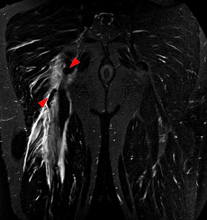
MRICoronal pelvic MRI, STIR sequence: tear of the hamstring tendons at their insertion on the ischial tuberosity, with a retracted tendon stump (arrowheads).
Source : Hellerhoff, Wikimedia Commons · CC BY-SA
Three provocation tests are considered the most useful for reproducing the patient's familiar pain by loading the proximal tendon:¹,³
- Puranen-Orava test (active stretch): patient standing, foot on a high support (hip flexed to about 90°), knee extended, patient leans forwards. Reproduces pain at the tuberosity through active stretch.
- Bent-Knee Stretch Test (BKS): patient supine, hip and knee maximally flexed, the examiner rapidly takes the limb into maximal hip flexion + passive knee extension. Reproduces pain through maximal passive stretch.
- Modified Bent-Knee Stretch Test (MBKS): a slower, more controlled variant of the BKS, considered more sensitive and better tolerated. Reproduces pain through progressive stretch.³
Direct palpation of the ischial tuberosity often reproduces the pain, but this test lacks specificity: the area is tender in many asymptomatic people (ischiogluteal bursitis, anatomical variation). Side-to-side comparison is essential.¹,⁴
📋 Comparative table of PHT provocation tests
Level of evidence: evidence based essentially on clinical case series and expert reviews (Goom 2016, Cacchio 2013)
| Test | Modality | Estimated sensitivity | Estimated specificity | Level of evidence |
|---|---|---|---|---|
| Ischial tuberosity palpation | Direct digital pressure | High | Low (not very specific) | Expert opinion |
| Puranen-Orava | Standing active stretch | Moderate | Moderate | Clinical case series (Cacchio 2013) |
| Bent-Knee Stretch Test (BKS) | Rapid passive stretch | High | Moderate | Clinical case series (Cacchio 2013) |
| Modified BKS | Progressive passive stretch | High | Moderate to high | Clinical case series (Cacchio 2013) |
| Resisted isometric hip extension | Muscle strength test | Variable | Moderate | Expert opinion |
Sources: Cacchio A, et al. Reliability and validity of three pain provocation tests for chronic PHT. Br J Sports Med. 2012;46(12):883-887. Goom 2016 JOSPT. Caveat : precise sensitivity and specificity values vary between studies and do not rest on a robust recent meta-analysis. No single test constitutes a \"gold standard\".
The differential diagnosis is crucial in the deep gluteal region, a complex anatomical crossroads:¹,⁵
- Acute or subacute proximal avulsion: an often identifiable traumatic mechanism (sprinting, slipping into the splits, a fall), extensive bruising of the posterior thigh, a prognostic strength deficit (> 30%), intense acute pain. If suspected, urgent MRI because a complete avulsion may benefit from early surgery.⁶
- Deep gluteal syndrome: compression of the sciatic nerve in the subgluteal space. Characteristic feature: paraesthesia or numbness along the sciatic distribution, pain worsening on sitting but with a neurological component. Neural provocation tests (SLR, slump test) are often positive.⁷
- L5-S1 lumbar radiculopathy: a lumbar history, ascending pain, neurological signs (motor deficit, hypoaesthesia, reflex asymmetry). Lumbar examination and neurodynamic tests are essential.
- Hip disorders: femoroacetabular impingement (FAI), osteoarthritis, labral tear. Pain is often inguinal or follows the \"C-sign\" pattern, with positive FADIR/FABER tests. Radiographs and MRI if needed.⁸
- Proximal gluteal tendinopathy: lateral trochanteric pain, not ischial. Different provocation tests (Trendelenburg, single-leg stance).
- Isolated ischiogluteal bursitis: rare in isolation, more often associated with the tendinopathy. Ultrasound can help.
Red flags requiring urgent referral
- Acute traumatic mechanism (maximal sprint, fall into the splits) with sudden intense pain, extensive bruising of the posterior thigh and a major strength deficit → suspicion of proximal avulsion : MRI within ≤ 7 days and an orthopaedic surgical opinion. A complete avulsion (3 tendons) in an active patient may benefit from early repair.
- History of cancer, unexplained weight loss, night sweats, palpable mass → rule out a tumour (posterior thigh sarcoma, rare but serious).
- Severe night pain, fever, deterioration in general condition → rule out infection (rare: abscess, tropical pyomyositis of the hamstrings in travellers).
- Progressive neurological deficit (L5-S1 motor deficit, saddle anaesthesia, sphincter disturbance) → neurosurgical emergency (cauda equina).
- Apophyseal avulsion in the adolescent (open growth plate, sprinting mechanism) → suspicion of avulsion of the ischial tuberosity: radiographs and a paediatric surgical opinion.
The role of imaging is second line:¹,⁹
- MRI: the reference standard. It demonstrates tendon thickening, intratendinous oedema (T2 hyperintensity), peri-ischial bone oedema (sometimes) and partial tears. It rules out an avulsion and quantifies the chronic component. Limitation: abnormalities are present in 50% of asymptomatic people (corrected Zissen 2010 AJR), so interpretation must be made in clinical correlation.¹⁰
- Ultrasound: less sensitive than MRI for oedema but useful for visualising thickening, Doppler neovascularisation and the adjacent bursae. Operator-dependent. It allows injections to be guided.
- Radiography: of little value for the tendon but useful for ruling out a calcification, an adolescent bony avulsion or hip involvement.
🧮 Decision flow chart: suspected deep buttock pain
Recommended clinical approach according to Goom 2016, Beatty 2017 and expert consensus
Sources: Goom 2016 JOSPT, Beatty 2017 Curr Sports Med Rep, Lempainen 2015 KSSTA.
Should patients be classified by their stage on the tendon continuum?
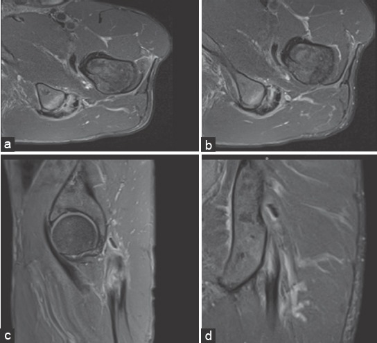
MRIAxial and coronal thigh MRI: heterogeneous thickening of the proximal common hamstring tendon with partial tear and bone marrow oedema of the ischial tuberosity.
Source : Costa et al., Journal of Orthopaedic Case Reports, 2016, figure 1 · CC BY-NC-SA
Yes: not to produce a rigid \"label\", but to tailor the load management strategy.⁷ The clinician tries to place the patient on the continuum:
- Reactive stage: young athlete, recent symptoms (< 4-6 weeks), triggered by an identifiable acute rise in load. The tendon is often swollen. Priority: temporary reduction of the aggravating load (compression and energy storage), then progressive reintroduction over a few weeks.
- Dysrepair or degenerative stage: older patient, long history (months to years), previous treatment failures. Priority: progressive heavy mechanical stimulus (HSR) to improve tendon capacity, rather than prolonged rest.
This logic explains a clinical paradox: a heavy loading programme applied too early to a reactive tendon can worsen symptoms, whereas a simple reduction in load is not enough for a degenerative tendon that needs a mechanical stimulus in order to adapt.¹,⁷
- The diagnosis of PHT is clinical : a combination of history (sitting and running aggravate) and positive provocation tests (Puranen-Orava, BKS, MBKS).
- No single gold standard : the battery of tests outweighs any one test. Published sensitivity and specificity values come from small series without a robust meta-analysis.
- The essential differential diagnoses: deep gluteal syndrome, acute avulsion, S1 radiculopathy, hip disorders, proximal gluteal tendinopathy. And, where the mechanism is abrupt and dated rather than gradual in onset, acute hamstring muscle tear (strain), whose timescales and recurrence risk bear no relation to those of PHT.
- Imaging (MRI > ultrasound) is second line, justified by red flags, diagnostic doubt or treatment failure.
- Classifying the patient by stage on the continuum (reactive vs degenerative) helps to calibrate the loading strategy: temporary reduction for a reactive tendon, heavy stimulus for a degenerative one.
Chapter 2 bibliography
- Goom TS, Malliaras P, Reiman MP, Purdam CR. Proximal Hamstring Tendinopathy: Clinical Aspects of Assessment and Management. J Orthop Sports Phys Ther. 2016;46(6):483-93. PMID 27084841
- Beatty NR, Félix I, Hettler J, Moley PJ, Wyss JF. Rehabilitation and Prevention of Proximal Hamstring Tendinopathy. Curr Sports Med Rep. 2017;16(3):162-171. PMID 28498225
- Cacchio A, Borra F, Severini G, et al. Reliability and validity of three pain provocation tests used for the diagnosis of chronic proximal hamstring tendinopathy. Br J Sports Med. 2012;46(12):883-887. PMID 22219215
- Nasser AM, Vicenzino B, Grimaldi A, Anderson J, Semciw AI. Proximal hamstring tendinopathy; expert physiotherapists' perspectives on diagnosis, management and prevention. Phys Ther Sport. 2021;48:67-75. PMID 33378733
- Lempainen L, Banke IJ, Johansson K, et al. Clinical principles in the management of hamstring injuries. Knee Surg Sports Traumatol Arthrosc. 2015;23(8):2449-2456. PMID 24556933
- Lempainen L, Sarimo J, Mattila K, Vaittinen S, Orava S. Proximal hamstring tendinopathy: results of surgical management and histopathologic findings. Am J Sports Med. 2009;37(4):727-734. PMID 19218559
- Cook JL, Purdam CR. Is tendon pathology a continuum? A pathology model to explain the clinical presentation of load-induced tendinopathy. Br J Sports Med. 2009;43(6):409-416. PMID 18812414
- Martin HD, Khoury A, Schroder R, Palmer IJ. Ischiofemoral Impingement and Hamstring Syndrome as Causes of Posterior Hip Pain: Where Do We Go Next? Clin Sports Med. 2016;35(3):469-486. PMID 27343396
- Cushman D, Rho ME. Conservative Treatment of Subacute Proximal Hamstring Tendinopathy Using Eccentric Exercises Performed With a Treadmill: A Case Report. J Orthop Sports Phys Ther. 2015;45(7):557-562. PMID 25996362
- Zissen MH, Wallace G, Stevens KJ, Fredericson M, Beaulieu CF. High hamstring tendinopathy: MRI and ultrasound imaging and therapeutic efficacy of percutaneous corticosteroid injection. AJR Am J Roentgenol. 2010;195(4):993-998. PMID 20858830
Which treatment strategies are the most effective for PHT?
The management of PHT rests on three principles: (1) load management (temporary reduction of compressive and energy-storage loads), (2) a structured strengthening progression (isometrics, then heavy slow, then plyometric), and (3) the cautious addition of adjuncts when progress plateaus (shockwave therapy first, injections as a last resort).¹,²,³
Where do you start? What is the hierarchy of interventions?
The consensus treatment pyramid always places education and load modification in the first line:¹,⁴
- Modify the sitting position: use a \"donut\" cushion or a gel cushion, sit on the edge of the seat (transferring pressure to the thighs), avoid prolonged sitting on hard seats, take standing breaks every 30-45 minutes.
- Temporarily avoid provocative stretching: classic stretches into full hip flexion (hands-to-toes, seated straight-leg stretches) increase compression. To be avoided in the initial phase, then reintroduced cautiously from phase 3 onwards.³
- Reduce the volume of energy-storage activities: sprints, hills, climbs and deep lunges are temporarily limited. \"Slow jogging\" on flat ground can be maintained if it is well tolerated.
- Education: explain the non-inflammatory overload nature of the problem, the potentially chronic course, and the importance of adhering to the progression (typically 3-6 months).
The complete-rest approach is harmful : it weakens the tendon further and increases the risk of re-exacerbation when activity resumes. The rule is: load without aggravating, guided by 24-hour pain monitoring (the Silbernagel rule).⁵
What is the place of progressive exercise and which phases does it comprise?
Progressive exercise is the intervention best supported by the evidence, although the overall quality of the studies remains moderate (the systematic reviews of Nasser 2021 IJSPT and Dizon 2023 Sports identify a lack of high-quality RCTs but a consistency of findings favouring progressive loading for pain and function).⁶,⁷ The sequence recommended by Goom 2016 has become the de facto clinical standard:¹
🏋️ Four-phase progressive exercise programme for PHT
Conceptual progression: indicative durations, individually adapted according to the 24-hour rule
Sources: Goom et al. JOSPT. 2016;46(6):483-493. PMID 27050674. Krueger K et al. IJSPT. 2020;15(5):844-852. PMID 33110701. Bohm 2015 Sports Med Open (tendon loading).
A few clinical points on each phase:
- Phase 1 (isometrics): closed-chain isometrics with the hip close to neutral (basic glute bridge) allow the posterior chain to be activated without compression against the ischium. Contractions are long (30-45 sec), repeated 5 times, 2-3 times a day. The post-isometric analgesic effect observed in other tendinopathies (patellar tendon) is sometimes cited for PHT by extrapolation, without specific direct high-level evidence.⁸
- Phase 2 (HSR): Heavy Slow Resistance (3 sec eccentric + 3 sec concentric) with increasing loads (RPE 7-8/10) is now the dominant approach. The exemplary case report by Krueger 2020 (a 31-year-old powerlifter) describes a 12-week progression leading to a return to the pre-injury level and beyond.⁹ Typical exercises: Romanian deadlift, single-leg deadlift, dumbbell hip thrust, machine leg curl. Range of motion is limited initially (avoiding maximal hip flexion), then widened.
- Phase 3 (energy storage and release): only for patients who need to run or jump. Graded plyometrics: A-skips → bounding on the spot → forward bounding → jump lunges. This phase is not required for sedentary patients.
- Phase 4 (return to running): low-intensity straight-line running → increasing volume → increasing intensity → short sprints → changes of direction. Running cadence can be increased by 5-10% to reduce compressive forces on the tendon (Lenhart 2014 JOSPT).¹⁰
Shockwave, injections, adjunctive therapies: what is the real efficacy?
Adjunctive modalities never replace a well-conducted exercise programme. They may be considered when progress stalls at 8-12 weeks.¹,²
- Extracorporeal shockwave therapy (ESWT): the systematic review by Korakakis 2018 BJSM (52(6):387-407) on lower limb tendinopathies concludes that ESWT is a reasonable option for chronic tendinopathies, including PHT. The pivotal RCT by Cacchio 2011 AJSM (39(1):146-153) in 40 professional athletes with chronic PHT showed superiority of radial shockwave therapy (4 sessions, 2500 impulses) over traditional conservative treatment at 12 months (success: 85% vs 10%).¹¹,¹² Caveat : a small, single-centre study in a specific population of professional athletes.
- Ultrasound-guided corticosteroid injections: may provide short-term relief (4-6 weeks; Zissen 2010 AJR reported 50% relief lasting > 1 month). However: increased risk of tendon rupture, not recommended routinely, to be reserved for exceptional cases (rehabilitation impossible for occupational reasons or immediate competition).⁵,¹³
- Platelet-rich plasma (PRP): data specific to PHT are limited. For tendinopathies in general, recent meta-analyses do not demonstrate robust superiority over placebo in good-quality RCTs.¹⁴ Not to be recommended routinely.
- Manual therapy, dry needling, transverse frictions: may be used as adjuncts for pain modulation, but without evidence of specific efficacy for PHT. Dry needling has sometimes been used as a complement (Jayaseelan 2014 JOSPT).¹⁵
- Relative rest, ice, systemic NSAIDs: NSAIDs can be used occasionally during an acute flare, but their chronic use is harmful (inhibition of tendon healing). Complete rest is contraindicated.
🧮 GRADE-style synthesis of interventions for PHT
Narrative synthesis of the available data: no formal GRADE system specific to PHT exists to date
| Intervention | Level of evidence | Expected clinical effect | Recommendation |
|---|---|---|---|
| Education + load management | Moderate to high (expert consensus) | Indispensable foundation | First line: always |
| Progressive exercise (4 phases) | Moderate (SR Nasser 2021, Dizon 2023) | Pain reduction, VISA-H improvement | First line: therapeutic mainstay |
| ESWT (shockwave therapy) | Moderate (Cacchio 2011, Korakakis 2018) | Pain reduction in chronic forms | Second line if plateau |
| Manual therapy / dry needling | Low (indirect evidence) | Short-term pain modulation | Occasional adjunct |
| Corticosteroid injection | Low (short term only, risks) | Relief ≤ 6 weeks | Reserved for exceptions |
| PRP | Low (insufficient PHT data) | Uncertain effect vs placebo | Not recommended routinely |
| Surgery (tenotomy / repair) | Low (case series) | ~80% good outcomes in the Lempainen series | Conservative failure ≥ 6 months |
Sources: Goom 2016 JOSPT, Nasser 2021 IJSPT, Dizon 2023 Sports, Korakakis 2018 BJSM, Cacchio 2011 AJSM, Lempainen 2009 AJSM.
Beyond the physical: how do you educate the patient and address psychological factors?
Education is a therapeutic tool in its own right, not a \"bonus\". Three key messages should be conveyed:¹,⁶
- Pain is not a precise \"tissue alarm signal\" : pain of 4/10 during exercise does not mean that things are getting worse. The rule is: acceptable pain during exercise + no worsening at 24 hours = carry on.
- Recovery takes a long time : 3-6 months on average for significant improvement; 9-12 months for chronic cases. Setting realistic expectations prevents discouragement and drop-out.
- Adherence is the number 1 predictor of success : a patient who completes 80% of their programme is twice as likely to improve their VISA-H as a patient who completes 30%.¹⁶
Psychosocial factors to screen for:
- Kinesiophobia (fear of movement): Tampa Scale of Kinesiophobia (TSK), clinical threshold > 37.
- Catastrophising : Pain Catastrophizing Scale (PCS), clinical threshold > 30.
- Limiting beliefs : \"my tendon is damaged, I will never be able to run again\". To be challenged with the evidence on tendon plasticity (Bohm 2015 Sports Med Open) and the success stories in case reports (Krueger 2020).
- Always begin with education + load management before any intensive exercise.
- Progressive exercise in 4 phases (isometrics → HSR → plyometrics → return to running) is the best supported intervention, with a moderate level of evidence.
- The shockwaves (ESWT) are the best supported adjunct when progress stalls, especially in chronic forms (Korakakis 2018, Cacchio 2011).
- The corticosteroid injections and PRP are not recommended routinely; long-term effects are unfavourable or unproven.
- Education about the nature of the condition, the timescales and the 24-hour rule is a treatment in its own right.
- Screen for kinesiophobia, catastrophising and limiting beliefs in order to tailor biopsychosocial support.
Chapter 3 bibliography
- Goom TS, Malliaras P, Reiman MP, Purdam CR. Proximal Hamstring Tendinopathy: Clinical Aspects of Assessment and Management. J Orthop Sports Phys Ther. 2016;46(6):483-93. PMID 27084841
- Beatty NR, Félix I, Hettler J, Moley PJ, Wyss JF. Rehabilitation and Prevention of Proximal Hamstring Tendinopathy. Curr Sports Med Rep. 2017;16(3):162-171. PMID 28498225
- Cook JL, Purdam CR. Is compressive load a factor in the development of tendinopathy? Br J Sports Med. 2012;46(3):163-168. PMID 22113234
- Cook JL, Purdam CR. The challenge of managing tendinopathy in competing athletes. Br J Sports Med. 2014;48(7):506-509. PMID 23666020
- Silbernagel KG, Thomeé R, Eriksson BI, Karlsson J. Continued sports activity, using a pain-monitoring model, during rehabilitation in patients with Achilles tendinopathy: a randomized controlled study. Am J Sports Med. 2007;35(6):897-906. PMID 17307888
- Nasser AM, Vicenzino B, Grimaldi A, Anderson J, Semciw AI. Proximal Hamstring Tendinopathy: A Systematic Review of Interventions. Int J Sports Phys Ther. 2021;16(2):288-305. PMID 33842025
- Dizon P, Jeanfavre M, Leff G, Norton R. Comparison of Conservative Interventions for Proximal Hamstring Tendinopathy: A Systematic Review and Recommendations for Rehabilitation. Sports (Basel). 2023;11(3):53. PMID 36976939
- Bohm S, Mersmann F, Arampatzis A. Human tendon adaptation in response to mechanical loading: a systematic review and meta-analysis of exercise intervention studies on healthy adults. Sports Med Open. 2015;1(1):7. PMID 27747846
- Krueger K, Washmuth NB, Williams TD. THE MANAGEMENT OF PROXIMAL HAMSTRING TENDINOPATHY IN A COMPETITIVE POWERLIFTER WITH HEAVY SLOW RESISTANCE TRAINING - A CASE REPORT. Int J Sports Phys Ther. 2020;15(5):814-822. PMID 33110701
- Lenhart RL, Thelen DG, Heiderscheit BC. Hip muscle loads during running at various step rates. J Orthop Sports Phys Ther. 2014;44(10):766-774. PMID 25156044
- Cacchio A, Rompe JD, Furia JP, Susi P, Santilli V, De Paulis F. Shockwave therapy for the treatment of chronic proximal hamstring tendinopathy in professional athletes. Am J Sports Med. 2011;39(1):146-153. PMID 20855554
- Korakakis V, Whiteley R, Tzavara A, Malliaropoulos N. The effectiveness of extracorporeal shockwave therapy in common lower limb conditions: a systematic review including quantification of patient-rated pain reduction. Br J Sports Med. 2018;52(6):387-407. PMID 28954794
- Zissen MH, Wallace G, Stevens KJ, Fredericson M, Beaulieu CF. High hamstring tendinopathy: MRI and ultrasound imaging and therapeutic efficacy of percutaneous corticosteroid injection. AJR Am J Roentgenol. 2010;195(4):993-998. PMID 20858830
- Andriolo L, Altamura SA, Reale D, Candrian C, Zaffagnini S, Filardo G. Nonsurgical Treatments of Patellar Tendinopathy: Multiple Injections of Platelet-Rich Plasma Are a Suitable Option: A Systematic Review and Meta-analysis. Am J Sports Med. 2019;47(4):1001-1018. PMID 29601207
- Jayaseelan DJ, Moats N, Ricardo CR. Rehabilitation of proximal hamstring tendinopathy utilizing eccentric training, lumbopelvic stabilization, and trigger point dry needling: 2 case reports. J Orthop Sports Phys Ther. 2014;44(3):198-205. PMID 24261928
- Vicenzino B, de Vos RJ, Alfredson H, et al. ICON 2019-International Scientific Tendinopathy Symposium Consensus: There are nine core health-related domains for tendinopathy (CORE DOMAINS): Delphi study of healthcare professionals and patients. Br J Sports Med. 2020;54(8):444-451. PMID 31685525
How do you secure lasting recovery and prevent recurrence of PHT?
The long-term management of PHT does not stop when pain disappears. It aims to restore a load capacity greater than functional demands, which is the condition for preventing recurrence.¹ The exact recurrence rate is not robustly established for PHT (few long-term prospective studies), but clinical experience and analogies with other tendinopathies suggest a persistent vulnerability of the tendon, particularly to load spikes.²
How can the patient be made an active partner in recovery through self-management?
Patient empowerment rests on three pillars: (1) understanding the condition, (2) self-monitoring of the response to load, and (3) the ability to modulate their activities according to that response.¹,³
Silbernagel's 24-hour rule: the key self-monitoring tool
Initially validated on the Achilles tendon (Silbernagel 2007 AJSM), the 24-hour rule is today applied by extrapolation to most tendinopathies, including PHT:⁴
- Acceptable pain during exercise: VAS ≤ 4/10.
- Acceptable pain after exercise: no worsening the next day compared with baseline.
- Acceptable pain on palpation: return to baseline within 24 hours.
If one of these criteria is exceeded: reduce the volume or intensity of the next session by 10-20%. If all are met: consider progressing. This system turns the patient into the \"co-pilot\" of their rehabilitation.⁴
Pacing: avoiding the boom-bust cycle
Pacing is the art of dosing activity to avoid peaks followed by crashes (the boom-bust) :⁵
- Identify the \"tolerable baseline\" : the daily activity volume that generates no worsening the next day.
- Increase in 10% steps : follow the rule of a maximum 10% weekly increase in running volume.
- Schedule active recovery days: 1-2 days a week without running but with HSR exercises maintained.
- Manage occasional spikes (competition, an unplanned hike) with a planned load reduction over the following 2-3 days.
Education about the non-linear nature of recovery
The improvement trajectory is rarely linear. The patient must be prepared for occasional painful flares, which do not mean a relapse if they stay within the limits of the 24-hour rule. Picturing progress as \"two steps forward, one step back\" reduces anxiety and drop-out.¹
When and how should a safe return to sport and running be planned?
Return to sport (RTS) must never be dictated by a fixed calendar.⁶ A premature return, based solely on the disappearance of pain at rest, is the number 1 risk factor for recurrence. RTS planning relies on objective functional criteria, following the logic of the BAMIC framework (British Athletics Muscle Injury Classification) developed for muscle injuries (Macdonald 2019 BJSM) and applicable by analogy to tendinopathies.⁷
Prerequisite criteria for a partial return
- No pain on palpation of the ischial tuberosity (or pain < 2/10 side to side).
- Full tolerance of HSR exercises at a load equivalent to pre-injury or to the healthy side.
- Isometric strength symmetry in hip extension (deficit < 10%), measured with a hand-held dynamometer.
- Tolerance of plyometric exercises at a basic level (A-skips, bounding on the spot) without a delayed reaction.
- VISA-H > 80/100 or an improvement of ≥ 20 points from baseline.
Phases of reintroduction to sport
- Straight-line running, low intensity (60-70% max HR): start at 1.5-3 km, then +10% per week according to tolerance. 3 sessions a week.
- Moderate-intensity running (70-85% max HR): introduction of short accelerations (15-30 m).
- Progressive sprints: short sprints (30 m), then increasing length, then increasing intensity (80% → 90% → 100%).
- Changes of direction, agility work: the final stage, with maximal multidirectional demands.
- Reintegration into team training : controlled portions (warm-up, technical drills) before game situations.
🏁 Decision tree: return to sport after PHT
Criterion-based return to sport, adapted from Goom 2016 and Macdonald 2019 (BAMIC)
Sources: Goom 2016 JOSPT, Macdonald 2019 BJSM (BAMIC), Hickey 2020 JOSPT (pain monitoring).
\"Taping the window\": maintaining HSR even after the return
Athletes returning to competition must maintain 1-2 HSR sessions a week as prevention. The tendon does not \"heal\" by becoming what it was before; it remains more vulnerable and requires continuous maintenance of its load capacity.¹,³
- Self-management through Silbernagel's 24-hour rule is the key tool for durability: pain ≤ 4/10 during exercise + no worsening the next day = progression is possible.
- Pacing avoids the boom-bust cycle through gradual increases (10%/wk) and planned active recovery days.
- The return to sport must be driven by functional criteria (palpation OK, HSR at pre-injury load, strength symmetry < 10%, VISA-H > 80) and never by a fixed calendar.
- The sporting progression follows 5 stages: easy running → moderate → sprints → agility → full match.
- Maintaining HSR 1-2x/wk as prevention after RTS is essential for durability.
Chapter 4 bibliography
- Goom TS, Malliaras P, Reiman MP, Purdam CR. Proximal Hamstring Tendinopathy: Clinical Aspects of Assessment and Management. J Orthop Sports Phys Ther. 2016;46(6):483-93. PMID 27084841
- Cook JL, Purdam CR. The challenge of managing tendinopathy in competing athletes. Br J Sports Med. 2014;48(7):506-509. PMID 23666020
- Beatty NR, Félix I, Hettler J, Moley PJ, Wyss JF. Rehabilitation and Prevention of Proximal Hamstring Tendinopathy. Curr Sports Med Rep. 2017;16(3):162-171. PMID 28498225
- Silbernagel KG, Thomeé R, Eriksson BI, Karlsson J. Continued sports activity, using a pain-monitoring model, during rehabilitation in patients with Achilles tendinopathy: a randomized controlled study. Am J Sports Med. 2007;35(6):897-906. PMID 17307888
- Mascaro A, Cos MA, Morral A, Roig A, Purdam C, Cook J. Load management in tendinopathy: Clinical progression for Achilles and patellar tendinopathy. Apunts Sports Medicine. 2018;53(197):19-27. DOI 10.1016/j.apunts.2017.11.005
- Hickey JT, Timmins RG, Maniar N, et al. Pain-Free Versus Pain-Threshold Rehabilitation Following Acute Hamstring Strain Injury: A Randomized Controlled Trial. J Orthop Sports Phys Ther. 2020;50(2):91-103. PMID 32005093
- Macdonald B, McAleer S, Kelly S, Chakraverty R, Johnston M, Pollock N. Hamstring rehabilitation in elite track and field athletes: applying the British Athletics Muscle Injury Classification in clinical practice. Br J Sports Med. 2019;53(23):1464-1473. PMID 31300391
- Heiderscheit BC, Sherry MA, Silder A, Chumanov ES, Thelen DG. Hamstring strain injuries: recommendations for diagnosis, rehabilitation, and injury prevention. J Orthop Sports Phys Ther. 2010;40(2):67-81. PMID 20118524
- Cacchio A, De Paulis F, Maffulli N. Development and validation of a new visa questionnaire (VISA-H) for patients with proximal hamstring tendinopathy. Br J Sports Med. 2014;48(6):448-452. PMID 23470447
What do real clinical cases teach us about PHT?
Clinical cases are a valuable source of learning for the clinician: they show how general principles take shape in individual trajectories, and bring to light subtleties that standardised protocols do not capture. All the cases presented here are peer-reviewed publications with a searchable PMID.
Analysis of a \"classic\" case: a long-distance runner (Campos-Villegas 2024)
The case report by Campos-Villegas et al. published in the International Journal of Sports Physical Therapy in 2024 describes the management of a 30-year-old recreational runner with deep right buttock pain that had come on insidiously after a 10 km race.¹ Symptoms were aggravated by running on sloping ground and by prolonged sitting, particularly while driving.
Diagnostic approach: palpation reproducing pain at the ischial tuberosity, positive provocation tests (Puranen-Orava, Bent-Knee Stretch), no neurological signs. Clinical diagnosis of PHT made without initial imaging.
Rehabilitation programme: a progressive loading protocol over several phases, including initial isometric exercises, then controlled eccentric and concentric strengthening, with assessment by the VISA-H score. The progression incorporated the 24-hour rule to adjust loads.¹
Results: a progressive return to running after several weeks of rehabilitation, with significant improvement in VISA-H and in functional tolerance. The case illustrates the value of an individualised progression that respects the principles of load management.¹
The diagnostic challenge: when PHT coexists with other conditions
The case report by Lewis 2020 (Journal of Chiropractic Medicine) describes a 53-year-old Tae Kwon Do athlete, who had practised for 10 years and had stopped all activity for a year because of chronic, insidious-onset pain in the left buttock, radiating to the hip and posterior thigh.² Initial pain score: 7/10. Major functional impact (brisk walking, sitting).
Diagnostic approach: radiographs of the lumbopelvic complex were negative. Clinical examination localised the pain to the left ischial tuberosity. MRI revealed bilateral trochanteric bursitis + left hamstring tendinopathy.
Management: a conservative approach combining therapeutic ultrasound over the hamstring enthesis, manual sacroiliac manipulation, and progressive rehabilitation. Complete resolution of symptoms.
Lessons: this case illustrates two crucial points:
- The combination of conditions is common in veteran athletes (female, age, repeated stress). The clinician must look beyond the symptomatic tendon alone.
- The associated sacroiliac dysfunction can increase the stresses on the ischial enthesis and must be addressed in parallel.²
Complex case studies: a powerlifter on HSR and an elite tennis player on pain monitoring
Case A: a competitive powerlifter on Heavy Slow Resistance (Krueger 2020 IJSPT)
This case report describes a 31-year-old powerlifter with chronic PHT, with pain at the ischial tuberosity and hip weakness limiting his lifts and preventing him from sitting for more than 30 minutes.³
Programme: 12 weeks of targeted Heavy Slow Resistance, with progression of the load intensity. The main exercises were lifting variations (squat, deadlift) with controlled range of motion and slow tempo.
Results:
- Marked reduction in pain from week 4 onwards.
- At 12 weeks: performance exceeding the pre-injury level (100 kg back squat, 160 kg conventional deadlift, 90 kg Romanian deadlift).
- No recurrence at 1-year follow-up.³
This case demonstrates that heavy load ≠ dangerous in tendinopathy. On the contrary, in a well-selected patient (young, motivated, strength athlete), HSR is the most effective intervention for raising tendon capacity beyond the pre-injury level.
Case B: an elite tennis player with the pain-monitoring model (Petersen 2022 JOSPT Cases)
This case report published in JOSPT Cases describes the application of the pain-monitoring model in a 24-year-old elite tennis player with right-sided PHT.⁴ The model had initially been validated on the Achilles tendon (Silbernagel 2007) but had never been the subject of a dedicated publication for PHT.
Approach: rehabilitation based mainly on progressive strengthening exercises and training load management guided by the pain-monitoring model (acceptable pain ≤ 5/10 during exercise AND no worsening the next day).
Results after 24 weeks:
- Return to unrestricted training.
- VISA-H: 76/100 (baseline not reported, but a clear improvement).
- OSTRC severity score: 0 (asymptomatic).⁴
This case validates the extrapolation of the Achilles pain-monitoring model to PHT and illustrates the importance of regular self-assessment by the elite patient.
Critical notes on clinical cases in PHT
Several limitations must be kept in mind when interpreting case reports:
- Publication bias: published cases tend to be success stories. Patients whose rehabilitation fails are under-represented.
- Limited generalisability: a protocol that succeeded in a 31-year-old powerlifter (Krueger 2020) does not apply as it stands to a 55-year-old female runner with hypermobility.
- No control group: it is impossible to attribute the improvement formally to any particular intervention.
- Heterogeneity of outcomes: some report VISA-H, others VAS, others sport-specific performance. The Nasser 2021 IJSPT meta-analysis highlights this lack of standardisation which complicates synthesis.⁵
- The Campos-Villegas 2024 case in a recreational runner validates structured progression with progressive loading and the 24-hour rule.
- The Krueger 2020 case in a powerlifter demonstrates that HSR with heavy loads allows a return beyond the pre-injury level when the patient is motivated and well selected.
- The Petersen 2022 case in an elite tennis player validates the extrapolation of the pain-monitoring model to PHT.
- The Lewis 2020 case in a veteran Tae Kwon Do athlete is a reminder that associated conditions (sacroiliac dysfunction, bursitis) must be addressed in parallel.
- Case reports are a source of clinical inspiration but are subject to publication bias and to a lack of direct generalisability.
Chapter 5 bibliography
- Campos-Villegas C, Ortega-Pérez de Villar L, Gámez-Payá J, Alarcón-Jiménez J, de Bernardo N. Clinical Progression and Load Management For Proximal Hamstring Tendinopathy In A Long-Distance Runner: A Case Report. Int J Sports Phys Ther. 2024;19(5):609-617. PMID 38707848
- Lewis CL. Chronic High Hamstring Tendinopathy and Sacroiliac Segmental Dysfunction in a Mature Tae Kwon Do Athlete: A Case Study. J Chiropr Med. 2020;19(3):201-207. PMID 32952477
- Krueger K, Washmuth NB, Williams TD. THE MANAGEMENT OF PROXIMAL HAMSTRING TENDINOPATHY IN A COMPETITIVE POWERLIFTER WITH HEAVY SLOW RESISTANCE TRAINING - A CASE REPORT. Int J Sports Phys Ther. 2020;15(5):814-822. PMID 33110701
- Pettersen SD, Sigvardsen H, Tjelta LI. Applying the Pain-Monitoring Model in a Female Elite Tennis Player With Proximal Hamstring Tendinopathy: A Case Report. JOSPT Cases. 2022;2(4):238-244. DOI 10.2519/josptcases.2022.11372
- Nasser AM, Vicenzino B, Grimaldi A, Anderson J, Semciw AI. Proximal Hamstring Tendinopathy: A Systematic Review of Interventions. Int J Sports Phys Ther. 2021;16(2):288-305. PMID 33842025
- Cushman D, Rho ME. Conservative Treatment of Subacute Proximal Hamstring Tendinopathy Using Eccentric Exercises Performed With a Treadmill: A Case Report. J Orthop Sports Phys Ther. 2015;45(7):557-562. PMID 25996362
- Jayaseelan DJ, Moats N, Ricardo CR. Rehabilitation of proximal hamstring tendinopathy utilizing eccentric training, lumbopelvic stabilization, and trigger point dry needling: 2 case reports. J Orthop Sports Phys Ther. 2014;44(3):198-205. PMID 24261928
- White KE. High hamstring tendinopathy in 3 female long distance runners. J Chiropr Med. 2011;10(2):93-99. PMID 22014904
- McCormack JR, Underwood FB, Slaven EJ, Cappaert TA. The management of bilateral high hamstring tendinopathy with ASTYM treatment and eccentric exercise: a case report. J Man Manip Ther. 2012;20(3):142-146. PMID 23904753
How do you apply these recommendations concretely in your practice?
Integrating evidence into daily practice is the main challenge for the modern physiotherapist. Beyond technical knowledge, excellence rests on the capacity for clinical reasoning, on interprofessional collaboration and on objective measurement of outcomes.
When and to which other professionals should you refer?
The direct-access physiotherapist plays a key triage role. Vigilance for red flags is the most critical safety skill:¹
- To the emergency department, or urgently to the GP: acute trauma with a mechanism of slipping into the splits, extensive bruising of the posterior thigh, major motor deficit → suspicion of acute proximal avulsion. MRI within ≤ 7 days and an orthopaedic surgical opinion. A complete avulsion in an active person may benefit from early surgical repair (Lempainen 2009).²
- To the orthopaedic surgeon / sports physician: failure of well-conducted conservative treatment over 6-12 months; suspicion of an unoperated partial avulsion documented on MRI; refractory disabling pain in a person wishing to return to competition.
- To the rheumatologist / physician in internal medicine: an atypical picture (bilateral pain of simultaneous onset, systemic signs, inflammatory context) → rule out a spondyloarthropathy or an axial-peripheral enthesopathy.
- To the neurologist: associated neurological signs (L5/S1 sensory deficit, motor deficit, saddle anaesthesia) → rule out a compressive radiculopathy or cauda equina syndrome.
- To the psychologist / pain specialist: predominant psychosocial factors (catastrophising, kinesiophobia, anxiety, depression). A biopsychosocial approach combining CBT and exercise is superior to either approach alone for chronic musculoskeletal pain.³,⁴
- To the podiatrist / biomechanist: a runner with objectively measurable biomechanical faults (excessive overpronation, marked overstriding). Running analysis and possible orthotic adjustment can reduce the stresses on the enthesis.
How do you measure outcomes and overcome barriers to implementation?
Recommended objective measures
Progress assessment must be standardised, multidimensional and reproducible:⁵
- VISA-H (Victorian Institute of Sport Assessment: Hamstring): a questionnaire validated in 2014 by Cacchio (BJSM), 8 items, score 0-100 (100 = asymptomatic). Sensitive to change. Available in French (Causeret 2019).⁵
- Sit-to-pain duration : the maximum tolerable sitting time without pain > 2/10. A simple measure, representative of functional impact.
- Isometric hip extension strength test : hand-held dynamometer, supine with the hip flexed to 30° / 60° / 90°. Allows side-to-side symmetry to be tracked.
- VAS / NPRS : pain on palpation, the patient's typical running pain, post-exercise pain.
- OSTRC severity score (Oslo Sports Trauma Research Center): for athletes, assesses the impact on sports participation.
Understanding the GRADE pyramid of evidence
The GRADE system (Grading of Recommendations, Assessment, Development and Evaluation) is the international reference scale for appraising the quality of evidence. It distinguishes four levels of certainty:⁶
📊 GRADE pyramid: levels of evidence applied to PHT
Horizontal cards (never text INSIDE the triangle): maximum legibility
Sources: GRADE Working Group (Schunemann 2013). Applied to the PHT context by the authors.
Barriers to implementation, and levers
The common barriers to evidence-based practice in physiotherapy have been documented:
- Lack of time for collecting PROMs → build it into the welcome time or before the session with a paper form or a dedicated app.
- Difficulty interpreting studies → take part in journal clubs, follow critical summaries (Physio Network, La Clinique Du Coureur, Bjsmclub).
- Weak organisational support → promote outcome measurement to patients and payers as a quality indicator.
- Resistance to changing practice → mentoring by experienced clinicians and specialised continuing education are powerful levers.
Shared decision-making: bringing in the patient's values
Evidence-based practice is not the mechanical application of recipes. It brings together three equal components : (1) the best available evidence, (2) the physiotherapist's clinical expertise, and (3) the patient's values and preferences. A powerlifter patient will prefer heavy HSR; an older sedentary patient will prefer a gentler programme. The clinician adapts without betraying the spirit of the approach.
CTA section: take the next step
Want to go further?
Deepen your evidence-based practice in tendinopathy: explore our other syntheses on lateral epicondylalgia, the Achilles tendon and gluteal tendinopathies, which share common principles (Cook-Purdam continuum, progressive loading, ESWT).
- The red flag triage is the number 1 safety skill of the direct-access physiotherapist: acute avulsion, cancer, infection, progressive neurological deficit.
- Measuring outcomes with VISA-H, sit-to-pain duration and isometric strength makes progress objective and guides the decision.
- The GRADE system guides the interpretation of the evidence: most PHT recommendations rest on moderate-low evidence.
- The barriers to implementation (time, training, organisational support) can be overcome with organisational strategies and journal clubs.
- The shared decision-making brings the patient's values together with the scientific evidence and the clinician's expertise.
Chapter 6 bibliography
- Finucane LM, Downie A, Mercer C, et al. International Framework for Red Flags for Potential Serious Spinal Pathologies. J Orthop Sports Phys Ther. 2020;50(7):350-372. PMID 32438853
- Lempainen L, Sarimo J, Mattila K, Vaittinen S, Orava S. Proximal hamstring tendinopathy: results of surgical management and histopathologic findings. Am J Sports Med. 2009;37(4):727-734. PMID 19218559
- O'Sullivan PB, Caneiro JP, O'Keeffe M, et al. Cognitive Functional Therapy: An Integrated Behavioral Approach for the Targeted Management of Disabling Low Back Pain. Phys Ther. 2018;98(5):408-423. PMID 29669082
- Nicholas MK, Linton SJ, Watson PJ, Main CJ. Early identification and management of psychological risk factors (\"yellow flags\") in patients with low back pain: a reappraisal. Phys Ther. 2011;91(5):737-753. PMID 21451099
- Cacchio A, De Paulis F, Maffulli N. Development and validation of a new visa questionnaire (VISA-H) for patients with proximal hamstring tendinopathy. Br J Sports Med. 2014;48(6):448-452. PMID 23470447
- Guyatt GH, Oxman AD, Vist GE, et al. GRADE: an emerging consensus on rating quality of evidence and strength of recommendations. BMJ. 2008;336(7650):924-926. PMID 18436948
- Maher CG, Sherrington C, Herbert RD, Moseley AM, Elkins M. Reliability of the PEDro scale for rating quality of randomized controlled trials. Phys Ther. 2003;83(8):713-721. PMID 12882612
- Jaeschke R, Singer J, Guyatt GH. Measurement of health status: ascertaining the minimal clinically important difference. Control Clin Trials. 1989;10(4):407-415. PMID 2691207
- Nasser AM, Vicenzino B, Grimaldi A, Anderson J, Semciw AI. Proximal Hamstring Tendinopathy: A Systematic Review of Interventions. Int J Sports Phys Ther. 2021;16(2):288-305. PMID 33842025

