

















Clinical synthesis on femoroacetabular impingement (FAI) syndrome: telling the morphology (common, often silent) from the syndrome, understanding what the FADIR test and the surgery versus rehabilitation trials are worth, and placing physiotherapy. Every reference has been checked individually on PubMed.
📝 In brief: clinical synthesis
- Femoroacetabular impingement (FAI) syndrome is diagnosed as a triad : it requires appropriate symptoms, positive clinical signs AND imaging abnormalities. The term « syndrome » recentres the diagnosis on the patient's symptoms; an isolated radiological cam or pincer is not enough 1.
- Bone morphology is common in people without symptoms : the prevalence of cam deformity reaches 37 % of asymptomatic hips (54.8 % in athletes against 23.1 % in the general population) and pincer 67 %; imaging therefore never makes the diagnosis on its own 2.
- The FADIR test (flexion, adduction, internal rotation) is a tool for screening, not confirmation : its pooled sensitivity is high (0.94 to 0.99) but its specificity is low; a negative FADIR helps rule impingement out, a positive FADIR is not enough to confirm it 6.
- Cam morphology is a prospective risk factor for hip osteoarthritis : in the CHECK cohort, a moderate cam (alpha angle > 60°) multiplied the risk of end-stage osteoarthritis by 3.67 and a severe cam (> 83°) by 9.66, hence the value of early identification 3.
- The structured conservative rehabilitation of reference (Personalised Hip Therapy, developed for the FASHIoN trial) rests on four components led by the physiotherapist: detailed assessment, education and advice, help with pain relief, and an individualised, supervised and progressive exercise programme 8.
- In the randomised trial UK FASHIoN, arthroscopy and rehabilitation both improve quality of life (iHOT-33); at 12 months, surgery brings a modest additional benefit of 6.8 points (95 % CI 1.7–12.0; p=0.0093), close to the threshold of clinical relevance 9.
- A second trial (FAIT) confirms a functional advantage for arthroscopy in specialist care (HOS-ADL +10.0 points; p<0.001), but both approaches improve: the surgery versus conservative comparison remains debated and depends on the patient's profile 10.
🦴 Femoroacetabular impingement: what are we talking about?
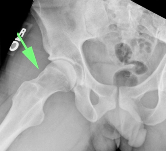
X-rayFrog-leg view radiograph of the right hip: dysplastic bump at the head-neck junction, arrow, cam-type morphology typical of femoroacetabular impingement.
Source : Royston et al., Radiology Case Reports, 2014, figure 2 · CC BY-NC-ND
📸 Having the « bump » does not mean being ill
Bone morphology (cam, pincer) is very common in people who hurt nowhere, especially in athletes. That is the heart of the message: image ≠ syndrome.
Prevalence of the morphologies in ASYMPTOMATIC volunteers (systematic review, 2,114 hips). The syndrome requires a TRIAD: symptoms + clinical signs + imaging 1. Source: Frank et al., 2015 (PMID 25636988).
Hip impingement, or femoroacetabular impingement (FAI), describes an abnormal, early and repeated contact between the proximal femur and the acetabular rim during hip movement, in particular flexion combined with adduction and internal rotation. But behind this mechanism lies a major conceptual trap for the clinician: FAI is not only a radiological image. Since 2016 the international community has clarified the vocabulary to distinguish an abnormality of bone shape , common and often silent, from the syndrome itself, which does cause suffering and justifies management. This distinction is not an academic subtlety: it shapes the physiotherapist's diagnostic reasoning and avoids « treating an X-ray » rather than a patient.
The reference definition: the Warwick consensus 1
The current reference framework is the Warwick Agreement, an international consensus published in 2016 1. It sets a simple and demanding rule: femoroacetabular impingement syndrome is a diagnosis of triad. To be made, it requires three elements at the same time:
- appropriate symptoms , typically mechanical hip or groin pain, worsened by prolonged or deep flexion positions;
- positive clinical signs , including the FADIR impingement test (flexion, adduction, internal rotation);
- imaging abnormalities , a documented cam and/or pincer morphology.
The word « syndrome » was added deliberately to the name. As the authors put it, « the term femoroacetabular impingement syndrome was introduced to reflect the central role of the patient's symptoms in the disorder » 1. In other words: without symptoms there is no syndrome, whatever the image. The same consensus adds that the treatments judged appropriate are conservative care, rehabilitation, and arthroscopic or open surgery 1 , placing physiotherapy from the outset among the first-line options and not as a mere fallback.
The three morphologies: cam, pincer, mixed
The impingement has its anatomical origin in two main types of bone deformity, which can coexist.
Cam morphology (cam). This is an asphericity of the femoral head-neck junction : the femoral head is no longer perfectly spherical and shows a « bump » at its periphery. It is quantified by the alpha angle 23. During flexion with internal rotation, this bulging zone abuts the acetabulum and shears the cartilage and the labrum from the inside. Cam morphology is the one that concentrates most of the long-term concern, because it is associated with the risk of osteoarthritis (see below).
Pincer morphology (pincer). Here the abnormality is acetabular : an excessive bony coverage of the femoral head by the acetabulum (global or focal), which « pinches » the labrum against the femoral neck at the end of range.
Mixed morphology. In a large proportion of cases the two mechanisms are combined in the same patient, cam and pincer together. It is this mixed form that is often met in specialist practice.
Key points
- Cam = an abnormality of the femur (head-neck asphericity, measured by the alpha angle).
- Pincer = an abnormality of the acetabulum (excessive bony coverage).
- Mixed = the two combined.
- The diagnosis of the syndrome requires the triad : symptoms + clinical signs + imaging 1.
The crucial distinction: morphology is not syndrome
This is probably the most important message of this section, and the one the physiotherapist must take on board before any reasoning. Bone morphology is extremely common in people who hurt nowhere. A systematic review of 26 studies and 2,114 asymptomatic hips (mean age ≈ 25 years) found a prevalence of cam deformity of 37 %, and a prevalence of pincer deformity of 67 % 2. In athletes the cam is even more widespread: 54.8 % against 23.1 % in the general population 2. The mean alpha angle in these painless hips was 54.1°, and, strikingly, a labral lesion was visible on MRI in 68.1 % of asymptomatic hips 2.
The clinical consequence is direct: an impingement image in someone with no complaint is a false friend. Finding a cam or a damaged labrum on imaging does not « make » the diagnosis and does not prove that the hip is the source of the symptoms.
We should also stay cautious about the figures themselves. Another systematic review (30 studies) showed that estimates of the prevalence of cam morphology ranged from 5 %–75 % of participants depending on the populations, without the authors being able to demonstrate a genuinely higher prevalence in expected subgroups such as athletes or people with hip pain, for lack of data of sufficient quality 8. In other words: the true prevalence of the cam remains uncertain, and that uncertainty is itself a further reason never to make the diagnosis on the image alone.
Epidemiology and the long-term stake: the risk of osteoarthritis
If cam morphology attracts so much attention, it is not only because of the immediate pain: it is because it is a prospective risk factor for hip osteoarthritis. The Dutch national prospective cohort CHECK (1,002 subjects, 5-year follow-up) established this robustly: a cam that is moderate (alpha angle > 60°) multiplied the risk of end-stage hip osteoarthritis by 3.67 (adjusted OR 3.67; 95 % CI 1.68–8.01), and a cam that is severe (alpha angle > 83°) by nearly ten (adjusted OR 9.66; 95 % CI 4.72–19.78) 3. The association was particularly predictive when the severe cam was combined with reduced internal rotation : these people are « strongly predisposed to rapid progression to end-stage osteoarthritis » 3.
A key point for the reasoning: this risk is carried by the bone morphology, distinct from the symptomatic syndrome. And above all, the authors stress that « cam impingement could be a modifiable risk factor, hence the importance of spotting it early » 3. That is precisely where part of the physiotherapist's added value lies: early identification and support of the young adult at risk.
What the physiotherapist needs to understand
From this section the clinician can retain four points that structure everything that follows.
1. Reason in triads, not in images. The diagnosis of FAI syndrome exists only if symptoms, clinical signs and imaging agree 1. Faced with a painful hip, the question is never « is there a cam? » but « does the cam explain these symptoms in this patient? ».
2. Use the clinical tests for what they are: screening tests. The FADIR is the examination best supported as a screening tool for FAI 5. Its pooled sensitivity is high (0.94 to 0.99), so that a negative test helps rule out impingement; but its low specificity means that a positive test is not enough to confirm the diagnosis 6. The authors conclude that these tests have only « screening value » 6. The FADIR points, it does not settle.
3. Physiotherapy has a recognised place, surgery included. The Warwick consensus places rehabilitation among the appropriate treatments 1, and the large randomised trials show that structured conservative management genuinely improves patients, even if surgery brings an additional benefit in patients referred to specialist care (a debate developed later in the article). The physiotherapist is not a plan B.
4. Early identification makes sense. Because the cam is a potentially modifiable risk factor for osteoarthritis 3, identifying a hip at risk early, a young sporting adult with a severe cam and limited internal rotation, falls squarely within the physiotherapist's field, in a logic of prevention as much as of treating the established syndrome.
🔎 How is it recognised on examination?
Femoroacetabular impingement is not an image on an X-ray: it is a syndrome. The Warwick international consensus 1 is unambiguous on this point: the diagnosis requires three elements together, appropriate symptoms, positive clinical signs and imaging abnormalities. The term « syndrome » was in fact introduced precisely to underline the central role of the patient's symptoms 1. In other words, the clinical examination is not a mere preamble to the MRI: it is one of the three pillars of the diagnosis, the one that reconnects the image to the patient who is suffering.
A cam on imaging, with no symptom and no clinical sign, is not an impingement syndrome.
Groin pain: the « C sign »
The most suggestive reason for consultation is deep groin pain, often hard to point to with a fingertip. Patients frequently describe it by cupping the hip between thumb and index finger, the hand forming a C just above the greater trochanter, the well-known « C sign ». This pain is typically mechanical: it wakes up on prolonged sitting, when driving, when squatting, or during sporting movements that mix flexion and rotation. It reflects the abnormal contact, at the extreme of range, between the femoral neck and the acetabular rim.
This symptomatic phase is what gives the picture its value: the bone morphology itself is extraordinarily ordinary. In a systematic review of 26 studies covering 2,114 hips that were asymptomatic (mean age 25 years), the prevalence of cam deformity reached 37 %, rising to 54.8 % in athletes against 23.1 % in the general population, and that of pincer deformity 67 % 2. More troubling still, a labral lesion was visible on MRI in 68.1 % of these hips with no complaint 2. A clinician reasoning from the image alone therefore runs a real risk of over-diagnosis. This article deals with the morphological cause and the impingement syndrome ; the labral lesion itself, what it means, how to know whether it really is the source of the pain, and what is done about it, has its own article : hip labral tear.
Key points
- FAI is a diagnosis of triad : symptoms + clinical signs + imaging 1. None of the three is enough on its own.
- The pain is most often in the groin, mechanical, provoked by sitting, squatting and movements in flexion with rotation (the « C sign »).
- Cam morphology is present in more than a third of people without pain 2 : the image does not make the diagnosis.
- The FADIR test is an excellent screening test (very sensitive), but a positive FADIR confirms nothing 6.
The FADIR test: sensitive, therefore useful for ruling out
The central test of the examination is the FADIR (flexion, adduction, internal rotation). With the patient supine, the clinician passively takes the hip to 90° of flexion, then adds adduction and internal rotation. Reproduction of the patient's usual groin pain marks a positive test. The movement mechanically brings the femoral neck close to the anterosuperior rim of the acetabulum: it is the manoeuvre that « loads » the presumed impingement zone.
Its value must be understood precisely so as not to over-interpret it. A systematic review with meta-analysis finds a pooled sensitivity that is very high, from 0.94 to 0.99, and a sensitivity of 0.96 for the flexion with internal rotation variant 6. In practice, this high sensitivity is what makes the test useful as a tool for screening : when the FADIR is negative it seriously helps rule impingement out. Conversely, its specificity is low, and when it is positive it is not enough to confirm the diagnosis. The authors conclude explicitly that these tests have only « screening value » (screening accuracy) 6.
The reader should also be told of a real heterogeneity in the literature on this point. Another systematic review reports FADIR sensitivities much more scattered depending on the reference imaging: from 0.08 to 1 (radiography) and from 0.33 to 1 (MRI), and concludes that « the overall usefulness of the FADIR in diagnosing FAI remains uncertain given only moderate sensitivity and specificity » 7. These two readings, very high sensitivity on one side and uncertain on the other, do not agree perfectly, and it would be dishonest to hide that. They nonetheless converge on the clinical message that counts: the FADIR points and screens, it does not settle. An umbrella review of 6 systematic reviews confirms that the FADIR is the test best supported as a screening tool, most clinical tests having a sensitivity higher than their specificity 5.
Limited internal rotation
Beyond reproducing the pain, the physiotherapist measures passive ranges with the hip flexed to 90°. A reduced internal rotation is a key sign of cam impingement: early bone contact mechanically limits the gliding of the head in the acetabulum. Comparing systematically with the other side is essential here; it is the asymmetry, more than the absolute value, that speaks.
This sign goes beyond diagnosis: it carries prognostic information. In the Dutch national prospective cohort CHECK (1,002 subjects, 5-year follow-up), the combination of a severe cam and reduced internal rotation strongly predisposed to rapid progression to end-stage hip osteoarthritis 3. A severe cam (alpha angle > 83°) already multiplied the risk of end-stage osteoarthritis by 9.66 (adjusted OR 9.66; 95 % CI 4.72–19.78), a moderate cam (> 60°) by 3.67 3. Spotting early, on examination, a hip with a cam and stiff internal rotation therefore means identifying ground to watch, the cam possibly being a modifiable risk factor 3.
What the physiotherapist tests, and what they do with it
The examination is not limited to the FADIR. In practice the assessment reproduces the first component of Personalised Hip Therapy, the rehabilitation protocol developed for the FASHIoN trial, one of whose four pillars is precisely a detailed assessment : stability, strength, ranges and impingement tests 8. In practice the clinician judges passive mobility (rotations, flexion, adduction), the strength and control of the glutes and external rotators, trunk stability, and seeks to reproduce, then to modulate, the patient's usual pain.
Interpretation follows a probabilistic logic, not a verdict. A typical picture, mechanical groin pain, a positive FADIR, limited and asymmetric internal rotation in an active young adult, raises the probability of impingement and justifies completing with imaging. A negative FADIR lowers that probability sharply and invites exploration of other causes of hip or groin pain. But neither the examination alone nor the image alone ever makes the diagnosis: it is the convergence of the triad that allows it 1. The physiotherapist's role is to hold the three threads together, bearing in mind that the morphology is common in the asymptomatic 2 and that the true prevalence of the cam is itself uncertain, estimated at 5 to 75 % depending on the populations without any reliable figure having been established 8.
The examination is not there to assert an impingement but to place its probability and to link the image to the complaint.
📸 Imaging does not make the diagnosis on its own
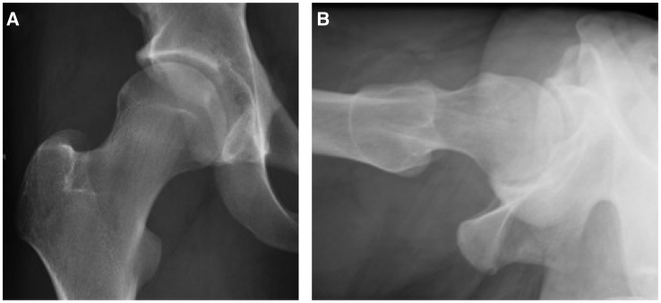
X-rayFrontal and lateral hip radiographs in a young athlete: bony prominence at the head-neck junction, typical of cam-type femoroacetabular impingement.
Source : Packer et al., Journal of hip preservation surgery, 2015, figure 1 · CC BY
⚠️ Cam morphology, on the other hand, counts for the future of the knee… sorry, of the hip
In people followed over time, a marked cam strongly multiplies the risk of end-stage hip osteoarthritis. It is a possibly modifiable factor, hence the value of spotting it early.
Adjusted odds ratios for end-stage hip osteoarthritis, Dutch national prospective cohort CHECK. Pincer morphology, for its part, is not a robust risk factor. Source: Agricola et al., 2013 (PMID 22730371).
This is probably the most counter-intuitive point about femoroacetabular impingement, and the one that changes physiotherapy practice most: an « abnormal » X-ray does not make the diagnosis. The Warwick international consensus 1 is explicit on this point. Femoroacetabular impingement syndrome (FAI syndrome) is defined as a triad : to make the diagnosis, the patient must have appropriate symptoms, positive clinical signs and imaging abnormalities all at once. The term « syndrome » was in fact introduced precisely to underline the central role of the patient's symptoms, to separate the disease from the mere shape of the bone. A cam or a pincer visible on a film, in someone who hurts nowhere, is not FAI syndrome: it is a morphological particularity.
What the X-ray measures: the alpha angle
Cam morphology corresponds to an asphericity of the junction between the head and the neck of the femur: instead of being nicely round, the head « overhangs » and loses its sphericity. It is quantified by the alpha angle, measured on the X-ray (or MRI or CT). The higher the angle, the more aspherical the junction. In the literature the commonly used thresholds are an alpha angle above 60° for a so-called moderate cam and above 83° for a severe cam 3. Pincer morphology, for its part, corresponds to excessive acetabular coverage of the femoral head.
These measurements are useful, but they describe a bone shape, not a pain. And that is where everything is decided: the same image can belong to a very troubled patient… or to someone who will never know they « have » a cam.
Cam morphology is common in the asymptomatic
This is the figure to keep in mind at every reading of imaging. In a systematic review of 26 studies covering 2,114 hips that were asymptomatic (mean age 25 years), the prevalence of cam deformity was 37 %, and much higher in athletes (54.8 %) than in the general population (23.1 %). The mean alpha angle of these painless hips was 54.1°. Pincer deformity reached 67 %. And a labral lesion was visible on MRI in 68.1 % of these hips with no complaint 2.
In other words: in one athlete out of two a cam will be found on imaging without it meaning anything clinically. Finding a « damaged » labrum on MRI in seven painless hips out of ten strongly relativises the value of such an image taken in isolation. If the diagnosis were made on imaging alone, FAI would be « diagnosed » in a large part of the healthy general population.
A prevalence to be interpreted with caution
We must stay honest about the uncertainty: prevalence figures vary enormously from study to study. Frank's review 2 already gave a range of 7 % to 100 % across the studies. Another systematic review of 30 studies 8 found estimates ranging from 5 % to 75 % of participants affected, without managing to demonstrate a higher prevalence in subgroups supposedly at risk such as athletes or people with hip pain, for lack of data of sufficient quality. The authors conclude that we do not currently have data solid enough to establish the true prevalence of cam morphology in the general population.
What can be drawn from this for clinical practice is not « the exact figure » but the robust message that runs through all these studies: the cam is a common morphological trait, often silent, which on its own cannot carry a diagnosis of syndrome.
Key points
- FAI syndrome is a triad 1 : symptoms + clinical signs + imaging. All three, not one.
- Imaging measures a bone shape (the alpha angle for the cam), not a pain.
- The cam is common in the asymptomatic: 37 % of painless hips, up to 54.8 % in athletes 2.
- The exact prevalence remains uncertain 8 : an isolated figure is not to be over-read.
- The FADIR test is a tool for screening, not for confirmation: negative it helps rule out, positive it proves nothing.
- Practical conclusion: the image is always read in the light of the clinical picture, never the other way round.
The clinical side: the FADIR screens, it does not confirm
If imaging is not enough, neither is the clinical test. The FADIR test (flexion, adduction, internal rotation) is the examination best supported for screening for FAI. Its pooled sensitivity is very high: from 0.94 to 0.99 in Reiman's meta-analysis 6, which also finds 0.96 for the flexion with internal rotation test. Such high sensitivity has a concrete virtue: a negative FADIR helps rule impingement out. Its specificity, on the other hand, is low; a positive FADIR on its own confirms nothing. The authors are categorical: these tests have only « screening » value (screening accuracy).
The literature is not perfectly homogeneous either: another systematic review 7 reported a FADIR sensitivity much more scattered depending on the reference imaging modality (from 0.08 to 1 confirmed by radiography, from 0.33 to 1 by MRI), concluding that its overall diagnostic value remains uncertain given only moderate sensitivity and specificity, while judging it useful as a low-risk screening tool. These divergences in figures, and the reader deserves to be told plainly, do not undermine the message: an isolated positive test does not make the diagnosis.
Combining image and clinical picture: why and how
The logic of the triad becomes clear when the two sides are crossed. The cam, taken in isolation, is too common to be specific. The FADIR, taken in isolation, is too unspecific to confirm. But each covers the other's weakness : the symptoms designate the hip that is suffering, the FADIR reproduces and locates the trouble, and imaging checks that there really is a coherent morphological substrate. It is the concordance of the three, not one of the three, that carries the diagnosis of FAI syndrome 1.
In practice, that means two symmetrical errors to avoid:
| Situation | Cautious interpretation |
|---|---|
| Cam on imaging, patient with no pain and no clinical sign | Not FAI syndrome. A simple, common morphology. One does not rehabilitate an X-ray. |
| An isolated positive FADIR, with no concordant imaging and no clear clinical picture | A screening signal to explore, not enough to conclude. A positive proves nothing on its own. |
| Appropriate symptoms + positive clinical signs + concordant imaging | FAI syndrome plausible under the Warwick triad: a management plan can be built. |
The morphology still keeps a prognostic meaning
Saying that the image does not make the diagnosis is not saying it is useless. Cam morphology has a well-documented prognostic value over the long term. In the Dutch national prospective cohort CHECK (1,002 subjects, 5-year follow-up), a moderate cam (alpha angle > 60°) multiplied the risk of end-stage hip osteoarthritis by 3.67 (adjusted OR 3.67; 95 % CI 1.68–8.01) and a severe cam (> 83°) by nearly 10 (adjusted OR 9.66; 95 % CI 4.72–19.78). A severe cam combined with reduced internal rotation particularly predisposed to rapid progression to osteoarthritis 3.
The authors stress that cam impingement could be a modifiable risk factor, hence the value of spotting it early. The clinical nuance is therefore twofold: imaging does not diagnose the syndrome on its own, but when it reveals a marked cam it identifies a profile that deserves educational attention (risk factors, joint hygiene, monitoring of symptomatic cams), without turning a painless person into a patient. That is the right place for imaging: one element of the reasoning, never the whole of it.
⚖️ Surgery or rehabilitation? What the trials say
⚖️ Surgery or rehabilitation? Both work, and the gap is modest
Two randomised trials give the advantage to arthroscopy, but only slightly: conservative rehabilitation also improves patients and remains a legitimate first-line option.
Mean differences in favour of arthroscopic surgery over conservative management: FASHIoN +6.8 points 9, FAIT +10.0 points 10. Both arms improve; the reference rehabilitation is « Personalised Hip Therapy ». After surgery, 87 % of athletes return to sport 11.
This is the question every patient asks when imaging has found a cam or a pincer: should it be operated on, or is physiotherapy enough? For a long time the debate fed mainly on surgical series with no control group. Since 2018, two well-made randomised controlled trials, the British UK FASHIoN and the FAIT trial (Palmer), have finally allowed hip arthroscopy to be compared directly with structured conservative management. Their results converge, but reading them calls for nuance: they do not say that surgery « cures » and that rehabilitation « fails ».
FASHIoN: arthroscopy does a little better, physiotherapy works too
The UK FASHIoN 9 trial randomised 348 patients with femoroacetabular impingement syndrome across 23 NHS hospitals, between hip arthroscopy (n=171) and a personalised conservative rehabilitation led by a physiotherapist, « Personalised Hip Therapy » (PHT), delivered in 6 to 10 sessions. The primary outcome was hip-related quality of life, measured by the iHOT-33 score at 12 months.
The most important result is often forgotten in the headlines: both groups improved. The iHOT-33 score went from 39.2 to 58.8 after arthroscopy, and from 35.6 to 49.7 after rehabilitation 9. Arthroscopy did bring an additional gain, an adjusted mean difference of 6.8 points (95 % CI 1.7–12.0; p=0.0093) in favour of surgery, which the authors judged clinically significant.
| iHOT-33 score | Arthroscopy (n=171) | PHT rehabilitation (n=177) |
|---|---|---|
| Before | 39.2 | 35.6 |
| At 12 months | 58.8 | 49.7 |
| Adjusted difference | 6.8 points (95 % CI 1.7–12.0; p=0.0093) in favour of surgery | |
This figure must nonetheless be put on the right scale. The 6.8-point difference only just exceeds the estimated minimal clinically important difference (about 6.1 points on the iHOT-33). In other words, the advantage of surgery is real but modest, on the edge of what a patient really perceives as an improvement. Presenting arthroscopy as clearly superior would be an over-interpretation.
FAIT: a more marked surgical advantage, but a particular context
The second multicentre randomised trial, FAIT 10, compared arthroscopy with a physiotherapy programme combined with activity modification, in patients referred to secondary or tertiary care. The primary outcome was the HOS-ADL functional score (activities of daily living) at 8 months.
Here again surgery wins, and more clearly than in FASHIoN: the HOS-ADL was 78.4 after arthroscopy against 69.2 after physiotherapy, an adjusted difference of 10.0 points (95 % CI 6.4–13.6; p<0.001) in favour of surgery 10. The authors conclude that these patients « achieve better outcomes with arthroscopy than with physiotherapy and activity modification ».
| Trial | Score / time point | Difference in favour of surgery |
|---|---|---|
| FASHIoN (2018) | iHOT-33 at 12 months | 6.8 pts (95 % CI 1.7–12.0) |
| FAIT (2019) | HOS-ADL at 8 months | 10.0 pts (95 % CI 6.4–13.6) |
Two reservations apply before generalising. First, the FAIT population was already heading for surgery : patients referred to specialist care, so a priori more severe or more selected than those seen in community practice. Then, as in FASHIoN, the conservative group also improved : physiotherapy is not a placebo, it improves patients; surgery simply adds an extra measure of function.
What these trials do not settle
It would be tempting to conclude « two trials, same direction, so operate ». Reality is more cautious, and it must be said plainly to the patient as to the referrer.
- The size remains debated. A gap of 6.8 points on a quality-of-life scale does not carry the same weight as a gap of 10 on a functional scale, and the two instruments do not measure the same thing. We are talking about an additional benefit, not a difference between « it works » and « it does not work ».
- Durability is unknown at this stage. The primary outcomes were measured at 8 and 12 months. These trials do not tell us what happens at 5 or 10 years, nor whether the surgical advantage holds, widens or fades.
- The profile of the patient who really benefits from surgery has not been identified. Group averages hide strong heterogeneity: some conservatively managed patients do very well, some operated patients remain limited.
The surgery versus conservative comparison therefore remains, in the authors' own words, an open question whose answer depends on each patient's profile 10. Our role as physiotherapists is not to « lose » a patient to the operating theatre, nor to delay useful surgery, but to set the balance out honestly.
Conservative management is not « doing nothing »
A frequent misunderstanding is to equate the conservative arm with simple abstention. Yet in FASHIoN the comparator was a precise protocol, Personalised Hip Therapy 8, structured around four components led by the physiotherapist: a detailed assessment, education and advice, help with pain relief, and an individualised, supervised and progressive exercise programme, targeting in particular the glutes, the external rotators and trunk stability, over 12 to 26 weeks. It is that rehabilitation, structured, which produces the gains observed, not vague advice.
Finally, recall the precondition common to both trials: their patients had a syndrome of impingement, that is, the complete triad of the Warwick consensus, appropriate symptoms, positive clinical signs and imaging abnormalities 1. An isolated cam or pincer, discovered on imaging in someone with few or no symptoms, belongs to neither FASHIoN nor FAIT: offering arthroscopy on the image alone would fall outside the framework these trials validated.
Key points
- Two randomised trials 9 show an advantage of arthroscopy over rehabilitation, but a modest one : +6.8 points (iHOT-33) and +10.0 points (HOS-ADL).
- In both trials, the conservatively managed patients also improve , and structured physiotherapy is a legitimate option, often first line.
- The FASHIoN advantage is close to the threshold of clinical relevance; the FAIT one concerns patients already referred to specialist care. It cannot be generalised to every patient.
- Durability beyond a year and the profile that really benefits from surgery remain uncertain : to be said honestly to the patient.
- The conservative comparator is not « doing nothing » but a precise protocol 8.
🎯 What rehabilitation should be offered?
Once the diagnosis has been made under the triad of the Warwick consensus, appropriate symptoms, positive clinical signs and imaging abnormalities 1 , the therapeutic question arises. The consensus itself lists among the appropriate treatments « conservative care, rehabilitation, and arthroscopic or open surgery » 1. In other words, physiotherapy is not a stopgap while waiting for the theatre: it is an option in its own right, and most often the first line to offer.
Rehabilitation first line: the reasoning
Two arguments argue for starting conservatively. First, the syndrome is defined by the symptoms, not by the bone: an isolated radiological cam or pincer does not make the disease, and the morphology is ordinary in the asymptomatic, 37 % of cams in a review of 2,114 hips with no complaint, up to 54.8 % in athletes 2. We therefore do not rehabilitate an image, we treat a patient who is in pain and whose function is limited. Then, even in the trials that favour surgery, the physiotherapy group genuinely improves , and we come back to that below. Well-conducted rehabilitation changes something; it deserves a fair trial before arthroscopy is considered.
Personalised Hip Therapy (PHT): the reference protocol
There is a structured conservative protocol specific to FAI, Personalised Hip Therapy (PHT), developed for the FASHIoN trial and led by physiotherapists 8. It is today the best formalised framework we have. It rests on four key components :
- A detailed assessment of the patient: stability, strength, joint ranges and impingement tests 8.
- Education and advice : explanations about posture and gait, and above all learning to avoid the provocative positions, deep flexion with adduction and internal rotation combined, precisely the positions that close the impingement 8.
- Help with pain relief : relieving the pain, the condition for active work to be possible 8.
- An individualised, progressive and supervised exercise programme over time, completed by home work 8.
In practical terms, the PHT is delivered over 12 to 26 weeks, in 6 to 10 sessions between physiotherapist and patient, backed by a home programme 8. This format, few supervised sessions and a lot of guided autonomy, is an important point to explain to the patient: the result depends largely on how diligent they are between appointments.
What does the exercise programme actually contain?
The PHT programme progresses from motor control to stretching and then strengthening, explicitly targeting three territories: the gluteal muscles, the hip external rotators and trunk (core) stability 8. These axes, which match the clinical logic of impingement, can be set out in detail.
Abductors and glutes
Strengthening the glutes (gluteus medius, gluteus maximus) is an explicit pillar of the programme 8. The functional aim: to improve pelvic control under load and on single-leg stance, in order to limit anterosuperior stress on the labrum-cartilage complex during walking, stairs and transitions.
External rotators and motor control
Work on the external rotators is targeted by name 8. Clinically, it aims to reduce the tendency to femoral internal rotation, the movement which, combined with flexion and adduction, reproduces the impingement and the positive FADIR. The motor control phase, placed early in the progression, seeks to reorganise hip and pelvic movement before load is added: the logic of « move well before moving hard ».
Trunk (core) stability
Trunk stability is the third targeted territory 8. Better lumbopelvic control conditions the correct positioning of the pelvis, and therefore the orientation of the acetabulum above the femoral head during effort.
Mobility and movement education
The PHT includes a stretching component in its progression 8. One point deserves caution: it is not about forcing range towards the impingement positions. Education about avoiding deep flexion with adduction and internal rotation 8 and the mobility work must stay consistent: gaining useful, painless range without going to « find » the mechanical stop that provokes the pain.
Key points: what the conservative programme contains
- Framework : Personalised Hip Therapy (PHT), 6 to 10 sessions over 12 to 26 weeks, led by the physiotherapist 8.
- Four components : detailed assessment, education and advice, help with pain relief, individualised, progressive and supervised exercise 8.
- Muscle targets : glutes, external rotators, trunk stability 8.
- Progression : from motor control to stretching and then strengthening 8.
- Key message to the patient : avoid the positions of deep flexion + adduction + internal rotation that close the impingement 8.
What level of evidence? What the large trials say
Two British multicentre randomised trials structure the discussion today. Both compare hip arthroscopy with conservative management, and both show the same thing in substance: both arms improve, and surgery brings an additional benefit of modest size.
In UK FASHIoN (348 patients, 23 NHS hospitals), at 12 months the iHOT-33 quality-of-life score improves in both groups: from 39.2 to 58.8 after arthroscopy, and from 35.6 to 49.7 after PHT 9. The adjusted difference is 6.8 points in favour of surgery (95 % CI 1.7–12.0; p=0.0093), judged clinically significant by the authors, but which only just exceeds the minimal clinically important difference used (6.1 points) 9.
In FAIT 10, at 8 months the HOS-ADL functional score is 78.4 after surgery against 69.2 after physiotherapy plus activity modification, an adjusted difference of 10.0 points (95 % CI 6.4–13.6; p<0.001) in favour of arthroscopy 10. The gap there is more marked than in FASHIoN.
| Trial | Score / time point | Conservative | Surgery | Difference in favour of surgery |
|---|---|---|---|---|
| UK FASHIoN 9 | iHOT-33 at 12 months | 35.6 → 49.7 | 39.2 → 58.8 | +6.8 pts (CI 1.7–12.0; p=0.0093) |
| FAIT 10 | HOS-ADL at 8 months | 69.2 | 78.4 | +10.0 pts (CI 6.4–13.6; p<0.001) |
How to interpret these figures with caution
It would be misleading to conclude « surgery is clearly superior ». Several reservations apply, and they must be said to the patient as to the reader.
- The gap is modest. In FASHIoN, 6.8 points barely reach the threshold of clinical relevance 9. The additional gain of arthroscopy over physiotherapy exists but remains small.
- Conservative works. In both trials the rehabilitation group genuinely improves 910. Structured physiotherapy is not a placebo.
- Recruitment biases in favour of surgery. In FAIT, the population was already referred to secondary or tertiary care, that is, directed towards specialist and even surgical management 10 , which limits generalisation to a patient seen early in community practice.
- Durability and the profile of responders remain debated. The size of the surgical benefit, its persistence over the long term and the identification of the patients who really gain from it are not settled by these trials.
In practice this supports a reasonable sequence: first offer a fair trial of structured PHT-type rehabilitation, and reserve the surgical discussion for patients who improve insufficiently, taking the background into account. That last point is not trivial: cam morphology is a prospective risk factor for hip osteoarthritis; in the CHECK cohort, a severe cam (alpha angle > 83°) multiplied the risk of end-stage osteoarthritis by nearly 10 (adjusted OR 9.66; 95 % CI 4.72–19.78) 3. Spotting and monitoring a symptomatic cam is therefore part of the long-term reasoning, even when the conservative route is chosen.
And after surgery? Post-operative rehabilitation
When arthroscopy is chosen, physiotherapy keeps a central role after the operation, particularly in athletes. The overall prognosis for return to sport is rather favourable: in a systematic review of 18 series, 87 % of athletes return to sport and 82 % regain their previous level after FAI surgery 11.
This good result is not guaranteed, however. The presence of diffuse hip osteoarthritis at the time of the operation can prevent return to sport 11. This finding reinforces two clinical messages: do not confuse symptomatic FAI in the young adult with an already osteoarthritic hip, and manage the problem before advanced cartilage lesions set in, whether the conservative route or surgery followed by rehabilitation is chosen.
In summary for the physiotherapist
FAI syndrome rehabilitation has a clear framework (the PHT), an identified content (motor control, glutes, external rotators, trunk, cautious mobility, education about provocative positions) and an honest but nuanced level of evidence: it genuinely improves patients, while being, in two large trials, slightly outdone by arthroscopy in populations directed towards specialist care 910. The defensible position is therefore a structured, supervised conservative first line, with regular review and the surgical discussion reserved for failures, without ever forgetting that the data remain debated and that the best choice depends on the patient in front of you.
📈 Prognosis, return to sport and the risk of osteoarthritis
Speaking of « prognosis » for femoroacetabular impingement (FAI) syndrome requires separating two questions the literature often mixes: the course of the symptoms (will the pain settle? will the patient get their sport back?) and the long-term joint risk (will the morphology damage the hip?). These two planes do not overlap. One can carry a marked radiological cam without pain, and suffer from symptomatic FAI with no visible osteoarthritis. Recall that the diagnosis is a triad, appropriate symptoms, positive clinical signs AND imaging abnormalities, and that the term « syndrome » was introduced precisely to recentre the reasoning on the patient's complaints rather than on the bone 1. This distinction is the thread running through everything that follows.
Symptomatic course: two routes that improve the patient
Good news to announce straight away: whichever strategy is chosen, the majority of patients treated improve. The two large randomised trials show it unambiguously. In the British UK FASHIoN trial (348 patients, 23 NHS hospitals), the hip-related quality-of-life score (iHOT-33) improved in both arms at 12 months: from 39.2 to 58.8 after arthroscopy, and from 35.6 to 49.7 after personalised conservative rehabilitation (Personalised Hip Therapy). The adjusted difference of 6.8 points (95 % CI 1.7–12.0; p=0.0093) favoured surgery and only just exceeded the threshold of clinical relevance used (6.1 points), but structured physiotherapy also produced a real gain 9. The second trial, FAIT 10, points the same way: at 8 months the HOS-ADL functional score was 78.4 after surgery against 69.2 after physiotherapy and activity modification, an adjusted difference of 10.0 points (95 % CI 6.4–13.6; p<0.001) in favour of arthroscopy.
Should we conclude that surgery is « the » solution? No, and that must be said honestly to the patient. The superiority of arthroscopy is real but modest in FASHIoN and more marked in FAIT, and both trials covered populations already directed towards specialist settings, often after failure or with a view to surgery. The conservative group improved in both cases. The surgery versus rehabilitation comparison therefore remains debated, and its result depends on the patient's profile more than on any universal rule. First-line physiotherapy keeps its full legitimacy.
The reference rehabilitation is not a passive placebo either: Personalised Hip Therapy (PHT) is a structured, specific protocol, led by the physiotherapist over 12 to 26 weeks (6 to 10 sessions) around four components, detailed assessment, education and advice, help with pain relief, and an individualised, progressive and supervised exercise programme targeting the glutes, the external rotators and trunk stability 8. It is that standard which held its own against arthroscopy in FASHIoN, and that gives the conservative message a solid basis.
Return to sport: favourable, but not guaranteed
In athletes, the available data concern mainly the surgical course. A systematic review of 18 case series reports that 87 % of athletes return to sport after FAI surgery and that 82 % regain their previous level 11. The figure is encouraging, but two reservations apply. First, these are case series, a modest level of evidence with no control group and a risk of selecting the good candidates. Second, returning to the previous level is not automatic: nearly one operated athlete in five does not regain their level. The same review identifies a clear unfavourable factor: the presence of diffuse hip osteoarthritis at the time of the operation compromises return to sport.
This last point has a direct clinical bearing: it argues for spotting and managing the impingement before extensive cartilage lesions set in. Once diffuse osteoarthritis is in place, the joint procedure loses functional value. In other words, the best sporting prognosis is decided upstream, on a joint that is still preserved.
Cam morphology and osteoarthritis: an established prospective risk
It is on this ground that the evidence is strongest, and most useful for framing follow-up. The Dutch national prospective cohort CHECK (1,002 subjects, 5-year follow-up) showed that cam morphology, present in subjects initially free of osteoarthritis, predicts hip osteoarthritis in the long term. A moderate cam (alpha angle >60°) multiplied the risk of end-stage osteoarthritis by 3.67 (adjusted OR 3.67; 95 % CI 1.68–8.01), and a severe cam (>83°) by nearly ten (adjusted OR 9.66; 95 % CI 4.72–19.78). For incident osteoarthritis, only the moderate cam was predictive (OR 2.42; 95 % CI 1.15–5.06). The excess risk is particularly strong when the severe cam is combined with reduced internal rotation: that combination predisposes to rapid progression to end-stage osteoarthritis 3.
| Cam morphology (alpha angle) | Risk of end-stage osteoarthritis at 5 years | Level of evidence |
|---|---|---|
| Moderate (>60°) | Adjusted OR 3.67 (CI 1.68–8.01) | Prospective cohort 3 |
| Severe (>83°) | Adjusted OR 9.66 (CI 4.72–19.78) | Prospective cohort 3 |
| Severe + reduced internal rotation | Rapid progression to osteoarthritis | Prospective cohort 3 |
Two cautions in reading apply. First, this risk is carried by the bone morphology, not by the symptomatic syndrome as such: it is the cam on imaging that predicts osteoarthritis, independently of the pain. Second, the cam is extremely common in people with no complaint, 37 % of asymptomatic hips in a review of 2,114 hips, up to 54.8 % in athletes against 23.1 % in the general population 2. The great majority of these carriers will not develop end-stage osteoarthritis. A high OR therefore does not mean individual fate: it describes a relative risk in a population, not a personal destiny. Telling an asymptomatic patient with a cam that they « will get osteoarthritis » would be an over-interpretation.
Is this risk factor modifiable? Honesty calls for caution
The CHECK authors stress that cam impingement « could be a modifiable risk factor », hence the value of early identification 3. The conditional is deliberate, and it should be passed on as such. That the cam is a prospective risk factor for osteoarthritis is established. That acting on it, by surgery or by rehabilitation, does prevent osteoarthritis in the long term is not, to date, demonstrated by the available trials: FASHIoN and FAIT measured quality of life and function at 8–12 months, not the incidence of osteoarthritis at 10 or 20 years 910. The reasoning « correcting the cam protects the joint » is biologically plausible but remains a hypothesis. It must not be presented to the patient as a preventive certainty.
Key points
- Symptoms vs joint: the prognosis of the pain and the risk of osteoarthritis are two distinct questions; a radiological cam is neither a diagnosis nor a sentence.
- Both routes work: arthroscopy and personalised rehabilitation both improve the patient; the surgical advantage is modest (FASHIoN) to moderate (FAIT) and its size remains debated by profile.
- Return to sport: ~87 % return after surgery, ~82 % at the previous level, but from case series; pre-existing diffuse osteoarthritis compromises the return 11.
- Risk of osteoarthritis: a moderate cam (OR 3.67) and above all a severe one (OR 9.66) predicts end-stage osteoarthritis at 5 years 3 , a population relative risk, not an individual fate.
- Prevention not proven: correcting the cam is plausible but no trial has shown that it prevents osteoarthritis in the long term, to be said honestly to the patient.
In clinical practice this body of evidence invites a measured position: reassure about the symptomatic course, most patients get better and structured physiotherapy is a solid first line; identify the hips at risk of osteoarthritis (severe cam, limited internal rotation, persistent symptoms) for close follow-up; and avoid two symmetrical excesses, dramatising an image in someone with few or no symptoms, or minimising a painful impingement accompanied by a high-risk morphology. Where the evidence is missing, in particular on the long-term preventive benefit, the clinician's role is to name the uncertainty, not to hide it.
🗂️ What do concrete case reports teach us?
The case that follows is published and carries its PubMed identifier. It is worth more than two invented stories, because it compares the two options in the same patient. They were built solely from the work cited in this article, and add no claim unsupported by the literature. Their purpose is educational: to make the clinical reasoning visible, in particular the central distinction between a morphology of bone and a syndrome that is symptomatic.
An impingement image is not a diagnosis: it is the patient, not the X-ray, that makes the syndrome.
Published case: the same patient, one hip operated and the other not
The case. Öhlin and colleagues report a young woman with bilateral femoroacetabular impingement syndrome, of cam morphology, managed surgically on one side and non-surgically on the other12.
The follow-up. Five years after the surgical treatment of the right hip and two years after the non-surgical treatment of the left hip, with clinical assessment12. The authors present both approaches as a success12.
Why this case is valuable, and what it does not say. It removes the variable that ruins every comparison between patients: the patient. Same age, same morphology, same functional demand, same adherence. What it suggests is modest but useful: cam morphology does not dictate the treatment. A hip with a cam can do well without surgery.
What the case does not say, on the other hand, is which of the two options is superior: a single patient cannot settle that, and the trials cited earlier in this article remain the arbiter. It only recalls that the question genuinely arises, including in a young and sporting person.
The underlying reminder is the one set out above: cam morphology is common in asymptomatic people4, and it is indeed the patient, not the X-ray, that makes the syndrome.
What this published case teaches fits in one sentence : sort well (morphology vs syndrome), do not over-read a screening test, and offer structured rehabilitation first line in the young athlete, while staying transparent about what the evidence cannot yet settle.
🧭 How is this applied in practice?
Moving from theory to the clinic assumes a simple rule: treat a patient, not an image. Femoroacetabular impingement syndrome is a diagnosis of triad , appropriate symptoms, positive clinical signs AND imaging abnormalities must coexist; the term « syndrome » was coined precisely to recentre the approach on the patient's complaints 1. An isolated radiological cam or pincer, with no symptom, does not constitute the syndrome. Here is how to apply that principle in reasoning on the ground.
A five-step clinical algorithm
The sequence below is not a rigid protocol but a reasoning framework, drawn directly from the four components of Personalised Hip Therapy 8 and from the Warwick triad logic 1.
- Recognise the picture. Hip or groin pain in a young to middle-aged adult, often sporting, worsened by deep flexion, prolonged squatting, low sitting or changes of direction. The mechanical, positional pattern points the way; it is not enough.
- Screen clinically. The FADIR test (flexion, adduction, internal rotation) is the examination best supported as a screening tool 5. Its pooled sensitivity is high 6 : when the FADIR is negative it mainly helps rule impingement out. Its specificity being low, when it is positive it confirms nothing.
- Assess in detail. Ranges (above all internal rotation, often reduced), strength of the glutes and external rotators, trunk control, lumbopelvic stability, impingement tests. This detailed assessment is the first component of the PHT 8.
- Compare with imaging, cautiously. A cam or pincer morphology only makes sense when related to concordant symptoms and signs. The morphology is common in the asymptomatic (see below), so the image confirms a clinical picture, it does not create it.
- Treat conservatively and in a structured way first. Education, help with pain relief, then an individualised, supervised and progressive exercise programme targeting the glutes, the external rotators and the core, from motor control to strengthening, over 12 to 26 weeks in 6 to 10 sessions completed by home work 8.
The key messages to convey to the patient
The quality of the education conditions adherence. Three ideas deserve simple explanation.
- « Your X-ray is not your pain. » Cam morphology exists in 37 % of asymptomatic hips (54.8 % in athletes against 23.1 % in the general population) and pincer in 67 % 2. A labral lesion is visible on MRI in 68.1 % of hips with no complaint. Defusing the image avoids kinesiophobia.
- « Rehabilitation really works. » In the FASHIoN trial, the physiotherapy group improved from 35.6 to 49.7 on the iHOT-33 score in 12 months 9. The conservative gain is real, not a mere waiting placebo.
- « Positions are adapted, not banned for life. » Advice on posture, gait and temporary limitation of the positions of deep flexion, adduction and internal rotation 8, for as long as it takes for tolerance to rise again through strengthening.
We treat a patient who is in pain, not an asphericity spotted on a film.
Common mistakes to avoid
Most clinical dead ends come from confusing morphology with syndrome.
- Making the diagnosis on imaging alone. This is the cardinal error the Warwick consensus aims to correct: with no concordant symptoms or clinical signs, a cam or a pincer is not FAI syndrome 1.
- Taking a positive FADIR for proof. The FADIR is sensitive but not very specific; it screens, it does not confirm 65. Interpreting it in isolation leads to over-diagnosis.
- Overestimating the prevalence to justify a treatment. Cam estimates range from 5 to 75 % across studies, with no demonstrated over-prevalence in athletes or in patients with pain, for lack of data of sufficient quality 8. Stay humble about the figures.
- Offering an unstructured programme. What makes the PHT effective is individualisation, progression and supervision 8. A simple sheet of generic exercises is not its equivalent.
- Referring to the surgeon too quickly. Surgery does better in the trials, but the gap is modest and the conservative route remains a legitimate first-line option (see below).
When to refer to the surgeon?
Here the literature is nuanced and it must be said plainly to the reader: the two reference randomised trials show an advantage for arthroscopy, but a moderate one, in patients already directed towards specialist care.
| Trial | Comparison | Main result | Level of evidence |
|---|---|---|---|
| UK FASHIoN 9 | Arthroscopy vs physiotherapy PHT, 348 patients, 23 hospitals | iHOT-33 at 12 months: +6.8 pts in favour of surgery (95 % CI 1.7–12.0; p=0.0093); both groups improve | Multicentre RCT, gap close to the clinical threshold (6.1 pts) |
| FAIT 10 | Arthroscopy vs physiotherapy + activity modification, 222 patients | HOS-ADL at 8 months: +10.0 pts in favour of surgery (95 % CI 6.4–13.6; p<0.001) | Multicentre RCT, population already referred to specialist care |
| Return to sport 11 | Systematic review, 18 series after surgery | 87 % return to sport, 82 % at the previous level; diffuse osteoarthritis = a brake | Case series, no comparator |
In practice, a surgical opinion is discussed when:
- a structured, well-conducted conservative programme (PHT type, 6 to 10 supervised sessions over several weeks) has not improved pain and function enough;
- the functional impact remains significant despite adherence to treatment;
- the patient, informed, wishes to discuss the additional, modest, gain that surgery can bring 910.
The uncertainty must nonetheless be presented honestly: in FASHIoN the difference only just exceeds clinical relevance, and in both trials physiotherapy also improves. The true size of the surgical benefit, its durability and the exact profile of the patients who gain most from it remain debated. The decision is shared, not automatic.
Do not forget the long-term stake
Spotting a symptomatic cam early is not only a question of immediate comfort. Cam morphology is a prospective risk factor for end-stage hip osteoarthritis: in the CHECK cohort, a moderate cam (alpha angle > 60°) multiplied that risk by 3.67 and a severe cam (> 83°) by 9.66, the combination of a severe cam with reduced internal rotation being particularly predictive 3. It is the bone morphology that carries this risk, not the symptomatic syndrome as such; the authors stress that the cam could be a modifiable factor, hence the value of early identification.
Key points
- Diagnosis = triad : concordant symptoms + clinical signs + imaging. An image alone is never enough 1.
- FADIR = screening, not confirmation: negative it rules out, positive it does not prove 65.
- Morphology ≠ syndrome : cam present in 37 % of asymptomatic hips, pincer in 67 % 2 ; the overall prevalence is very uncertain 8.
- Conservative first : a structured PHT (assessment, education, pain relief, individualised and progressive exercise) genuinely improves patients 89.
- Surgery : a real but modest advantage (FASHIoN +6.8 pts; FAIT +10.0 pts), to be discussed in shared decision-making after conservative failure 910.
- Long-term stake : the cam is a risk factor for osteoarthritis 3 , and early identification is useful.
Bibliography
Every reference checked individually on PubMed (clickable PMID). 11 sources. Click a superscript note marker in the text: the bibliography opens and highlights the source.
- Griffin DR, et al. (Warwick Agreement) (2016). British Journal of Sports Medicine. PMID 27629403. doi:10.1136/bjsports-2016-096743.
- Frank JM, Harris JD, Erickson BJ, et al. (2015). Arthroscopy. PMID 25636988. doi:10.1016/j.arthro.2014.11.042.
- Agricola R, Waarsing JH, Thomas GE, et al. (2013). Annals of the Rheumatic Diseases. PMID 22730371. doi:10.1136/annrheumdis-2012-201643.
- Dickenson E, Wall PDH, Robinson B, et al. (2016). Osteoarthritis and Cartilage. PMID 26778530. doi:10.1016/j.joca.2015.12.020.
- Fernandes DA, Melo GM, Contreras ES, et al. (2022). Clinical Journal of Sport Medicine. PMID 34534982. doi:10.1097/JSM.0000000000000978.
- Reiman MP, Goode AP, Cook CE, Hölmich P, Thorborg K (2015). British Journal of Sports Medicine. PMID 25515771. doi:10.1136/bjsports-2014-094302.
- Shanmugaraj A, Shell JR, Horner NS, et al. (2020). Clinical Journal of Sport Medicine. PMID 31855915. doi:10.1097/JSM.0000000000000575.
- Wall PD, Dickenson EJ, Robinson D, et al. (2016). British Journal of Sports Medicine. PMID 27629405. doi:10.1136/bjsports-2016-096368.
- Griffin DR, Dickenson EJ, Wall PDH, et al. (UK FASHIoN) (2018). Lancet. PMID 29893223. doi:10.1016/S0140-6736(18)31202-9.
- Palmer AJR, Gupta VA, Fernquest S, et al. (2019). BMJ. PMID 30733197. doi:10.1136/bmj.l185.
- Casartelli NC, Leunig M, Maffiuletti NA, Bizzini M (2015). British Journal of Sports Medicine. PMID 25841163. doi:10.1136/bjsports-2014-094414.
- Öhlin A, Ayeni OR, Swärd L, Karlsson J, Sansone M (2018). Bilateral femoroacetabular impingement syndrome managed with different approaches: a case report. Open Access Journal of Sports Medicine. PMID 30310334. doi:10.2147/OAJSM.S162304.
❓ Frequently asked questions
What exactly is femoroacetabular impingement (FAI) syndrome?
According to the Warwick international consensus, FAI is a triad diagnosis: it requires appropriate symptoms, positive clinical signs AND imaging abnormalities all at once. The term « syndrome » was introduced precisely to underline the central role of the patient's symptoms: an isolated cam or pincer morphology, with no symptom and no clinical sign, does not constitute the syndrome 1.
Does a bone deformity of the hip seen on imaging mean I have FAI?
No. Bone morphology is very common in people without pain: the prevalence of cam deformity is 37 % of asymptomatic hips (54.8 % in athletes against 23.1 % in the general population) and that of pincer is 67 %. A labral lesion is even visible on MRI in 68.1 % of asymptomatic hips. Imaging alone therefore never makes the diagnosis 2.
Does the FADIR test confirm a diagnosis of FAI?
No, it is a screening test and not a confirmatory one. Its pooled sensitivity is high (0.94 to 0.99), which mainly allows the diagnosis to be ruled out when it is negative, but its low specificity means that a positive test is not enough to confirm FAI. The authors conclude that these clinical tests have only screening value and must be combined with imaging and clinical reasoning 6.
Does cam morphology increase the risk of hip osteoarthritis?
Yes, it is a prospective risk factor. In the Dutch national prospective cohort CHECK (5-year follow-up), a moderate cam deformity (alpha angle > 60°) multiplied the risk of end-stage hip osteoarthritis by 3.67 (95 % CI 1.68–8.01) and a severe cam (> 83°) by 9.66 (95 % CI 4.72–19.78). A severe cam combined with reduced internal rotation strongly predisposes to rapid progression; since it may be a modifiable factor, early identification matters 3.
What does conservative rehabilitation for FAI involve?
The reference rehabilitation, Personalised Hip Therapy (PHT) developed for the FASHIoN trial, is led by the physiotherapist and rests on four key components: a detailed patient assessment, education and advice, help with pain relief, and an individualised, supervised exercise programme that progresses over time. It is delivered over 12 to 26 weeks in 6 to 10 sessions, completed by a home programme 8.
Should FAI be operated on, or is physiotherapy enough?
Both options improve the patient. In the UK FASHIoN randomised trial, the iHOT-33 quality-of-life score improved in both arms (arthroscopy 39.2→58.8; rehabilitation 35.6→49.7), surgery bringing a modest additional benefit of 6.8 points (95 % CI 1.7–12.0; p=0.0093), close to the threshold of clinical relevance 9. A second trial, FAIT, finds a functional advantage for surgery (HOS-ADL +10.0 points; p<0.001) in patients referred to secondary and tertiary care, but the physiotherapy group also improved: the choice depends on the patient's profile and is a matter for discussion 10.

