















In brief
Greater trochanteric pain syndrome (GTPS) is the most common cause of lateral hip pain in adults; it corresponds above all to a compressive tendinopathy of the gluteus medius and gluteus minimus tendons, and not to an isolated bursitis (49.9% tendinosis against 20.2% bursitis on ultrasound). Predominantly female (female-to-male ratio around 4:1) and of later onset (peak between 40 and 60 years), it presents as palpable trochanteric pain, worsened by night-time side-lying on the affected side. First-line management combines education and progressive abductor exercise: the LEAP trial shows 77.3% success at 52 weeks against 58.5% for corticosteroid injection.
Clinical synthesis based on LEAP 2018, recent meta-analyses (Bremer 2025, Harding 2024) and landmark reviews (Grimaldi & Fearon 2015, Speers 2017): gluteus medius and gluteus minimus tendinopathy.
Clinical synthesis
- GTPS (greater trochanteric pain syndrome) is above all a compressive tendinopathy of the gluteus medius and gluteus minimus tendons, not an isolated bursitis: across 877 patients scanned with ultrasound, Long 2013 found 49.9% tendinosis against only 20.2% bursitis, and almost never in isolation.
- A condition of women and of later life: female-to-male ratio around 4:1, peaking between 40 and 60 years (Fearon 2013, Segal 2007). Established risk factors: high BMI, associated low back pain, ipsilateral hip osteoarthritis, hip abductor weakness (Allison 2016).
- The diagnosis is clinical : lateral pain over the greater trochanter reproduced on palpation, worsened by night-time side-lying on the affected side, by prolonged single-leg stance, by walking uphill and by stairs.
- Reference clinical tests: 30-second single-leg stance (sensitivity 100%, specificity 97%, Lequesne 2008), FADER (flexion, adduction and external rotation; sensitivity up to 88%), reproduction of pain on direct palpation.
- Imaging (MRI or ultrasound) is not recommended as a first-line investigation : tendon abnormalities are very common in asymptomatic people, particularly with age (the correlation between imaging and symptoms is weak).
- First line, high level of evidence : patient education (avoiding compression) + progressive abductor exercise. The LEAP trial (Mellor 2018) shows 77.3% success at 52 weeks against 58.5% for corticosteroid injection.
- The 2025 meta-analysis (Bremer, Clinical Rehabilitation) confirms the long-term superiority of combined education + graded exercise. Shockwave therapy (ESWT) is second line for resistant cases (Harding 2024).
- PRP overtakes corticosteroids for function at 2 years (Fitzpatrick 2019, AJSM); endoscopic surgery is discussed only as a last resort, after more than 6 months of well-conducted conservative management has failed.
- Education is central: avoid crossing the legs, do not let the pelvis drop on the stance hip, place a pillow between the knees at night, avoid low seats (knees higher than hips).
- GTPS can mimic an L5 radiculopathy (abductor weakness, radiating lateral pain) or meralgia paraesthetica: a systematic neurological examination remains indispensable.
- Return to impact activities must be criterion-based (strength, motor control, 30-second Trendelenburg test), with a rule of 10% weekly increase in load to prevent recurrence.
- Psychosocial factors (kinesiophobia, catastrophising) predict chronicity (Plinsinga 2020): identifying them early is non-negotiable.
Contents
- What are the fundamentals to know about greater trochanteric pain syndrome (GTPS)?
- How do you assess and diagnose GTPS with confidence?
- Which treatment strategies are the most effective for GTPS?
- Where do you start? What is the recommended hierarchy of interventions?
- What is the place of exercise, and is there a superior approach?
- Manual therapy, shockwave therapy, injections: how effective are they really?
- Beyond the physical: how do you educate the patient and address psychological factors?
- How do you secure a lasting recovery and prevent GTPS from recurring?
- Women > 40: why is GTPS a sex-specific condition, and what are the clinical implications?
- What do concrete clinical cases teach us about GTPS?
- How do you apply these recommendations concretely in your practice?
1. What are the fundamentals to know about greater trochanteric pain syndrome (GTPS)?
1.1. How is this condition defined, who is affected and what are the risk factors?
GTPS is defined by pain reproducible on palpation of the greater trochanter, often combined with lateral hip pain aggravated by positions or activities that call for relative hip adduction: night-time side-lying on the affected side, prolonged single-leg stance, walking uphill or going down stairs.³ This clinical presentation is today attributed by consensus to a degenerative, non-inflammatory tendinopathy of the gluteal muscles, and not to a primary bursal inflammation.²- Lifetime prevalence : 10 to 25% of the general population experience at least one episode (reviews based on the Segal 2007 and Lievense 2005 cohorts).⁴⁻⁵
- Incidence in primary care : 1.8 patients per 1,000 per year (retrospective Lievense 2005 cohort, n = 164, mean age 55 years, 80% women).⁵
- Female-to-male ratio ≈ 4:1 (Fearon 2013, Speers 2017, Grimaldi 2015) with a peak of incidence between 40 and 60 years, particularly marked after the menopause.¹⁻³
📊 GTPS: what does imaging actually show?
Distribution of abnormalities across 877 consecutive patients (Long et al., AJR 2013)
Source: Long SS, Surrey DE, Nazarian LN. AJR Am J Roentgenol. 2013;201(5):1083-1086. Clinical implication : treatment focused on reducing inflammation in the bursa is inappropriate in 80% of cases.
- High BMI : one of the most powerful factors; a markedly increased risk in overweight and obese people (Segal 2007).⁴
- Female sex and age between 40 and 60 years, with a post-menopausal exacerbation (Fearon 2013, Speers 2017).¹⁻³
- Concomitant low back pain : an association documented by several cohorts; GTPS prevalence reaches 20 to 35% in patients referred for lumbar spine surgery (Tortolani 2002).⁶
- Ipsilateral osteoarthritis (hip or knee) (Segal 2007).⁴
- Hip abductor weakness : Allison 2016 (MSSE) shows, in comparison with asymptomatic controls, a significant reduction in isometric abductor strength on the symptomatic side AND on the contralateral side.⁷
- Increased pelvic width and the female Q angle (biomechanics): both promote relative adduction and tendon compression.²
🚩 Red flags in the face of lateral hip pain
- Severe non-mechanical night pain or progressive pain at rest → suspicion of malignancy (bone metastasis, lymphoma) or infection
- Recent history of cancer (breast, prostate, lung, kidney, thyroid, melanoma) → metastatic work-up before any management
- Fever, raised inflammatory markers, night sweats, unexplained weight loss → look for bone infection, septic arthritis, malignancy
- Recent high-energy trauma or sudden disabling pain in an elderly / osteoporotic patient → femoral neck fracture or trochanteric stress fracture
- Progressive neurological deficit (weakness, L4-S1 dermatomal dysaesthesia, cauda equina) → serious spinal pathology
- Deep swelling, asymmetry of soft-tissue mass on inspection / palpation → MRI work-up + orthopaedic referral
- Complete failure of well-conducted conservative treatment ≥ 6 months → look for a complete tendon tear (MRI) or an associated joint pathology
⚠️ Any red flag → prompt medical referral (general practitioner, emergency department, rheumatology or orthopaedic surgery) before physiotherapy management.
1.2. What happens inside the body, and how does GTPS evolve naturally?
The central pathophysiology of GTPS is tendon compression 🔬 : the gluteus medius and gluteus minimus tendons are compressed against the lateral and posterosuperior facets of the greater trochanter, mainly by the iliotibial band that overlies them.²·⁸ This compression is maximal when the hip is in relative adduction , that is, whenever the centre of gravity of the femoral head is displaced medially relative to the weight-bearing foot: prolonged single-leg stance, standing with the weight hitched onto one hip, side-lying on the affected side without a pillow, or sitting with the legs crossed.²·³🦵 Mechanism: why relative adduction crushes the gluteal tendons
At-risk positions versus protective positions (the “compressive load” concept, Grimaldi 2015)
Adapted from Grimaldi A & Fearon A. JOSPT. 2015;45(11):910-922. The fundamental biomechanical lever of treatment: shift the patient’s positions towards tendon decompression.
- Tendinopathy reactive (acute response to overload, non-inflammatory, reversible thickening);
- Tendon « in disrepair » (dysrepair): a disorganised attempt at healing;
- Tendinopathy degenerative : advanced structural changes with loss of collagen fibre organisation, neovascularisation and sensory hyperinnervation contributing to pain.
- GTPS is above all a compressive tendinopathy of the gluteus medius and gluteus minimus, and not an isolated bursitis (Long 2013: 49.9% tendinosis against 20.2% bursitis).
- A condition of women and of later life: female-to-male ratio ≈ 4:1, peak between 40 and 60 years. Key risk factors: high BMI, associated low back pain, hip osteoarthritis, abductor weakness.
- Mechanism = compression of the tendons by the iliotibial band, aggravated by positions of relative hip adduction.
- Potentially chronic course: 36% of symptoms still present at 5 years without active treatment. Central sensitisation may set in in refractory cases.
Bibliography - Chapter 1
- Speers CJ, Bhogal GS. Greater trochanteric pain syndrome: a review of diagnosis and management in general practice. Br J Gen Pract. 2017;67(663):479-480. PMID 28963433.
- Grimaldi A, Fearon A. Gluteal Tendinopathy: Integrating Pathomechanics and Clinical Features in Its Management. J Orthop Sports Phys Ther. 2015;45(11):910-922. PMID 26381486. doi:10.2519/jospt.2015.5829.
- Fearon AM, Scarvell JM, Neeman T, Cook JL, Cormick W, Smith PN. Greater trochanteric pain syndrome: defining the clinical syndrome. Br J Sports Med. 2013;47(10):649-653. PMID 22983121.
- Segal NA, Felson DT, Torner JC, et al. Greater trochanteric pain syndrome: epidemiology and associated factors. Arch Phys Med Rehabil. 2007;88(8):988-992. PMID 17678660.
- Lievense A, Bierma-Zeinstra S, Schouten B, Bohnen A, Verhaar J, Koes B. Prognosis of trochanteric pain in primary care. Br J Gen Pract. 2005;55(512):199-204. PMID 15808035.
- Tortolani PJ, Carbone JJ, Quartararo LG. Greater trochanteric pain syndrome in patients referred to orthopedic spine specialists. Spine J. 2002;2(4):251-254. PMID 14589475.
- Allison K, Vicenzino B, Wrigley TV, Grimaldi A, Hodges PW, Bennell KL. Hip Abductor Muscle Weakness in Individuals with Gluteal Tendinopathy. Med Sci Sports Exerc. 2016;48(3):346-352. PMID 26418561.
- Cook JL, Purdam CR. Is tendon pathology a continuum? A pathology model to explain the clinical presentation of load-induced tendinopathy. Br J Sports Med. 2009;43(6):409-416. PMID 18812414.
- Plinsinga ML, Coombes BK, Mellor R, Vicenzino B. Individuals with Persistent Greater Trochanteric Pain Syndrome Exhibit Impaired Pain Modulation, as well as Poorer Physical and Psychological Health, Compared with Pain-Free Individuals: A Cross-Sectional Study. Pain Med. 2020;21(11):2964-2974. PMID 32232468.
- Long SS, Surrey DE, Nazarian LN. Sonography of greater trochanteric pain syndrome and the rarity of primary bursitis. AJR Am J Roentgenol. 2013;201(5):1083-1086. AJR doi:10.2214/AJR.12.10038.
2. How do you assess and diagnose GTPS with confidence?
2.1. Which questions should you ask to understand the patient and their history? 🧐
The history, the most discriminating step, should set out to reconstruct a pathognomonic clinical pattern :- “Does your pain wake you at night, especially when you sleep on the painful side?” This is answered yes by the majority of patients with GTPS.¹
- “Which activities bring on or aggravate the pain?” Typically: prolonged walking, going up and down stairs, single-leg stance (putting on trousers), sitting with the legs crossed or on a low seat.³
- “Have you changed your activity habits recently?” A rapid increase in walking volume, recent weight gain or a return to sport are typical contexts.¹
- “Where exactly do you feel the pain?” Typically over the greater trochanter, sometimes radiating along the lateral aspect of the thigh without clearly descending below the knee (a discriminating sign versus L5 radiculopathy).⁴
- “Do you have associated low back pain?” A frequent comorbidity, but an essential one to distinguish (see the differential below).
2.2. Which clinical tests should you perform, and which other conditions must you rule out? 🤸
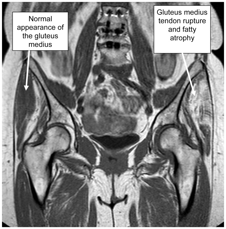
MRICoronal pelvic MRI: labels directly on the image, normal gluteus medius tendon on the right, tendinous rupture with fatty atrophy on the left, greater trochanteric pain syndrome.
Source : Kaltenborn et al., PLoS ONE, 2014, figure 3 · CC BY
| Test | Description | Performance | Level of evidence |
|---|---|---|---|
| Direct palpation of the greater trochanter | Reproduction of the familiar pain by pressure on the posterosuperior facet | High sensitivity but low specificity (palpation is tender in many asymptomatic people too) | Moderate |
| Single-leg stance (30 s) | The patient stands on the affected leg for 30 s; the test is positive if the lateral pain is reproduced | Sens. 100% / Spec. 97.3% for MRI-confirmed gluteal tendinopathy (Lequesne 2008 cohort, n=17 GTPS versus 17 controls) | Moderate (limited sample) |
| FADER (flexion, adduction, external rotation) | Patient supine: hip flexed to 90°, maximal adduction, external rotation | Sensitivity up to 88% (Grimaldi 2017, cohort with MRI as the reference) | Moderate |
| Resisted hip abduction test in side-lying | Patient in side-lying, abduction against manual resistance | Variable sensitivity, acceptable specificity | Moderate |
| Modified Trendelenburg (30 s) | Pelvic drop on the non-weight-bearing side ≥ 2 cm during 30 s of single-leg stance | An indicator of functional abductor weakness | Low (indirect test) |
🧭 Diagnostic decision tree for lateral hip pain
From clinical suspicion to confirmation: after the JOSPT 2023 CPG + Grimaldi 2015
Algorithm synthesised from Grimaldi 2015 (JOSPT) and from current practice: imaging has no place as a first-line investigation in a patient without red flags.
- L4-L5 radiculopathy : pain along the lateral aspect of the thigh that may travel down to the foot, radicular motor deficit, dermatomal dysaesthesia, positive neurodynamic signs (Slump, SLR). Several cases have been reported of a false radiculopathy that was in fact GTPS, and the reverse.⁴
- Hip osteoarthritis : pain rather in the groin, morning stiffness, restricted range (especially internal rotation), positive FADIR test.⁶
- Femoroacetabular impingement (FAI) : a young, athletic patient, predominantly anterior pain, positive FADIR.
- Meralgia paraesthetica : neurological symptoms of burning or paraesthesia in the territory of the lateral femoral cutaneous nerve (anterolateral thigh); Tinel's test over the anterior superior iliac spine is sometimes positive.⁷
- Piriformis syndrome or sacroiliac pathology: posterior buttock pain rather than lateral pain.
- Proximal hamstring tendinopathy : low buttock pain, brought on by prolonged sitting.
- Stress fracture of the femoral neck or of the trochanter (endurance athlete, woman with RED-S, elderly osteoporotic patient): progressive pain of increasing intensity, sometimes with a sudden loss of function.
2.3. Should patients with GTPS be classified, and what is gained from it? 🎯
A functional classification is useful for dosing the therapeutic load. The tendinopathy continuum model of Cook & Purdam (BJSM 2009, revisited in 2016) remains the operational reference:⁸- Reactive tendinopathy (often a young adult, recent onset < 6 weeks): pain of rapid onset after overload. Management favours a temporary reduction in load and pain-free isometric exercise. Early heavy strengthening is thought to be harmful.
- Tendon in disrepair (3-6 months): the intermediate phase; progressive introduction of isotonic exercise without high compressive loads.
- Degenerative tendinopathy (often > 6 months, in an older patient): an approach based on progressive heavy strengthening (HSR: heavy slow resistance) to improve functional capacity, even if the tendon structure is not restored.
- GTPS diagnosis = a clinical cluster : lateral trochanteric pain reproduced on palpation + positive 30 s single-leg stance + positive FADER + compatible history.
- Imaging is pointless as a first-line investigation, except with red flags or after conservative failure. The major risk: over-interpretation of asymptomatic abnormalities.
- A non-negotiable differential diagnosis: L5 radiculopathy, hip osteoarthritis, FAI, meralgia paraesthetica, stress fracture.
- Staging (reactive / disrepair / degenerative) to dose the rehabilitation : isometric work in the acute phase, HSR in the chronic phase.
Bibliography - Chapter 2
- Grimaldi A, Mellor R, Hodges P, Bennell K, Wajswelner H, Vicenzino B. Gluteal Tendinopathy: A Review of Mechanisms, Assessment and Management. Sports Med. 2015;45(8):1107-1119. PMID 25969366.
- Long SS, Surrey DE, Nazarian LN. Sonography of greater trochanteric pain syndrome and the rarity of primary bursitis. AJR Am J Roentgenol. 2013;201(5):1083-1086. AJR.
- Speers CJ, Bhogal GS. Greater trochanteric pain syndrome: a review of diagnosis and management in general practice. Br J Gen Pract. 2017;67(663):479-480. PMID 28963433.
- Tortolani PJ, Carbone JJ, Quartararo LG. Greater trochanteric pain syndrome in patients referred to orthopedic spine specialists. Spine J. 2002;2(4):251-254. PMID 14589475.
- Lequesne M, Mathieu P, Vuillemin-Bodaghi V, Bard H, Djian P. Gluteal tendinopathy in refractory greater trochanter pain syndrome: diagnostic value of two clinical tests. Arthritis Rheum. 2008;59(2):241-246. PMID 18240186.
- Fearon AM, Scarvell JM, Neeman T, Cook JL, Cormick W, Smith PN. Greater trochanteric pain syndrome: defining the clinical syndrome. Br J Sports Med. 2013;47(10):649-653. PMID 22983121.
- Grimaldi A, Mellor R, Nicolson PJA, Hodges P, Bennell K, Vicenzino B. Utility of clinical tests to diagnose MRI-confirmed gluteal tendinopathy in patients presenting with lateral hip pain. Br J Sports Med. 2017;51(6):519-524. PMID 27633027.
- Cook JL, Purdam CR. Is tendon pathology a continuum? A pathology model to explain the clinical presentation of load-induced tendinopathy. Br J Sports Med. 2009;43(6):409-416. PMID 18812414.
- Cook JL, Rio E, Purdam CR, Docking SI. Revisiting the continuum model of tendinopathy: what is its merit in clinical practice and research? Br J Sports Med. 2016;50(19):1187-1191. PMID 27127294.
3. Which treatment strategies are the most effective for GTPS?
3.1. Where do you start? What is the recommended hierarchy of interventions?
The consensus hierarchy (LEAP, Speers 2017, Grimaldi 2015, Bremer 2025):¹⁻⁴- First line (always): education + graded exercise. High level of evidence.
- Second line for cases that resist at 8-12 weeks : extracorporeal shockwave therapy (ESWT, Harding 2024).⁵
- Third line : corticosteroid injection, only for short-term relief (≤ 6 weeks) while waiting for exercise to take over. Harmful effect on tendon integrity when repeated (Coombes 2010, Lancet).⁶
- Alternative to corticosteroids : PRP (platelet-rich plasma). Fitzpatrick 2019 (AJSM, RCT n=80) shows an improvement maintained at 2 years, unlike corticosteroids, whose benefit is lost by 6 weeks.⁷
- Last line : surgery (endoscopic repair of the gluteal tendons, Walker-Santiago 2020) where at least 6 months of conservative management has failed, or where a complete tendon tear is documented.⁸
📊 LEAP trial: education + exercise versus corticosteroids versus “wait and see” at 52 weeks
% of patients reporting a global perceived improvement (n=204, multicentre RCT, Mellor 2018)
Source: Mellor R et al. BMJ. 2018;361:k1662. PMID 29720374. Success = “much better” or “recovered” on the GROC scale. Conclusion : the active approach almost doubles the odds of success versus corticosteroids.
3.2. What is the place of exercise, and is there a superior approach?
Exercise is not merely one component of treatment: it is the intervention with the highest level of evidence for durable results. The Bremer 2025 meta-analysis (searches up to August 2024, trials of medium to high quality) concludes unambiguously: “A combined approach of graded exercises and education has the best evidence for the management of gluteal tendinopathy and should be adopted as the core approach”.² 💪- Phase 1: pain modulation and decompression (weeks 1-2) :
- Education on managing compressive positions (see the self-management section) the absolute priority.
- Abduction isometrics in supine (without compressive load): 30-45 s holds × 4-5 repetitions, 1-2 times a day.
- Avoidance of positions of prolonged adduction.
- Phase 2: targeted isotonic strengthening (weeks 3-6) :
- Single-leg glute bridge (progressing towards 20-30 quality repetitions);
- Side-lying abduction without letting the thigh drop into adduction, with a resistance band or a light weight;
- modified “clam shell” without lateral compression;
- Hip hike (raising one half of the pelvis) in controlled single-leg stance.
- Phase 3: functional strengthening (weeks 6-12) :
- Partial single-leg squat with control of pelvis-knee alignment;
- Controlled step-ups and step-downs;
- Progressive uphill walking;
- Heavy slow resistance (HSR) with increasing load for degenerative patients.
- Phase 4: return to specific activities (beyond 12 weeks) : running, sport, hiking; see chapter 4.
3.3. Manual therapy, shockwave therapy, injections: how effective are they really?
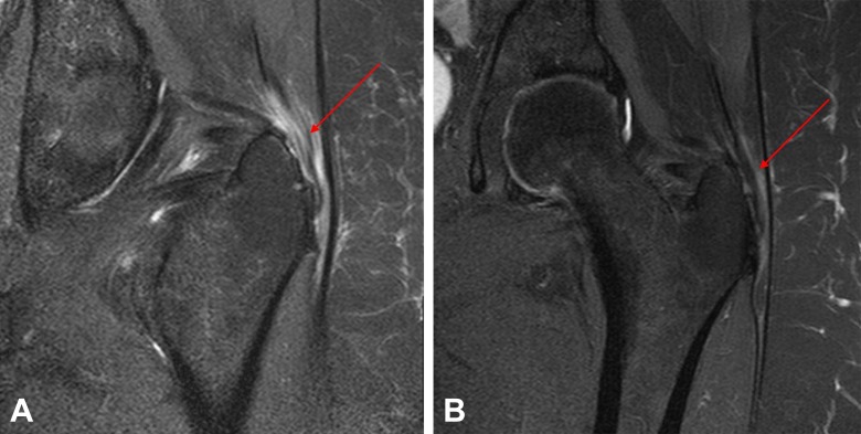
MRICoronal hip MRI before and after treatment: thickening of the gluteus medius tendon with intratendinous hyperintensity, arrow, visible reduction after autologous tenocyte injection.
Source : Bucher et al., Orthopaedic Journal of Sports Medicine, 2017, figure 4 · CC BY-NC-ND
| Modality | Level of evidence | Expected effect | Place in practice |
|---|---|---|---|
| Education + graded exercise | High (LEAP, Bremer 2025) | 77% success at 52 weeks | 1st line, indispensable |
| Shockwave therapy (ESWT) | Moderate (Harding 2024 SR/MA) | Significant pain reduction and functional improvement versus placebo / corticosteroids | 2nd line if there is failure at 8-12 weeks |
| PRP (platelet-rich plasma) | Moderate (Fitzpatrick 2019 RCT) | Benefit maintained at 2 years versus corticosteroids; superior effect at 12 weeks | Alternative to corticosteroids |
| Corticosteroid injection | Moderate (short term only) | Clear improvement at 4-6 weeks, loss of benefit at 12 weeks, inferior to exercise at 52 weeks | 3rd line: transient relief |
| Dry needling | Low (Brennan 2017 noninferiority) | Non-inferiority versus corticosteroid in the short term in one RCT | An adjunct option; limited evidence |
| Manual therapy | Low (few specific RCTs) | Useful for associated restrictions (lumbar spine, hip mobility) | An adjunct that makes exercise easier |
| Oral NSAIDs | Low | Short-term symptomatic relief | Caution (tendinosis > tendinitis, anti-inflammatory bias) |
| Endoscopic surgery | Moderate (Walker-Santiago 2020 SR) | Good recovery at 2 years, complications > PRP | Last resort, or a complete tear |
📐 GRADE hierarchy of interventions for GTPS
Horizontal pyramid (a synthesis of Bremer 2025, Mellor 2018, Harding 2024)
GRADE = Grading of Recommendations Assessment, Development and Evaluation. Source: a synthesis of the Bremer 2025 and Harding 2024 meta-analyses. Endoscopic surgery remains reserved for failures of ≥ 6 months.
3.4. Beyond the physical: how do you educate the patient and address psychological factors?
🧠 The biopsychosocial approach is non-negotiable in chronic GTPS. Plinsinga 2020 (Pain Med) showed that patients with persistent GTPS present with:⁹- An impairment of conditioned pain modulation (CPM) , a marker of central sensitisation.
- An impairment of psychological health (anxiety, depression, quality of life).
- Raised catastrophising and kinesiophobia scores.
- First line (high level of evidence) : patient education + progressive abductor exercise. LEAP 2018: 77.3% success versus 58.5% for corticosteroids versus 51.9% for “wait and see” at 52 weeks.
- A clear hierarchy : exercise → shockwave therapy → injections (short-term corticosteroids, or PRP for durability) → surgery as a last resort.
- Avoid compression : systematic education about the positions to avoid (legs crossed, hip hitched, side-lying without a pillow).
- Think biopsychosocial from the initial assessment onwards: kinesiophobia and catastrophising predict failure (Plinsinga 2020).
Bibliography - Chapter 3
- Mellor R, Bennell K, Grimaldi A, et al. Education plus exercise versus corticosteroid injection use versus a wait and see approach on global outcome and pain from gluteal tendinopathy: prospective, single blinded, randomised clinical trial (LEAP). BMJ. 2018;361:k1662. PMID 29720374.
- Bremer T, Nicklen P, Fearon A, Morrissey D. The efficacy of gluteal tendinopathy treatments: A systematic review. Clin Rehabil. 2025. doi:10.1177/02692155251327298.
- Speers CJ, Bhogal GS. Greater trochanteric pain syndrome: a review of diagnosis and management in general practice. Br J Gen Pract. 2017;67(663):479-480. PMID 28963433.
- Grimaldi A, Mellor R, Hodges P, Bennell K, Wajswelner H, Vicenzino B. Gluteal Tendinopathy: A Review of Mechanisms, Assessment and Management. Sports Med. 2015;45(8):1107-1119. PMID 25969366.
- Mani-Babu S, Morrissey D, Waugh C, et al. The effectiveness of extracorporeal shock wave therapy in lower limb tendinopathy: a systematic review. Am J Sports Med. 2015;43(3):752-761. PMID 24817008.
- Coombes BK, Bisset L, Vicenzino B. Efficacy and safety of corticosteroid injections and other injections for management of tendinopathy: a systematic review of randomised controlled trials. Lancet. 2010;376(9754):1751-1767. PMID 20970844.
- Fitzpatrick J, Bulsara MK, O'Donnell J, Zheng MH. Leucocyte-Rich Platelet-Rich Plasma Treatment of Gluteus Medius and Minimus Tendinopathy: A Double-Blind Randomized Controlled Trial With 2-Year Follow-up. Am J Sports Med. 2019;47(5):1130-1137. PMID 30840831.
- Walker-Santiago R, Wojnowski NM, Lall AC, et al. Platelet-Rich Plasma Versus Surgery for the Management of Recalcitrant Greater Trochanteric Pain Syndrome: A Systematic Review. Arthroscopy. 2020;36(3):875-888. PMID 31882270.
- Plinsinga ML, Coombes BK, Mellor R, Vicenzino B. Individuals with Persistent Greater Trochanteric Pain Syndrome Exhibit Impaired Pain Modulation, as well as Poorer Physical and Psychological Health. Pain Med. 2020;21(11):2964-2974. PMID 32232468.
- Brennan KL, Allen BC, Maldonado YM. Dry Needling Versus Cortisone Injection in the Treatment of Greater Trochanteric Pain Syndrome: A Noninferiority Randomized Clinical Trial. J Orthop Sports Phys Ther. 2017;47(4):232-239. PMID 28257614.
- Harding J, et al. Is shockwave therapy effective in the management of greater trochanteric pain syndrome? A systematic review and meta-analysis. Musculoskeletal Care. 2024. doi:10.1002/msc.1892.
4. How do you secure a lasting recovery and prevent GTPS from recurring?
4.1. How do you make the patient an active participant in their own recovery through self-management?
Therapeutic education outperforms passive approaches in the long term (LEAP: 77% success for “education+exercise” versus 58% for “injection”, at 52 weeks).² The patient must acquire 4 key skills:- Day-to-day management of compressive loads 🧑🏫 :
- Do not cross the legs when sitting;
- Avoid low seats (knees higher than hips);
- Standing: spread the weight over both legs, avoid prolonged “hip-hitched” standing;
- Sleep: prefer the side that is not painful, with a pillow between the knees ; avoid side-lying on the affected side.³
- Pain monitoring ⚖️ : a simple rule the patient can apply alone: pain during an activity may reach 4-5/10 provided it does not worsen over the following 24 h (adapted Silbernagel model).⁴
- Adherence to the home programme 💪 : consistency > intensity. A short daily session beats a long weekly session that gets missed.
- Identifying psychosocial factors 🧠 : recognising one's own catastrophising thoughts or avoidance behaviours; open dialogue with the clinician about movement-related fears.
🏠 8 validated key self-management habits (a synthesis of Grimaldi 2015, Mellor 2018)
To be taught systematically from the first session
Adapted from Grimaldi A. JOSPT 2015 and from the LEAP protocol (Mellor 2018). To be handed out as a patient roadmap.
4.2. When and how do you plan a safe return to sport and to activity? 🏃
Return to impact activities must be criterion-based, not calendar-based.⁵ The validated hierarchy:- Restoration of pain-free everyday function : 30 min of walking, pain-free stair climbing, prolonged standing tolerated.
- Strength and motor control criteria :
- 20-30 repetitions of the single-leg glute bridge without compensation;
- 30 s Trendelenburg test with no drop of ≥ 2 cm;
- Isometric abductor strength symmetry ≥ 90% (dynamometer measurement).
- Progressive reintroduction of specific loads (a runner, for example):
- Weeks 1-2: alternating walk/run programme (1 min running + 4 min walking × 5);
- Weeks 3-6: increase the volume by ≤ 10% per week ⚠️ (the Gabbett 2016 rule: beyond that, injury risk rises);⁶
- Weeks 6+: introduce only one new variable at a time (hills, speed, surface).
- Long-term maintenance : 1-2 gluteal strengthening sessions a week for life in at-risk patients (post-menopausal women, recreational runners).
🚩 Red flags specific to the return to activity
- Waking at night in pain after resuming activity → reduce the load immediately
- A rise in pain > 4/10 still present 24 h after exercise → adapt the programme
- Onset of a limp or a persistent Trendelenburg sign → go back to analytical exercises
- Pain travelling below the knee (L5-S1 radiation) → reconsider a radicular differential diagnosis
- A sensation of a “snap” or a sudden loss of function → suspected tendon tear → MRI
- For a durable recovery from GTPS, the approach must be active and patient-centred.
- ✅ Education is the key : avoid compressive positions (legs crossed, hip hitched, side-lying on the affected side without a pillow).
- 🏋️ Exercise is the treatment : progressive, consistent strengthening of the abductors.
- 📈 A graded, criterion-based return : the 10% weekly increase rule (Gabbett 2016) to prevent recurrence.
- 🧠 Self-management is the final goal : the patient becomes the expert on their own condition.
Bibliography - Chapter 4
- Grimaldi A, Mellor R, Hodges P, Bennell K, Wajswelner H, Vicenzino B. Gluteal Tendinopathy: A Review of Mechanisms, Assessment and Management. Sports Med. 2015;45(8):1107-1119. PMID 25969366.
- Mellor R, Bennell K, Grimaldi A, et al. LEAP trial. BMJ. 2018;361:k1662. PMID 29720374.
- Speers CJ, Bhogal GS. Greater trochanteric pain syndrome. Br J Gen Pract. 2017;67(663):479-480. PMID 28963433.
- Silbernagel KG, Thomeé R, Eriksson BI, Karlsson J. Continued sports activity, using a pain-monitoring model, during rehabilitation in patients with Achilles tendinopathy: a randomized controlled study. Am J Sports Med. 2007;35(6):897-906. PMID 17307888.
- Bremer T, Nicklen P, Fearon A, Morrissey D. The efficacy of gluteal tendinopathy treatments: A systematic review. Clin Rehabil. 2025. doi:10.1177/02692155251327298.
- Gabbett TJ. The training-injury prevention paradox: should athletes be training smarter and harder? Br J Sports Med. 2016;50(5):273-280. PMID 26758673.
- Plinsinga ML, Coombes BK, Mellor R, Vicenzino B. Individuals with Persistent GTPS Exhibit Impaired Pain Modulation. Pain Med. 2020;21(11):2964-2974. PMID 32232468.
- Allison K, Vicenzino B, Wrigley TV, Grimaldi A, Hodges PW, Bennell KL. Hip Abductor Muscle Weakness in Individuals with Gluteal Tendinopathy. Med Sci Sports Exerc. 2016;48(3):346-352. PMID 26418561.
5. Women > 40: why is GTPS a sex-specific condition?
5.1. Pelvic anatomy and biomechanics: why are women more exposed?
Several female biomechanical features converge to increase the compressive stresses on the gluteus medius and gluteus minimus tendons at their trochanteric insertion:²⁻³- A wider pelvis : the bi-trochanteric distance is on average 5 to 10% greater in women, which increases the lever arm in relative adduction and the tension in the iliotibial band that overlies the gluteal tendons.²
- An increased Q angle (quadriceps angle) : more pronounced dynamic knee valgus during walking, squatting and running, contributing to a hip adduction and internal rotation that compresses the lateral gluteal tendons.
- Femoral neck geometry : mean femoral anteversion 4 to 6 ° greater, which alters the path of the fascia lata over the greater trochanter.
- Relative abductor weakness : the Allison 2016 cohort (MSSE) showed significantly lower isometric abductor strength in women with GTPS than in asymptomatic controls, even on the pain-free side , which suggests a baseline vulnerability before the condition is ever triggered.⁴
5.2. Menopause, oestrogens and tendon health: an unfavourable combination?
The menopausal transition overlaps the incidence peak of GTPS, and that is no coincidence. 🌡️ Oestrogens take part in regulating the metabolism of tendon collagen: their abrupt fall (along with that of circulating androgens) degrades the quality of the tendon matrix, increasing susceptibility to overload tendinopathies.⁵⁻⁶- Reduced type I collagen synthesis and increasingly impaired cross-link maturation → a tendon less resilient to compressive loads.
- Altered fibre architecture : disturbed tenocyte turnover.
- Low-grade systemic inflammation (oestrogen deprivation): heightened sensitivity to repeated micro-trauma.
- Changes in body composition : increased abdominopelvic fat mass, frequent weight gain, global muscular deconditioning.
⚖️ Cumulative sex-specific risk factors in women > 40
Strength of the associations (estimated ORs or clinical consensus, Segal 2007, Allison 2016, Fearon 2013)
ORs consolidated from Segal 2007 (n=3026), Fearon 2013, Allison 2016. For the menopause, an indirect estimate based on the chronological incidence peak (40-60 years). The OR estimates are indicative.
5.3. How do you stratify risk and adapt management?
🎯 It is useful to identify 3 broad clinical subgroups of women > 40 with GTPS, because the optimal treatment strategy differs:| Subgroup | Typical profile | Treatment priority | Prognosis |
|---|---|---|---|
| A. Sedentary post-menopausal woman + high BMI | 55-65 years, BMI ≥ 28, global deconditioning, low back pain often associated, low activity level | A global approach: education + weight loss + physical reconditioning. A very gradual start (isometrics in an unloaded position) | Good with adherence; high risk of chronicity if the sedentary lifestyle persists |
| B. Active masters woman (recreational/sporting) | 45-60 years, runner or hiker, normal BMI, recent increase in training volume | Temporary load modification + progressive abductor strengthening. Return-to-running plan at ≤ 10% weekly increase | Excellent (recovery in 12-16 weeks) |
| C. Woman with chronic “complicated GTPS” | > 6 months of symptoms, failed management, signs of central sensitisation (kinesiophobia, catastrophising), frequent psychological comorbidities | A reinforced biopsychosocial approach. Adjuncts: shockwave therapy, PRP if these fail. Work on central modulation (pain education, graded exposure) | Variable; requires a multidisciplinary team |
- 📋 Systematically screen for other tendinopathies : lateral epicondylitis, rotator cuff tendinopathy, plantar fasciitis, all more frequent after the menopause.
- 🦴 Assess bone status (DXA where indicated): in a post-menopausal woman with progressive trochanteric pain, a stress fracture or an underlying bone fragility must be ruled out.
- 💊 Discuss whether to seek a gynaecological opinion for women with menopausal symptoms (HRT, raloxifene, and so on): a shared decision outside the physiotherapy remit.
- 🥗 A metabolic approach : suitable nutrition, sufficient protein, vitamin D, regular physical activity. BMI is one of the two major modifiable risk factors.
- GTPS shows a marked sex bias (F:M ≈ 4:1) explained by pelvic anatomy (wide pelvis, Q angle, femoral anteversion) and by the menopausal transition (its impact on tendon health).
- Stratify into 3 subgroups : (A) sedentary post-menopausal + high BMI, (B) active masters, (C) complicated chronic. The treatment strategy differs for each.
- Screen for associated tendinopathies (cuff, epicondyle, plantar), for bone fragility and discuss gynaecological referral according to the overall menopausal picture.
- BMI and a sedentary lifestyle are the major modifiable risk factors : managing them goes beyond the strict physiotherapy remit and calls for a multidisciplinary approach.
Bibliography - Chapter 5
- Fearon AM, Scarvell JM, Neeman T, Cook JL, Cormick W, Smith PN. Greater trochanteric pain syndrome: defining the clinical syndrome. Br J Sports Med. 2013;47(10):649-653. PMID 22983121.
- Birnbaum K, Siebert CH, Pandorf T, Schopphoff E, Prescher A, Niethard FU. Anatomical and biomechanical investigations of the iliotibial tract. Surg Radiol Anat. 2004;26(6):433-446. PMID 15378277.
- Grimaldi A, Mellor R, Hodges P, Bennell K, Wajswelner H, Vicenzino B. Gluteal Tendinopathy: A Review of Mechanisms, Assessment and Management. Sports Med. 2015;45(8):1107-1119. PMID 25969366.
- Allison K, Vicenzino B, Wrigley TV, Grimaldi A, Hodges PW, Bennell KL. Hip Abductor Muscle Weakness in Individuals with Gluteal Tendinopathy. Med Sci Sports Exerc. 2016;48(3):346-352. PMID 26418561.
- Hansen M, Kjaer M. Sex hormones and tendon. Adv Exp Med Biol. 2016;920:139-149. PMID 27535256.
- Magnusson SP, Hansen M, Langberg H, et al. The adaptability of tendon to loading differs in men and women. Int J Exp Pathol. 2007;88(4):237-240. PMID 17696904.
- Segal NA, Felson DT, Torner JC, et al. Greater trochanteric pain syndrome: epidemiology and associated factors. Arch Phys Med Rehabil. 2007;88(8):988-992. PMID 17678660.
- Speers CJ, Bhogal GS. Greater trochanteric pain syndrome. Br J Gen Pract. 2017;67(663):479-480. PMID 28963433.
6. What do concrete clinical cases teach us about GTPS? 📖
6.1. Analysis of a “classic” case: from assessment to resolution
The typical GTPS case matches the average profile of the LEAP trial (Mellor 2018, BMJ):¹- A woman aged 50 to 65, BMI often > 27, lateral hip pain of insidious onset over several months.
- Night pain when lying on the affected side, pain on climbing stairs, pain on prolonged walking.
- Failure of an initial treatment (NSAIDs, relative rest, sometimes an earlier injection that worked only briefly).
- Physical examination: trochanteric palpation reproduces the pain, positive 30 s single-leg stance, positive FADER.
- Initial assessment (1 session): history, clinical examination with the test cluster, identification of modifiable risk factors, screening for red and yellow flags.
- Patient education (built into every session): dismantling the “bursitis” myth, the principles of decompression, position management.
- Supervised exercise programme over 8 weeks (14 sessions in LEAP): isometric phase → isotonic → functional.
- Daily home programme alongside it: 10-15 min a day.
- Reassessment at 6 and 12 weeks : load adjusted to the response, introduction of the return to activity.
6.2. The diagnostic challenge: when GTPS mimics another condition
GTPS is a great imitator. Several published clinical cases report atypical presentations in which the symptoms perfectly mimicked other conditions. ⚠️ Case 1: GTPS mimicking meralgia paraesthetica 🔬 A presentation with burning and paraesthesia over the anterolateral aspect of the thigh may at first point towards compression of the lateral femoral cutaneous nerve (meralgia paraesthetica). When treatments aimed at the nerve remain ineffective, a meticulous clinical reassessment may reveal that palpation of the greater trochanter reproduces the familiar pain and that treatment focused on the gluteal tendinopathy resolves the symptoms. The implication: lateral thigh pain is not always neurogenic; always check trochanteric palpation.² Case 2: GTPS from a gluteus medius tear mimicking an L5 radiculopathy 🔬 Weakness of hip abduction (a positive Trendelenburg sign) together with pain radiating down the lateral aspect of the lower limb is a classic picture of L5 radiculopathy. However, an inconclusive lumbar MRI should raise the possibility of a full-thickness tear of the gluteus medius tendon, which can produce a genuine Trendelenburg sign and a misleading pattern of radiating cutaneous pain. Hip MRI is then the key to the diagnosis, and surgical repair can make the “pseudo-radicular” symptoms disappear.³🚩 Frequent diagnostic traps in GTPS
- Positive Trendelenburg + lateral pain travelling below the knee → do not settle for a radicular diagnosis before ruling out a gluteus medius tendon tear (hip MRI)
- Anterolateral thigh paraesthesia resistant to treatment for meralgia → always palpate the greater trochanter
- GTPS in a young, athletic man with no obvious risk factor → look for a stress fracture, FAI, piriformis syndrome or iliopectineal impingement
- Sudden unilateral pain after a fall in an elderly patient → think first of a fracture of the greater trochanter or of the femoral neck
- Bilateral, symmetrical GTPS of progressive onset in a young adult → suspect axial spondyloarthritis, rheumatoid arthritis, or fibromyalgia as a comorbidity
6.3. A complex case study: the surgically repaired tendon tear
Some patients do not respond to well-conducted conservative management over > 6 months. In those cases, it is necessary to investigate further and consider options of 2nd/3rd line. 🔧 Typical profile :- Woman ≥ 55-65 years with chronic GTPS, conservative failure ≥ 6 months;
- Marked, persistent abductor weakness (positive Trendelenburg) despite rehabilitation;
- MRI showing a high-grade partial or full-thickness tear of the gluteus medius tendon.
- Focused shockwave therapy : where symptoms dominate but there is no complete tear. The Harding 2024 meta-analysis (Musculoskeletal Care) confirms a significant benefit on pain versus placebo in GTPS.⁴
- PRP (Fitzpatrick 2019, AJSM) : RCT n=80, superiority at 2 years over corticosteroids for function and pain.⁵
- Endoscopic tendon repair surgery (Walker-Santiago 2020, systematic review): arthroscopic reattachment of the gluteus medius and minimus. Progressive restoration of strength and resolution of pain in the majority of cases at 1-2 years after surgery, but it demands rigorous post-operative rehabilitation (8-12 months).⁶
- Special cases after total hip replacement : persistent trochanteric pain after hip arthroplasty → look for a local mechanism (implant impingement, an intra-articular fragment, low-grade infection); do not settle for a diagnosis of “secondary GTPS”.
- The typical LEAP case (a woman of 50-65, high BMI, insidious pain) responds well to an education + exercise protocol over 8-12 weeks: 77% success at 1 year.
- Traps : meralgia paraesthetica, L5 radiculopathy, stress fracture, piriformis syndrome, FAI. Familiar pain on trochanteric palpation remains the diagnostic pivot.
- In the event of failure at ≥ 6 months : hip MRI to rule out a tear, then shockwave therapy / PRP / endoscopic surgery as a therapeutic escalation.
- Always look for a secondary mechanical cause in persistent GTPS after arthroplasty or after trauma.
Bibliography - Chapter 6
- Mellor R, Bennell K, Grimaldi A, et al. LEAP trial. BMJ. 2018;361:k1662. PMID 29720374.
- Grimaldi A, Mellor R, Hodges P, Bennell K, Wajswelner H, Vicenzino B. Gluteal Tendinopathy: A Review of Mechanisms, Assessment and Management. Sports Med. 2015;45(8):1107-1119. PMID 25969366.
- Lequesne M, Mathieu P, Vuillemin-Bodaghi V, Bard H, Djian P. Gluteal tendinopathy in refractory greater trochanter pain syndrome: diagnostic value of two clinical tests. Arthritis Rheum. 2008;59(2):241-246. PMID 18240186.
- Harding J, et al. Is shockwave therapy effective in the management of greater trochanteric pain syndrome? A systematic review and meta-analysis. Musculoskeletal Care. 2024. doi:10.1002/msc.1892.
- Fitzpatrick J, Bulsara MK, O'Donnell J, Zheng MH. Leucocyte-Rich PRP Treatment of Gluteus Medius and Minimus Tendinopathy: A Double-Blind RCT With 2-Year Follow-up. Am J Sports Med. 2019;47(5):1130-1137. PMID 30840831.
- Walker-Santiago R, Wojnowski NM, Lall AC, et al. Platelet-Rich Plasma Versus Surgery for the Management of Recalcitrant Greater Trochanteric Pain Syndrome: A Systematic Review. Arthroscopy. 2020;36(3):875-888. PMID 31882270.
- Speers CJ, Bhogal GS. Greater trochanteric pain syndrome: a review of diagnosis and management in general practice. Br J Gen Pract. 2017;67(663):479-480. PMID 28963433.
- Long SS, Surrey DE, Nazarian LN. Sonography of greater trochanteric pain syndrome and the rarity of primary bursitis. AJR Am J Roentgenol. 2013;201(5):1083-1086. AJR.
7. How do you apply these recommendations concretely in your practice?
7.1. When, and to which other health professionals, should you refer?
A fundamental competence of the modern physiotherapist includes recognising the limits of their own scope of practice. Identifying red flags (Finucane 2020) is a non-negotiable prerequisite.¹ Criteria for medical referral 🚨 :- Red flags (see chapter 1): recent history of cancer, severe non-mechanical night pain, fever, unexplained weight loss, progressive neurological deficit, high-energy trauma, suspected stress fracture.
- Failure of well-conducted conservative treatment at ≥ 8-12 weeks → see a doctor to discuss shockwave therapy / PRP / imaging / injection.
- Suspected complete tendon tear (major Trendelenburg, marked abductor weakness) → hip MRI + orthopaedic opinion.
- Significant psychiatric comorbidities (major depression, disabling anxiety disorders) → referral to a psychologist or to the GP.
- Suspected systemic disease (spondyloarthritis, rheumatoid arthritis, fibromyalgia) → rheumatologist.
- Progressive, symmetrical bilateral GTPS → full rheumatological work-up.
- The GP : for a short NSAID prescription if needed, test requests, specialist referral.
- Sports physician / rehabilitation physician : for recreational and competitive athletes, shockwave therapy, PRP.
- Rheumatologist : suspected inflammatory disease, rheumatological comorbidities.
- Orthopaedic surgeon : suspected tendon tear, prolonged conservative failure.
- Dietitian / nutritionist : high BMI (one of the major modifiable risk factors).
- Clinical psychologist / pain specialist : kinesiophobia, catastrophising, anxiety and depressive comorbidities, signs of central sensitisation.
7.2. How do you measure outcomes and overcome the barriers to implementation?
📊 The PROMs (patient-reported outcome measures) are standardised questionnaires that quantify progress from the patient's point of view. For GTPS, the most relevant are:- VISA-G (Victorian Institute of Sport Assessment - Gluteal): 8 items, validated specifically for gluteal tendinopathy, with demonstrated responsiveness to change.²
- HOOS (Hip disability and Osteoarthritis Outcome Score): a generic hip measure, 5 subscales (pain, symptoms, activities of daily living, sport, quality of life).
- Numerical Pain Rating Scale (NPRS) 0-10 : simple, validated.
- GROC (Global Rating of Change): the patient's own perceived rating of change (-7 to +7), used in LEAP.
- FABQ (Fear-Avoidance Beliefs Questionnaire): screening for kinesiophobia.
🛠️ Overcoming the barriers to implementing EBP
A synthesis of the barriers most often cited by physiotherapists, and the levers for improvement
A synthesis of several reviews on the barriers to EBP in physiotherapy. The practice of shared decision-making (Shared Decision-Making) is a powerful lever for aligning patient expectations with the scientific data.
- Present the 3 evidence-based options to the patient: (1) education + exercise (1st line, long-term success but requires commitment), (2) corticosteroid injection (fast but transient), (3) watchful waiting alone (an option if the acute episode is not very disabling).
- Explain the real figures (LEAP 2018) in language the patient can understand.
- Identify the values and preferences of the patient (do they prefer immediate relief or a lasting solution? Which activities do they want to get back to?).
- Build together the treatment plan, with concrete goals and criteria for reassessment.
- Referral is crucial: red flags → doctor or emergency department, yellow flags → psychologist, conservative failure at ≥ 6-8 weeks → doctor for escalation.
- The interprofessional collaboration (doctor, rheumatologist, surgeon, dietitian, psychologist) is essential in complex cases.
- Measure outcomes with validated PROMs (the specific VISA-G, the generic HOOS, NPRS, GROC).
- The barriers to EBP (time, skills, resources) are real but can be overcome through continuing education, digital tools and shared decision-making.
Bibliography - Chapter 7
- Finucane LM, Downie A, Mercer C, et al. International Framework for Red Flags for Potential Serious Spinal Pathologies. J Orthop Sports Phys Ther. 2020;50(7):350-372. PMID 32438853. doi:10.2519/jospt.2020.9971.
- Fearon AM, Ganderton C, Scarvell JM, et al. Development and validation of a VISA tendinopathy questionnaire for greater trochanteric pain syndrome, the VISA-G. Man Ther. 2015;20(6):805-813. PMID 25870117.
- Mellor R, Bennell K, Grimaldi A, et al. LEAP trial. BMJ. 2018;361:k1662. PMID 29720374.
- Bremer T, Nicklen P, Fearon A, Morrissey D. The efficacy of gluteal tendinopathy treatments: A systematic review. Clin Rehabil. 2025. doi:10.1177/02692155251327298.
- Plinsinga ML, Coombes BK, Mellor R, Vicenzino B. Individuals with Persistent GTPS Exhibit Impaired Pain Modulation. Pain Med. 2020;21(11):2964-2974. PMID 32232468.
- Grimaldi A, Mellor R, Hodges P, Bennell K, Wajswelner H, Vicenzino B. Gluteal Tendinopathy: A Review of Mechanisms, Assessment and Management. Sports Med. 2015;45(8):1107-1119. PMID 25969366.
- Speers CJ, Bhogal GS. Greater trochanteric pain syndrome. Br J Gen Pract. 2017;67(663):479-480. PMID 28963433.
- Hoffmann TC, Légaré F, Simmons MB, et al. Shared decision making: what do clinicians need to know and why should they bother? Med J Aust. 2014;201(1):35-39. PMID 24999896.
What next, after this read?
This article is part of a collection of evidence-based clinical syntheses. A question, some feedback, a correction to suggest? Contact us directly through the WhatsApp button at the bottom right of the screen.

