














On-siteDPCFIFPL

Neurological rehabilitation
Neurological disorders in musculoskeletal practice: screen, assess and treat
Miguel Galan
Go further
Meralgia paraesthetica: the territory of the lateral femoral cutaneous nerve, the differential with femoral neuralgia, and what rehabilitation is worth.

A burning sensation or tingling in the anterolateral thigh, never below the knee, suggests meralgia, purely sensory, confirmed by the pelvic compression test.
58%of meralgia patients have an identifiable mechanical risk factor
What follows covers each of these points in detail, with sources. It is there if you need it.
This topic is taught in a course: Neurological disorders in musculoskeletal practice: screen, assess and treat, on-site with Miguel Galan.
1 / 14
13 article chapters · 54 min in total
Understanding◔ 4 min
It is a purely sensory mononeuropathy of the lateral femoral cutaneous nerve.
Territory◔ 4 min
The territory stops above the knee: nothing in the leg, ankle or foot.
Differentiating◔ 7 min
A root carries motor fibres, a cutaneous nerve does not: that is what decides.
Causes◔ 7 min
In its common form, it is a disease of external constraint that can be identified.
Investigations◔ 3 min
The review literature is consistent: the diagnosis remains clinical.
Serious forms◔ 3 min
The comfort of an easy diagnosis is the best ally of delayed diagnosis.
Course◔ 2 min
The rate of spontaneous resolution is very often quoted, but rarely referenced.
Rehabilitation◔ 7 min
Seven randomised trials exist on this condition, of which only one tests a physiotherapy technique.
Injection◔ 6 min
The treatment ladder follows an order: conservative first, injection, then surgery as a last resort.
Pregnancy, children◔ 3 min
Pregnancy carries the highest odds ratio, at 12.0, of all the factors measured.
Case reports◔ 3 min
A case report illustrates, it does not demonstrate: it is the lowest level of evidence.
In practice◔ 2 min
A hand drawing an oval on the thigh, never crossing the knee, is worth an examination.
Questions◔ 3 min
The nerve is exclusively sensory: it innervates no muscle.
Train on this
Neurological rehabilitation
Miguel Galan
Next comes practice: the course that teaches this topic, with Miguel Galan.
Our courses on this topic may be covered by DPC or FIFPL, the French continuing-education funding schemes, depending on your profession and situation.
A diagnosis that is easy to make when you think of it, invisible when you do not. Meralgia paraesthetica is a nerve trunk lesion, not a radicular one: purely sensory, with no motor deficit or absent reflex, in a defined territory of the anterolateral thigh that never runs below the knee. That is what separates it from femoral neuralgia and from L2-L3 radiculopathy, with which it is confused, and it is what this synthesis sets out first. For the radicular conditions themselves, see femoral neuralgia and sciatica from disc herniation.
In brief
Meralgia paraesthetica in three figures
What the literature establishes on frequency, spontaneous course and the level of evidence available.
Sources: van Slobbe AM et al. (2004), J Neurol, PMID 15015008; Khalil N et al. (2012), Cochrane Database of Systematic Reviews, PMID 23235604; Dengler NF (2023), Neurological Research, PMID 36520581.
In this chapter: the nerve trunk definition of the lesion, the nerve's course and its considerable anatomical variability, what “purely sensory” means concretely at examination, and the structural reasons why this diagnosis is missed.
Meralgia paraesthetica is a mononeuropathy of the lateral femoral cutaneous nerve. The term comes from the Greek meros (thigh) and algos (pain), and the original description is attributed to Bernhardt and to Roth at the end of the nineteenth century, hence the name Bernhardt-Roth disease that is still met.
The point that governs everything else is this: this nerve is exclusively sensory. It arises from the L2 and L3 roots, runs down medial to the psoas muscle, crosses the iliacus muscle, then leaves the pelvis near the anterior superior iliac spine before reaching the skin of the anterolateral thigh.11 It innervates no muscle. It takes part in no tendon reflex.
A purely sensory nerve cannot produce a motor deficit. Any weakness, any wasting, any absent knee reflex in a patient labelled “meralgia” is a diagnostic error, not a severe form.
That proposition is the diagnostic key to this whole article, and it is constant in the review literature: the diagnosis rests on the description of a sensory disturbance, often painful, of the anterolateral thigh, with normal strength and reflexes.3
The classic compression site is the passage under the inguinal ligament, immediately next to the anterior superior iliac spine. An anatomical study of 33 cadavers measured a mean distance of 8.8 mm between the anterior superior iliac spine and the nerve ; the nerve was less than 2 cm from the medial tip of the spine in about 90 % of cases, and less than 1 cm in 76 % of cases. The mean angle between the inguinal ligament and the nerve was 83.3 degrees.8
These means hide major dispersion, and it is that dispersion which explains both the nerve's susceptibility and treatment failures. A surgical series of 148 operated patients described, alongside the usual arrangement (a single trunk, deep to the superficial fascia and the inguinal ligament, running downwards and lateral to the spine), less frequent but real variants: early bifurcation, epifascial position, inferomedial direction, and exit from the pelvis through a bony iliac canal. Notably: in 13 cases out of 148, that is 8.8 %, the nerve was not found during the operation.9
A study of 20 embalmed cadavers specified the extent of that dispersion: the nerve could lie from 6.5 cm medial to the anterior superior iliac spine to 6 cm lateral to it, and in the latter case sit in a groove of the iliac crest. The same work described a complete fascial canal surrounding the nerve along its whole course, from the inguinal ligament to beyond its terminal branches, as well as variants in which the nerve arises from the femoral nerve and cases of duplication.10
Four landmarks that change the clinical reading
Anatomy, unexplained share, weight of excess weight, and the surgical setting.
Sources: Lee SH et al. (2017), Muscle & Nerve, PMID 27543938; van Slobbe AM et al. (2004), J Neurol, PMID 15015008; Mondelli M et al. (2007), Acta Neurologica Scandinavica, PMID 17661798; Yang SH et al. (2005), Spine, PMID 16166883.
Three mechanisms combine, and none is a matter of competence.
The site immediately suggests the spine. Thigh pain in an adult in their fifties spontaneously points to a disc herniation or lumbar osteoarthritis, all the more so because the patient often describes worsening on walking and standing, exactly as in neurogenic claudication. The reflex is to request lumbar imaging, which finds something in almost any adult of that age, and the case closes on a radiological diagnosis.
The nerve is purely sensory, so the standard neurological examination is normal. An assessment looking for a motor deficit and an absent reflex concludes that there is no neurological involvement, whereas the disturbance really is neurological: it is simply invisible to the two items tested first.
The cause is often in front of us and not recorded. A belt, tight trousers, a load carried at the waist, recent weight gain: these features appear in no routine questionnaire, and the patient does not report them spontaneously because they attach no medical value to them.
Key points
In this chapter: the map of the sensory territory and its two borders, the four verbal descriptions that should alert you, the position that makes it worse and the one that relieves it, and the most specific sign of all, intolerance of clothing rubbing.
The territory of the lateral femoral cutaneous nerve occupies the anterolateral aspect of the thigh. It has two borders the clinician must know, because it is those that make the diagnosis.
Upwards, it does not extend above the inguinal crease. True inguinal pain, groin pain, is not a meralgia: it points to the hip or to the psoas.
Downwards, it stops above the knee. It is the most useful border of all. The lateral femoral cutaneous nerve innervates nothing below the tibiofemoral joint line: not the leg, not the ankle, not the foot. Paraesthesia running below the knee is not a meralgia, whatever the rest of the picture.
The sensory territory, and what it is confused with
Anterior view of the right lower limb. On the left, the territory of the lateral femoral cutaneous nerve. On the right, the comparison with the two neighbouring radicular conditions.
Teaching diagram, not to scale. The territory of the lateral femoral cutaneous nerve is described after Onat SS et al. (2016), Pain Physician, PMID 27228536, and Grossman MG et al. (2001), Journal of the American Academy of Orthopaedic Surgeons, PMID 11575913. The radicular distributions are those measured by Kido T et al. (2016) in 58 patients operated on for upper lumbar disc herniation, Journal of Orthopaedic Science, PMID 27053156.
The verbal descriptions are fairly stereotyped. The review articles invariably report the same quartet: burning, numbness, tingling, pain of the anterolateral thigh.56 A military medicine review adds a symptom often left out of the lists: itching, which belongs to the register of dysaesthesia and not to that of dermatology.20
The most suggestive sign is nevertheless not in that list, because it is not said spontaneously: hypersensitivity to clothing rubbing. The patient cannot bear the contact of trousers, of the sheet, of a hand resting on the thigh. They rarely express it themselves because they do not file it under “symptom”. It has to be asked for, and the question must be precise: does the feel of fabric on this area bother you?
Pain that worsens on standing and settles on sitting, in a thigh territory that does not run below the knee, with intolerance of clothing rubbing: the diagnosis is made before you even touch the patient.
The positional behaviour is mechanically logical and clinically valuable. On prolonged standing and in hip extension, the inguinal ligament tensions and the pincer closes on the nerve: the symptoms appear or worsen. In hip flexion, that is when sitting, the tension eases and the trouble lessens.
The military medicine review describes exactly that profile: symptoms present on standing and walking, liable to be relieved by adopting other postures.20 A physical medicine review completes the picture on the triggering factors, citing stretching of the nerve by prolonged hyperextension of the trunk or thigh, prolonged standing, leg length inequality, external compression by a belt, weight gain and tight clothing. It also notes that the complaints typically disappear after weight loss or removal of the underlying cause.11
The walking trap
Worsening on walking and standing, relieved on sitting, is also the definition of the neurogenic claudication of lumbar stenosis. The two pictures sound alike in the history. What separates them is the territory (a meralgia does not run below the knee), the absence of motor deficit, and the unilateral, strictly defined character of the area. One article in the corpus covers stenosis: see degenerative lumbar stenosis.
Key points
In this chapter: why the territory alone is not enough, the complete differential table, what the bedside tests are really worth in figures, and the decision tree that follows.
This is the heart of this article, and the point where it differs from the two neighbouring pages on the site. Meralgia paraesthetica is a nerve trunk lesion: the nerve is damaged along its peripheral course, downstream of the plexus. Femoral neuralgia and L2-L3 radiculopathy are radicular lesions: the lesion sits at the root, upstream, where motor and sensory fibres still run together.
Everything else follows from that difference in site, and one thing alone is truly decisive: a root carries motor fibres, a cutaneous nerve does not.
You often read that meralgia is recognised by its anterolateral distribution. That is true but insufficient, and a Japanese study demonstrates it with figures. Kido and colleagues analysed 58 patients operated on for a far lateral lumbar disc herniation, grouped by level. In the L2-L3 herniation group, pain or numbness sat in the thigh, above the knee, in every single patient.51
In other words, an L2-L3 radiculopathy also gives thigh pain that does not run below the knee. The territory, on its own, does not separate the two diagnoses. It does on the other hand separate meralgia from L3-L4 femoral neuralgia very well, since in that latter group 80 % of patients had pain or numbness on the medial side of the knee.51
What decides is the motor and reflex examination. In the same series, weakness was found in 60 to 95 % of patients for iliopsoas and in 27 to 70 % for quadriceps depending on the level involved, and the knee reflex was reduced or absent in 27 to 100 % of patients depending on the group.51 Those proportions are not 100 % everywhere, and that is a useful piece of honesty: the absence of a motor deficit does not formally rule out a radiculopathy. But the presence of a motor deficit formally rules out a meralgia, since the nerve has no motor fibres to damage.
| Feature | Meralgia paraesthetica | L3-L4 femoral neuralgia | L2-L3 radiculopathy |
|---|---|---|---|
| Site of the lesion | Nerve trunk : peripheral nerve, under the inguinal ligament | Radicular: L3 or L4 root | Radicular: L2 or L3 root |
| Nature of the disturbance | Purely sensory, without exception | Sensorimotor | Sensorimotor |
| Territory | Anterolateral thigh, well defined, stops above the knee | Anterior thigh, then medial side of the knee in 80 % of cases | Thigh above the knee in 100 % of the patients in the series |
| Runs below the knee | Never | Possible, medial side | Not expected |
| Motor deficit | Absent by definition | Quadriceps and iliopsoas frequently affected | Iliopsoas above all |
| Knee reflex | Normal | Reduced or absent in a large share of patients | May be altered |
| Worse with | Prolonged standing, hip extension, walking, tight clothing | Variable, often posture and exertion | Variable |
| Relieved by | Hip flexion, sitting, loosening the belt | Variable | Variable |
| Clothing rubbing | Poorly tolerated, highly suggestive | Nothing particular | Nothing particular |
| Key bedside test | Pelvic compression: Se 95 %, Sp 93.3 % | Femoral nerve stretch test, positive in 91 to 95 % of the patients in the series | Femoral nerve stretch test |
| Effect of a local anaesthetic block at the ASIS | Clear relief, of diagnostic value | No effect | No effect |
Table to be scrolled horizontally on a small screen.
Table sources: the purely sensory nature and the normality of strength and reflexes are established by Chalk and Namiranian (2024)3 and by Ahmed and colleagues (2025)4 ; the distributions and the proportions of radicular deficits by Kido and colleagues (2016)51 ; the values of the pelvic compression test by Nouraei and colleagues (2007)12 ; the diagnostic value of the anaesthetic block by Grossman and colleagues (2001)7.
The pelvic compression test is the test specific to meralgia, and it is simple. Patient side-lying, symptomatic side uppermost, the examiner applies downward pressure on the pelvis and holds it for about 45 seconds. The manoeuvre releases the tension in the inguinal ligament and decompresses the nerve: the test is positive if the symptoms lessen. It is a test whose logic is the reverse of the usual provocation tests, which explains why it is forgotten.
Nouraei and colleagues, in a series of 45 patients, report a sensitivity of 95 % and a specificity of 93.3 %, and conclude explicitly that this test helps to distinguish meralgia from lumbosacral radicular pain.12 An independent case-control study of 30 patients whose meralgia was electrophysiologically proven finds slightly more modest but concordant values: 86.7 % sensitivity and 93.0 % specificity, with identical figures for the neurodynamic test and slightly lower ones for Tinel's sign at the anterior superior iliac spine.13
Diagnostic value of the tests, from the bedside to imaging
Measured sensitivity and specificity. The clinical tests beat somatosensory evoked potentials.
Sources: Nouraei SA et al. (2007), Neurosurgery, PMID 17415207; Paneyala S et al. (2024), Case Reports in Medicine, PMID 39687528; Chhabra A et al. (2013), Skeletal Radiology, PMID 23306718; Seror P (2004), Muscle & Nerve, PMID 14755498. SEP: somatosensory evoked potentials. Chhabra's values are the floors reported for the two readers.
Reading this graph honestly
These values come from small series (45 and 30 patients) and not from large-scale diagnostic accuracy studies. They are concordant, which is reassuring, but they are not as solid as a figure from a meta-analysis. The most reliable line in this table is in fact the most disappointing: evoked potentials on stimulation at the anterior superior iliac spine have a sensitivity of 5 %, and the author concludes that this montage has no diagnostic value.14
Decision tree for pain in the anterolateral thigh
Three questions in this order. The first two rule out, the third confirms.
Tree built from Nouraei SA et al. (2007), PMID 17415207; Chalk C and Namiranian D (2024), PMID 38697741; Ahmed MS et al. (2025), PMID 39673032; de Ruiter GCW et al. (2023) for the secondary causes branch, PMID 37148363.
Red flags of the diagnostic chapter
Key points
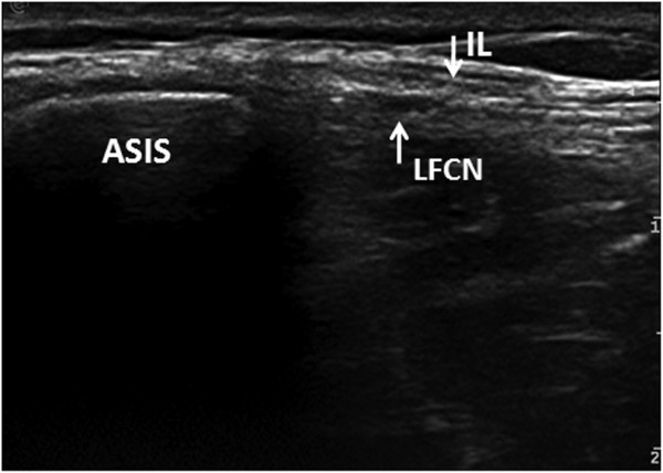
UltrasoundTransverse ultrasound showing the lateral femoral cutaneous nerve passing directly through the inguinal ligament near the anterior superior iliac spine, an anatomical course predisposing to compression.
Source : Zhu et al., BMC musculoskeletal disorders, 2012, figure 4 · CC BY
In this chapter: the inventory of external compressions, what obesity and pregnancy really weigh in odds ratios, iatrogenic and positional causes, and why well-targeted advice is worth more here than technique.
This is the chapter that changes the prognosis. Meralgia is, in its common form, a disease of external constraint : something presses on the nerve, and that something is most often identifiable and removable. A physical and rehabilitation medicine review draws up the inventory: trauma or overuse, pelvic and retroperitoneal tumours, stretching of the nerve by prolonged hyperextension of the trunk or thigh, leg length inequality, prolonged standing, external compression by a belt, weight gain, tight clothing.11
Two case-control studies give the figures. In the Dutch general practice cohort, meralgia is significantly associated with carpal tunnel syndrome, with an odds ratio of 7.7 (95 % CI 1.9 to 31.1), and with pregnancy, odds ratio 12.0 (95 % CI 1.2 to 118.0). The authors draw an elegant hypothesis from it: meralgia would result from the combination of a general susceptibility to entrapment syndromes and a local triggering factor, which is precisely what the association with carpal tunnel suggests.1
The confidence interval of the odds ratio for pregnancy, from 1.2 to 118.0, is extraordinarily wide: the association is real but its magnitude is very poorly estimated, and that must be said rather than quoting the 12.0 as though it were precise.
For obesity, an Italian case-control study of 104 cases matched with 416 controls establishes that obesity doubles the risk : odds ratio 2.04 (95 % CI 1.13 to 3.67) against neurological controls and 2.5 (95 % CI 1.4 to 4.5) against dermatological controls. Mean body mass index was 28.0 in cases against 26.0 and 25.5 in controls. The authors attribute the mechanism to the increased pressure linked to abdominal protrusion.18
Measured risk factors, in odds ratios
Logarithmic scale. The vertical line marks the absence of association. The bar shows the 95 % confidence interval.
Sources: van Slobbe AM et al. (2004), J Neurol, PMID 15015008; Mondelli M et al. (2007), Acta Neurologica Scandinavica, PMID 17661798; Yang SH et al. (2005), Spine, PMID 16166883. The upper bound of 118.0 for pregnancy reflects a small sample: the association exists, its magnitude remains very imprecise.
A systematic review devoted to unusual causes of meralgia identified 66 publications, 37 of them on traumatic injury to the nerve. Its finding is clear: the commonest traumatic cause is iatrogenic, whether from procedures around the anterior superior iliac spine, intra-abdominal operations or the operative positioning itself.26
The iliac crest bone graft harvesting deserves a particular mention because it is common and because the physiotherapist sees these patients postoperatively. A craniofacial surgery review recalls that the nerve's location and its great anatomical variability expose it to compression, to scar fibrosis and to direct injury during such harvesting.25
The operative position is quantified better still, and the figures are high. A prospective study of 252 patients operated on the spine through a posterior approach on a Relton-Hall frame reports postoperative meralgia in 60 patients, that is 23.8 %. The affected patients had a higher body mass index (23.6 against 22.4) and a longer operating time (3.7 against 3.2 hours).21 A Japanese series of 446 operations in the prone position finds 10.3 % meralgias, with a striking difference between spinal surgery (13.7 %) and craniotomy (1.6 %), and identifies a new factor: more marked preoperative thoracic kyphosis (38.9 degrees against 23.1).22
Frequency of meralgia by setting
Logarithmic scale: three orders of magnitude separate the general population from the postoperative spinal setting.
Sources: van Slobbe AM et al. (2004), PMID 15015008; Knapik JJ et al. (2017), Journal of Special Operations Medicine, PMID 28285487; Zhao X et al. (2026), Obesity Surgery, PMID 42484825; Yoshida S et al. (2021), Journal of Clinical Neuroscience, PMID 34119283; Tejwani SG et al. (2006), Journal of Pediatric Orthopaedics, PMID 16791074; Yang SH et al. (2005), Spine, PMID 16166883.
A word on bariatric surgery, which concerns a population in whom obesity is precisely the risk factor: a retrospective cohort of 2,635 sleeve gastrectomies reports 26 postoperative meralgias, that is 9.86 per thousand, and identifies preoperative body mass index as an independent risk factor, with an odds ratio of 1.036 per unit (95 % CI 1.001 to 1.072).24
A Taiwanese hospital study of 50 patients meeting the clinical and electrodiagnostic criteria found an identifiable risk factor in 29 patients, that is 58 %. Overweight or obese patients were more vulnerable to occupational factors (50 % against 19 %). The authors' conclusion is sober: the cause often remains obscure.19
Those two figures, 58 % identifiable causes on one side and 79 % unexplained attributable risk on the other,1 honestly mark out the ground. A removable cause is found in roughly one patient in two. That is a great deal, because in those cases removing the factor is the treatment, and it costs nothing. It is not everything, and that has to be said to the patient.
In one patient in two, the history finds the cause and advice is enough. In the other, no technique will make up for the absence of a target: that is where therapeutic caution begins.
Key points
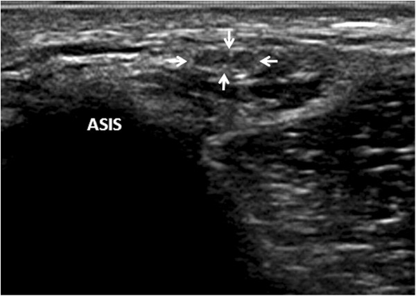
UltrasoundTransverse ultrasound near the anterior superior iliac spine showing a neuroma of the lateral femoral cutaneous nerve, indicated by the arrow.
Source : Zhu et al., BMC musculoskeletal disorders, 2012, figure 5 · CC BY
In this chapter: why the diagnosis stays clinical, what electrophysiology can and cannot do, the place of ultrasound and MR neurography, and the only investigation that really decides, the anaesthetic block.
The position of the review literature is consistent: the diagnosis is clinical. The 2023 German review puts it thus: it rests mainly on the typical symptoms combined with a positive response to an injection; in atypical cases, electrophysiological testing, neurosonography and MRI can help.2 The 2025 narrative review is blunter still: there is no obviously superior diagnostic strategy.4
The reference chapter of the Handbook of Clinical Neurology is explicit: sensory conduction studies and somatosensory evoked potentials can support the diagnosis, but both have technical limitations, with low sensitivity and specificity.3
Seror's work quantifies that limit. Across 21 patients and 21 controls, evoked potentials obtained by stimulation at the anterior superior iliac spine had a sensitivity of 5 % for a specificity of 95 %: in other words, that montage has no diagnostic value. On stimulation of the distal third of the lateral thigh, sensitivity rose to 52 % for a specificity of 76 %. The author's recommendation is restrictive: reserve that latter montage for obese patients in whom sensory conduction cannot be measured.14
More recent work is more encouraging, provided a particular technique is used. By recording potentials with distal electrodes about 30 cm below the anterior superior iliac spine, and stimulating both 10 cm below the spine and at the spine itself, Tataroglu and colleagues found abnormalities in all 34 patients studied, compared with 38 healthy controls. Slowing of sensory conduction at the inguinal canal and loss of response were the commonest abnormalities, 44.7 % and 31.6 % respectively.15
The practical lesson is not that electrophysiology is useless, but that its yield depends entirely on the montage used, and that a negative test done with an insensitive technique does not refute the diagnosis.
The 3 tesla MR neurography has been the subject of a study of 11 patients and 28 controls, with two independent blinded readers: sensitivity and negative predictive value of at least 71 %, specificity and positive predictive value of at least 94 %, and diagnostic accuracy of at least 90 % for both readers. Agreement between readers was moderate for signal abnormalities and poor for the diagnosis of neuroma.16 It is a good test, but in a sample of 11 patients and with imperfect inter-reader agreement.
Ultrasound has a double value, diagnostic and interventional: it allows a nerve that we have seen can sit anywhere across a 12 cm span to be located, and an injection to be guided.11
Skin biopsy with measurement of intraepidermal nerve fibre density The skin biopsy with measurement of intraepidermal nerve fibre density also reduced in the asymptomatic thigh in two of them, while it was normal in the distal leg in four.17 That subclinical contralateral involvement is a useful reminder that susceptibility can be bilateral even when the complaint is not.
What the physiotherapist should make of it
None of these investigations is a prerequisite for management. A physiotherapist who receives a patient with a typical picture and no red flag does not have to wait for laboratory confirmation to start: the first useful intervention is identifying and removing the compressive factor, and it is risk-free. An atypical, bilateral or resistant picture, on the other hand, justifies seeking an opinion, and it is then useful to know that negative electrophysiology does not close the debate.
In this chapter: what the secondary forms cover, their real frequency in a surgical series, the pictures that should alert you, and the published cases where a meralgia was the first sign of a cancer.
Common meralgia is benign. That is precisely what makes it necessary to know how to recognise the one that is not, because the comfort of an easy diagnosis is the best ally of delayed diagnosis.
The systematic review of de Ruiter and colleagues provides the framework. Of 66 publications on unusual causes, 37 concerned traumatic injury to the nerve and 29 compression by a tumour or a mass. In their own surgical series of 187 cases operated on between April 2014 and September 2022, they count 14 traumatic lesions and 4 cases linked to a mass. Their conclusion fits into one sentence: it is important to consider a traumatic cause or compression by a mass in patients presenting with meralgia.26
Four cases out of 187, that is about 2 %, is few. It is also enough for the question to arise at every consultation, because the cost of a missed diagnosis bears no relation to that of a question asked.
The older literature had already raised the problem: a 1968 article in the American Journal of Surgery is soberly entitled “meralgia paresthetica, a clue to retroperitoneal malignant tumour”.27
A case published in 1997 in the Archives of Physical Medicine and Rehabilitation is more telling still, and it concerns our specialty directly. A 60-year-old man presents with the clinical picture of meralgia paraesthetica. Investigations find a malignant secondary deposit in the iliac crest, a metastasis from a lung adenocarcinoma. The authors conclude that this neuropathy, most often linked to a benign lesion, can sometimes be the presenting symptom of a malignancy.28
Not all secondary forms are tumoral. A case reported in 2026 describes a 30-year-old man, healthy until then, developing bilateral burning of the anterolateral thighs in the wake of a self-limiting febrile illness, with an associated right piriformis syndrome. Conduction studies confirmed bilateral involvement of the lateral femoral cutaneous nerve, and pelvic MRI oedema and hypertrophy of the right piriformis. The authors stress the need to consider a post-infectious origin.50
Red flags: when a meralgia is not one, or is not alone
In this chapter: the only sourced figure for spontaneous resolution, the speed of recovery of the postoperative forms, the share of patients who will remain symptomatic, and why that prognosis calls for therapeutic caution.
The rate of spontaneous resolution of meralgia is a figure very often quoted and rarely referenced. There is nevertheless a precise source for it, and only one: the Cochrane review of Khalil, Nicotra and Rakowicz. Having found no randomised or quasi-randomised trial after three search cycles in 2008, 2010 and 2012, the authors fell back on good-quality observational studies. Only one natural history study met the criteria, and it reports spontaneous improvement in 20 patients out of 29, that is 69 %.29
That is the figure to quote, and its fragility must be quoted with it: 29 patients, a single study. Any claim along the lines of “meralgia resolves on its own in 85 to 90 % of cases” comes from repetition and not from measurement.
The postoperative forms, for their part, are better documented and the prognosis there is frankly good. In the prospective series of 252 spinal operations, recovery took 10.5 days on average, 32 of the 60 affected patients (53 %) recovered in the first week, and all of them in under two months. The authors describe that complication as common but benign.21 In children operated on for scoliosis, the symptoms resolved on average before the six-week visit, with a range of 2 to 24 weeks.23
That leaves the refractory share. The chapter of the Handbook of Clinical Neurology puts it bluntly: most cases are self-limiting, but a small proportion of patients keep refractory and disabling symptoms.3 The Nouraei series gives an order of magnitude on the surgical side: across 45 patients, 25 were successfully treated conservatively and 20 needed an operation; in those operated on, the actuarial rates of survival free of further surgery were 91 % at two years and 78 % at five years.12
Seven patients in ten improve without anything being done. It is the strongest argument in favour of patient management, and the biggest trap for anyone who would attribute to their technique what time would have done alone.
Key points
In this chapter: the first intervention, which is not a technique; the only randomised physiotherapy trial that exists and a critical reading of it; the real level of evidence for neural mobilisation, which is not the one people think; and the table of modalities ranked by the evidence they have.
You have to start by measuring the ground. Querying the Europe PMC database on 15 August 2026, there are 1,095 publications indexed on meralgia paraesthetica, of which 18 carry the randomised controlled trial label. Reading those 18 records one by one, about half turn out not to concern meralgia but work in which the term appears marginally. Seven randomised trials genuinely devoted to this condition remain, of which only one tests a manual physiotherapy technique. That gap is a gap in indexing as much as a gap in the literature, but it is consistent with the finding of the reviews: no level 1 evidence exists for idiopathic meralgia.30
It consists of finding what is pressing and making it stop. Loosening or moving the belt, giving up rigid-waisted trousers, redistributing a load carried at the waist, changing prolonged standing, starting weight loss where indicated. The physical medicine review puts it directly: the complaints typically disappear after weight loss, abdominal muscle strengthening, or removal of the underlying cause.11
That intervention rests on no randomised trial, and probably never will: nobody will randomise patients between “we take off the belt that is compressing” and “we leave it on”. Its formal level of evidence is therefore very low, and its clinical priority is nevertheless first. It is a case where the hierarchy of evidence and the hierarchy of action legitimately diverge.
El-Din Mahmoud, El Meligie and Yehia randomised 30 women with postpartum meralgia between a muscle energy technique added to conventional exercises and conventional exercises alone, at three weekly sessions of 30 to 40 minutes for four weeks.31
The results in favour of the treated group are clear on three outcomes:
And a result that is not there, which the authors report honestly: no difference between the groups on the pelvic compression test (p = 0.41).31
Reading this trial as a clinician
Three reservations, which do not cancel the result but bound its reach. The sample is 30 patients, fifteen per arm. The population is exclusively postpartum, so in a setting where the compressive cause regresses spontaneously with time, which works in favour of both groups. And the effect sizes, all above 1.7, are unusually large for a manual intervention: in small samples, that profile should prompt caution rather than enthusiasm. The most instructive detail remains the absence of a difference on the pelvic compression test, that is on the measure most specific to the compression itself: the treatment improved pain and conduction, without demonstrating that it had decompressed anything.
This is the point where the gap between common practice and the literature is widest, and it needs to be precise.
There is no randomised trial of neural mobilisation in meralgia paraesthetica. What is invoked to justify it is an extrapolation from other compressive neuropathies, and that extrapolation is fragile.
The systematic review with meta-analysis by Basson and colleagues, published in the Journal of Orthopaedic and Sports Physical Therapy, included 40 randomised trials, 17 of them at low risk of bias. Its results are mixed: benefit in chronic low back pain (disability, mean difference of 9.26 points on the Oswestry scale; pain, mean difference of 1.78 points) and in chronic neck-arm pain (1.89 points). But for most clinical outcomes in carpal tunnel syndrome, neural mobilisation was not effective (p greater than 0.11), even though favourable neurophysiological effects were observed, such as reduction of intraneural oedema. For the other conditions, the authors conclude that the effect remains uncertain.32
Carpal tunnel syndrome is the most relevant comparator for meralgia, since it is the other major entrapment neuropathy, and it is precisely there that neural mobilisation disappoints on clinical outcomes. See also, in the corpus, carpal tunnel syndrome.
A more recent systematic review, devoted to treatment of the nerve's mechanical interface , that is to joint and tissue techniques applied to the structures surrounding the nerve, reaches an unambiguous finding on our subject: the 11 studies included all concerned carpal tunnel syndrome.33 . None on meralgia.
Neural mobilisation is not contraindicated in meralgia; it is simply unevaluated. Saying so to the patient costs one sentence and is better than selling them a level of evidence that does not exist.
A three-arm Turkish randomised trial compared an ultrasound-guided nerve injection, ten sessions of transcutaneous electrical nerve stimulation and ten sessions of sham stimulation, in patients whose diagnosis was confirmed clinically and electrophysiologically; 54 of the 62 patients completed the study. The between-group differences favoured the injection on the painDETECT questionnaire and the Semmes-Weinstein monofilament test, at fifteen days and one month. On the other hand, no difference between groups on the visual analogue scale, quality of life and sleep quality. A decisive detail: the painDETECT score fell significantly in all three groups, including the sham group.34
A double-blind placebo-controlled randomised trial, published in 2026, tested high-intensity laser : 62 patients randomised, 54 analysed, twelve sessions over four weeks. At twelve weeks, the treated group did better than the sham group on pain (p = 0.006), on function measured with the Lower Extremity Functional Scale (p = 0.043) and on sensory conduction velocity (p = 0.027). The authors themselves qualify this: the neurophysiological changes were small and stayed within normal limits, which calls for cautious interpretation. Mild, transient adverse effects were commoner in the treated group (16.1 % against 0 %).35
Physiotherapy modalities, ranked by the level of evidence available
Ranked by the evidence, and not by clinical priority. The two do not coincide, and that is the message of this chapter.
Sources: Mogahed HG et al. (2026), American Journal of Physical Medicine and Rehabilitation, PMID 42307460; El-Din Mahmoud LS et al. (2023), Journal of Back and Musculoskeletal Rehabilitation, PMID 36617775; Onat SS et al. (2016), Pain Physician, PMID 27228536; Mondelli M et al. (2007), PMID 17661798; Kiliç S et al. (2020), Pain Physician, PMID 32517391; Basson A et al. (2017), JOSPT, PMID 28704626; Iogna Prat P et al. (2024), Musculoskeletal Science and Practice, PMID 38217928. Ranked by level of evidence, on GRADE logic: no modality reaches a moderate or high level in this condition.
The paradox of this table
The modality best supported by a trial is high-intensity laser, and it is not what you should do first. The modality most useful in practice, removing the compressive factor, comes third because it will never be randomised. Ranking by evidence is not ranking by action : the evidence says what we know, not what should be done first. Faced with a patient whose belt is compressing the nerve, the order stays: remove the cause, explain the favourable course expected, and add a technique only if the picture persists.
Key points
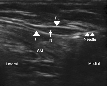
UltrasoundShort-axis ultrasound of the lateral femoral cutaneous nerve between the fascia lata and the fascia iliaca, with the puncture needle in place for an ultrasound-guided block, a technique used in meralgia paraesthetica.
Source : Kim et al., The Korean journal of pain, 2011, figure 2 · CC BY-NC
In this chapter: the accepted treatment ladder, what injections are really worth when compared with placebo, the unsettled debate between neurolysis and neurectomy, and the concrete referral criteria.
The treatment ladder is a matter of consensus in the reviews, and it is simple: conservative treatment first, injection next, surgery as a last resort, the surgery being either a neurolysis or a neurectomy.4
Three randomised trials and a meta-analysis allow precision, and the result is more nuanced than the reputation of injections suggests.
The 2024 meta-analysis of corticosteroid efficacy concludes to a significant benefit on complete pain relief and on the pain score at fifteen days (p = 0.02), but not at one month (p = 0.79). The authors' conclusion is explicitly bounded in time: injections play a role, above all in the short term.38
The methodologically most rigorous trial is also the most negative. Kloosterziel and colleagues randomised 20 patients double-blind between a nerve-stimulator-guided injection of methylprednisolone and lidocaine and an injection of saline. Result: the placebo group showed a significant reduction in pain (visual analogue scale from 6.8 to 4.3 at twelve weeks, p = 0.014), while the reduction in the corticosteroid group did not reach significance (from 7.4 to 4.8, p = 0.053), with no difference between the groups. The authors conclude that they found no objective evidence of benefit, while stressing the small sample.37
A double-blind randomised trial compared an ultrasound-guided injection of local anaesthetic alone with the same injection plus betamethasone, in 32 patients. No significant difference between the groups : the pain scores fell in both, and the improvement continued in the following weeks, reaching 2.47 in the corticosteroid group and 3.13 in the anaesthetic group at four weeks.36 In other words, in that trial, the corticosteroid adds nothing to the local anaesthetic.
A more recent trial compared hydrodissection with 5 % dextrose with hydrodissection with corticosteroid in 56 patients followed for six months. Dextrose did better at four and six months on pain and quality of life, with a better clinical response at six months, and no adverse effects against six in the corticosteroid group.39
The meta-analysis of Lu and colleagues pooled 25 articles and 670 patients: 78 treated by injection (12 %), 496 by neurolysis (74 %) and 96 by neurectomy (14 %). The rates of complete relief of pain differ markedly: 85 % after neurectomy (95 % CI 71 to 96), 63 % after neurolysis (56 to 71) and 22 % after injection (13 to 33), all comparisons being significant. The reoperation rate was 0 % after neurectomy, 12 % after neurolysis and 81 % after injection, and complications, which were comparable, ran from 0 to 5 %.40
A radiology meta-analysis comparing ultrasound-guided injection with surgery in 149 patients finds no significant difference : 85 % success (49 out of 57) after ultrasound-guided injection against 80 % (74 out of 92) after surgery. The authors specify that no comparative study and no randomised trial existed.41
The same treatments, two outcomes, two verdicts
On the left, cure or improvement. On the right, COMPLETE pain relief. The choice of outcome changes the ranking.
Sources: Khalil N, Nicotra A, Rakowicz W (2012), Cochrane Database of Systematic Reviews, PMID 23235604; Lu VM et al. (2021), Journal of Neurosurgery, PMID 33450741. The two columns do not measure the same thing and must not be compared line by line: “improvement” and “complete relief” are two very different thresholds.
Between neurolysis and neurectomy, the literature does not decide. A systematic review devoted to that question assessed each study using the American Academy of Neurology algorithm: none was a randomised trial, and all were level 4 evidence. The authors' conclusion: the data are insufficient to recommend one technique over the other.42 The 2023 review confirms that decompression and neurectomy have never been compared in a randomised design for idiopathic meralgia.30
What can be said to the patient therefore comes down to a trade-off and not to evidence. Neurectomy relieves more often and completely, with no reoperation, but it permanently sacrifices the nerve and leaves a permanent area of anaesthesia over the anterolateral thigh. Neurolysis preserves the nerve but gives complete relief less often, with 12 % reoperations. A pain medicine review presenting four patients treated conservatively or interventionally discusses precisely those respective advantages and drawbacks, and recalls that the first objective remains eliminating the underlying cause when it is known.43
| Situation | What to do | Support |
|---|---|---|
| Typical picture, compressive cause identified | Remove the factor, educate, review at 4 to 6 weeks. No referral at the outset. | 69 % spontaneous improvement29 |
| Immediate postoperative meralgia | Reassure, explain the expected timeline. Review at 8 weeks. | Complete recovery in all in under 2 months21 |
| Persistent diagnostic doubt | Opinion for a diagnostic anaesthetic block, with or without ultrasound. | A positive response to injection, the pivot of the diagnosis27 |
| Persistence beyond 3 months despite removal of the cause | Opinion for an ultrasound-guided injection. Announce a real but short benefit. | Significant effect at 15 days, not at 1 month38 |
| Failed injections, significant functional impact | Surgical opinion. Explain that neurolysis and neurectomy have not been separated. | All studies level 442 |
| Any red flag at all | Medical opinion without delay, without waiting for the review. | 29 publications of compression by a mass26 |
Table to be scrolled horizontally on a small screen.
Key points
In this chapter: why pregnancy is the risk factor with the highest odds ratio, what is particular about postpartum meralgia, and the paediatric picture, little known, bilateral and long misdiagnosed.
Pregnancy carries the highest odds ratio of all the factors measured, 12.0, even though its very wide confidence interval (1.2 to 118.0) means the magnitude must not be over-interpreted.1 The mechanism is twofold: an increase in abdominal volume and postural changes, in particular the increased lumbar lordosis and the relative hip extension in late pregnancy, two elements that tension the inguinal ligament.
An obstetric review devoted jointly to carpal tunnel syndrome and meralgia during pregnancy recalls that these two common neuropathies cause significant discomfort but can be safely diagnosed and treated during pregnancy.44 The association of the two conditions in the same patient is not fortuitous: it is exactly the association measured by van Slobbe, which supports the hypothesis of a general susceptibility to entrapment syndromes.1
Postpartum meralgia has a practical peculiarity: it is the only population in which a physiotherapy technique has been evaluated by a randomised trial.31 A case report also describes a meralgia occurring after caesarean section under spinal anaesthesia, in a 29-year-old woman, with a normal neurological examination and lumbar MRI, the diagnosis confirmed by electromyography and nerve conduction, and the symptoms disappearing within a month on conservative treatment.49
Meralgia in children exists and differs markedly from that of the adult. An old but precise series in the Journal of Bone and Joint Surgery followed 20 children and adolescents, that is 30 lesions since ten had bilateral involvement. The initial symptom was severe pain markedly limiting activities. The mean age at onset was ten years, with extremes of one to seventeen years. The most instructive point: the diagnosis was missed initially in ten of the twenty patients, which led to multiple unnecessary investigations, and the mean duration of symptoms before the first specialist consultation was twenty-four months.45
A contemporary series confirms the profile: 24 patients, mean age 12.7 years, 92 % girls, 63 % bilateral involvement, and 38 % referred with another diagnosis. Mean body mass index was 20.96, that is normal, which clearly distinguishes this population from the obese adult. All patients were first treated with physiotherapy, anti-inflammatories and a local injection for diagnostic purposes; the injection relieved every patient immediately, for a mean duration of only eleven days, and 71 % were eventually operated on.46
What paediatrics changes in the reasoning
In children, bilaterality is common and not alarming (63 % in the recent series), whereas it is a red flag in the adult. Body mass index is normal, so the absence of obesity does not rule out the diagnosis. And the rate of recourse to surgery is high, which suggests that the paediatric form responds less well to conservative treatment than the adult form. A physiotherapist seeing an adolescent with anterolateral thigh burning for months should therefore think of it early: the older series shows twenty-four months of mean diagnostic delay.45
Red flags of this chapter
In this chapter: four real cases, all indexed and identifiable, chosen because each corrects a received idea. No fictional case, no reconstructed vignette.
The cases that follow are genuine published reports, cited with their identifier. They illustrate, they do not demonstrate: the case report is the lowest level of evidence, and in a condition that improves spontaneously in 69 % of cases, a success reported in one patient does not prove the effect of the treatment. They keep their value for recognising presentations and avoiding errors.
A 22-year-old patient, in the sixteenth week of pregnancy, attends with low back pain, bilateral anterolateral thigh paraesthesia and groin pain of one month's duration. Examination finds no motor deficit in the lower limbs and intact reflexes. The diagnosis is made on the clinical history and a battery of functional tests.
Treatment comprises six sessions over six weeks, combining active release techniques applied to the right sacroiliac complex and quadratus lumborum, active release and post-isometric relaxation techniques on the iliopsoas, and a home exercise programme of lumbopelvic mobility, stabilisation and relaxation. After six treatments, the patient reports complete resolution of the low back pain and of the left lower limb symptoms, and 90 % improvement on the right. At one year's follow-up, she is pain-free.47
What this case teaches: meralgia in pregnancy can be bilateral without being worrying, and the treatment that worked here did not target the nerve directly but the regional structures, sacroiliac, quadratus lumborum and iliopsoas. What it does not teach: at sixteen weeks of pregnancy and over six weeks, the spontaneous course is a major confounder.
Two cases published in 2026 concern a 61-year-old woman symptomatic for three weeks and a 48-year-old man symptomatic for two months. In both cases, electromyography confirmed involvement of the lateral femoral cutaneous nerve, and previous non-conservative treatments had failed.
The first received neurodynamic mobilisation in sliding and tensioning, oscillatory techniques and myofascial techniques. The second received a muscle energy technique, tissue, fascial and visceral techniques, a neurodynamic exercise and education. The first patient was asymptomatic at one year, the second at six months.48
What these cases teach: they are the only detailed descriptions of neurodynamic mobilisation applied to an electrically confirmed meralgia, in a literature that has no trial of it at all. What they do not teach: two patients, with no comparator, in a condition with high spontaneous resolution. The authors themselves claim only a potential role.
A 60-year-old man presents with the complete clinical picture of meralgia paraesthetica: pain, paraesthesia and sensory loss of the anterolateral thigh, with no motor deficit. Investigations reveal a malignant secondary deposit in the iliac crest, a metastasis from a lung adenocarcinoma.28
What this case teaches: the semiology of meralgia is perfectly compatible with a serious cause. The absence of a motor deficit, which confirms the nerve trunk lesion, says nothing about its cause. That is exactly why the absence of an identifiable compressive factor must remain an open question rather than a comfortable diagnosis of exclusion.
How to read a case series on this condition
Three of the four cases above report resolution after treatment. In a condition in which 69 % of patients improve with nothing,29 that is exactly the success rate you would expect without treatment. A case report cannot distinguish the effect of care from that of time, and honesty consists of saying so rather than counting successes.
In this chapter: the four-step consultation framework, what to tell the patient, and the errors not to make.
Five errors not to make
No. The lateral femoral cutaneous nerve is exclusively sensory : it innervates no muscle. Genuine weakness of the quadriceps or iliopsoas points to a radicular lesion or to the femoral nerve, not to a meralgia.3 Some patients describe a sense of the leg giving way: it is linked to the pain and the sensory change, not to a strength deficit, and the examination shows it.
It depends on the form. Meralgias occurring after surgery in the prone position recover fast: 10.5 days on average, 53 % in the first week and all of them in under two months.21 For the common forms, the only natural history study available reports spontaneous improvement in 69 % of patients, without specifying a uniform timeline.29 A minority keep refractory symptoms.3
Not for a typical picture. The diagnosis is clinical, and a lumbar MRI in a patient of that age will almost always find degenerative changes unrelated to the complaint, which risks a false diagnosis. Imaging is justified in the face of diagnostic doubt, a red flag or a motor deficit.24
Yes, but that changes the reasoning depending on age. In adults, bilateral involvement from the outset, outside pregnancy or marked obesity, calls for a pelvic, retroperitoneal or systemic cause to be sought.26 In children, on the contrary, bilaterality is usual: 63 % in a recent series and 50 % in an older one.4546
It is a serious lead, without being guaranteed. Obesity doubles the risk,18 and the reviews report that the complaints typically disappear after weight loss or removal of the cause.11 But no trial has tested weight loss as an intervention in this condition: the evidence is indirect, and it must be presented as such.
In the short term, yes; as a durable solution, that is more debatable. The meta-analysis shows a benefit at fifteen days that is not maintained at one month.38 On the outcome of complete relief, injection reaches only 22 %, with 81 % reoperations.40 And in the most rigorous placebo-controlled trial, the placebo group improved significantly, the corticosteroid group did not.37 Their diagnostic value, on the other hand, remains solid.2
There are no specific data on return to sport in meralgia, and that should be said rather than inventing a protocol. Mechanical reasoning still applies: activities that keep the hip in prolonged extension or that require a tight belt are the most likely to keep the symptoms going. Compression by rucksack hip belts, weapon belts and harnesses is documented in military medicine.20
Yes, especially if the compressive factor reappears. In patients operated on with decompression, the rates of survival free of further surgery were 91 % at two years and 78 % at five years, which means that about one patient in five needed a further procedure by five years.12
Femoral neuralgia is a radicular lesion that comes with motor signs and can change the knee reflex, and whose pain reaches the medial side of the knee in 80 % of patients.51 Meralgia is a nerve trunk lesion and purely sensory, with no motor or reflex sign, and its territory does not run below the knee. The full detail is in the differential table of this article, and the radicular condition itself is covered in femoral neuralgia.
The 51 references below were verified against two independent databases: the complete record, the untruncated author list and the pagination come from the PubMed E-utilities API, and the title, journal, year and authors were cross-checked on CrossRef from the DOI. Three references published in Pain Physician carry no DOI, that journal not depositing them: they are cited by their PMID alone.
Behind this article
An author who explains, a reviewer who checks.
How we write and check our content

Physiotherapist · co-founder of Physio Learning
Marked for life by his first four-hour lecture without a single image, he took a master’s in instructional design so that it would never happen to anyone again. He hunts down publication bias and unreadable slides with the same intransigence.

Head of scientific content
Physiotherapist specialising in neuro-musculoskeletal practice and holder of a master’s in public health. He checks the methodological rigour of every article: primary sources, levels of evidence, no exceptions.
