

















In brief
Degenerative lumbar spinal stenosis (LSS) is an anatomical narrowing of the spinal canal, the lateral recesses or the foramina, linked to facet osteoarthritis, hypertrophy of the ligamentum flavum and disc degeneration. Its cardinal symptom is neurogenic claudication: pain, heaviness or paraesthesia in the lower limbs triggered by walking or by prolonged standing, and relieved by lumbar flexion or by sitting (the « shopping trolley sign »); it affects mainly people over 65. First-line management is conservative and multimodal: education, supervised exercise and adjunct manual therapy. Its clinical prevalence in the general population is about 11 %.
A clinical synthesis based on the most recent meta-analyses and international consensus statements, Katz JAMA 2022, NASS Kreiner 2013, Ammendolia 2022 BMJ Open, Comer 2024 SR/ICA, Delphi Tomkins-Lane 2016, N-CLASS Genevay 2018, SPORT 8-year Lurie 2015.
Clinical synthesis
- Degenerative lumbar spinal stenosis (LSS) is an anatomical narrowing of the spinal canal, the lateral recesses or the foramina, linked to facet osteoarthritis, hypertrophy of the ligamentum flavum and disc degeneration (Katz JAMA 2022).
- The clinical prevalence in the general population is about 11 % (CI 4-18 %); it rises to 25-29 % in primary and secondary care (Jensen 2020, a meta-analysis of 41 studies).
- The cardinal symptom is neurogenic claudication: pain, heaviness or paraesthesia in the lower limbs on walking or on prolonged standing, relieved by lumbar flexion or by sitting (the « shopping trolley sign »).
- The natural course is rather favourable : most patients stay stable or improve. Only 10-25 % worsen clinically (Ammendolia 2022, Lurie BMJ 2016).
- The diagnosis is above all clinical: the Cook 2011 cluster (5 items), the ISSLS Delphi consensus of Tomkins-Lane 2016 and the N-CLASS criteria of Genevay 2018 structure the identification.
- The correlation between radiological severity on MRI and the symptoms is weak ; about 30 % of asymptomatic people over 60 have a visible stenosis (Brinjikji 2015).
- The major differential diagnosis remains vascular claudication ; the ankle-brachial pressure index rules it out.
- First-line management is conservative and multimodal : education, supervised exercise, adjunct manual therapy (Ammendolia 2022, NASS Kreiner 2013).
- No type of exercise has been shown superior; supervision and lumbar flexion are the commonest components of the programmes, while those that appear more often in successful interventions are stretching, trunk strengthening, fitness work above all on a bicycle, and psychologically informed approaches (Comer 2024 SR/ICA Clinical Rehabilitation, 13 trials).
- The passive modalities (ultrasound, traction, TENS, heat) have no demonstrated effectiveness in addition to exercise (Ammendolia 2022, a BMJ Open systematic review).
- The LSS is the leading cause of spinal surgery after 65 (Deyo 2010 JAMA); surgery rates rose sharply between 2002 and 2007 without a proportionate improvement in results.
- The RCTs (Delitto 2015 Ann Intern Med, SPORT Lurie 2015) show that structured physiotherapy reaches results comparable to surgical decompression at 2 years in many patients.
- In older people, the assessment must include frailty (Fried 2001) and sarcopenia (EWGSOP2 Cruz-Jentoft 2019), major prognostic factors.
- The red flags (cauda equina syndrome, progressive deficit, unexplained weight loss) require urgent medical referral (Finucane 2020 IFOMPT).
- Self-management through therapeutic education, keeping up a regular exercise programme and managing kinesiophobia are the pillars of preventing recurrence.
- Measure outcomes with validated PROMs (ZCQ Stucki 1996, ODI, Tampa Scale) is essential for documenting progress and guiding shared decision-making.
Contents
- What are the fundamentals to know about degenerative lumbar spinal stenosis?
- How is degenerative lumbar spinal stenosis assessed and diagnosed with certainty?
- Which treatment strategies are the most effective for degenerative lumbar spinal stenosis?
- How is durable recovery ensured and recurrence prevented?
- A frail older person and LSS: when to refer for surgery?
- What do concrete clinical cases teach us about degenerative lumbar spinal stenosis?
- How do you apply these recommendations concretely in your practice?
What are the fundamentals to know about degenerative lumbar spinal stenosis?
How is this condition defined, who is affected and what are the risk factors?
The degenerative lumbar spinal stenosis (LSS) is an anatomical condition characterised by a narrowing of the central spinal canal, the lateral recesses or the intervertebral foramina, leading to potential compression of the cauda equina and of the emerging lumbar nerve roots.¹ That narrowing is the consequence of a progressive age-related degenerative process.¹,² LSS is today the leading indication for spinal surgery in adults over 65 in the United States.³
The prevalence of this condition rises significantly with age. The reference meta-analysis of Jensen et al. (2020), covering 41 studies and 55 population samples, clearly separates the radiological prevalence (visible on imaging) from the clinical prevalence (symptomatic):⁴
- Clinical prevalence in the general population: about 11 % (confidence interval 4–18 %)
- Prevalence in primary care: 25 % (CI 19–32 %)
- Prevalence in secondary care: 29 % (CI 22–36 %)
- Prevalence in mixed primary and secondary consultations: 39 %
This distinction is crucial: a great many people show signs of narrowing on imaging with no clinical symptom at all.⁴,⁵ The diagnosis can therefore never rest on MRI alone.
The cardinal and most distinctive symptom of LSS is neurogenic claudication.¹,⁶ It is pain, numbness, heaviness or weakness radiating into the buttocks and/or the lower limbs, typically triggered by walking or prolonged standing (positions of lumbar extension) and relieved by rest, by lumbar flexion or by sitting. That characteristic postural relief is illustrated by the « shopping trolley sign »: the patient leans forward on a supermarket trolley in order to walk without pain.⁶,⁷
📊 Clinical prevalence of LSS by care setting (Jensen 2020, meta-analysis)
A meta-analysis of 41 studies, 55 samples, showing the gradient from the general population to specialist care
Source: Jensen RK, Jensen TS, Koes B, Hartvigsen J. Eur Spine J. 2020;29(9):2143-2163. PMID 32095908.
Several risk factors have been identified:
- Advanced age is the main non-modifiable risk factor, because of the accumulation of degenerative changes.¹,⁶
- Some constitutional factors, such as a congenitally narrow spinal canal, can predispose someone to develop a symptomatic stenosis earlier in life.⁶
- The occupations with a high mechanical load and certain genetic variations (collagen, metalloproteases) are also thought to contribute.¹,⁸
- The association with the metabolic syndrome (obesity, diabetes, hypertension) remains debated and is not confirmed by a quality meta-analysis; the LOHAS cohort data suggest on the contrary that LSS sometimes precedes the appearance of the metabolic syndrome.⁹
What happens in the body and how does degenerative lumbar spinal stenosis progress naturally?
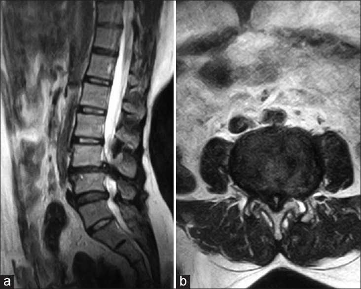
MRISagittal and axial lumbar MRI: L3-L4 disc degeneration with bulging, facet joint hypertrophy and ligamentum flavum thickening, causing lumbar canal stenosis.
Source : Shrinuvasan et al., Journal of Neurosciences in Rural Practice, 2016, figure 4 · CC BY-NC-SA
The pathophysiology of LSS is a multifactorial process, often described as a « degenerative cascade » affecting the whole vertebral motion segment.⁶ The three major contributors to the reduction in canal space are:
- Hypertrophy of the facet joints : osteoarthritis of the zygapophyseal joints leads to bone growth (osteophytes) that encroaches on the canal and the lateral recesses.¹,⁶
- Bulging or protrusion of the intervertebral disc : loss of height and dehydration of the disc make it bulge into the canal.⁸
- Thickening and buckling of the ligamentum flavum : this ligament, at the back of the canal, loses elasticity with age and thickens, further reducing the space available for the neural structures.¹,⁶
🔬 The degenerative cascade leading to LSS
Three main mechanisms converge on canal narrowing and neurogenic claudication
Sources: Katz JN et al. JAMA. 2022;327(17):1688-1699. PMID 35503342 · Kobayashi S. World J Orthop. 2014;5(2):134-145. PMID 24829876.
The combination of these phenomena compresses the neural elements and their blood supply. That mechanical compression and the venous ischaemia it produces are considered the main mechanisms behind the symptoms of neurogenic claudication.⁸,¹⁰ Lumbar flexion increases the canal diameter by 2 to 4 mm, which explains the characteristic postural relief.¹⁰
🚶 Contrary to received wisdom, the natural history of LSS is often benign. The management review by Lurie & Tomkins-Lane (BMJ 2016) and the Ammendolia 2022 systematic review (BMJ Open), an update of their 2013 Cochrane review (23 new trials identified), converge: over 1 to 5 years of follow-up, about a third of patients improve, half stay stable, and only 10 to 25 % experience clinically significant worsening.²,⁹ The appearance of severe, progressive neurological deficits (such as a cauda equina syndrome) remains a rare event in the course of this chronic condition.²,¹⁰
🧘 This knowledge of the natural history is fundamental for physiotherapy management: it allows the clinician to reassure patients and to justify an initial conservative approach, centred on managing symptoms and improving function rather than on correcting a radiological image.²,¹¹
Key points
- LSS is an anatomical narrowing of the spinal canal, but the presence of symptoms (neurogenic claudication) is necessary for a clinical diagnosis.
- Clinical prevalence in the general population is 11 % (Jensen 2020 meta-analysis), rising to 25-29 % in primary and secondary care.
- The neurogenic claudication relieved by lumbar flexion is the cardinal symptom.
- The natural history is often favourable or stable : only 10-25 % of patients worsen clinically, which supports conservative management first line.
Chapter 1 bibliography
- Katz JN, Zimmerman ZE, Mass H, Makhni MC. Diagnosis and Management of Lumbar Spinal Stenosis: A Review. JAMA. 2022;327(17):1688-1699. PMID 35503342. doi:10.1001/jama.2022.5921.
- Lurie J, Tomkins-Lane C. Management of lumbar spinal stenosis. BMJ. 2016;352:h6234. PMID 26727925.
- Deyo RA, Mirza SK, Martin BI, Kreuter W, Goodman DC, Jarvik JG. Trends, major medical complications, and charges associated with surgery for lumbar spinal stenosis in older adults. JAMA. 2010;303(13):1259-1265. PMID 20371784.
- Jensen RK, Jensen TS, Koes B, Hartvigsen J. Prevalence of lumbar spinal stenosis in general and clinical populations: a systematic review and meta-analysis. Eur Spine J. 2020;29(9):2143-2163. PMID 32095908.
- Brinjikji W, Luetmer PH, Comstock B, et al. Systematic literature review of imaging features of spinal degeneration in asymptomatic populations. AJNR Am J Neuroradiol. 2015;36(4):811-816. PMID 25430861.
- Katz JN, Harris MB. Lumbar Spinal Stenosis. N Engl J Med. 2008;358(8):818-825. PMID 18287604.
- Tomkins-Lane C, Melloh M, Lurie J, et al. ISSLS Prize Winner: Consensus on the Clinical Diagnosis of Lumbar Spinal Stenosis: Results of an International Delphi Study. Spine (Phila Pa 1976). 2016;41(15):1239-1246. PMID 26839989.
- Genevay S, Atlas SJ. Lumbar spinal stenosis. Best Pract Res Clin Rheumatol. 2010;24(2):253-265. PMID 20227646.
- Ammendolia C, Hofkirchner C, Plener J, et al. Non-operative treatment for lumbar spinal stenosis with neurogenic claudication: an updated systematic review. BMJ Open. 2022;12(1):e057724. PMID 35046008.
- Kobayashi S. Pathophysiology, diagnosis and treatment of intermittent claudication in patients with lumbar canal stenosis. World J Orthop. 2014;5(2):134-145. PMID 24829876.
- Zaina F, Tomkins-Lane C, Carragee E, Negrini S. Surgical versus non-surgical treatment for lumbar spinal stenosis. Cochrane Database Syst Rev. 2016;(1):CD010264. PMID 26824399.
How is degenerative lumbar spinal stenosis assessed and diagnosed with certainty?
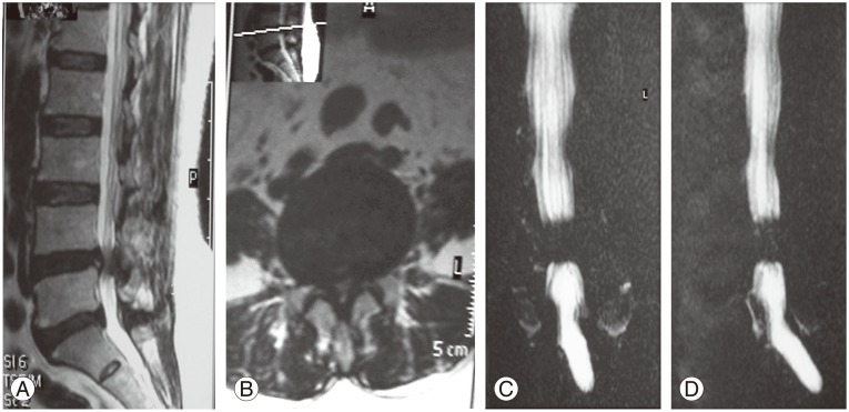
MRISagittal and axial lumbar MRI: severe canal stenosis at L4-L5 due to disc protrusion and ligamentum flavum hypertrophy, confirmed by magnetic resonance myelography.
Source : Al-Tameemi et al., Asian Spine Journal, 2017, figure 1 · CC BY-NC
The diagnosis of LSS is a process that is essentially clinical, in which imaging confirms the hypotheses formed during the history and the physical examination.¹ Diagnostic certainty rests on the coherent convergence between the reported symptoms, the observed clinical signs and the radiological data. 🧐
Which questions should you ask to understand the patient and their history?
The history is the cornerstone. The aim is to bring out the most pathognomonic symptom: neurogenic claudication.¹,² The international ISSLS Delphi consensus of Tomkins-Lane et al. (2016, ISSLS Prize Winner) identified the key questions at 80 % diagnostic certainty :³
- Do the symptoms appear on walking or on prolonged standing? 🚶
- Are they relieved by sitting or by leaning forward? (the « shopping trolley sign »)
- Are they bilateral? (often, but asymmetric)
- Is the leg pain worse than the back pain?
- Is the patient over 50 to 60?
- Is there relief on lumbar flexion?
The Cook et al. (2011) diagnostic cluster, validated on 1,448 consecutive patients with low back pain with or without leg pain, offers a set of 5 items which, combined, significantly raise the probability of LSS:⁴
| Item of the Cook 2011 cluster | Meaning |
|---|---|
| Bilateral symptoms | Bilateral compression of the central canal |
| Leg pain > back pain | A neural rather than a purely lumbar origin |
| Pain on walking or standing | A position of lumbar extension |
| Relief on sitting | Lumbar flexion increases the canal diameter |
| Age > 48 years | Consistent with the degenerative age |
The more items are present, the stronger the diagnostic probability (a rising likelihood ratio).⁴
Which clinical tests should be done and which other conditions must be ruled out?
The clinical examination aims to reproduce or relieve the symptoms and to rule out other diagnoses. The systematic review by de Schepper et al. (Spine 2013) confirms that most individual tests have low to moderate diagnostic accuracy.⁵ It is their grouping into clusters that significantly raises diagnostic performance.⁴
The most relevant tests:
- Two-stage treadmill stoop test (Fritz 1997) : walking on a level then an inclined treadmill (which induces lumbar flexion). A longer walking time on the inclined treadmill is positive for neurogenic claudication. The original study covered 45 subjects (26 with stenosis, 19 without).⁶
- Lumbar extension test (30 to 60 s) : reproduction of radicular symptoms in a position of extension.⁵
- Neurological examination : assessment of the tendon reflexes, motor strength (manual testing) and sensation in the L1-S2 dermatomes. The deficits are often subtle or absent at rest.⁵
- Neurodynamic tests (SLR, slump): useful for distinguishing a focal radicular lesion (disc herniation) from a more diffuse neurogenic claudication.
The main differential diagnosis to rule out is vascular claudication (peripheral arterial disease of the lower limbs). Vascular pain is generally cramping, relieved by simply stopping walking, with no need to sit or bend forward, and can be assessed objectively by measuring the ankle-brachial pressure index.⁷ An index < 0.9 favours peripheral arterial disease.
⚖️ Neurogenic versus vascular claudication, the differential diagnosis
Discriminating criteria to ask about systematically
Sources: Katz JN et al. JAMA. 2022;327(17):1688-1699 · Lurie J, Tomkins-Lane C. BMJ. 2016;352:h6234. Measuring the ankle-brachial pressure index objectively confirms peripheral arterial disease if it is < 0.9.
Other differential diagnoses to consider: peripheral neuropathy (diabetic, alcoholic), hip osteoarthritis (pain on weight-bearing relieved by rest), a radicular syndrome of discal origin (typically unilateral L5 or S1 pain), rheumatoid arthritis, fibromyalgia.
🚩 Red flags specific to the lumbar spine (Finucane 2020 IFOMPT)
- Cauda equina syndrome : saddle anaesthesia, sphincter disturbance (urinary retention or incontinence) → a neurosurgical emergency
- Progressive or bilateral motor deficit → urgent MRI
- Unexplained weight loss + a history of cancer + non-mechanical night pain → a cancer work-up
- Fever + low back pain + immunosuppression or intravenous drug use → suspected spondylodiscitis or epidural abscess
- High-energy trauma or severe osteoporosis + sudden pain → suspected vertebral fracture
- Progressive low back pain in someone < 20 or > 55 with no obvious mechanical cause → systematic reassessment
⚠️ Any red flag requires prompt medical referral (general practitioner, emergency department, rheumatologist or neurosurgeon) BEFORE any physiotherapy management.
Magnetic resonance imaging (MRI) is the reference investigation for visualising the anatomical stenosis and confirming compression of the neural structures.¹ It is crucial to recall, however, that the correlation between the degree of stenosis on MRI and the intensity of the symptoms is often weak.⁸ The systematic review by Brinjikji et al. (AJNR 2015), covering 33 articles and more than 3,000 asymptomatic subjects, shows that the prevalence of « moderate to severe stenosis » on MRI in subjects with no symptoms reaches:⁸
- 20 % at 40
- ≈ 30 % at 60
- > 50 % at 80
The diagnosis must therefore NEVER rest on MRI alone, but on the correlation between imaging and a compatible clinical picture.¹,⁸
Should patients be classified, and what are the benefits?
Yes, classifying patients is a step increasingly recommended for refining management and improving clinical research.⁹ Classification makes it possible to go beyond a binary diagnosis (present or absent) and describe severity and functional impact. ✅
The main tools:
- Zurich Claudication Questionnaire (ZCQ) / Swiss Spinal Stenosis Questionnaire (SSSQ) of Stucki et al. (1996): 18 items, 3 subscales (symptom severity, physical function, satisfaction after treatment). Its measurement properties were established in a cohort of patients who were candidates for surgical decompression: test-retest reliability from 0.82 to 0.96, internal consistency from 0.64 to 0.92 and high responsiveness. The authors present it as a specific instrument intended to complete the generic measures, not to replace them.¹⁰
- N-CLASS criteria of Genevay et al. (2018) : a clinical classification system for neurogenic claudication caused by LSS, developed by international consensus. Seven weighted items; a score ≥ 7 indicates a very probable LSS.¹¹
- Oswestry Disability Index (ODI) : a generic PROM for low back pain, responsive to change; an MCID of about 10 points is generally used.
- Roland-Morris Disability Questionnaire (RMDQ) : 24 dichotomous items, an alternative to the ODI.
Benefits of classification:
- Standardising the assessment and allowing objective follow-up of progress.
- Stratifying patients by severity to guide the treatment decision (conservative versus surgical referral).
- Facilitating clinical research by grouping patients into homogeneous subgroups.
Criticism and controversy
The greatest challenge remains the clinical-radiological paradox.⁸ A radiologist can describe « severe stenosis » on MRI, but if the patient has no characteristic neurogenic claudication, the LSS is not symptomatic. That dissonance feeds the risk of over-diagnosis and of inappropriate treatment (surgical in particular) based on imaging alone.¹,⁸ ⚠️
There is also no single « gold standard » for the diagnosis. The absence of a clinical test with high sensitivity AND specificity obliges clinicians to rely on a bundle of clues, which introduces a degree of subjectivity.⁵ Research is moving towards clinical prediction models (Cook 2011), international Delphi consensus statements (Tomkins-Lane 2016) and operationalised criteria (N-CLASS Genevay 2018), but synthesising the information remains an act of clinical reasoning.³,⁴,¹¹
Key points
- The diagnosis of LSS is above all clinical, based on identifying neurogenic claudication (leg pain relieved by flexion and by sitting).
- MRI is a tool of confirmation and not of screening: the imaging-symptom correlation is weak (Brinjikji 2015: 30 % of asymptomatic 60-year-olds have moderate to severe stenosis).
- No clinical test is perfect in isolation. Using clusters (Cook 2011), operationalised criteria (N-CLASS Genevay 2018) and the ISSLS Delphi consensus (Tomkins-Lane 2016) raises diagnostic certainty.
- The main differential diagnosis to rule out is vascular claudication : an ankle-brachial index < 0.9 favours peripheral arterial disease.
- Classification (ZCQ Stucki 1996) makes it possible to quantify the severity, guide the treatment and standardise research.
Chapter 2 bibliography
- Katz JN, Zimmerman ZE, Mass H, Makhni MC. Diagnosis and Management of Lumbar Spinal Stenosis: A Review. JAMA. 2022;327(17):1688-1699. PMID 35503342.
- Genevay S, Atlas SJ. Lumbar spinal stenosis. Best Pract Res Clin Rheumatol. 2010;24(2):253-265. PMID 20227646.
- Tomkins-Lane C, Melloh M, Lurie J, et al. ISSLS Prize Winner: Consensus on the Clinical Diagnosis of Lumbar Spinal Stenosis: Results of an International Delphi Study. Spine (Phila Pa 1976). 2016;41(15):1239-1246. PMID 26839989.
- Cook C, Brown C, Michael K, et al. The clinical value of a cluster of patient history and observational findings as a diagnostic support tool for lumbar spine stenosis. Physiother Res Int. 2011;16(3):170-178. PMID 21077266.
- de Schepper EIT, Overdevest GM, Suri P, et al. Diagnosis of lumbar spinal stenosis: an updated systematic review of the accuracy of diagnostic tests. Spine (Phila Pa 1976). 2013;38(8):E469-E481. PMID 23385136.
- Fritz JM, Erhard RE, Delitto A, Welch WC, Nowakowski PE. Preliminary results of the use of a two-stage treadmill test as a clinical diagnostic tool in the differential diagnosis of lumbar spinal stenosis. J Spinal Disord. 1997;10(5):410-416. PMID 9355058.
- Lurie J, Tomkins-Lane C. Management of lumbar spinal stenosis. BMJ. 2016;352:h6234. PMID 26727925.
- Brinjikji W, Luetmer PH, Comstock B, et al. Systematic literature review of imaging features of spinal degeneration in asymptomatic populations. AJNR Am J Neuroradiol. 2015;36(4):811-816. PMID 25430861.
- Kreiner DS, Shaffer WO, Baisden JL, et al. An evidence-based clinical guideline for the diagnosis and treatment of degenerative lumbar spinal stenosis (update). Spine J. 2013;13(7):734-743. PMID 23830297.
- Stucki G, Daltroy L, Liang MH, Lipson SJ, Fossel AH, Katz JN. Measurement properties of a self-administered outcome measure in lumbar spinal stenosis. Spine (Phila Pa 1976). 1996;21(7):796-803. PMID 8779009.
- Genevay S, Courvoisier DS, Konstantinou K, et al. Clinical classification criteria for neurogenic claudication caused by lumbar spinal stenosis. The N-CLASS criteria. Spine J. 2018;18(6):941-947. PMID 29031994.
- Finucane LM, Downie A, Mercer C, et al. International Framework for Red Flags for Potential Serious Spinal Pathologies. J Orthop Sports Phys Ther. 2020;50(7):350-372. PMID 32438853.
Which treatment strategies are the most effective for degenerative lumbar spinal stenosis?
Where do you start? What is the recommended hierarchy of interventions?
Faced with a diagnosis of LSS, the guidelines and the evidence converge on a conservative approach first line for most patients with mild to moderate symptoms.¹,² That initial strategy rests on multimodal management, more effective than isolated interventions.²,³
The systematic review by Ammendolia et al. (2022, BMJ Open), an update of their 2013 Cochrane review, with 23 new trials identified for a corpus of more than 50 RCTs, shows that the evidence on non-surgical treatment remains of low to very low quality overall, but that the active approaches (supervised exercise, education) outperform the passive modalities.² The pivotal RCT of Delitto et al. (Ann Intern Med 2015) showed that structured physiotherapy reaches results comparable to surgical decompression at 2 years of follow-up for many patients.⁴
🪜 Hierarchy of the interventions recommended for LSS
A stratified approach based on NASS Kreiner 2013 + Ammendolia 2022 BMJ Open + Delitto 2015 RCT
Sources: Kreiner DS et al. NASS. Spine J. 2013;13(7):734-743 · Ammendolia C et al. An updated systematic review. BMJ Open. 2022;12(1):e057724 · Delitto A et al. Ann Intern Med. 2015;162(7):465-473.
Surgery is generally considered only after a well-conducted conservative treatment programme of at least 3 to 6 months has failed, or in the presence of progressive, severe neurological deficits (cauda equina syndrome, a progressive motor deficit, intolerable pain despite optimal medical treatment).¹,⁵
What is the place of exercise, and is there a superior approach?
Exercise is the cornerstone of the conservative treatment of LSS.⁶ Traditionally the programmes have concentrated on lumbar flexion exercises, on the biomechanical principle that this position increases the diameter of the spinal canal and of the intervertebral foramina, relieving the neural compression.⁷
Recent evidence strongly qualifies that view, however. The systematic review and intervention component analysis (SR/ICA) of Comer et al. (Clinical Rehabilitation 2024) , 13 trials reporting 23 interventions in 1,440 participants, compared the components of successful and unsuccessful exercise programmes for LSS with neurogenic claudication. Results:⁸
- The commonest components of the programmes are supervision and lumbar flexion exercises ; balance exercises are rarely included.
- The components that appear more often in successful interventions are stretching, plus trunk or whole-body strengthening, plus fitness exercise, cycling in particular, and psychologically informed approaches.
- No single type of exercise is shown superior, and the authors state that no conclusion could be drawn about supervision or about dose, the small number of trials and their heterogeneity limiting interpretation.
📊 Components of the exercise programmes for LSS (Comer 2024 SR/ICA)
An analysis of 13 RCTs, 23 interventions, 1,440 participants: frequency in the programmes, and the components more frequent in successful interventions
Source: Comer C, Williamson E, McIlroy S, Srikesavan C, Dalton S, Melendez-Torres GJ, Lamb SE. Exercise treatments for lumbar spinal stenosis: A systematic review and intervention component analysis of randomised controlled trials. Clin Rehabil. 2024;38(3):361-374. PMID 37715644.
Effective programmes typically include:
- General endurance exercise : treadmill walking with an incline (encouraging slight flexion), a stationary bicycle (recommended for its flexion effect). It improves cardiovascular fitness and walking capacity.⁸,⁹
- Strengthening : the trunk, hip and lower limb muscles; it improves lumbar support and walking biomechanics.³
- Exercise in water : water offers support that reduces spinal load, allowing exercise with less pain.⁶
- Segmental mobility and stretching (psoas, hamstrings, erector spinae).
The final choice must be individualised, based on the patient's preferences, tolerance and target functional activities. The aim is not to « correct » the stenosis but to make the patient stronger and more resilient in the face of their structural limitations.²,¹⁰
Manual therapies and modalities: what is their real effectiveness?
Manual therapies (joint mobilisation, spinal manipulation, neurodynamics) are frequently included. The Ammendolia 2022 systematic review (BMJ Open) and the comparative RCT of Schneider et al. (JAMA Network Open 2019, n=259) suggest that manual therapies can bring short-term benefits on pain and mobility, above all when combined with an exercise programme.²,³
On the passive modalities and technologies, on the other hand, the literature is categorical. The Ammendolia 2022 review concludes that there is low to very low quality evidence for the use of traction, ultrasound, heat or electrotherapy (TENS).² Adding these modalities to an exercise programme has not shown any significant additional benefit.² Their routine use is not recommended, and resources should be directed to the active and educational approaches.²
| Modality | Level of evidence (GRADE) | Recommendation |
|---|---|---|
| Multimodal supervised exercise | Moderate | Recommended (1st line) |
| Therapeutic education | Moderate | Recommended (1st line) |
| Adjunct manual therapy | Moderate, short term | Recommended alongside exercise |
| Epidural corticosteroid injections | Moderate, short term | An option, a transient benefit |
| Ultrasound / TENS / heat | Low / very low | Not recommended routinely |
| Lumbar traction | Low | Not recommended |
| Surgical decompression (physiotherapy failure) | High for selected patients | Indicated if there is a progressive deficit or failure at ≥ 3-6 months |
Beyond the physical: how do you educate the patient and address psychological factors?
🧠 Patient education is a non-negotiable therapeutic component and of capital importance. It aims to transform the patient's understanding of their condition. It is fundamental to explain that:
- LSS is an age-related condition, often slowly progressive and with a rather favourable natural course (Lurie BMJ 2016).⁹
- The imaging findings (MRI) correlate weakly with the intensity of the symptoms and with functional capacity (Brinjikji 2015).¹¹
- Terms such as « degeneration », « pinching » and « wear » should be « defused » : they describe a normal age-related process, present in many people with no symptoms.¹¹
Education must also cover the day-to-day management of symptoms : recognising the aggravating activities (prolonged standing, carrying loads in extension) and modifying them (using a trolley to lean forward while shopping, alternating standing and sitting, frequent breaks).¹²
Acting on the psychosocial factors is just as crucial. Kinesiophobia (fear of movement), catastrophising and activity avoidance can create a vicious circle of deconditioning and disability more disabling than the stenosis itself.¹³ Strategies from cognitive behavioural therapy, SMART goal setting, restructuring negative thoughts and graded exposure, have proved effective at improving long-term results in patients with chronic low back pain, a model that transfers to LSS.¹³
Criticism and controversy
The main debate remains the place of surgery relative to conservative treatment. While surgery can offer faster and greater relief of symptoms in the short term, particularly for leg pain, several high-quality studies show that the differences between the two approaches tend to fade after 2 to 4 years.⁴,⁵ Conservative treatment also avoids the risks inherent in an operation, which are not negligible in a population that is often elderly and has comorbidities.³
Another controversy lies in the real mechanism of action of conservative treatment. For decades the thinking was purely biomechanical: flexion exercises « open » the canal. The Comer 2024 SR/ICA, showing no superiority of any single type of exercise, suggests that the benefits come from more general effects: better overall fitness, increased muscle strength, modulation of pain perception and a greater sense of self-efficacy.⁸,¹⁰ Successful treatment is therefore less a mechanical « repair » than an optimisation of function and a reprogramming of the patient's relationship with their pain and their body.
Key points
- Non-surgical, multimodal management (education + exercise + manual therapy) is the recommended first line of treatment (NASS 2013, Ammendolia 2022, Delitto 2015).
- No type of exercise is shown superior. Supervision and flexion are the commonest components of the programmes, with no conclusion possible about supervision or dose; those that appear more often in successful interventions are stretching, trunk strengthening, cycling and psychologically informed approaches (Comer 2024 SR/ICA, 13 trials).
- Manual therapies offer short-term relief, but the passive modalities (ultrasound, traction, TENS) have no demonstrated effectiveness.
- Therapeutic education and addressing the psychosocial factors (kinesiophobia, catastrophising) are essential for durable results.
Chapter 3 bibliography
- Kreiner DS, Shaffer WO, Baisden JL, et al. An evidence-based clinical guideline for the diagnosis and treatment of degenerative lumbar spinal stenosis (update). Spine J. 2013;13(7):734-743. PMID 23830297.
- Ammendolia C, Hofkirchner C, Plener J, et al. Non-operative treatment for lumbar spinal stenosis with neurogenic claudication: an updated systematic review. BMJ Open. 2022;12(1):e057724. PMID 35046008.
- Schneider MJ, Ammendolia C, Murphy DR, et al. Comparative Clinical Effectiveness of Nonsurgical Treatment Methods in Patients With Lumbar Spinal Stenosis: A Randomized Clinical Trial. JAMA Netw Open. 2019;2(1):e186828. PMID 30646197.
- Delitto A, Piva SR, Moore CG, et al. Surgery versus nonsurgical treatment of lumbar spinal stenosis: a randomized trial. Ann Intern Med. 2015;162(7):465-473. PMID 25844995.
- Zaina F, Tomkins-Lane C, Carragee E, Negrini S. Surgical versus non-surgical treatment for lumbar spinal stenosis. Cochrane Database Syst Rev. 2016;(1):CD010264. PMID 26824399.
- Backstrom KM, Whitman JM, Flynn TW. Lumbar spinal stenosis-diagnosis and management of the aging spine. Man Ther. 2011;16(4):308-317. PMID 21367646.
- Bodack MP, Monteiro M. Therapeutic exercise in the treatment of patients with lumbar spinal stenosis. Clin Orthop Relat Res. 2001;(384):144-52. PMID 11249159.
- Comer C, Williamson E, McIlroy S, et al. Exercise treatments for lumbar spinal stenosis: A systematic review and intervention component analysis of randomised controlled trials. Clin Rehabil. 2024;38(3):361-374. PMID 37715644.
- Lurie J, Tomkins-Lane C. Management of lumbar spinal stenosis. BMJ. 2016;352:h6234. PMID 26727925.
- Katz JN, Zimmerman ZE, Mass H, Makhni MC. Diagnosis and Management of Lumbar Spinal Stenosis: A Review. JAMA. 2022;327(17):1688-1699. PMID 35503342.
- Brinjikji W, Luetmer PH, Comstock B, et al. Systematic literature review of imaging features of spinal degeneration in asymptomatic populations. AJNR Am J Neuroradiol. 2015;36(4):811-816. PMID 25430861.
- Tomkins-Lane C, Melloh M, Lurie J, et al. ISSLS Prize Winner: Consensus on the Clinical Diagnosis of Lumbar Spinal Stenosis. Spine (Phila Pa 1976). 2016;41(15):1239-1246. PMID 26839989.
- Louw A, Diener I, Butler DS, Puentedura EJ. The effect of neuroscience education on pain, disability, anxiety, and stress in chronic musculoskeletal pain. Arch Phys Med Rehabil. 2011;92(12):2041-2056. PMID 22133255.
How is durable recovery ensured and recurrence prevented in degenerative lumbar spinal stenosis?
The management of LSS does not stop when the acute symptoms resolve. The real key to success lies in setting up long-term strategies aimed at maintaining the functional gains, minimising the risk of recurrence and durably improving quality of life.¹ Two axes dominate: empowering the patient through self-management and rigorous planning of the return to activity.²
How is the patient made an active participant in their own recovery through self-management?
Self-management (self-management) is an active process by which the patient, guided by the clinician, learns to manage their symptoms, their treatment and the physical and psychosocial consequences of their condition.³ It is not a simple handover of exercises but a collaborative approach resting on therapeutic patient education. 🧠
Effective self-management is built around several pillars:
- Understanding the condition : the patient must understand the mechanical nature of the symptoms (neurogenic claudication), the influence of lumbar posture (flexion relieves, extension aggravates) and the often benign natural course (Lurie BMJ 2016).⁴,⁵ That knowledge makes it possible to defuse the symptoms and to give the patient positioning strategies.⁶
- Engagement in a personalised exercise programme : long-term adherence is one of the most important predictors of the success of conservative treatment.⁷ The programme includes lumbar flexion exercises, strengthening (trunk and lower limbs) and endurance activities (cycling, walking on an inclined treadmill).⁸ Supervised plus home programmes combine the short-term benefits with lasting change.
- Activity modification and ergonomics : identify the aggravating positions (prolonged standing, carrying loads in extension) and adapt them (using a trolley when shopping, alternating standing and sitting, frequent breaks in a forward-leaning position).⁹,¹⁰
- Managing flares : the patient must recognise the signs of a flare and know how to react: relative rest (not absolute), temporary activity modification, applying ice, step 1 analgesics if needed, a gradual return.⁷
- Fighting kinesiophobia : fear of movement is a major barrier to recovery.⁵,¹¹ A gradual, safe return strengthens the sense of self-efficacy, a powerful driver of adherence to treatment.¹²
🔄 The 5 pillars of durable self-management in LSS
A virtuous circle of education → engagement → adaptation → flare management → confidence
Sources: Lorig KR, Holman H. Ann Behav Med. 2003;26(1):1-7 (the self-management concept) · Aggarwal VR et al. Eur J Pain. 2019;23(5):849-865 (the effectiveness of therapeutic education in chronic orofacial pain, a model that transfers to LSS).
When and how do you plan a safe return to activity?
The return to physical activity (sport, demanding leisure) must be a gradual, individualised process based on functional criteria rather than on a rigid calendar. 🏆 The aim is to regain a satisfying level of activity without provoking a recurrence of the neurogenic symptoms.¹³
Planning proceeds in several steps:
- Day-to-day symptom control : before a return to activity is considered, the patient must be able to carry out their daily activities with minimal pain and without neurogenic claudication over functional distances (≥ 500 m).⁹
- Restoring physical capacity : adequate strength and neuromuscular control at the trunk, hips and lower limbs.⁸ Functional tests (6-minute walk test, sit-to-stand, single-leg balance) to document it.
- Psychological assessment : the patient's confidence is essential. Scales: Tampa Scale for Kinesiophobia (TSK-17 or TSK-11), Fear-Avoidance Beliefs Questionnaire (FABQ).¹²
A « traffic light » system 🚦 to guide the progression:
| Light | Symptom response | Action |
|---|---|---|
| 🟢 Green | Pain ≤ 3/10, no neurological symptoms | Continue, increase gradually |
| 🟡 Amber | Pain 4-5/10, mild and transient neurological symptoms | Maintain or reduce the intensity |
| 🔴 Red | Pain > 5/10, marked or persistent neurological symptoms | Stop, reassess with the therapist |
The choice of activities is crucial. Prefer sports that are low impact and encourage lumbar flexion :
- 🚴 Cycling (above all with straight or semi-raised handlebars, not too low, giving slight flexion)
- 🏊 Swimming (front crawl, backstroke)
- 🚶 Walking on an inclined treadmill or with Nordic poles
- 🧘 Gentle yoga, Pilates (with adaptations)
Activities involving significant lumbar extension or rotation (running on a hard surface, golf, tennis) should be reintroduced later and with caution.¹³
Criticism and controversies
Although the self-management model is widely validated, several grey areas persist. First, long-term adherence to home exercise programmes remains the Achilles heel of many conservative approaches.¹⁴ Studies show a significant fall in adherence after a few months, which compromises the durability of the results. Building in strategies from the behavioural sciences (motivational interviewing, SMART goal setting) is promising but still insufficiently implemented.¹²
Second, the definition of the « best » exercise programme is still debated. While multimodal approaches combining strengthening, endurance and manual therapy are favoured, the optimal dose (frequency, intensity, duration) is not clearly established.⁷,⁸ The Comer 2024 SR/ICA identified the components more frequent in successful interventions, but concludes explicitly that no conclusion can be drawn about supervision or about dose, and therefore does not allow a universal standardised protocol to be prescribed.⁸
Finally, the place of conservative treatment relative to surgery remains a subject of discussion, above all in patients with severe, persistent symptoms. While the current data show that structured physiotherapy can produce results similar to surgery in the medium and long term,⁴,⁵ the challenge is to identify precisely and early the patients who will not respond to conservative treatment and for whom a surgical option should be considered sooner.
Key points
- Durable recovery rests on empowering the patient through education and learning self-management strategies. 💪
- Keeping up a regular exercise programme that is suited to the patient is the key factor in preventing recurrence.
- The return to activity must be gradual, guided by the symptoms (the traffic light system) and based on functional criteria.
- Managing the psychological factors (kinesiophobia, catastrophising) is as important as the physical rehabilitation.
- The activities to prefer : cycling, swimming, walking in flexion (inclined treadmill, Nordic walking). Activities to reintroduce cautiously: running on a hard surface, golf, tennis.
Chapter 4 bibliography
- Genevay S, Atlas SJ. Lumbar spinal stenosis. Best Pract Res Clin Rheumatol. 2010;24(2):253-265. PMID 20227646.
- Schneider MJ, Ammendolia C, Murphy DR, et al. Comparative Clinical Effectiveness of Nonsurgical Treatment Methods in Patients With Lumbar Spinal Stenosis: A Randomized Clinical Trial. JAMA Netw Open. 2019;2(1):e186828. PMID 30646197.
- Lorig KR, Holman H. Self-management education: history, definition, outcomes, and mechanisms. Ann Behav Med. 2003;26(1):1-7. PMID 12867348.
- Delitto A, Piva SR, Moore CG, et al. Surgery versus nonsurgical treatment of lumbar spinal stenosis: a randomized trial. Ann Intern Med. 2015;162(7):465-473. PMID 25844995.
- Lurie JD, Tosteson TD, Tosteson A, et al. Long-term outcomes of lumbar spinal stenosis: eight-year results of the Spine Patient Outcomes Research Trial (SPORT). Spine (Phila Pa 1976). 2015;40(2):63-76. PMID 25569524.
- Louw A, Diener I, Butler DS, Puentedura EJ. The effect of neuroscience education on pain, disability, anxiety, and stress in chronic musculoskeletal pain. Arch Phys Med Rehabil. 2011;92(12):2041-2056. PMID 22133255.
- Ammendolia C, Hofkirchner C, Plener J, et al. Non-operative treatment for lumbar spinal stenosis with neurogenic claudication: an updated systematic review. BMJ Open. 2022;12(1):e057724. PMID 35046008.
- Comer C, Williamson E, McIlroy S, et al. Exercise treatments for lumbar spinal stenosis: A systematic review and intervention component analysis of randomised controlled trials. Clin Rehabil. 2024;38(3):361-374. PMID 37715644.
- Lurie J, Tomkins-Lane C. Management of lumbar spinal stenosis. BMJ. 2016;352:h6234. PMID 26727925.
- Katz JN, Zimmerman ZE, Mass H, Makhni MC. Diagnosis and Management of Lumbar Spinal Stenosis: A Review. JAMA. 2022;327(17):1688-1699. PMID 35503342.
- Shiri R, Coggon D, Falah-Hassani K. Exercise for the Prevention of Low Back Pain: Systematic Review and Meta-Analysis of Controlled Trials. Am J Epidemiol. 2018;187(5):1093-1101. PMID 29053873.
- Aggarwal VR, Fu Y, Main CJ, Wu J. The effectiveness of self-management interventions in adults with chronic orofacial pain: A systematic review, meta-analysis and meta-regression. Eur J Pain. 2019;23(5):849-865. PMID 30620145.
- Kreiner DS, Shaffer WO, Baisden JL, et al. An evidence-based clinical guideline for the diagnosis and treatment of degenerative lumbar spinal stenosis (update). Spine J. 2013;13(7):734-743. PMID 23830297.
- Jordan JL, Holden MA, Mason EE, Foster NE. Interventions to improve adherence to exercise for chronic musculoskeletal pain in adults. Cochrane Database Syst Rev. 2010;(1):CD005956. PMID 20091582.
A frail older person and LSS: when to refer for surgery?
LSS is a mainly geriatric condition, and the treatment decision in a frail older person calls for a radically different approach from that in a young one. This section offers an evidence-based framework for building the modern concepts of frailty and sarcopenia into the care strategy, and for clarifying the surgical indications on the basis of the reference RCTs.
How are frailty and sarcopenia built into the treatment decision?
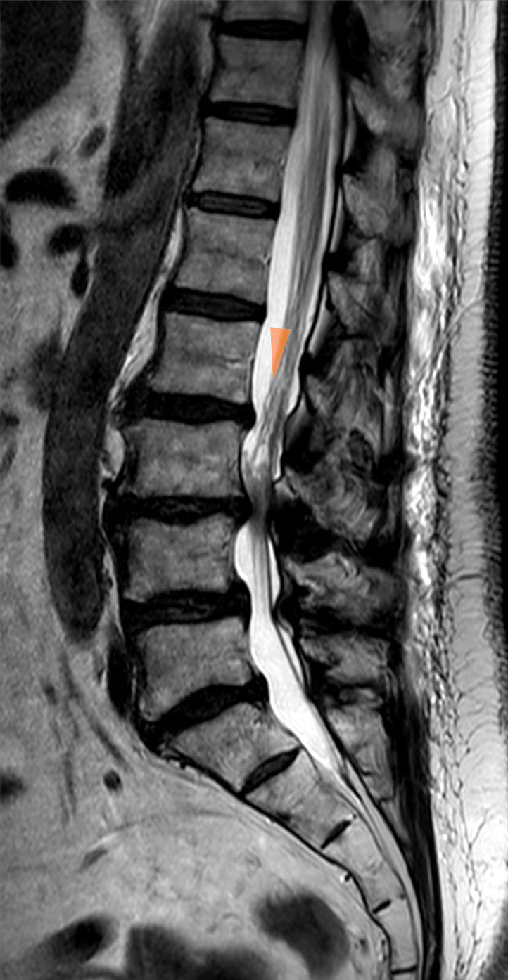
MRISagittal T2 lumbar MRI: severe canal stenosis at L3-L4 with redundancy of the cauda equina roots above the narrowed level, arrow pointing to the stenosis, in an 82-year-old woman.
Source : Hellerhoff, Wikimedia Commons · CC BY-SA
What we call frailty is a geriatric syndrome characterised by a reduction in physiological reserve, increasing vulnerability to adverse events (falls, hospital admission, functional decline, mortality). The frailty phenotype of Fried et al. (2001) rests on 5 criteria:¹
- Involuntary weight loss ≥ 4.5 kg a year
- Muscle weakness (reduced grip strength)
- Self-reported fatigue (exhaustion)
- Slow walking speed (< 0.8 m/s over 4 m)
- Low physical activity
A frail patient: ≥ 3 positive criteria. Pre-frail: 1-2 criteria. Robust: 0 criteria.
What we call sarcopenia, redefined in 2019 by the European consensus EWGSOP2 (Cruz-Jentoft et al.), is a progressive, generalised muscle syndrome combining loss of muscle strength (primary), loss of muscle mass and reduced physical performance.² The diagnosis in clinical practice:
- Find : suspicion through the SARC-F questionnaire (5 items)
- Assess : grip strength or the 5-times chair-stand test
- Confirm : measurement of appendicular muscle mass (DXA, BIA)
- Severity : walking speed, TUG (Timed Up & Go), SPPB
⚖️ Decision algorithm: LSS in older people by frailty profile
Match the aggressiveness of treatment to the patient's overall profile, not to the MRI alone
Sources: Fried LP et al. J Gerontol A Biol Sci Med Sci. 2001;56(3):M146-M156. PMID 11253156 · Cruz-Jentoft AJ et al. EWGSOP2. Age Ageing. 2019;48(1):16-31. PMID 30312372.
Why build these concepts in?
- In older people, frailty strongly predicts perioperative complications (mortality, cardiovascular complications, infection, functional decline after surgery, prolonged length of stay).¹,
- Sarcopenia is associated with an increased risk of falls, of poor recovery after surgery and of mortality.²
- Comprehensive geriatric assessment before spinal surgery makes it possible to identify the patients at high risk and to put « prehabilitation » in place (nutrition, exercise, medical optimisation).
Surgery or conservative treatment: what do the RCTs and the guidelines say?
LSS is the leading cause of spinal surgery in adults over 65 in the United States.³ The data of Deyo et al. (JAMA 2010) are eloquent: between 2002 and 2007 the rate of complex surgery (lumbar fusion) for LSS rose fifteen-fold in Medicare beneficiaries over 65, with no proportionate improvement in functional results, but with a significant increase in major complications and in costs.³
The two reference RCTs that compare surgery with conservative treatment:
- SPORT (Spine Patient Outcomes Research Trial) , Lurie et al. 2015: 8-year follow-up, showing significant and persistent benefits of surgery in patients with stenosis who chose surgery, but with substantial crossover (the « as-treated » analyses differ from the « intention-to-treat » ones).⁴
- Delitto et al. (Ann Intern Med 2015) : a multicentre RCT, 169 patients, comparing surgical decompression with structured physiotherapy (≥ twice a week for 6 weeks). Result: physiotherapy reaches results similar to surgery at 2 years on physical function (SF-36 PF), with no statistically significant difference.⁵
🔬 Surgery versus physiotherapy for LSS, a synthesis of the key RCTs (2 to 8 years of follow-up)
The effect of surgery on physical function against structured conservative treatment
Sources: Lurie JD et al. SPORT 8-year. Spine. 2015;40(2):63-76. PMID 25569524 · Delitto A et al. Ann Intern Med. 2015;162(7):465-473. PMID 25844995. Note: SPORT has a high crossover rate (~40 %), which makes intention-to-treat and as-treated interpretation difficult.
The Cochrane review of Zaina et al. (2016) on the surgery versus non-surgical comparison concludes cautiously that there is low-quality evidence in favour of a short-term benefit of surgery on pain and function, but that the long-term benefits are less clear, and that the perioperative complications (10-24 % across studies) must be taken into account.⁶
Consensus indications for surgery (NASS Kreiner 2013, Katz JAMA 2022):
- Cauda equina syndrome : an absolute neurosurgical emergency 🚨
- A progressive motor neurological deficit that can be documented
- Failure of a well-conducted conservative treatment of ≥ 3-6 months (structured physiotherapy + education)
- Disabling pain persisting despite optimal medical treatment and significantly affecting quality of life
- Informed patient preference after sharing the information on benefits and risks (shared decision-making)
The decompression alone is generally preferred to lumbar fusion in older patients, unless there is a degenerative spondylolisthesis with documented instability. Fusion adds an increased risk of complications with no systematic clinical benefit.⁷
Criticism and controversies
The persistent grey areas:
- The high crossover rate in SPORT (≈ 40 %) complicates interpretation: the « as-treated » analyses show a clear benefit of surgery, but the « intention-to-treat » analyses are less conclusive.⁴
- Heterogeneity of the physiotherapy programmes in the RCTs: some protocols are of low intensity (1-2 sessions, unsupervised home exercise), which may underestimate the benefit of real conservative treatment when it is well conducted.
- The question of prehabilitation before surgery: preoperative optimisation of nutrition, muscle strength and comorbidity management improves postoperative results but remains poorly standardised.
- Comprehensive geriatric assessment before spinal surgery is not yet routine in everyday practice, although it could significantly improve patient selection and results.
Key points
- LSS is the leading cause of spinal surgery after 65 (Deyo 2010), with an explosion in the use of lumbar fusion without a proportionate clinical benefit.
- The assessment must build in frailty (Fried 2001) and sarcopenia (EWGSOP2 Cruz-Jentoft 2019), not only imaging.
- The reference RCTs (SPORT Lurie 2015, Delitto 2015) show that structured physiotherapy catches up with surgery at 2 years for many patients (statistically small differences).
- Clear surgical indications: cauda equina, a progressive motor deficit, failure at ≥ 3-6 months of physiotherapy, disabling pain together with the patient's informed preference.
- The decompression alone is preferred to fusion unless instability is documented.
Chapter 5 bibliography
- Fried LP, Tangen CM, Walston J, et al. Frailty in older adults: evidence for a phenotype. J Gerontol A Biol Sci Med Sci. 2001;56(3):M146-M156. PMID 11253156.
- Cruz-Jentoft AJ, Bahat G, Bauer J, et al. Sarcopenia: revised European consensus on definition and diagnosis (EWGSOP2). Age Ageing. 2019;48(1):16-31. PMID 30312372.
- Deyo RA, Mirza SK, Martin BI, Kreuter W, Goodman DC, Jarvik JG. Trends, major medical complications, and charges associated with surgery for lumbar spinal stenosis in older adults. JAMA. 2010;303(13):1259-1265. PMID 20371784.
- Lurie JD, Tosteson TD, Tosteson A, et al. Long-term outcomes of lumbar spinal stenosis: eight-year results of the Spine Patient Outcomes Research Trial (SPORT). Spine (Phila Pa 1976). 2015;40(2):63-76. PMID 25569524.
- Delitto A, Piva SR, Moore CG, et al. Surgery versus nonsurgical treatment of lumbar spinal stenosis: a randomized trial. Ann Intern Med. 2015;162(7):465-473. PMID 25844995.
- Zaina F, Tomkins-Lane C, Carragee E, Negrini S. Surgical versus non-surgical treatment for lumbar spinal stenosis. Cochrane Database Syst Rev. 2016;(1):CD010264. PMID 26824399.
- Försth P, Ólafsson G, Carlsson T, et al. A Randomized, Controlled Trial of Fusion Surgery for Lumbar Spinal Stenosis. N Engl J Med. 2016;374(15):1413-1423. PMID 27074066.
- Katz JN, Zimmerman ZE, Mass H, Makhni MC. Diagnosis and Management of Lumbar Spinal Stenosis: A Review. JAMA. 2022;327(17):1688-1699. PMID 35503342.
- Kreiner DS, Shaffer WO, Baisden JL, et al. An evidence-based clinical guideline for the diagnosis and treatment of degenerative lumbar spinal stenosis (update). Spine J. 2013;13(7):734-743. PMID 23830297.
- Katz JN, Harris MB. Lumbar Spinal Stenosis. N Engl J Med. 2008;358(8):818-825. PMID 18287604.
What do concrete clinical cases teach us about degenerative lumbar spinal stenosis?
Analysing clinical cases published in the scientific literature offers a pragmatic, detailed view of LSS. Beyond the large statistical studies, these reports illustrate the variability of presentations, the challenges of differential diagnosis and the effectiveness of personalised therapeutic approaches.
Analysis of a « classic » case: from assessment to conservative resolution
The pivotal RCT of Schneider et al. (JAMA Network Open 2019) compared three conservative interventions in 259 consecutive patients with symptomatic LSS:¹
- Group 1: medical care + home exercise (n=88)
- Group 2: individual multimodal physiotherapy (manual therapy + supervised exercise + education, n=87)
- Group 3: supervised group exercise (n=84)
The typical profile of the patients included: mean age 72, 55 % women, with a mean walking distance before the intervention of 230 metres (ZCQ symptom score 3.2/5).
Results at 2 months: multimodal physiotherapy obtained the best statistically significant results on walking capacity (6-minute walk test: +59 metres against +29 m in group 1, p=0.02) and on function (ZCQ function score: -0.57 against -0.35, p=0.01). At 6 months the benefits were maintained.¹
Clinical application: a well-conducted structured physiotherapy programme (6 sessions on average over 6 weeks) can produce significant functional gains even in the typical older patient. The effective content combines:
- Manual therapy of the lumbar spine and the lower limbs (mobilisation)
- Supervised exercise: lumbar flexion, trunk and lower limb strengthening, endurance (cycling)
- A home exercise programme
- Education about the condition, the aggravating positions and activity modification
The diagnostic challenge: when lumbar stenosis mimics (or is mimicked by) another condition
LSS is a « great imitator ». As the Katz 2022 JAMA review recalls, the picture can be confused with:²
- Hip osteoarthritis : pain on weight-bearing relieved by rest (against walking for LSS). The FABER test and hip radiographs make the distinction.
- Vascular claudication (peripheral arterial disease) : cramping pain relieved by stopping without sitting down. An index < 0.9 confirms it.
- Peripheral neuropathy (diabetic, alcoholic, B12): bilateral non-postural symptoms, objective signs on monofilament and tuning fork. EMG/NCS if in doubt.
- Trochanteric bursitis or gluteus medius tendinopathy : lateral hip pain, FADIR and Trendelenburg tests, direct palpation.
- A radicular syndrome of discal origin at L5 or S1 : unilateral pain typical of one root, relieved by lying down, reproduced by the straight leg raise. MRI makes the distinction.
- Fibromyalgia : persistent diffuse pain, tender points, a frequent comorbidity with LSS.
Conversely, LSS can be masked by other dominant conditions :
- In a patient with severe hip osteoarthritis operated on : the LSS can be unmasked after hip replacement when the hip limitation disappears.
- In a patient with Parkinson's disease or dementia : the altered posture and gait analysis can mask the typical neurogenic claudication.
👉 Practical approach : in any older patient with « lower limb pain worsened by activity », systematically include in the assessment:
- The claudication questionnaire (« where, when, what relieves it »)
- The ankle-brachial index (failing that, the peripheral pulses)
- Hip mobility (FABER, FADIR)
- The segmental neurological examination
- The treadmill stoop test (if available)
Study of a complex case: LSS with degenerative scoliosis or multiple comorbidities
Management becomes more complex when spinal or systemic comorbidities are present. The narrative review of Genevay and Atlas (2010) stresses that degenerative lumbar scoliosis, plus degenerative spondylolisthesis and multiple conditions (hypertension, diabetes, sarcopenia, depression) profoundly change the treatment strategy.³
The typical profile of a complex case:
- A patient aged 75-85, with clinically symptomatic degenerative lumbar stenosis
- Degenerative lumbar scoliosis (Cobb angle > 10°)
- Grade I L4-L5 spondylolisthesis or more
- Comorbidities : hypertension, type 2 diabetes, depression, osteoporosis, sarcopenia
- Polypharmacy (≥ 5 medicines)
- Frailty (Fried 2-3 criteria)
For these patients, the systematic reviews converge on the following:
- The structured physiotherapy remains indicated first line, but adapted (reduced intensity, closer supervision, exercise in unloading, ideally in water).⁴,⁵
- Multidisciplinary assessment is essential: geriatrician, rheumatologist, neurosurgeon, physiotherapist, occupational therapist, psychologist if needed.
- The surgical decision must formally build in frailty (Fried), sarcopenia (EWGSOP2), the comorbidities (Charlson Comorbidity Index) and the patient's preferences.⁶
- If surgery is considered, prehabilitation (4-6 weeks of nutritional optimisation, exercise and comorbidity management) improves the results.
The data from the Zaina 2016 Cochrane review and from SPORT (Lurie 2015) show that even complex cases with structural deformity can respond well to a well-targeted programme, offering a viable alternative to surgery for many patients.⁵,⁷
Criticism and controversies: the right place for clinical cases
While clinical cases have great educational value, they represent the lowest level of scientific evidence (level 5 Oxford CEBM, GRADE « very low »).⁸ The success of an intervention in one patient does not guarantee its effectiveness in another. Their main use is to generate hypotheses and illustrate the complexity of clinical reasoning. 🤔
A major controversy in the field of LSS remains the weak correlation between radiological signs and clinical symptoms.⁹ That means treatment must never be based on imaging alone, but on a rigorous synthesis of the patient's history, symptoms, physical examination and overall profile (frailty, comorbidities).
Key points
- The typical cases show that a multimodal conservative approach (multimodal physiotherapy + education + home exercise) can produce significant functional gains even in an older patient (Schneider RCT 2019).
- LSS is a « great imitator » : it can present as, or be confused with, hip osteoarthritis, peripheral arterial disease, peripheral neuropathy, trochanteric bursitis or a disc herniation. A rigorous differential diagnosis is essential.
- The presence of a stenosis on imaging does not confirm the diagnosis on its own. Correlating imaging with the clinical picture remains essential.
- Complex cases (LSS + scoliosis + comorbidities) call for a multidisciplinary approach and can respond well to a well-targeted conservative programme.
- A case report = level 5 (the lowest). It illustrates, it never demonstrates. Follow the meta-analyses where they diverge.
Chapter 6 bibliography
- Schneider MJ, Ammendolia C, Murphy DR, et al. Comparative Clinical Effectiveness of Nonsurgical Treatment Methods in Patients With Lumbar Spinal Stenosis: A Randomized Clinical Trial. JAMA Netw Open. 2019;2(1):e186828. PMID 30646197.
- Katz JN, Zimmerman ZE, Mass H, Makhni MC. Diagnosis and Management of Lumbar Spinal Stenosis: A Review. JAMA. 2022;327(17):1688-1699. PMID 35503342.
- Genevay S, Atlas SJ. Lumbar spinal stenosis. Best Pract Res Clin Rheumatol. 2010;24(2):253-265. PMID 20227646.
- Ammendolia C, Hofkirchner C, Plener J, et al. Non-operative treatment for lumbar spinal stenosis with neurogenic claudication: an updated systematic review. BMJ Open. 2022;12(1):e057724. PMID 35046008.
- Zaina F, Tomkins-Lane C, Carragee E, Negrini S. Surgical versus non-surgical treatment for lumbar spinal stenosis. Cochrane Database Syst Rev. 2016;(1):CD010264. PMID 26824399.
- Cruz-Jentoft AJ, Bahat G, Bauer J, et al. Sarcopenia: revised European consensus on definition and diagnosis (EWGSOP2). Age Ageing. 2019;48(1):16-31. PMID 30312372.
- Lurie JD, Tosteson TD, Tosteson A, et al. Long-term outcomes of lumbar spinal stenosis: eight-year results of the Spine Patient Outcomes Research Trial (SPORT). Spine (Phila Pa 1976). 2015;40(2):63-76. PMID 25569524.
- Burns PB, Rohrich RJ, Chung KC. The levels of evidence and their role in evidence-based medicine. Plast Reconstr Surg. 2011;128(1):305-310. PMID 21701348.
- Brinjikji W, Luetmer PH, Comstock B, et al. Systematic literature review of imaging features of spinal degeneration in asymptomatic populations. AJNR Am J Neuroradiol. 2015;36(4):811-816. PMID 25430861.
How do you apply these recommendations concretely in your practice?
Applying the recommendations that come out of research is the bridge between science and better patient outcomes. 🧭 It requires knowing not only what to do, but also how to integrate it, when to collaborate and how to measure the impact.
When and to which other health professionals should the patient be referred?
One of the fundamental skills of the modern physiotherapist is recognising the limits of their scope of practice and identifying the situations that need interprofessional collaboration. 🤝
Identifying the red flags is the first non-negotiable step. The international IFOMPT framework (Finucane et al. JOSPT 2020) structures this approach for potentially serious spinal conditions.¹ It recalls that red flags taken in isolation have a low positive predictive value, but that their clustering and persistence over time significantly increase the suspicion.¹,²
🚩 Red flags specific to LSS that must be known
- Cauda equina syndrome : saddle anaesthesia, sphincter disturbance (recent urinary retention, faecal incontinence), bilateral lower limb weakness → an IMMEDIATE neurosurgical emergency
- A progressive motor deficit that can be documented (muscle testing dropping a grade over days or weeks) → urgent MRI + a neurosurgical opinion
- A recent history of cancer + progressive night-time low back pain → a cancer work-up (looking for vertebral metastases)
- Unexplained weight loss (> 10 % of body weight in 6 months) + persistent low back pain → a cancer or chronic disease work-up
- Fever + low back pain + immunosuppression, intravenous drug use or recent sepsis → suspected spondylodiscitis or epidural abscess
- Recent high-energy trauma OR severe osteoporosis + sudden pain → a vertebral fracture
- Progressive low back pain in someone > 75 with no obvious mechanical cause → systematic reassessment (myeloma, osteoporotic fracture)
- No improvement after 6 weeks of well-conducted treatment → diagnostic reassessment
⚠️ Any red flag requires prompt medical referral (general practitioner, emergency department, rheumatologist, neurosurgeon) BEFORE physiotherapy continues.
Beyond emergencies, referral should be considered for:
- Yellow flags (psychosocial factors): severe kinesiophobia (TSK > 41), catastrophising (PCS > 30), depression, anxiety → referral to a psychologist or doctor for CBT, EMDR or integrated care.³
- No response to treatment after 6-12 weeks of well-conducted physiotherapy (≥ 2 sessions a week) with patient adherence → reassessment by the general practitioner, a rehabilitation physician or a rheumatologist.
- Surgical assessment : a progressive deficit, persistent disabling pain, failure at ≥ 3-6 months → a neurosurgeon specialising in the spine.
- Standardised geriatric assessment in a frail older person considering surgery → a geriatrician and a multidisciplinary team.
The interprofessional collaboration is fundamental.⁴ Effective collaboration rests on structured communication, shared goals and a clear understanding of each professional's role.⁴ Advanced practice physiotherapists play a growing role in first-line triage.
How do you measure outcomes and overcome the barriers to implementation?
To know whether a treatment works, it must be measured. 📈 The systematic use of standardised measurement tools is the cornerstone of reflective, patient-centred practice.
The PROMs (patient-reported outcome measures) specific to LSS:
| PROM | Items | Subscales | MCID | Reference |
|---|---|---|---|---|
| ZCQ / SSSQ (specific to LSS) | 18 | Severity, function, satisfaction | ~0.5 per subscale | Stucki 1996 |
| ODI (Oswestry Disability Index) | 10 | Overall score 0-100 | 10 points | Fairbank 1980 |
| RMDQ (Roland-Morris) | 24 | Score 0-24 | 2-3 points | Roland 1983 |
| NPRS (Numeric Pain Rating) | 1 | 0-10 pain | 2 points / 30 % | Childs 2005 |
| 6-minute walk test | Performance | Distance in metres | ~30-50 m | ATS 2002 |
| TSK-11 (kinesiophobia) | 11 | Score 11-44 | 4 points | Woby 2005 |
Using PROMs at the start, during and at the end of treatment makes it possible to follow progress objectively, to support shared decision-making and to justify continuing or changing the treatment plan.
📐 The GRADE pyramid of levels of evidence applied to LSS
Ranking the sources: what a systematic review or meta-analysis says is not equivalent to a case report
Source: adapted from the GRADE Working Group + Oxford CEBM Levels of Evidence (2011). In practice, ranking your sources avoids mistaken conclusions based on low-level publications.
Implementation nonetheless meets significant obstacles. 🚧 The main barriers identified:
- A lack of time in a context of heavy clinical pressure.⁵
- A lack of skills for searching, appraising and integrating the scientific literature.⁵
- Lack of organisational support and of resources (access to databases, protected time).⁵
- Cultural inertia anchored in tradition rather than evidence.⁵
Effective strategies for overcoming these obstacles:
- Interactive continuing education (practical workshops) rather than simply reading guidelines.⁶
- Audit and feedback : comparing your practice with quality standards (Cochrane Ivers 2012).⁶
- Clinical mentoring and communities of practice for peer learning.⁵
- Clinical decision support tools and reminders in the electronic patient record at the point of care.⁶
Criticism and controversies
Several points of friction deserve reflection:
First, the PROM paradox. Their value is universally recognised but their implementation remains low, for lack of perceived time. The risk is that they become an administrative box-ticking exercise rather than a real tool for clinical dialogue.
Second, the focus on red flags generates an illusion of safety. Although screening for them is essential, excessive confidence in lists can mislead: a patient can have none of the classic red flags and still have a serious condition.¹ Conversely, an obsessive hunt can lead to over-prescribing imaging.²
Finally, the persistent gap between knowing and doing remains a major challenge. The cultural shift towards reflective, evidence-based practice runs into the inertia of habit, budget constraints and a perceived resistance to standardisation as a threat to clinical autonomy.⁵
Key points
- Screening for red flags (Finucane 2020 IFOMPT) and yellow flags is crucial for the safety and effectiveness of care, and sometimes calls for referral.
- The interprofessional collaboration (general practitioner, rheumatologist, rehabilitation physician, neurosurgeon, geriatrician, psychologist) is essential for complex cases.
- Using validated PROMs (ZCQ Stucki 1996, specific to LSS, ODI, NPRS, 6-minute walk test, TSK-11) is essential for following progress and guiding shared decision-making.
- Rank the sources by the GRADE pyramid : follow the systematic reviews and meta-analyses (1a) first, not the case reports (5).
- For successful implementation, adopt active strategies : interactive continuing education, mentoring, audit and feedback.
Chapter 7 bibliography
- Finucane LM, Downie A, Mercer C, et al. International Framework for Red Flags for Potential Serious Spinal Pathologies. J Orthop Sports Phys Ther. 2020;50(7):350-372. PMID 32438853.
- Verhagen AP, Downie A, Popal N, Maher C, Koes BW. Red flags presented in current low back pain guidelines: a review. Eur Spine J. 2016;25(9):2788-2802. PMID 27376890.
- Kamper SJ, Apeldoorn AT, Chiarotto A, et al. Multidisciplinary biopsychosocial rehabilitation for chronic low back pain: Cochrane systematic review and meta-analysis. BMJ. 2015;350:h444. PMID 25180773.
- Foster NE, Anema JR, Cherkin D, et al. Prevention and treatment of low back pain: evidence, challenges, and promising directions. Lancet. 2018;391(10137):2368-2383. PMID 29573872.
- Alshehri MA, Alalawi A, Alhasan H, Stokes E. Physiotherapists' behaviour, attitudes, awareness, knowledge and barriers in relation to evidence-based practice implementation in Saudi Arabia: a cross-sectional study. Int J Evid Based Healthc. 2017;15(3):127-141. PMID 28399014.
- Ivers N, Jamtvedt G, Flottorp S, et al. Audit and feedback: effects on professional practice and healthcare outcomes. Cochrane Database Syst Rev. 2012;(6):CD000259. PMID 22696318.
- Stucki G, Daltroy L, Liang MH, Lipson SJ, Fossel AH, Katz JN. Measurement properties of a self-administered outcome measure in lumbar spinal stenosis. Spine (Phila Pa 1976). 1996;21(7):796-803. PMID 8779009.
- Tomkins-Lane C, Melloh M, Lurie J, et al. ISSLS Prize Winner: Consensus on the Clinical Diagnosis of Lumbar Spinal Stenosis. Spine (Phila Pa 1976). 2016;41(15):1239-1246. PMID 26839989.
- Genevay S, Courvoisier DS, Konstantinou K, et al. Clinical classification criteria for neurogenic claudication caused by lumbar spinal stenosis. The N-CLASS criteria. Spine J. 2018;18(6):941-947. PMID 29031994.
- Burns PB, Rohrich RJ, Chung KC. The levels of evidence and their role in evidence-based medicine. Plast Reconstr Surg. 2011;128(1):305-310. PMID 21701348.
What next after reading this?
This article is part of a collection of evidence-based clinical syntheses. A question, a comment, a correction to suggest? Contact us directly through the WhatsApp button at the bottom right of the screen.

