


















In brief
Lumbar fusion deliberately abolishes movement at one or more segments in order to abolish the pain that movement produced. Three consequences follow, and they govern the whole of rehabilitation. The first is psychological: 65.2 % of these patients have measurable kinesiophobia after the operation (Pan 2025), and their satisfaction depends more on the gap between outcome and expectation than on the outcome itself (Zhang 2024). The second is mechanical: the stiffened level transfers its loads onto its neighbours, and adjacent segment degeneration progresses at about 5.9 % per year, its symptomatic form at 1.8 % per year (Zhang 2016). The third is documentary: there is no single protocol, and what is "allowed" depends more on the surgeon who operated than on any consensus of evidence. The operative report therefore remains the first item of the assessment.
This synthesis is addressed to the physiotherapist who receives a patient operated on for lumbar fusion and must decide, before any exercise, what can be moved, what must be protected, and what can honestly be announced. It completes the series the site devotes to the postoperative management of the major arthroplasties (total hip arthroplasty, total knee arthroplasty and total shoulder arthroplasty), adding the spine, which was missing from it. The difference is not one of degree: an arthroplasty restores a movement, a fusion abolishes one. Everything else follows from that.
It also begins where the site's articles on the conditions that lead to theatre leave off. Non-specific low back pain, sciatica from disc herniation, facet joint arthropathy, degenerative lumbar stenosis and degenerative spondylolisthesis each describe a presentation and its conservative management. This page describes what happens afterwards, when that management has failed and the construct is in place.
Three figures that frame the subject
Surgical volume, the psychological burden carried by the operated patient, and the long-term mechanical stake.
Sources: Martin 2019 (PMID 30074971), Pan 2025 (PMID 40212616), Zhang 2016 (PMID 26836484). No French national data on the incidence of lumbar fusion is published in a peer-reviewed journal: the volume figures quoted here are those of the American hospital registry, and they do not transpose as they stand.
Clinical synthesis
- This patient is not one more low back pain patient. They arrive with years of pain behind them, physical activity that had already collapsed before the operation, 83 % do not meet the WHO guidelines and 16 % take fewer than 5,000 steps a day (Lotzke 2018), and fear of movement present in two thirds of those operated on (Pan 2025). Treating that as a preamble to the “real” work is to miss the main lever on outcome.
- Surgery relieves the leg better than the back, and the patient often does not know it. In the Norwegian trial that compared fusion with a cognitive intervention plus exercises, lower limb pain decreased more after surgery, whereas avoidance beliefs decreased more without surgery (Brox 2003). A badly calibrated expectation is costly: for an identical functional outcome, 43 % satisfied among patients with high expectations against 85 % among those with low expectations (Zhang 2024).
- There is no single protocol, and saying so is more honest than inventing one. The 18 trials in the reference meta-analysis range from a single session to daily sessions for a month (Bogaert 2022). A survey of 703 spine surgeons finds activity restrictions prescribed in 79 % of cases and braces in 34 %, over heterogeneous durations that the authors themselves put down to habit rather than to evidence (Hamouda 2026).
- Timing is not the right question, content is. The only trial that has compared two start dates head-on finds the six-week start inferior to the twelve-week start (Oestergaard 2012), and its economic evaluation confirms it (Oestergaard 2013). But motor control and cognitive work started in the first few days are safe and worthwhile: less pain at three months and less persistent pain syndrome at one year, 11 cases out of 214 against 20 out of 181 (Nie 2025).
- What works is multimodal, not muscular. Rehabilitation combining exercise with a cognitive-behavioural approach beats exercise alone on disability and on fear of movement, with a low level of evidence and an effect that fades beyond one year (Bogaert 2022). Monticone's trial, conducted in 130 patients operated on for spondylolisthesis and stenosis, still holds one year after the end of treatment (Monticone 2014).
- The adjacent segment is the long-term stake, and it justifies the work on control and on spinal care. The imaging finding is common and poorly correlated with symptoms; the clinical disease is rarer but costly. The factor most consistently associated is neither sex nor segmental alignment, it is the number of levels fused (Zhang 2016).
- Postoperative red flags are not the same as the preoperative ones. Pain that changes in nature, a fever, a discharge, a new neurological deficit: surgical site infection affects 1.53 % of patients operated on the lumbar spine (Ying 2022), and cauda equina syndrome can appear after the operation, at the adjacent level (Chang 2024).
What is a lumbar fusion, and who ends up on the table?
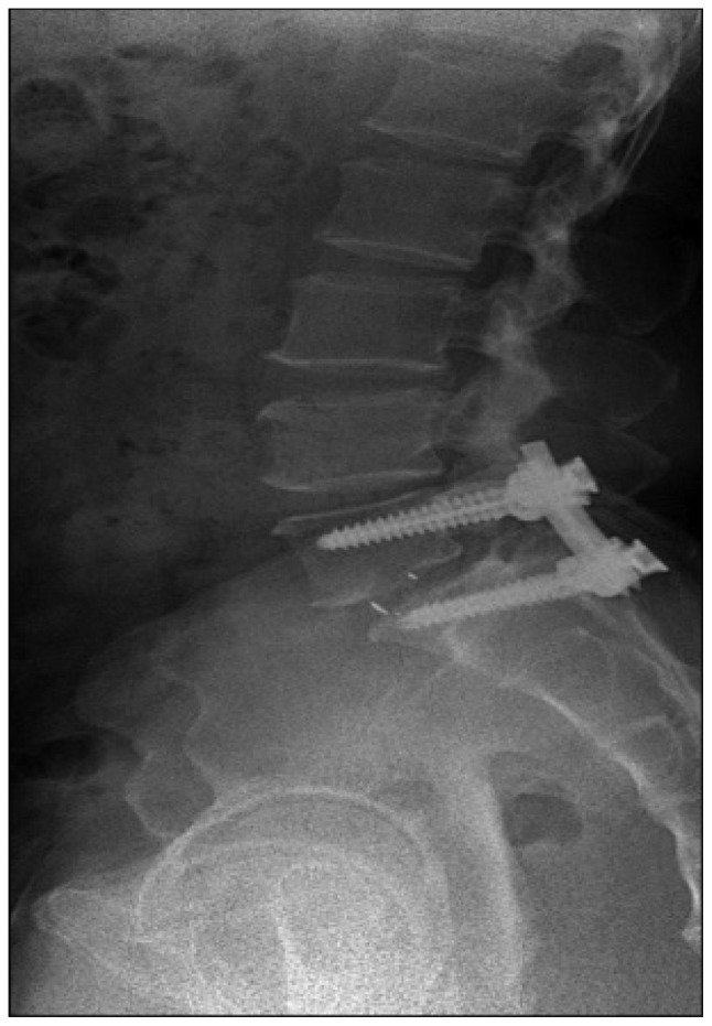
X-rayLateral lumbar spine radiograph, twelve months after posterior fusion: pedicle screws and rods in place, with an interbody cage visible between the vertebral bodies.
Source : Kim et al., Journal of Korean Neurosurgical Society, 2015, figure 4 · CC BY-NC
A fusion does not repair a segment: it abolishes it as a joint. Understanding what the surgeon removed, added and crossed governs everything the physiotherapist can do in the three months that follow.
Fusing is not repairing
Lumbar fusion consists in fusing two vertebrae, sometimes more, into a single block of bone. The disc is most often removed and replaced by a cage filled with graft, instrumentation with pedicle screws and rods holds the assembly immobile while the bone consolidates, and movement at the segment disappears permanently. The reasoning is simple: if a segment produces pain because it moves badly, depriving it of movement removes the source.
That reasoning works better when the target is an identifiable mechanical instability than when it is isolated discogenic pain. In Brox's Norwegian randomised trial, 64 patients with low back pain lasting more than a year and disc degeneration at L4-L5 or L5-S1 were randomly assigned between instrumented fusion and a cognitive intervention programme with exercises. At one year, disability measured by the Oswestry fell from 41 to 26 after surgery and from 42 to 30 after the cognitive intervention: a mean difference of 2.3 points (95 % confidence interval −6.7 to 11.4; p = 0.33), that is to say none. The success rate judged by an independent observer was 70 % after surgery and 76 % after conservative treatment, and the early complication rate in the surgical group was 18 %PMID 12973134.
One observation from this trial deserves to be kept as it stands, because it announces the whole of the next chapter: lower limb pain decreased more after surgery, whereas avoidance beliefs and the fingertip-to-floor distance improved more without surgery. In other words, the operation acts on what compresses the nerve, and rehabilitation on what prevents movement. These are not the same targets, and the patient almost always confuses them.
The indications, and the uneven strength of their evidence
Not all fusions are equal in the light of the evidence, and the physiotherapist has every interest in knowing which one is in front of them. Degenerative spondylolisthesis is the best-supported indication: the SLIP trial, conducted in 66 patients aged 50 to 80 years with grade I spondylolisthesis and symptomatic stenosis, showed a greater gain in physical quality of life with laminectomy plus fusion compared with laminectomy alone, 15.2 against 9.5 points of SF-36 at two years (difference 5.7; 95 % CI 0.1 to 11.3; p = 0.046), a gain maintained at three and four years. The cumulative reoperation rate was 14 % after fusion against 34 % after decompression alonePMID 27074067. It should be noted that the difference on the Oswestry did not reach significance (p = 0.06): the benefit lies in overall physical health more than in back-related disability.
At the other extreme, isolated degenerative disc disease remains the most debated indication, and registries show that practice takes account of it. In the American hospital registry National Inpatient Sample, the volume of elective lumbar fusions rose from 122,679 procedures in 2004 to 199,140 in 2015, an increase of 62.3 % in volume and of 32.1 % relative to the adult population. The steepest rise concerns people over 65 years (+138.7 % in volume, +73.2 % in rate). The two indications that grew most in absolute terms are spondylolisthesis (+47,390 procedures, that is +111 %) and degenerative scoliosis (+16,129, that is +186.6 %), precisely those whose evidence of effectiveness is the strongest; the share of indications with weaker evidence (disc disease, herniation, stenosis without instability) still represented 42.3 % of the total in 2015, but slightly declining over the last few yearsPMID 30074971.
What made lumbar fusion grow, indication by indication
Change in the volume of elective lumbar fusions in the United States between 2004 and 2015, by surgical indication. The two indications best supported by evidence are also those that grew most.
Source: Martin 2019, Spine, analysis of the National Inpatient Sample 2004-2015 (PMID 30074971). American data: they describe a trend, not French practice.
Four families of surgical approach, and what they change for us
The acronym written in the operative report is not a technician's detail. The approach determines which muscles were crossed, and therefore which part of the assessment will be lastingly false. A review of twelve studies and 529 patients compared postoperative muscle volumes: paraspinal atrophy is significantly more marked after fusion than after surgery without fusion (p = 0.0001), after a posterior approach than after an anterior approach (p = 0.0001), and after conventional surgery than after minimally invasive surgery (p = 0.001), whereas no difference appears between minimally invasive and conventional decompressionPMID 26840699. It is fusion by the posterior approach, and it alone, that costs muscle.
| Surgical approach | What the surgeon crosses | What the physiotherapist must take from it |
|---|---|---|
| PLIF, TLIF posterior or transforaminal approach | The paraspinal muscles, detached or retracted on either side of the spinous processes. The TLIF passes on one side only and through one facet joint, the PLIF through both. | A midline scar that is painful on early mobilisation. Multifidus deficient for a long time, and not only because of pain. Trunk strength tests measure a surgical lesion as much as deconditioning. |
| ALIF anterior approach | The abdominal wall and the retroperitoneal space, with the great vessels retracted. The posterior spine is spared. | Posterior muscles intact: motor control starts earlier and higher. On the other hand the abdominal wall has been crossed, and ileus, wall pain and discomfort on coughing dominate the first few days. |
| LLIF, OLIF lateral or oblique approach | The psoas, crossed or skirted, in contact with the lumbar plexus. | Psoas weakness and reduced sensation over the front of the thigh are common in the immediate postoperative period: this is most often transient, but it falsifies any hip flexion test. To be distinguished from a new radicular deficit. |
| Instrumented posterolateral fusion without interbody cage | The same posterior planes, with graft laid on the facet joints and the transverse processes. | The same muscular cost as the PLIF. Consolidation depends on a posterior graft that is less well vascularised: this is one of the constructs where loading instructions are often the most cautious. |
An animal model sheds light on what the assessment really measures. In the rabbit, an approach that retracts the multifidus produces an oedema signal on MRI that peaks at three weeks and only returns to normal at twenty-four weeks; histology shows necrosis and inflammation at one week, fibrosis at three and six weeks, fatty degeneration at twelve and twenty-four weeks, and damage to the neuromuscular junctions that worsens over timePMID 24352778. The result is experimental and animal; it does not transpose figure for figure. But it gives a useful order of magnitude: part of the weakness observed at three months is denervation, not laziness.
In humans, volumetric measurement after single-level posterior interbody fusion finds a significant reduction in multifidus volume at one year, bilateral and confirmed on MRI as on computed tomography, with only a trend towards a decrease for the erector spinae, which lies more laterally; the psoas, for its part, does not change. Counter-intuitively, in the group examined on MRI, the loss was all the more marked when the patient was youngPMID 32024500.
Part of what is taken for deconditioning at three months is a muscular scar. It can be worked on; it cannot be held against the patient.
What must have been read before the first session
Five lines of the operative report change what is done, and none of them can be guessed:
- The levels fused and their number. It is the factor most consistently associated with the risk of adjacent segment deteriorationPMID 26836484, and it is also what determines the mechanical lever that the neighbouring levels will be subjected to.
- The surgical approach. It says which muscles are damaged, and therefore which assessment measures will be uninterpretable in the first months.
- The presence of an associated decompression. A plain fusion and a fusion with extended laminectomy do not carry the same risk of compressive haematoma in the first hours, nor the same delay to neurological recovery.
- The graft and its harvest site. An iliac harvest adds crest pain that is often confused with sacroiliac or trochanteric pain, and that has a course of its own.
- The surgeon's explicit instructions on the brace, the permitted load and the delay before physiotherapy. Where a general rule and a named instruction diverge, it is the instruction that prevails, always.
Why is the fusion patient not a low back pain patient like any other?
This chapter is not an atmospheric note to be placed before the exercises. It is the chapter where the largest part of the outcome is decided, and the trials that made the difference are precisely those that treated it as session content.
They arrive with years behind them, and a body that has already stopped
Fusion is never a first-line option. When the patient walks into the clinic after surgery, they most often have several years of pain behind them, injections, physiotherapy sessions that were not enough, and a surgical decision taken as a last resort. That past can be measured: among 118 patients monitored by accelerometry before their fusion, 96 of them (83 %) did not meet the World Health Organization physical activity guidelines, and 19 (16 %) took fewer than 5,000 steps a day, the threshold usually used to speak of a sedentary lifestyle. The daily step count was lower the higher the fear of movement and the disabilityPMID 30305065.
The practical consequence is immediate. The patient who comes out of theatre does not set off from a normal activity level to which they should be brought back: they set off from a level that had already collapsed months earlier, and the operation has done nothing to raise it. A postoperative programme content to “gradually resume usual activities” would therefore aim at a target that no longer existed before the incision.
Fear of movement concerns two thirds of those operated on
This is the figure that should change the way the first session opens. Among 178 consecutive patients operated on for posterior lumbar fusion, 116 (that is 65.2 %) had kinesiophobia defined by a score of 37 or more on the 17-item Tampa scale. On multivariate analysis, this fear was associated with age, pain intensity, the Beck depression score, low self-efficacy, the number of levels operated on, the volume of postoperative drainage, the degree of preoperative nerve root compression, and a small paraspinal muscle cross-sectional areaPMID 40212616.
This list says something important: the kinesiophobia of the operated patient is not only a personality trait imported from before. It is correlated with markers of the surgical insult itself: number of levels, drainage, residual muscle mass. It is also built during and after the operation, which means that it is accessible to what is done in the session.
And it has measurable consequences later on. In the Swedish cohort from the PREPARE trial, fear of movement measured before the operation significantly predicted the proportion of time spent in sedentary behaviour at six and twelve months after surgery (β = 0.013; 95 % CI 0.004 to 0.022; p = 0.007), whereas neither catastrophising nor exercise self-efficacy showed any associationPMID 37950235. In practical terms, this points to one screening tool rather than another.
A useful threshold completes the set: in 180 patients followed during a four-week motor and cognitive-behavioural rehabilitation programme after fusion, the minimal clinically important difference on the Tampa scale was estimated at more than 6 points for the total score, more than 4 for the “harm” subscale and more than 2 for the “activity avoidance” subscalePMID 27827516. It is this figure that makes it possible to say whether the work on fear has produced something, rather than to assume it.
The profile of the patient who arrives at the clinic after a fusion
Four measured findings, each from its own cohort, describing a starting point very far from that of an ordinary low back pain patient.
Sources: Pan 2025 (PMID 40212616), Lotzke 2018 (PMID 30305065), Zhang 2024 (PMID 37797842), Park 2026 (PMID 42215594). The last figure covers all lumbar surgery in a Korean national database, fusions and decompressions together: it does not apply specifically to fusion.
The leg does better than the back, and nobody had warned them
This is the commonest disappointment, and it has a real basis. In Brox's trial, lower limb pain decreased more in the surgical arm than in the conservative arm, whereas avoidance beliefs and flexibility improved more in the conservative armPMID 12973134. Surgery decompresses what was compressing; it does not teach movement again.
The nuance deserves to be added, because the common belief goes further than the data. Among 781 patients operated on by minimally invasive TLIF for degenerative spondylolisthesis, grouped according to whether the predominant pain was back pain, radicular pain or equivalent, no significant difference appeared in pain, disability, quality of life, satisfaction, attainment of the minimal clinically important difference or return to work: the only exception being a poorer Oswestry at one month in the group with predominant back painPMID 34008509. In other words: when the indication is right, predominant back pain is not a contraindication to success. What misleads the patient is not the location of their pain, it is the speed at which each component recedes.
Residual back pain, for its part, is common and documented: after endoscopic decompression for lumbar stenosis, 98 patients out of 202 (48.5 %) still had back pain of 25 mm or more on a visual analogue scale at one year, even though the group mean had improved markedly, from 66.7 to 29.7 mm. The factors predicting this residual pain were Modic type 1 changes, preoperative back pain of 70 mm or more and female sexPMID 34781941. This series concerns decompression without fusion: it does not transpose directly, but it gives the texture of the phenomenon, a mean that improves a great deal and half the patients who keep something.
Expectation is the most profitable determinant of satisfaction
This is the study to know for this chapter. Patients due to undergo a one- to two-level fusion, recruited in four centres, completed before the operation a standard Oswestry and a modified Oswestry asking, item by item, what they expected one year later. The difference between the two was used to classify them as having high, moderate or low expectations. One year later, the disability scores and their improvement were comparable between the three groups; satisfaction was not comparable at all. In the high-expectation group, 43 % declared themselves satisfied, against 71 % in the intermediate group and 85 % in the low-expectation group (p = 0.041)PMID 37797842.
For an identical functional outcome, the satisfaction gap between high and low expectations is 42 points. It is not the outcome that satisfies, it is the gap between the outcome and what was promised.
A second study, covering 157 patients operated on for lumbar stenosis with or without fusion, adds a detail that contradicts a widespread intuition: dissatisfaction at six months was associated with preoperative anxiety (odds ratio 3.95; 95 % CI 1.16 to 13.46; p = 0.03), with low preoperative mental health and, counter-intuitively, with less severe preoperative back pain, but not with depression, not with catastrophising and not with avoidance beliefsPMID 34015209. The patient with the most to lose is not always the one who was suffering most.
In the long term, satisfaction nevertheless remains high when the indication is right. Among 328 patients operated on for grade 2 spondylolisthesis and followed for five years in a multicentre quality registry, 208 of the 241 evaluable patients declared themselves satisfied, that is 86 %. On multivariate analysis, satisfaction was associated with improvement in disability and in radicular pain, and with no demographic characteristic, comorbidity or baseline surgical parameterPMID 42066348. What the patient obtains counts; what they were at the outset, much less.
What this changes in the conduct of the session
Three concrete translations follow from the above, and they belong in the first sessions rather than at the end of the programme:
- Measure fear, not only pain. The Tampa scale is completed in three minutes and gives a figure that will move, with an interpretable threshold of more than 6 pointsPMID 27827516. Without it, work on kinesiophobia remains an intention.
- Name the back-leg dissociation before it becomes a disappointment. Telling the patient, from the first session, that sciatic pain often recedes faster and more completely than back pain turns a future proof of failure into an expected event.
- Reframe the objective in terms of capacity rather than disappearance of pain. This is exactly the mechanism of the gap measured by Zhang: expectations are not lowered so as to disappoint less, they are moved towards what surgery and rehabilitation actually produce.
Key points from this chapter
- Two thirds of fusion patients have measurable fear of movement, and this fear is partly built by the operation itself, and therefore modifiable.
- Four patients in five were already failing to meet activity guidelines before going into theatre: the postoperative starting point is lower than is assumed.
- For an identical functional outcome, satisfaction varies from 43 to 85 % according to preoperative expectations. Expectation is an object of care, not a matter of atmosphere.
- Radicular pain recedes faster than back pain. Saying so beforehand prevents the patient from reading the lag as a failure.
What can I move, at what point, and what really threatens the construct?
This is the question every physiotherapist asks in front of a recent fusion. The honest answer comes in two parts: there is no single protocol, and what puts consolidation at risk is almost never what the patient does in the session.
The variability of protocols is not a gap in the literature, it is its result
The reference meta-analysis on rehabilitation after lumbar fusion brought together 18 randomised trials and 1,402 patients. Its authors note that the dose and intensity of the interventions range from a single session to daily sessions for a month, and that what is called “usual care” in the control arm most often amounts to information and postoperative mobilisationPMID 35258644. An earlier systematic review, which had retained only level I or II evidence, adds an observation less often quoted and yet decisive: the different categories of clinicians do not use the same clinical terminology, so that two protocols described in the same words do not cover the same actsPMID 28291412.
This heterogeneity is found in practice, and it is quantified. An international survey conducted in 2024 among AOSpine members, with 322 usable responses on degenerative lumbar fusion surgery, identifies as the leading obstacle to implementing enhanced recovery programmes “the absence of clear protocols and guidelines”, cited by 56.2 % of respondents, ahead of staff shortages (53.8 %) and coordination difficulties (43.0 %). The three components judged most decisive for recovery are opioid-sparing multimodal analgesia (73.3 %), early mobilisation (63.8 %) and preoperative education (58.6 %), and these are also the three where surgeons consider they have the most room for improvement, preoperative education coming first at 62.6 %PMID 41817204.
A second survey, this time covering 703 spine surgeons, quantifies the gap between practice and evidence after lumbar disc surgery: 34 % prescribe a brace, half of them for four weeks, and 79 % recommend activity restrictions. The durations usually quoted are two weeks for prolonged standing, prolonged sitting and driving, three months for bending, twisting, lifting and low-intensity physical activity, and six months for intense activity and contact sports. The authors themselves describe these figures as illustrating a “persistent gap between evidence and practice”PMID 41666866. This survey concerns disc surgery, not fusion: the timings for a fusion cannot be drawn from it. What it establishes, on the other hand, transposes without difficulty: postoperative instructions for the lumbar spine are, to a large extent, a matter of surgical school.
When a physiotherapist asks “what is allowed after a fusion”, they are asking a question the literature does not answer and which the operating surgeon answers in one sentence. The right reflex is not to search further: it is to call.
The brace: prescribed one time in three, supported by no evidence
This is one of the rare points where the literature is clear. A systematic review of four randomised trials concludes that a brace after surgery for degenerative lumbar spine disease improves neither disability, nor pain, nor quality of life compared with no brace, and that no difference appears either in fusion rate, complications or the need for revision surgery: all of it on a low to moderate level of evidencePMID 31676949. An updated meta-analysis covering five trials and 362 patients reaches the same conclusion, with no significant difference in early Oswestry, in visual analogue scale at follow-up, in complication rate (14.9 % with a brace against 17.4 % without) or in fusion ratesPMID 38744484.
The management line does not, however, follow mechanically. If the patient is wearing a brace, it is because their surgeon prescribed it: the physiotherapist has neither the standing nor the information needed to remove it on their own initiative, since a fragile construct, severe osteoporosis or a posterior graft may justify a caution that the operative report does not always spell out. What the evidence allows is not to add a brace where there is none, not to prolong its wear beyond the instruction, and not to present the orthosis to the patient as the guarantee of their consolidation. That last point is not trivial when two thirds of operated patients have measurable fear of movement.
What really threatens consolidation
The worrying question, put plainly, is: can an exercise break the construct or stop the graft from taking? None of the published interventional trials reports such an event attributed to rehabilitation. The trial that started strengthening earliest, at three weeks after surgery, in patients aged 45 to 70 years, with two weekly sessions for nine weeks focused on activating the lumbopelvic stabilisers, states explicitly that no hardware loosening or failure was observed in the trained groupPMID 29914580.
What the literature identifies as risk factors for nonunion and mechanical failure belongs to another register. Smoking comes first: a meta-analysis of 29 studies finds a significantly higher incidence of nonunion in smokers, with a crude odds ratio of 1.97 (95 % CI 1.55 to 2.52; p < 0.001), also 1.97 in the lumbar subgroup, and still 1.38 after adjustment (95 % CI 1.12 to 1.72). Smoking also goes with less return to work (OR 0.70) and, in the lumbar subgroup, with lower satisfaction (OR 0.24). A point worth making to the patient: former smokers who had stopped at least a year before the operation were no longer distinguishable from non-smokers, either for nonunion or for painPMID 41066602.
Bone density comes next. A systematic review of 71 studies and 12,278 patients finds an association between low bone mineral density and implant failure (cage subsidence, screw loosening), often diagnosed at the same time as a nonunion, together with an increased risk of proximal junctional kyphosis from fracture. The heterogeneity of methods precluded any meta-analysis, and the authors stress that osteoporosis remains largely under-diagnosed and under-treated in this populationPMID 38962714.
These two findings have a direct translation into the session, and it is more useful than a list of prohibitions. The physiotherapist following an operated smoker has a documented lever in front of them; the one following a 72-year-old woman with no known bone assessment has a question to put to the general practitioner. These two actions weigh more heavily on consolidation than the choice between two core exercises. The site covers this second point in its article on osteoporosis and the prevention of fragility fractures.
What surgeons prescribe, and what the evidence says about it
Three common postoperative instructions for the lumbar spine, set against their level of scientific support.
Sources: Hamouda 2026, 703 surgeons, after disc surgery (PMID 41666866); Jonkman 2026, 322 responses, lumbar fusion (PMID 41817204); Nasi 2020 (PMID 31676949) and Feng 2024 (PMID 38744484) for the brace; Kernc 2018 (PMID 29914580) for the safety of strengthening at three weeks.
How to put the question to the surgeon so as to get a usable answer
Since the answer is patient-specific, one may as well ask for what is needed. Four questions are enough, and they fit into a letter of a few lines:
- What load can the patient carry, and from what date? An answer in kilograms and in weeks is worth more than a “no heavy lifting” that the patient will translate as “nothing”.
- Is active lumbar flexion allowed, and how far? This is the instruction most often passed on ambiguously, and the one that governs dressing, washing and picking objects up, and therefore real independence.
- Is the brace worn permanently, only when upright, or can it be weaned? And on what timescale?
- Is there a feature of the construct that calls for extra caution? Osteoporosis, isolated posterior graft, revision surgery, adjacent level already degenerated.
The answer to these four questions makes up the safety framework. Within that framework, what the physiotherapist decides is a matter for their reasoning, and that is the subject of the next two chapters.
Key points from this chapter
- There is no single protocol after lumbar fusion. The main obstacle cited by surgeons themselves is the absence of clear guidelines (56.2 %).
- The restriction periods commonly prescribed (two weeks, three months, six months) are landmarks of surgical custom, and the authors who measured them say so explicitly.
- The brace improves nothing measurable, but it is not removed without the opinion of whoever prescribed it.
- What threatens consolidation is smoking (nonunion OR 1.97) and low bone density, not a well-dosed motor control exercise.
- Four questions to the surgeon usefully replace any general rule: load, flexion, brace, feature of the construct.
Does early rehabilitation have evidence, and what evidence?
Yes, and it is more interesting than a simple green light. It says that the question of timing has been badly framed: it is not the start date that decides, it is what is put into it.
The only trial that compared two dates finds the early one inferior
One has to start there, because it is the result most often passed over in silence. A Danish multicentre randomised trial allocated 82 patients operated on for instrumented lumbar fusion for degenerative disc disease or spondylolisthesis between a rehabilitation start at six weeks and a start at twelve weeks. The programme was rigorously identical in the two arms: four group rehabilitation sessions and home exercises focused on active stability. Only the date changed.
At one year, the median reduction in the Oswestry was 6 points in the “six weeks” group against 20 points in the “twelve weeks” group (p < 0.05). Back pain followed the same direction, −2.2 against −3.3. No difference appeared in return to work at one year. The authors conclude, without hedging, that the early start produced inferior resultsPMID 22565381. The economic evaluation conducted alongside drives the point home: the early strategy tended to cost €6,869 more, for functional disability significantly worse by 9 points (95 % CI −18 to −3), and its probability of being cost-effective did not reach 10 % at the usual thresholdsPMID 23928716. The systematic review of level I and II evidence published four years later took up this conclusion as it stood: starting at twelve weeks gives better results at lower cost than starting at six weeksPMID 28291412.
The trial is small, single-centre in its main recruitment, and its result is counter-intuitive. It remains, to this day, the only direct comparison of two start dates for the same programme after lumbar fusion. It cannot be set aside because it is unwelcome.
But “early” does not mean the same thing in the other trials
The contradiction is only apparent, and it dissolves as soon as one looks at what was started early in each trial.
In 107 patients operated on for stenosis, spondylosis, spondylolisthesis or disc disease, a psychomotor therapy targeting pain cognitions, behaviours and motor control was compared with a conventional exercise therapy, both delivered during the first three months. Psychomotor therapy improved functional disability, self-efficacy, outcome expectancy and fear of movement significantly more, at every follow-up point up to two to three years. The authors conclude that postoperative rehabilitation can be safely conducted during the first three months, provided it includes measures targeting psychological as well as motor functions. They honestly report a numerically higher reoperation rate in the psychomotor arm, while placing it within the usual rangePMID 20354468.
In 27 patients aged 45 to 70 years, strengthening started at three weeks, at two sessions a week for nine weeks, focused on activating the lumbopelvic stabilisers, produced a greater improvement in walking speed than the standard protocol (p < 0.01), together with significant progress in all isometric trunk measures, in standing reach height and in the pre-activation pattern. No hardware loosening or failure was observed. At eighteen months, no training effect remainedPMID 29914580.
The largest and most recent trial on this question included 395 patients operated on by modified TLIF for degenerative lumbar disease. The experimental arm received a programme that was sequential: motor control from the first postoperative day, then stabilisation work from the fifth week onwards. The control arm received the same stabilisation work, but from the fifth week only, without an initial motor control phase. At three months, back pain was lower in the sequential arm, local pressure pain thresholds higher, temporal summation lower and fatty infiltration of the erector spinae less marked. At one year, persistent spinal pain syndrome type 2 affected 11 patients out of 214 in the sequential arm against 20 out of 181 in the conventional arm (p < 0.05), that is 5.1 % against 11.0 %, with resting pain and avoidance beliefs also lowerPMID 39453543.
Finally, a Chinese trial of 52 patients compared progressive functional training delivered in the first twelve weeks with an identical programme started at the twelfth week. The six-minute walk distance was greater in the early group at three months (95 % CI 32.51 to 101.88 m; p < 0.001), but the difference had disappeared at six months, and no difference appeared in disability, quality of life or multifidus cross-sectional area. Nine patients (17.3 %) had an adverse event, without the distribution between the groups being specified in the abstractPMID 40249395.
Five trials, one and the same badly framed question
What each trial actually started, and at what date. What is early and beneficial is cognitive or motor; what is early and harmful is a group strengthening programme brought forward by six weeks.
Sources: Nie 2025 (PMID 39453543), Kernc 2018 (PMID 29914580), Abbott 2010 (PMID 20354468), Lu 2025 (PMID 40249395), Oestergaard 2012 (PMID 22565381).
The programme that fails when brought forward by six weeks is a group strengthening programme. The programmes that succeed when started on the first day are motor control and cognitive work. It is not the date that was tested, it is the content.
Content: what sets apart the programmes that work
The Belgian meta-analysis of 2022 is the most complete on this question. Across 18 trials and 1,402 patients, it establishes that in the short term exercise does better than usual care for disability (standardised mean difference −0.41; 95 % CI −0.71 to −0.10, four studies) and pain (−0.36; −0.65 to −0.08, five studies), and above all that multimodal rehabilitation, most often exercise plus cognitive-behavioural training, does better than exercise alone for disability (−0.31; −0.49 to −0.13, six studies) and for pain-related fear (−0.64; −1.11 to −0.17, four studies). Two major reservations accompany these results: the quality of evidence is low according to GRADE, and the effects disappear beyond one year. The trend for return to work is favourable without reaching significance (pooled relative risk 1.30; 95 % CI 0.99 to 1.69)PMID 35258644.
The earlier British meta-analysis, narrower but convergent, found larger effect sizes for what it called “complex rehabilitation”, exercise plus cognitive-behavioural therapy, against usual care: short-term disability −0.85 (95 % CI −1.41 to −0.29) and fear-avoidance −1.07 (−1.33 to −0.80), effects maintained beyond twelve months. It rested, however, on only three identified studies, two of them meta-analysable, that is 237 patients, with once again a low quality of evidencePMID 26555833. A third meta-analysis, covering 15 trials, 528 rehabilitated patients and 498 controls, arrives at a coherent division of roles: the psychological component lowers pain-related fear, the multimodal component lowers disability, and exercise lowers painPMID 37016875.
A fourth review imposes its own nuance and deserves to be quoted for that reason. Across eight studies in systematic review and three in meta-analysis, it does not find any superiority of cognitive therapy added to exercise on long-term pain or on quality of life, the confidence intervals crossing the line of no effect; the advantage on long-term disability and kinesiophobia is supported only by low-quality data. It also notes that the positive effects of core stability work on strength and endurance observed in the short term are not found in long-term studiesPMID 35398574. The reasonable conclusion is therefore not that the multimodal approach is proven, but that it is the least poorly supported option.
The individual trials that moved the line most
Three trials deserve to be known by name, because they describe interventions that can be reproduced in the clinic.
Monticone 2014. One hundred and thirty patients operated on for fusion for degenerative spondylolisthesis or lumbar stenosis, allocated between a programme combining exercise and cognitive-behavioural therapy and a programme of exercise alone, over four weeks. The Oswestry showed a massive group effect (F = 95.78; p < 0.001) and a significant group × time interaction, in favour of the experimental arm, for pain, catastrophising, kinesiophobia and quality of life. The effects still held one year after the end of treatmentPMID 23836299.
Archer 2016. Eighty-six adults operated on for laminectomy with or without fusion, selected before the operation on the basis of high kinesiophobia on the Tampa scale, then randomised at six weeks after surgery between cognitive-behavioural-based physiotherapy and an educational programme. The cognitive-behavioural arm achieved significantly greater reductions in pain and disability, and better results on general health and on physical performance tests (sit-to-stand, timed up and go, ten-metre walk), at three monthsPMID 26476267. The remarkable point is the targeting: the cognitive-behavioural approach was not given to everyone, it was given to those who were afraid.
Greenwood 2019, the REFS trial. Fifty-two patients, ten weekly group sessions combining education, low-technicality cardiovascular work, limb and spine strengthening, and peer support. The short-term reduction in disability reached −13.27 points against −2.42 with usual care, and was maintained in the long term (−14.72 against −7.57). After adjustment, the improvement was significant for disability (p = 0.014) and for pain self-efficacy (p = 0.007). The cost of the programme was £275 per participantPMID 30788599. This is a feasibility study, not a superiority trial: its interest lies in showing that a low-cost group format produces figures of the right order of magnitude.
Rehabilitation modalities after lumbar fusion, by level of evidence
Cards stacked from the best supported to the least supported. No modality reaches a high level of evidence: that is the real state of the field.
Sources: Bogaert 2022 (PMID 35258644), Manni 2023 (PMID 37845718), Nie 2025 (PMID 39453543), Özden 2022 (PMID 35398574), Janssen 2021 (PMID 33356804), Nasi 2020 (PMID 31676949). The levels shown reproduce the gradings published by these reviews; they do not result from an independent appraisal.
Prehabilitation: an attractive idea, negative trials
It would be logical for preparing an inactive and frightened patient before their operation to improve what follows. The trials do not show it. A dedicated meta-analysis concludes that preoperative cognitive-behavioural interventions do no better than usual care, with pooled effects of −2.0 (95 % CI −4.4 to 0.4) for physical function, −1.9 (−5.2 to 1.4) for back pain and −0.4 (−4.1 to 0.4) for radicular pain, on a very low to low certainty of evidence; the data on preoperative exercise were too scarce to concludePMID 33356804.
The PREPARE trial, conducted in 118 patients in a university hospital and two spine clinics, found no significant difference on its primary outcome, disability, up to six months after surgery. The largest effect sizes at six months, favouring the active arm, concerned physical activity intensity, the number of steps a day and single-leg balancePMID 30951604. The long-term follow-up is harsher still: no difference between the groups at twelve or at twenty-four months, with the exception of a single-leg balance test at one year favouring the control group. The authors also note that physical activity did not improve over time, despite significantly better physical capacity and functional scoresPMID 38753831.
This last point is the real lesson of PREPARE, and it is postoperative: the patient is better, they are more capable, and they do not move any more than before. Activity behaviour does not automatically follow recovered capacity. That is exactly the target of a Danish trial of 144 patients that tested nine sessions of graded activity and pain education after fusion: no effect on sedentary behaviour at three months, but a significant difference at twelve months, of −25.4 minutes a day (95 % CI −49.1 to −1.7), which the authors themselves describe as statistically significant but perhaps not clinically relevant. The intervention was safePMID 38685291.
Key points from this chapter
- Only one trial has compared two start dates for an identical programme: the six-week start did worse than the twelve-week start, and cost more. That result holds.
- The interventions started in the first days or the first weeks that do improve something are not strengthening programmes: they are motor control and cognitive-behavioural work.
- Multimodal rehabilitation beats exercise alone on disability and on fear of movement, with a low level of evidence and an effect that fades beyond one year.
- Isolated cognitive-behavioural prehabilitation has not proved itself, including at twenty-four-month follow-up.
- The patient recovers capacity without recovering activity. That gap is a rehabilitation target in its own right.
How to progress on criteria rather than on a calendar?
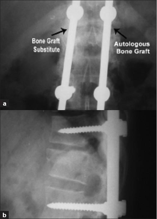
X-rayAnteroposterior and lateral lumbar spine radiographs one year after posterolateral fusion: continuous bridging bone mass between the instrumented levels, with the graft incorporated on both sides.
Source : Bansal et al., Indian journal of orthopaedics, 2009, figure 3 · CC BY
If the published timings are a matter of surgical school and if the trial that tested a date found the early one inferior, then a dated progression is not defensible. A criterion-based progression is, provided the criteria are measured and not felt.
Why the calendar fails here more than elsewhere
Three reasons add up. First, surgical variability is real: a single-level TLIF in a 45-year-old patient and a three-level fusion with iliac graft in a 74-year-old osteoporotic woman share neither the same safety framework nor the same speed of consolidation. Next, the variability of the starting point is greater still: four patients in five were already below the activity guidelines before the operation, and one in six took fewer than 5,000 steps a dayPMID 30305065. Finally, fear of movement, present in two thirds of those operated on, completely dissociates what the patient can do from what they doPMID 40212616.
This dissociation is not a hypothesis. The twenty-four-month follow-up of the PREPARE trial measured significantly improved physical capacity and functional scores, and physical activity that, for its part, did not progressPMID 38753831. A calendar that advances the programme because six weeks have gone by therefore advances at the mercy of two variables it does not measure.
Tests that have been validated in this precise population
This is what makes criterion-based progression practicable here rather than elsewhere: there are physical capacity measures whose responsiveness and minimal important change threshold have been determined in lumbar fusion patients, and not extrapolated from another population. In 118 patients tested eight to twelve weeks before the operation and again six months afterwards, the one-minute stair climb, the 50-foot walk and the timed up-and-go showed adequate responsiveness, with at least 80 % of the prespecified hypotheses confirmed. The five-minute walk, for its part, failed (40 % of hypotheses confirmed). The minimal important change thresholds for improvement are set at 20.0 steps for the one-minute stair climb, at −0.6 second for the 50-foot walk and at −1.3 second for the timed up-and-goPMID 30927913.
Three tests, three thresholds, a few minutes of assessment. That is the objective foundation missing from most dated protocols.
| Measure | Minimal important change threshold | Validated in which population |
|---|---|---|
| Stair climb, 1 minute | 20.0 steps | 118 lumbar fusion patients, before and 6 months after. Adequate responsiveness. |
| 50-foot walk (15.24 m) | −0.6 second | Same cohort. Adequate responsiveness. |
| Timed up-and-go | −1.3 second | Same cohort. Adequate responsiveness. |
| 5-minute walk | 45.5 m, but to be used with caution | Same cohort: responsiveness insufficient (40 % of hypotheses confirmed). |
| Tampa scale (TSK) | > 6 points on the total score > 4 on the “harm” subscale > 2 on “activity avoidance” | 180 patients in motor and cognitive-behavioural rehabilitation after fusion. |
| Oswestry Disability Index | ≥ 14.3 points | Threshold used in the multicentre quality registry for grade 2 spondylolisthesis, at 60 months. |
| Numerical rating scale, radicular pain | ≥ 1.7 point | Same registry. Back pain: ≥ 1.6 point. |
Sources: Jakobsson 2019 (PMID 30927913), Monticone 2017 (PMID 27827516), Alan 2026 (PMID 42066348).
Four phases, defined by what must have been achieved to move on to the next
The timeline that follows sets no date for moving on. The time markers given in brackets are orders of magnitude observed in the trials cited, not instructions: a patient may cross a criterion earlier, and many cross it later. The safety framework (permitted load, flexion, brace) remains in every case the one set by the surgeon.
Progression timeline after lumbar fusion, by criteria
Each phase closes on what has been measured, not on a date reached. The times in brackets are indicative.
Thresholds from Jakobsson 2019 (PMID 30927913), Monticone 2017 (PMID 27827516) and Alan 2026 (PMID 42066348). The time markers are orders of magnitude observed in the trials cited in this article; none of them has been validated as a criterion for moving on. The safety framework remains the surgeon's.
Two warnings on how to use this timeline
First warning: it does not replace the surgical instruction, it sits inside it. A criterion that has been met does not authorise a load the surgeon has forbidden. If the operative report limits lifting to five kilograms for three months, phase 3 is run under that limit, with variants that do not require the load: work in partial unloading, increasing volume rather than intensity, blood flow restriction where it is well mastered.
Second warning: none of these thresholds has been validated as a criterion for moving on. They are minimal important change thresholds, that is, markers saying “this patient has genuinely progressed”, measured in this population. Turning them into gateways to the next phase is clinical reasoning, not a trial result. It is better than a calendar, but it does not have the same status as evidence.
Key points from this chapter
- Three tests have been validated as sensitive to change in these particular patients: the one-minute stair climb, the 50 foot walk, timed up-and-go. The five-minute walk has not.
- The usable thresholds are quantified: 20 steps, −0.6 s, −1.3 s, more than 6 points on the Tampa scale, 14.3 points on the Oswestry.
- Criterion-based progression sits inside the surgical safety framework; it never widens it.
- Capacity is not enough: the criterion for the final phase concerns an activity actually kept up, not a test passed.
What is adjacent segment syndrome, and what can the physiotherapist really do about it?
This is the long-term stake of fusion, the one played out in years rather than months. It justifies part of the motor control and spinal care work, provided one is honest about what that work has demonstrated, and about what it has not.
Two words that must stop being confused
The literature carefully distinguishes two entities that everyday language merges. Adjacent segment degeneration is an imaging finding: disc space narrowing, facet joint osteoarthritis, radiological instability at the level above or below the construct. Adjacent segment disease is a clinical entity: symptoms attributable to that level, marked enough to justify management, or even revision surgery.
The distinction is not academic: the most cited literature review on the subject establishes that asymptomatic radiological degeneration is common and that it does not correlate with functional outcomesPMID 15534420, a finding taken up by the 2016 meta-analysis, which notes that radiological degeneration shows no strong correlation with clinical outcomesPMID 26836484. A patient who reports “wear above the fusion” seen on a follow-up radiograph is therefore not necessarily reporting bad news, and that is the first thing to tell them.
How many? The figures depend on what is being counted
Estimates vary by a factor of ten depending on the definition used, the length of follow-up and the population. Presenting them side by side is more useful than choosing one of them.
Four sources, four ways of counting the adjacent segment
The differences do not reflect a contradiction but different definitions and different follow-up durations. The right-hand column shows what was actually counted.
Sources: Zhang 2016 (PMID 26836484), Donnally 2020 (PMID 32445805), Cannizzaro 2023 (PMID 36345970), Yuan 2022 (PMID 36376871). An older review reported radiological degeneration of 5.2 to 100 % depending on the series, for a symptomatic form of 5.2 to 18.5 % (Park 2004,PMID 15534420).
The mechanism, and what it honestly allows us to say
The biomechanical reasoning is simple and long-standing: after fusion, intradiscal pressure, facet joint loading and mobility increase at the neighbouring levelsPMID 15534420. One segment no longer moves: those around it move more, and under higher stress. It is intuitive, it has been measured on cadavers, and it is enough to explain why the question arises.
This reasoning nevertheless runs into a serious objection, which has to be known so as not to over-interpret. The 2020 meta-analysis that compared fusions with motion-preserving techniques found no statistically significant difference between the two in the incidence of degeneration (36.4 % versus 19.2 %; p = 0.06), of clinical disease (10.7 % versus 4.42 %; p = 0.25) or of revisions (7.40 % versus 1.80 %; p = 0.19); only a fixed-effects analysis found an advantage for motion-preserving techniquesPMID 32445805. The respective share of the mechanical stress induced by the fusion and of the simple progression of degenerative disease with age has not been settled. The 2004 review said as much already: age-related spinal degeneration is regarded as a major contributor.
The identified risk factors bear this out. The one most consistently associated is the number of levels fused: the 2016 meta-analysis concludes that limiting the number of levels weighs more heavily on risk than the choice of fusion strategy, and that neither sex, nor age, nor segmental sagittal alignment, nor fusion method, nor instrumentation was associated with risk in its dataPMID 26836484. The 2023 meta-analysis adds advanced age and obesityPMID 36345970, and the cohort of 718 minimally invasive TLIFs identifies bone density, body mass index and pre-existing disc degeneration of the adjacent levelPMID 36376871.
No controlled trial has demonstrated that a rehabilitation programme reduces the incidence of adjacent segment syndrome. What follows is reasoning from the risk factors, and it must be presented as such.
What the physiotherapist can do, and the status of each of these actions
Three registers need to be kept apart, and they do not carry the same weight.
First register: acting on documented risk factors, by means whose effectiveness is established elsewhere. Obesity and low bone density are among the risk factors for the adjacent segment; physical activity acts on both, and that action is well established. It is not proof that rehabilitation prevents adjacent segment problems, but it is a defensible chain of reasoning. It is also the most concrete lever the practice has.
Second register: restoring a distribution of movement that does not overload the neighbours. A patient whose hip and thoracic spine are stiff will ask more of the two mobile levels flanking the fusion. Working on hip extension, thoracic rotation and pelvic girdle–shoulder girdle dissociation is consistent with the mechanism described; no study has measured that this changes the incidence. It is applied mechanical reasoning, not data.
Third register: educating without frightening. This is where the distinction made at the start regains its full value. Telling a patient they “will wear out the neighbouring levels” is both imprecise and counterproductive in someone two thirds of whose fellow patients have measurable kinesiophobia. The exact wording is more reassuring: an image of degeneration will probably appear, it is common and does not predict symptoms; the form that genuinely causes trouble is markedly rarer; and it comes late, 62.7 months on average in the cohort most precise on this pointPMID 36376871.
The patient who consults years later for new pain above their construct calls for full clinical reasoning, not for a diagnosis by deduction. The presentations to consider are those the site already covers: stenosis of the level above, facet joint arthropathy, sciatica from disc herniation, or sacroiliac pain whose frequency increases after lumbosacral fusion. Pain above a fusion is not automatically adjacent segment syndrome.
Key points from this chapter
- Radiological degeneration and clinical disease are two different things: the first is common and poorly correlated with symptoms, the second is rarer.
- The most solid annualised rates are 5.9 % per year for the imaging finding and 1.8 % per year for disease requiring revision.
- The risk factor found most consistently is the number of levels fused; to it are added age, obesity, low bone density and pre-existing degeneration of the neighbouring level.
- The share due to the stress induced by the fusion and the share due to natural ageing have not been separated: the comparison with motion-preserving techniques does not reach significance.
- No trial has shown that rehabilitation prevents this syndrome. What we do rests on reasoning about risk factors, and it must be put to the patient as such.
Which postoperative red flags should bring the session to a halt?
The red flags of the operated spine are not those of the untouched spine. Pain that increases is not alarming in itself; pain that changes in nature is. A fever on day 3 is open to discussion; a fever at three weeks with a weeping wound is not.
The signs that stop the session and prompt a call to the surgeon
- A new or worsening neurological deficit. Loss of segmental strength, foot drop, areflexia appearing since the last session. After minimally invasive lumbar decompression, postoperative epidural haematoma occurs in 1.4 % of cases, and 64.3 % of those patients present with a neurological deficitPMID 32361024. The prognosis depends on the delay between the onset of symptoms and surgical evacuation: it is counted in hoursPMID 25537329.
- Sphincter disturbance, saddle anaesthesia, urinary retention. Cauda equina syndrome does not belong to the period before the operation alone. A published case describes it on the eleventh postoperative day, on a migrated herniation of the adjacent level, in a patient discharged improved four days earlierPMID 34474611. It is a surgical emergency, and the site gives it a whole article: this box flags the problem, it does not deal with it.
- Fever, discharge, redness or wound dehiscence. Surgical site infection affects 1.53 % of patients operated on the lumbar spine by a posterior approach in a series of 7,240 patientsPMID 34410502, and 2.9 % in a meta-analysis of 26 studies and 41,624 patients undergoing spinal surgery at all levels combinedPMID 35212281.
- A pain that changes in nature and not in intensity. Mechanical pain that becomes constant, nocturnal, unaffected by position, or that wakes the patient after having settled. This is the picture that must raise the possibility of spondylodiscitis, including late and including at the adjacent level: a published case describes an adjacent segment infection revealed two months after a three-level fusion, in a patient initially relieved, with blood cultures that remained negativePMID 32395359.
- A sudden pain occurring during a movement, with a sense of cracking and loss of function. It calls for stopping and for an opinion, if only to rule out a fracture, screw loosening or cage subsidence: events whose risk is increased when bone density is lowPMID 38962714.
- Unilateral calf pain, swelling, breathlessness. Thromboembolic disease remains a complication of any major surgery in a patient whose mobility has fallen. It is not treated in the practice.
The rule that simplifies: faced with one of these signs, the exercise is not adapted, the session is stopped and the surgical team is contacted. The only cost of an unjustified alert is a phone call; the cost of a delay on a compressive haematoma or a cauda equina is neurological and permanent.
Surgical site infection: rare, but rarely early in the way it presents
The figure to remember is an order of magnitude around 1.5 to 3 %, depending on the population and the definition. In the series of 7,240 patients operated on the lumbar spine by a posterior approach, the overall incidence was 1.53 %, with two findings useful to the physiotherapist: patients operated on for lumbar stenosis had the highest incidence (2.39 %; p < 0.001), and the number of cases differed significantly from one surgeon to anotherPMID 34410502. The meta-analysis of 26 studies finds 2.9 % and identifies among the associated factors the number of levels fused, osteoporosis, diabetes, hypertension, transfusion and previous surgery; by contrast, neither age, nor sex, nor smoking, nor body mass index, nor operating time was associated with itPMID 35212281.
A series of 448 consecutive open TLIFs sets out the factors retained in multivariate analysis: thickness of the subcutaneous fat panniculus, high preoperative ASA score, low preoperative serum albumin and prolonged drainage durationPMID 31394358. The physiotherapist following an undernourished, diabetic patient operated on at three levels therefore has reason to look at the wound at every session in the first weeks, and not merely to ask how things are going.
What is not a red flag, and what one must know not to dramatise
The counterpart matters just as much. Three situations often cause needless alarm:
- Stiffness and pain of the scar on early mobilisation. After a posterior approach, the paravertebral muscles have been retracted or detached, and the atrophy measured is significantly more marked than after an anterior approach or a minimally invasive techniquePMID 26840699. It is a muscular scar, not a sign of failure.
- Weakness of hip flexion and reduced sensation over the front of the thigh after a lateral approach. The route crosses or skirts the psoas, in contact with the lumbar plexus; these signs are most often transient. They call to be recorded, dated and monitored, not for the programme to be stopped, as long as they are receding.
- An image of degeneration at the adjacent level on a follow-up radiograph. It is common and does not correlate with clinical outcomesPMID 15534420. Commenting on it as a complication, in a patient whose fear of movement is already high, costs more than it gains.
| What the patient presents with | What it may point to | Usual timing | What to do |
|---|---|---|---|
| New motor deficit, rapid worsening | Compressive epidural haematoma, nerve root compression | Hours to days after the operation | Immediate stop, surgical contact the same day. The prognosis depends on the time to evacuation. |
| Sphincter disturbance, saddle anaesthesia | Cauda equina syndrome, including from herniation of the adjacent level | Described as early as the 11th postoperative day | Emergency. Referral without delay. |
| Fever, discharge, redness, dehiscence | Surgical site infection, superficial or deep | First weeks, sometimes later | Stop the session, prompt medical opinion. Do not massage or mobilise the area. |
| Pain that has become constant and nocturnal, after a period of improvement | Spondylodiscitis, including of the adjacent segment | Described at 2 months in a published case | Medical opinion with imaging. Blood cultures may remain negative. |
| Sudden pain in the course of a movement, with loss of function | Fracture, screw loosening, cage subsidence | Variable, increased if bone density is low | Stop, seek an opinion. Do not repeat the same exercise before an answer comes. |
| Iliac crest pain away from the incision | Bone graft harvest site | From the immediate postoperative period | Not a red flag. To be distinguished from sacroiliac or trochanteric pain. |
Key points from this chapter
- The most discriminating sign is not the intensity of the pain but its change in nature: constant, nocturnal, indifferent to position.
- Compressive epidural haematoma plays out in hours; neurological deficit is the commonest mode of presentation.
- Cauda equina syndrome can appear after the operation, at the adjacent level, in a patient discharged improved.
- Surgical site infection runs at around 1.5 to 3 %, and lumbar stenosis is the indication most exposed to it.
- Conversely, scar stiffness, psoas weakness after a lateral approach and an image of adjacent degeneration are not red flags.
What can honestly be predicted: pain, work, sport?
Since satisfaction depends on the gap between the result and the expectation, the question of prognosis is not an optional extra: it is a therapeutic act. It still has to be answered with figures that exist.
Pain and disability: a majority who do well, a minority left with something
The two versions of the same result deserve to be stated together. On the favourable side: of 328 patients operated on for a grade 2 spondylolisthesis and followed for five years, 86 % declared themselves satisfied, with mean low back pain at 2.6 out of 10, radicular pain at 2.3 and an Oswestry of 8.9 among the satisfied, against 5.3, 5.2 and 19.5 respectively among the dissatisfiedPMID 42066348. On the unfavourable side: the authors of the British meta-analysis recall in the introduction to their work that 40 % of patients still have significant back-related disability twelve months after a lumbar fusionPMID 26555833, and the REFS trial opens on the observation that 40 % of those operated on describe themselves as uncertain about or dissatisfied with their resultPMID 30788599.
These figures do not contradict one another: the first concerns a well-founded indication with five years of follow-up, the others mixed populations at one year. What they say together is usable in consultation: the majority are clearly better, a substantial minority keep something, and knowing this in advance protects against a catastrophic reading of the smallest residual symptom.
Individual prognosis, by contrast, is predicted badly. A systematic review of sixteen prospective studies and 8,388 patients, looking for prognostic factors after lumbar fusion, concludes that no moderate- or high-certainty evidence exists. Only two factors reach low certainty: more severe preoperative radicular pain predicts greater improvement in that radicular pain, and being in work before the operation predicts less postoperative disability at one or two yearsPMID 34705106. Any individual prediction more precise than that goes beyond what the literature allows.
Return to work: three months, and a predictor that is not surgical
This is where the data are most usable. Of 1,805 employed patients drawn from the Canadian CSORN surgical registry and operated on with elective one- or two-level lumbar surgery, 71 % returned to work, with a median of 61 days across all procedures. The breakdown by procedure speaks louder: 46 days after laminectomy, 51 after discectomy, 90 days after fusionPMID 34560636. A second analysis of the same registry, restricted to the 1,290 patients who were working before the operation, finds a return rate of 82 % at one year and a median of about ten weeks after fusion, and above all, this median did not vary with the intensity of the work, whereas it did vary after surgery without fusion. Return rates ranged from 84 % for sedentary jobs to 77 % for heavy to very heavy jobsPMID 35725390.
The strongest predictor is neither the level operated on nor the technique. Patients who were working before the operation were nearly twice as likely to return within 90 days (hazard ratio 1.984; 95 % CI 1.680 to 2.344; p < 0.001), whereas those who were employed but not working were four times more likely never to return (odds ratio 4.076; 95 % CI 3.087 to 5.383; p < 0.001). The other factors associated with failure to return were a symptom duration of more than two years, a high number of comorbidities, a low level of education and an ongoing compensation claimPMID 34560636. Smoking adds to these: the meta-analysis on tobacco finds a lower rate of return to work in smokers (odds ratio 0.70; 95 % CI 0.54 to 0.90)PMID 41066602.
Return to work after elective lumbar surgery
Canadian CSORN registry, 1,805 employed patients. Fusion lengthens the median time to return by about six weeks compared with a procedure without fusion.
Source: Singh 2022, CSORN registry (PMID 34560636). Supplemented by Singh 2024 on 1,290 patients working before the operation: 82 % returning at one year, a median of about ten weeks after fusion, independent of job intensity (PMID 35725390). Canadian data: the French insurance context differs.
The most powerful determinant of return to work is settled before the incision, and it is not surgical: it is still being in work. A prolonged period off work before the operation multiplies the risk of never returning by four.
Sport: sparse data, and no validated criteria
It has to be said plainly: for lumbar fusion there is no equivalent of the return-to-sport test batteries developed for the anterior cruciate ligament. The available data are limited to a few case series.
The most cited concerns golf. Of 353 patients operated on with a one- or two-level fusion by a single surgeon, 200 answered a questionnaire, among them 34 golfers with a mean age of 57 years. In 79 % of them, preoperative pain significantly limited play. In the year following the operation, 65 % had resumed practice and 52 % the course; 29 % reported that pain still limited their game; 25 patients, that is 77 %, were playing as much as or more than before the operation, and among those who gave their handicap, 80 % had kept or improved itPMID 27879299. This is a level-4 case series, in a rotational sport but one without impact or contact: it permits no extrapolation to running, team sports or sporting load-carrying.
In the absence of validated criteria, the decision is built on three supports: the surgeon's explicit agreement, demonstrated tolerance of progressive loading in the target discipline, and the quality of motor control under fatigue. Practice surveys place the resumption of intense activities and contact sports at around six months after disc surgeryPMID 41666866 : a rule of thumb, not data, and one that does not even concern fusion.
A protective factor the physiotherapist can activate
The Korean national cohort covering 1,029,500 adults operated on the lumbar spine between 2009 and 2023 estimates the incidence of persistent pain syndrome at 26.7 %, combining two definitions: later revision surgery, or the coding of a post-laminectomy syndrome. The independent risk factors include age, peripheral polyneuropathy, obesity, diabetes, hypertension and current smoking. Three factors are protective instead: higher income, employment-linked insurance and, the one that concerns us, moderate physical activity, between 500 and 1,499 MET-minutes per weekPMID 42215594.
This is an association in a reimbursement database, not a causal effect demonstrated by a trial, and the figure covers lumbar surgery as a whole. But the order of magnitude speaks: 500 to 1,499 MET-minutes per week correspond roughly to 150 to 300 minutes of moderate-intensity activity, that is, the lower end of the general guidelines, and an achievable target for a patient 83 % of whose peers were not reaching it before the operation.
Key points from this chapter
- The majority do clearly better, 86 % satisfied at five years on a well-founded indication, and a substantial minority keep a disability, of the order of 40 % at one year in mixed populations.
- No moderate- or high-certainty evidence allows the result of a given patient to be predicted. Only two factors reach low certainty: the severity of preoperative radicular pain and being in work before the operation.
- The median return to work after fusion is 90 days, against 46 to 51 after a procedure without fusion, and it does not depend on the intensity of the job.
- The strongest predictor of return is preoperative and not surgical: still being in work before the operation.
- Return to sport has no validated criterion. The only usable series concerns golf: 52 % returning to the course within the year.
- Moderate physical activity is associated with less persistent pain at the scale of a national population. It is an association, not proof of an effect, but it points to a realistic target.
What do published case reports teach us?
Four real cases, all identified by their reference. Two describe rehabilitation conducted in difficult patients, two describe complications that declared themselves after an uneventful discharge from hospital. Together they map out the margin of uncertainty within which we work.
First case: a multilevel fusion, ten months of pain, and a programme that progresses on a test
A 68-year-old man is referred for physiotherapy ten months after a multilevel spinal fusion carried out for stenosis. He reports high levels of persistent pain and minimal activity, a direct consequence of that pain. The programme runs in three phases: pool exercise first, then progression to walking on land, the whole combined with transcutaneous nerve stimulation used for up to eight hours a day. Progression from one phase to the next is not decided on a time period: it is determined by the results of a shuttle test. The outcome measures are the daily pain level and the walking distance, followed as a rolling five-day average. Over that period, pain falls and the distance covered increases, with a trend towards lower pain as distance increases, and this despite poor medication adherence and high post-surgical painPMID 20690876.
What this case teaches the physiotherapist. Three things, two of which depend on neither the pool nor the nerve stimulation. First, progression was driven by a measurement, not by a calendar: exactly the logic of the previous chapter. Next, follow-up covered a capacity variable (the distance) as much as pain, which made it possible to see progress even on days when pain did not move. Finally, the patient was taken on ten months after the operation, a reminder that late management is not lost management. The authors themselves stress that there is no randomised trial comparing physiotherapy modalities for chronic low back and radicular pain associated with a multilevel fusion: it is an isolated case, and it demonstrates the effectiveness of none of its components taken separately.
Second case: a cauda equina syndrome on the eleventh postoperative day
A 55-year-old woman presents with low back pain with bilateral radiation resistant to six months of conservative treatment. Imaging finds a degenerative spondylolisthesis with stenosis at L3-L4 and L4-L5, and a simple disc bulge at L2-L3. She undergoes a full decompressive laminectomy with posterior fusion at L3-L4 and L4-L5. The stenosis symptoms improve markedly, and she is discharged on the seventh postoperative day.
Four days after discharge, that is on the eleventh postoperative day, she attends the emergency department with a bilateral motor deficit of sudden onset in the lower limbs, with urinary disturbance and difficulty defecating. Follow-up MRI shows a large disc herniation migrated backwards and a detached posterior longitudinal ligament, at L2-L3: a hyperacute deterioration of the adjacent segment causing a cauda equina syndrome. Emergency revision surgery, partial L2-L3 laminectomy and removal of the migrated fragment, allows the signs to regress. At one year, the patient is wellPMID 34474611.
What this case teaches the physiotherapist. It destroys two common shortcuts. The first: “the adjacent segment is a ten-year problem”, here, eleven days. The second: “a patient discharged improved is a patient out of danger”. This patient was better on discharge; the swing was sudden and complete. The authors themselves describe this presentation as extremely rare, and it is: this herniation is not looked for in every operated patient. What is retained is that questions about the sphincters and perineal sensation keep their full meaning after the operation, including in a patient who is getting better.
Third case: pain that returns at two months, with negative blood cultures
A 69-year-old woman with multilevel lumbar degenerative disease undergoes a three-level fusion. The symptoms that prompted the operation are relieved by the decompression. Two months later, severe low back pain reappears and forces readmission. MRI shows signs of spondylitis and discitis at the adjacent segment. No organism is identified, despite blood cultures taken before any antibiotic therapy. After prolonged anti-infective treatment with vancomycin, the patient is relieved and discharged. The pain returns nonetheless after prolonged oral antibiotic therapy, and a further admission takes place six months after the initial operation: CT shows frank destruction of the vertebral endplates at the adjacent level. Revision surgery with debridement and autologous iliac graft finally makes it possible to control the inflammatory markers and to obtain fusion of the infected spacePMID 32395359.
What this case teaches the physiotherapist. It illustrates precisely the red flag that is hardest to recognise: pain that returns after a period of clear improvement. The sequence (relief, then reappearance of severe pain at two months) is exactly the one a physiotherapist sees in the session, before anyone else. The case is also a reminder that negative blood cultures rule nothing out, which forbids taking reassurance from a blood work-up the patient reports as “normal”. Finally, it shows that the adjacent location is not reserved for degeneration: infection can settle there too.
Fourth case: graded exercise in a depressed patient after failed surgery
A 53-year-old man, a former serviceman, operated on with a lumbar discectomy and laminectomy with instrumented fusion, is admitted for recurrence of low back pain and pain radiating into the left lower limb. The failure of the surgery has triggered a major depressive episode, a comorbidity that in turn worsens the pain prognosis. After six weeks of routine treatment with no notable improvement, a three-week programme of graded exercise combined with manual therapy in movement is put in place. The scores improve: SF-36 physical component from 15.0 to 37.2, mental component from 21.9 to 30.1, numerical pain scale from 50 to 30. The daily walking distance increases significantly over the three weeks, in parallel with the therapeutic intensityPMID 38651058.
What this case teaches the physiotherapist, and what it does not. The acupuncture-in-movement component described here is not transposable to French practice, and a single case allows the improvement to be attributed to none of its components. What is transposable is the structure: six weeks of conventional management with no result, then a change of logic towards graded exposure with an explicitly planned progression of intensity, and a capacity measure, the walking distance, followed day after day. It is also one of the rare published cases that tackles depression head-on as an element of the post-surgical pain picture rather than as an incidental comorbidity.
What four cases do not prove
Nothing, in the strict sense. A case report demonstrates neither effectiveness, nor frequency, nor causality; it merely establishes that something happened at least once, and it is published precisely because it is rare or instructive. The two complication cases in this chapter must not therefore turn every session into anxious screening: the cauda equina on the eleventh day is described by its own authors as extremely rare, and adjacent segment infection is described as a rarely reported complication. What they bring is more modest and more solid: they fix the shape danger takes when it occurs, and that shape is recognisable, a sudden deficit, pain that returns after having settled. The two rehabilitation cases, symmetrically, do not prove that a protocol works; they show that late management, and management of a difficult patient, remains worthwhile, and that progression on measurement beats progression on the calendar.
How to apply all this from the first session?
This chapter does not offer a protocol: none exists. It offers a sequence of decisions, in the order in which they really arise, and indicating each time what each one rests on.
Before touching the patient: five lines to find, four questions to ask
The operative report gives the levels, the approach, the presence of a decompression, the graft and its site, and the explicit instructions. What it almost never gives are the four answers that structure the safety framework: the permitted load and its date, the flexion range allowed, the status of the brace, and whether any particular caution is linked to the construct. These four questions are put to the surgeon, in writing, and their answer is worth more than any general rule, because the literature that would produce that general rule does not existPMID 41817204.
At the first session: four measures that will serve as a reference
They take about ten minutes and make everything that follows readable.
- The timed up-and-go, whose minimal important change threshold in this population is −1.3 secondsPMID 30927913. It is the quickest and most reproducible measure in the practice.
- The Tampa scale, with its threshold of 37 for defining kinesiophobiaPMID 40212616 and its change threshold of more than 6 pointsPMID 27827516. Two thirds of patients will exceed the threshold: that is not a reason not to measure, it is the reason to measure.
- The Oswestry, a common reference with the surgeon, with a clinically important improvement threshold of 14.3 pointsPMID 42066348.
- Two separate numerical scales, back and leg. Separate, because it is their divergence that informs: thresholds of 1.6 and 1.7 points respectivelyPMID 42066348. A patient whose leg improves and whose back stalls is not a patient who is failing, it is a patient following the expected trajectory.
What has to be said, and which counts as much as what is prescribed
Four messages, all backed by data cited in this article, and all to be delivered early:
- “Your leg will probably get better, and faster, than your back.” Said beforehand, it is a prognosis. Said afterwards, it is an excuse. The divergence is real and documentedPMID 12973134.
- “The goal is what you will be able to do, not the number for your pain.” This is the direct translation of the 42 point satisfaction gap according to preoperative expectationsPMID 37797842.
- “Moving does not damage the construct.” No trial has reported hardware failure attributed to rehabilitation, including with strengthening started at three weeksPMID 29914580. This message is given within the loading framework set by the surgeon, never against it.
- “If you smoke, stopping now counts more than any exercise.” Odds ratio for nonunion 1.97, and complete catch-up in former smokers who have stopped for at least a yearPMID 41066602.
Choosing exercises with full knowledge of the facts
Session content is ranked by what the evidence supports, and not by habit:
| What goes into the session | Why | What it rests on |
|---|---|---|
| An explicit cognitive-behavioural component, not just corridor education | This is what distinguishes effective programmes from exercise alone, on disability and on fear of movement. | Meta-analysis of 18 trials, effects −0.31 and −0.64; Monticone 2014 and Archer 2016 trials. |
| Low-load motor control, early | The only large trial to have tested it finds less pain at three months and half as much persistent pain at one year. | Nie 2025, 395 patients, motor control from day 1 then stabilisation at week 5. |
| Walking, dosed and monitored | A capacity measure, graded exposure, and the target of the only protective factor identified at population level. | Jakobsson 2019 for the measure; Park 2026 for protective moderate activity. |
| Trunk strengthening, without expecting more of it than it gives | Real gains in strength and endurance in the short term, not found in the long term. | Özden 2022; Kernc 2018, whose effects had disappeared at 18 months. |
| Hip and thoracic spine mobility | Mechanical reasoning about the distribution of stress towards the neighbouring levels. No study measures an effect on the incidence of adjacent segment problems. | Mechanism described in Park 2004. Status: reasoning, not evidence. |
| A quantified and negotiated activity goal | Recovered capacity does not convert spontaneously into activity: this is the clearest finding of the 24-month follow-up of PREPARE. | Kemani 2024; Tegner 2024 for the modest effect of an intervention targeting sedentary behaviour. |
The operational summary
A lumbar fusion patient is rehabilitated within a safety framework that is patient-specific, set by the surgeon, and along a progression driven by measures rather than by dates. The best-supported content combines exercise and cognitive-behavioural work; the least useful content is isolated strengthening expected to be an end in itself. The two symmetrical errors to avoid are treating this patient as an ordinary low back pain patient, ignoring the two thirds with kinesiophobia and the pre-existing inactivity, and treating them as a fragile operated patient, confirming through excess caution the very fear that should be reduced.
Frequently asked questions
When can rehabilitation start after lumbar fusion?
It depends on what is meant by rehabilitation. Walking and low-load motor control start in the first few days, and this has been measured as safe: strengthening started at three weeks produced no hardware looseningPMID 29914580, and a sequential programme started on the first postoperative day reduced pain at three months and persistent pain syndrome at one yearPMID 39453543. By contrast, the only trial that compared two start dates for the same group strengthening programme found the six-week start inferior to the twelve-week startPMID 22565381. The dividing line is not the date, it is the content.
Which movements are forbidden after lumbar fusion?
No list commands consensus, and that is a fact to know rather than a gap to fill. An international survey of 703 spine surgeons found activity restrictions prescribed in 79 % of cases after disc surgery, with durations ranging from two weeks for prolonged standing and driving to three months for bending, twisting and liftingPMID 41666866. These durations reflect surgical habit more than evidence, and the authors say so themselves. The instruction of the surgeon who operated on this particular patient therefore takes precedence over any general rule.
Is a brace needed after lumbar fusion?
The available evidence does not support it: four randomised trials in a systematic reviewPMID 31676949 then five trials and 362 patients in an updated meta-analysisPMID 38744484 find no benefit on disability, pain, complications or fusion rate. Thirty-four per cent of surgeons prescribe one anyway, a figure measured after disc surgery and not after fusion. If the patient is wearing one, it is not removed on the physiotherapist's own initiative: the surgeon is asked.
What is adjacent segment syndrome?
It is the deterioration of the mobile level situated above or below the fused construct. A meta-analysis of 31 studies and 4,206 patients estimates the incidence at 5.9 % per year for radiological degeneration and 1.8 % per year for the symptomatic form requiring revisionPMID 26836484. The factor most consistently associated with it is the number of levels fused. The imaging finding, which is common and poorly correlated with symptoms, must be distinguished from the clinical disease.
Does rehabilitation prevent adjacent segment syndrome?
No controlled trial has shown that it does. What rehabilitation can do is act on identified risk factors (obesity, low bone density, inactivity), by means whose effectiveness is established elsewhere, and redistribute mobility towards the hip and the thoracic spine, which is consistent with the described mechanism without having been measured. It has to be presented to the patient in those terms, and not as a proven protection.
Why is my patient disappointed when the surgery succeeded?
Because satisfaction depends on the gap between the outcome and the expectation, not on the outcome alone. Among patients operated on for a one- to two-level fusion, those with the highest preoperative expectations were satisfied in only 43 % of cases against 85 % among those with low expectations, even though their disability scores at one year were comparablePMID 37797842.
When will my patient be able to return to work?
Among 1,805 employed patients in the Canadian CSORN registry, 71 % returned to work, with a median of 90 days after fusion against 46 to 51 days after surgery without fusionPMID 34560636. A second analysis finds a median of about ten weeks, independent of job intensityPMID 35725390. The strongest predictor is not surgical: not working before the operation multiplies the risk of never returning by four.
Which signs should interrupt the session?
A new or worsening neurological deficit, bladder or bowel dysfunction or saddle anaesthesia, pain that changes in nature rather than in intensity, a fever or discharge from the wound, and night pain that wakes the patient after it had settled. Cauda equina syndrome can occur after the operation: a published case describes it on the eleventh postoperative day, from a herniation at the adjacent level, in a patient discharged improvedPMID 34474611. A compressive epidural haematoma plays out in hours, not in daysPMID 25537329.
Is supervised physiotherapy really useful after fusion?
Yes, with a low level of evidence and an effect that fades. The meta-analysis of 18 trials and 1,402 patients finds a benefit of exercise on disability and pain in the short term, and a superiority of multimodal rehabilitation over exercise alone for disability and fear of movement; the differences disappear beyond one yearPMID 35258644. That is not an argument for doing nothing, it is an argument for doing something other than strengthening alone.
Can my patient return to sport?
The data are scarce and mostly concern low-demand sports. In a series of 34 golfers operated on for a one- or two-level fusion, 65 % had returned to practice and 52 % to playing a round within the year, and 77 % played as much as or more than beforePMID 27879299. There is no validated return criterion for lumbar fusion: the decision is made on load tolerance, the quality of motor control and the surgeon's agreement.
Should the patient be prepared before the operation?
The idea is attractive and the trials are disappointing. A dedicated meta-analysis concludes that cognitive-behavioural prehabilitation programmes do not improve postoperative outcomes, with very low to low certainty of evidencePMID 33356804, and the PREPARE trial finds no benefit either at six monthsPMID 30951604 or at twenty-four monthsPMID 38753831. That does not make the preoperative period useless: 83 % of these patients do not meet activity guidelines before the operationPMID 30305065, and still being at work is the best predictor of return.
My patient has pain above the scar three years on. Is it necessarily the adjacent segment?
No. Pain above a fusion calls for full clinical reasoning, and the presentations to consider are those the site already covers: stenosis at the level above, facet joint arthropathy, radicular pain from disc herniation, sacroiliac pain, or a thoracolumbar junction syndrome when the pain refers towards the iliac crest or the groin. Adjacent radiological degeneration, for its part, is common and does not correlate with symptomsPMID 15534420.
How do I find out what the surgeon actually did?
By reading the operative report, which remains the source of truth. Five elements change the session: the levels fused and their number, the surgical approach, the presence of an associated decompression, the existence of a graft and its harvest site, and the surgeon's explicit instructions. Where a general rule and a named instruction diverge, the instruction prevails.
References and verification
The references in this article were checked one by one against PubMed metadata, through the NCBI E-utilities API, on 15 August 2026: full author list, journal, year, volume, issue, pagination and DOI. The figures quoted come from structured abstracts or full texts, never from a reconstruction. The ICD-11 codes in the structured data were checked against the simple tabulation list published by the World Health Organization. Since no French national incidence data for lumbar fusion are published in a peer-reviewed journal, no French figure for surgical volume is put forward here.
To go further on the conditions that lead to fusion, the site covers degenerative lumbar spondylolisthesis, degenerative lumbar stenosis, facet joint arthropathy, sciatica from disc herniation and non-specific low back pain. For the one emergency that requires a session to be stopped without delay, see cauda equina syndrome.

