















In brief
Non-specific low back pain is pain located between the costal margin and the gluteal folds, with or without radiation, for which no specific pathological or anatomical cause can be identified; this diagnosis of exclusion accounts for 90 to 95 % of low back pain seen in primary care. The leading cause of disability worldwide, its chronicity is predicted above all by psychosocial factors (catastrophising, kinesiophobia, depression) and sustained by central sensitisation rather than by a structural lesion. Once red flags have been ruled out, first-line treatment is non-pharmacological: education, advice to stay active, self-management and exercise, with no type of exercise proving superior. GBD 2021 estimates that 619 million people were affected in 2020.
Clinical synthesis based on the most recent meta-analyses and pivotal trials: GBD 2021, RESTORE Lancet 2023, the Lancet Series 2018, NICE NG59 and prospective data from 2025-2026.
Clinical summary
- The non-specific low back pain is a diagnosis of exclusion that accounts for 90–95 % of low back pain cases in primary care.1,9
- It is the leading cause of disability worldwide. GBD 2021 estimates 619 million people affected in 2020, with a projection of 843 million in 2050.2
- The psychosocial factors (catastrophising, kinesiophobia, depression) are the strongest predictors of progression to chronicity.10,11
- Chronicity is sustained by mechanisms of central sensitisation and nociplastic plasticity rather than by a persistent structural lesion.3
- The recurrence rate within the year is high (≈ 33 % at 1 year, up to 73 % depending on the definition used).14
- The initial assessment must systematically rule out red flags (fracture, infection, tumour, cauda equina) before concluding that the low back pain is non-specific.4,5
- Bardin's diagnostic triage: non-specific 90-95 %, radicular 5-10 %, specific pathology < 1 %.6
- Imaging is not indicated routinely in the absence of a red flag: it may reveal abnormalities unrelated to the pain and generate a nocebo effect.1,7
- Stratification by risk group (STarT Back Tool) improves clinical outcomes and cost-effectiveness.8
- The first-line treatment is non-pharmacological: education, advice to stay active, self-management (APTA 2021, NICE NG59).12,15
- Therapeutic exercise is a pivotal intervention, but no type is superior to another: the choice should follow the patient's preferences.16,17
- The manual therapy offers modest short-term relief. It complements the active approach (exercise + education), it does not replace it.18
- The Cognitive Functional Therapy (CFT) (the RESTORE trial, Lancet 2023) clinically reduces pain and disability in disabled chronic patients, with effects maintained at 3 years.19,20
- Self-management combining pain neuroscience education (PNE) and exercise is the cornerstone of recurrence prevention.21,22
- Return to sport must be based on functional and psychological criteria, never on a fixed timetable.23
- Serious conditions (leaking aortic aneurysm, pyogenic spondylodiscitis, vertebral metastases) can mimic common low back pain: vigilance must be constant.4,5
- Refer to the doctor if a red flag is present; to a specialist psychologist if yellow flags are marked and persistent.11,15
- The PROMs (validated instruments such as the Oswestry Disability Index and the NPRS) are essential for documenting progress and supporting shared decision-making.12
Contents
- What are the fundamentals to know about non-specific low back pain?
- How can non-specific low back pain be assessed and diagnosed with confidence?
- Which treatment strategies are most effective for non-specific low back pain?
- How do you secure lasting recovery and prevent recurrence of non-specific low back pain?
- What do real case studies teach us about non-specific low back pain?
- How do you apply these recommendations concretely in your practice?
What are the fundamentals to know about non-specific low back pain?
How is this condition defined, who does it affect and what are the risk factors?
Non-specific low back pain is defined as pain and discomfort located below the costal margin and above the inferior gluteal folds, with or without pain referred into the leg, for which no specific pathological or anatomical cause (infection, tumour, osteoporotic fracture, structural deformity, inflammatory disorder, radicular syndrome or cauda equina syndrome) can be identified.¹ This definition by exclusion covers the overwhelming majority of cases in primary care: roughly 90–95 % according to Bardin's consolidated diagnostic triage (Med J Aust 2017): non-specific 90-95 %, radicular 5-10 %, specific pathology < 1 %.²,⁶ Epidemiologically, low back pain has been the leading global cause of years lived with disability (YLDs) for nearly three decades.¹ The Global Burden of Disease 2021 analysis estimates that 619 million people were living with low back pain in 2020, with a projection of 843 million by 2050 because of population growth and ageing.² Later estimates (Wu 2020, Ann Transl Med) confirm an age-standardised point prevalence of about 7.5 %, although the absolute number of cases rose from 377.5 M (1990) to 577 M (2017).³ 🌍📊 Global burden of low back pain: GBD 2021 (Lancet Rheumatol 2023)
Number of people living with low back pain (in millions), 1990 → 2050 projection
Source: GBD 2021 Low Back Pain Collaborators. Lancet Rheumatol. 2023;5(6):e316-e329. PMID 37273833.
- Individual and anthropometric factors : increasing age, female sex, high body mass index, previous low back pain. Smoking is associated with a pooled OR of 1.30 in the Shiri 2010 meta-analysis (Am J Med).⁴
- Psychosocial factors : the most powerful predictor of progression to chronicity and of persistent disability. Pain catastrophising, kinesiophobia (fear of movement), anxiety, depression, perceived stress and low expectations of recovery. Wertli's systematic review (Spine J 2014) confirms that fear-avoidance beliefs are a robust prognostic factor in primary care patients.¹⁰,¹¹
- Lifestyle and work-related factors : low levels of physical activity, sleep disturbance, heavy physical demands (lifting, awkward postures, whole-body vibration).¹
🧠 Hierarchy of prognostic factors for chronicity (2018-2023 synthesis)
Strength of association (relative effect size) between risk factors and progression to chronicity
Qualitative synthesis: the quantitative reading of the ORs varies with the definitions used. Sources: Hartvigsen Lancet 2018; Wertli Spine J 2014; Shiri Am J Med 2010; Chiarotto NEJM 2022.1,10,4,9
What happens in the body and how does low back pain evolve naturally?
The pathophysiology of non-specific low back pain cannot be reduced to a structural lesion. While acute pain (< 6 weeks) is often initiated by a nociceptive mechanism (activation of pain receptors in muscles, ligaments, facet joints and the annulus fibrosus), progression to chronicity (> 3 months) involves profound neuroplastic changes in the central nervous system.1,9 A key mechanism is central sensitisation, which may be nociplastic in the 2017 IASP terminology, reconceptualised by Nijs et al. (J Clin Med 2021) under the heading of nociplastic pain phenotyping. It is a hyperexcitability of central nervous system neurons that amplifies pain processing: stimuli that are normally not painful may be perceived as painful (allodynia) and painful stimuli are perceived as more intense (hyperalgesia).3 🧠 This phenomenon partly explains why chronic pain persists after tissue healing and why its intensity correlates poorly with imaging findings.1,9- Rapid improvement : most patients experience a significant improvement in pain and function within the first 6 weeks. The Itz 2013 review (Eur J Pain) nevertheless reported that ≈ 65 % of patients still report pain at 1 year, which challenges the idea that complete resolution is common.13
- Recurrence : the Stanton 2008 prospective cohort (Spine, n = 973) showed a 12-month recurrence rate of ≈ 33 % under the strict definition, and 73 % under the broad definition: far lower than historical estimates but clinically significant.14
- Chronicity : about 20 % of patients with acute low back pain go on to develop chronic low back pain beyond 3 months; the prognosis is less favourable in the presence of catastrophising, radiation into the leg, low self-efficacy and depression.1,10,11
- The non-specific low back pain is a diagnosis of exclusion that accounts for 90-95 % of low back pain cases in primary care.1,6
- It is the leading cause of disability worldwide, affecting 619 M people in 2020, projected to reach 843 M in 2050.2
- Psychosocial factors (catastrophising, kinesiophobia, depression) are the strongest predictors of progression to chronicity.10,11
- Chronicity is sustained by central / nociplastic sensitisation rather than by a persistent structural lesion.3
- The recurrence rate at 1 year is about 33 % (Stanton 2008): far lower than historical estimates but clinically major.14
Bibliography
- Hartvigsen J, Hancock MJ, Kongsted A, et al. What low back pain is and why we need to pay attention. Lancet. 2018;391(10137):2356-2367. PMID 29573870.
- GBD 2021 Low Back Pain Collaborators. Global, regional, and national burden of low back pain, 1990-2020, its attributable risk factors, and projections to 2050: a systematic analysis of the Global Burden of Disease Study 2021. Lancet Rheumatol. 2023;5(6):e316-e329. PMID 37273833.
- Nijs J, Lahousse A, Kapreli E, et al. Nociplastic Pain Criteria or Recognition of Central Sensitization? Pain Phenotyping in the Past, Present and Future. J Clin Med. 2021;10(15):3203. PMID 34361986.
- Shiri R, Karppinen J, Leino-Arjas P, Solovieva S, Viikari-Juntura E. The association between smoking and low back pain: a meta-analysis. Am J Med. 2010;123(1):87.e7-35. PMID 20102998.
- Maher C, Underwood M, Buchbinder R. Non-specific low back pain. Lancet. 2017;389(10070):736-747. PMID 27745712.
- Bardin LD, King P, Maher CG. Diagnostic triage for low back pain: a practical approach for primary care. Med J Aust. 2017;206(6):268-273. PMID 28359011.
- Foster NE, Anema JR, Cherkin D, et al. Prevention and treatment of low back pain: evidence, challenges, and promising directions. Lancet. 2018;391(10137):2368-2383. PMID 29573872.
- Hill JC, Whitehurst DG, Lewis M, et al. Comparison of stratified primary care management for low back pain with current best practice (STarT Back): a randomised controlled trial. Lancet. 2011;378(9802):1560-1571. PMID 21963002.
- Chiarotto A, Koes BW. Nonspecific Low Back Pain. N Engl J Med. 2022;386(18):1732-1740. PMID 35507483.
- Wertli MM, Rasmussen-Barr E, Weiser S, Bachmann LM, Brunner F. The role of fear avoidance beliefs as a prognostic factor for outcome in patients with nonspecific low back pain: a systematic review. Spine J. 2014;14(5):816-836.e4. PMID 24412032.
- Wertli MM, Rasmussen-Barr E, Held U, Weiser S, Bachmann LM, Brunner F. Fear-avoidance beliefs, a moderator of treatment efficacy in patients with low back pain: a systematic review. Spine J. 2014;14(11):2658-2678. PMID 24614254.
- George SZ, Fritz JM, Silfies SP, et al. Interventions for the Management of Acute and Chronic Low Back Pain: Revision 2021. J Orthop Sports Phys Ther. 2021;51(11):CPG1-CPG60. PMID 34719942.
- Itz CJ, Geurts JW, van Kleef M, Nelemans P. Clinical course of non-specific low back pain: a systematic review of prospective cohort studies set in primary care. Eur J Pain. 2013;17(1):5-15. PMID 22641374.
- Stanton TR, Henschke N, Maher CG, Refshauge KM, Latimer J, McAuley JH. After an episode of acute low back pain, recurrence is unpredictable and not as common as previously thought. Spine (Phila Pa 1976). 2008;33(26):2923-2928. PMID 19092626.
- Wu A, March L, Zheng X, et al. Global low back pain prevalence and years lived with disability from 1990 to 2017: estimates from the Global Burden of Disease Study 2017. Ann Transl Med. 2020;8(6):299. PMID 32355743.
How can non-specific low back pain be assessed and diagnosed with confidence?
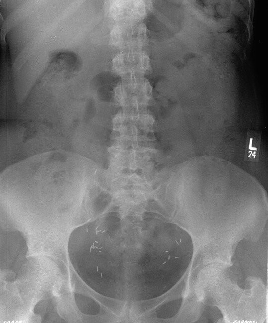
X-rayAP lumbar spine radiograph interpreted as normal, illustrating the frequent absence of visible structural abnormality in non-specific low back pain.
Source : Pringle et al., Chiropractic & osteopathy, 2006, figure 1 · CC BY
Which questions should you ask to understand the patient and their history?
History taking is the cornerstone of diagnosis. It must identify the relevant factors while building a solid therapeutic alliance. The initial objective is rigorous triage to screen for red flags : signs or symptoms that may point to serious underlying disease (fracture, infection, tumour, cauda equina syndrome).¹,⁶ Although the prevalence of these conditions is low (< 1 to 5 % of primary care cases depending on the setting), identifying them is crucial.²Red flags to screen for systematically
- Vertebral fracture : recent significant trauma (fall, road traffic accident); prolonged corticosteroid therapy; age > 70 years; known osteoporosis.6,7
- Malignancy / metastases : a personal history of cancer (the highest sensitivity, Henschke Cochrane 2013); unexplained weight loss; non-mechanical night pain; age > 50 years; no improvement after 4-6 weeks.6,7,8
- Infection (spondylodiscitis, epidural abscess) : fever, chills; immunosuppression; intravenous drug use; recent spinal surgery; constant non-mechanical pain.1,6
- Cauda equina syndrome 🚨 (surgical emergency): urinary retention with overflow voiding, sphincter incontinence, saddle anaesthesia, progressive bilateral motor deficit.1,3
- Abdominal aortic aneurysm : deep, pulsating low back pain, abdominal mass; older patient, smoker, hypertensive.10
- Inflammatory spondyloarthritis : age < 40 years, morning stiffness > 30 min, improvement with activity, waking in the second half of the night, family history of HLA-B27.9
- Beliefs and cognitions : catastrophising, kinesiophobia, negative expectations of recovery.11
- Emotional responses : symptoms of depression or anxiety.12
- Pain-related behaviours : avoidance strategies, overuse of healthcare, low self-efficacy.12
Which clinical tests should be performed and which other conditions must be ruled out?
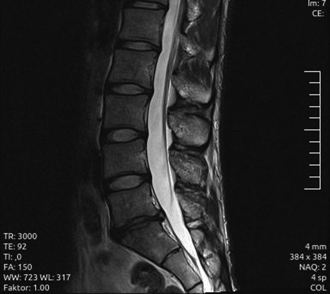
MRISagittal lumbar MRI showing severe disc degeneration at L5-S1 with a disc protrusion, without associated nerve compression.
Source : Külling et al., Orthopaedic journal of sports medicine, 2014, figure 1 · CC BY-NC-ND
🧭 Diagnostic triage of low back pain into 3 categories (Bardin 2017)
From the initial presentation to categorisation: the recommended clinical pathway
Source: Bardin LD, King P, Maher CG. Med J Aust. 2017;206(6):268-273. PMID 28359011.
- Inspection of posture and spinal curvatures.
- Assessment of active range of motion of the lumbar spine (flexion, extension, side bending, rotation): quality of movement and symptom response.
- Targeted neurological examination when radiculopathy is suspected: motor strength (myotomes L2 to S1), sensation (dermatomes), reflexes (knee jerk L4, ankle jerk S1).⁵
- Neural tension tests : the straight leg raise test (Straight Leg Raise, Lasègue): generally high sensitivity but low specificity for lower disc-root conflict. The crossed SLR (pain in the symptomatic leg when the opposite leg is raised) is more specific.5,9
Should patients with low back pain be classified, and for what benefit?
Stratifying patients moves beyond the “one size fits all” model and points towards targeted management.¹³ ✅ The most studied tool is the STarT Back Screening Tool (a 9-item self-report questionnaire), which sorts patients into 3 risk groups (low / medium / high) for developing chronic disability.¹³📊 STarT Back stratification: 3 risk groups + matched care
Hill JC et al. Lancet 2011: RCT n = 851, improves disability at 12 months and cost-effectiveness
Source: Hill JC et al. Lancet. 2011;378(9802):1560-1571. PMID 21963002.
Critique and controversy
First, the validity of isolated red flags is low. The Henschke review (Cochrane 2013) already concluded that for most red flags there is insufficient evidence to make recommendations about their diagnostic accuracy.⁸ The Verhagen 2017 review (Pain) reinforces this point for the malignancy flags. Their strength comes from consideration of their combination and of the clinical follow-up.7,8 Second, actually implementing STarT Back stratification remains a challenge. Moving from a biomedical model to a stratified approach demands training and a change of paradigm. Its effectiveness appears more marked in some health systems (UK) than in others, suggesting an effect of the way care is organised. Finally, the very concept of “non-specific low back pain” is debated. Some experts argue that the term masks real heterogeneity and holds back the search for meaningful subgroups. Research is moving towards more precise phenotypes that combine clinical, psychosocial and biological data.1,3- The assessment must systematically look for and rule out red flags before concluding that the low back pain is non-specific.6,7
- The Bardin diagnostic triage structures the reasoning: non-specific 90-95 % / radicular 5-10 % / specific pathology < 1 %.6
- The assessment of yellow flags (catastrophising, kinesiophobia) is as important as the physical examination for predicting chronicity.10,11
- Imaging is not indicated routinely without a red flag: APTA 2021, NICE NG59 and Stochkendahl 2018 all agree on this point.3,4,15
- The STarT Back Tool (3 risk groups) improves clinical outcomes and cost-effectiveness.13
Bibliography
- Hartvigsen J, Hancock MJ, Kongsted A, et al. What low back pain is and why we need to pay attention. Lancet. 2018;391(10137):2356-2367. PMID 29573870.
- Oliveira CB, Maher CG, Pinto RZ, et al. Clinical practice guidelines for the management of non-specific low back pain in primary care: an updated overview. Eur Spine J. 2018;27(11):2791-2803. PMID 29971708.
- George SZ, Fritz JM, Silfies SP, et al. Interventions for the Management of Acute and Chronic Low Back Pain: Revision 2021. J Orthop Sports Phys Ther. 2021;51(11):CPG1-CPG60. PMID 34719942.
- National Institute for Health and Care Excellence (NICE). Low back pain and sciatica in over 16s: assessment and management. NICE Guideline NG59. London: NICE; 2016 (updated December 2020). www.nice.org.uk/guidance/ng59.
- Stochkendahl MJ, Kjaer P, Hartvigsen J, et al. National Clinical Guidelines for non-surgical treatment of patients with recent onset low back pain or lumbar radiculopathy. Eur Spine J. 2018;27(1):60-75. PMID 28429142.
- Bardin LD, King P, Maher CG. Diagnostic triage for low back pain: a practical approach for primary care. Med J Aust. 2017;206(6):268-273. PMID 28359011.
- Downie A, Williams CM, Henschke N, et al. Red flags to screen for malignancy and fracture in patients with low back pain: systematic review. BMJ. 2013;347:f7095. PMID 24335669.
- Premkumar A, Godfrey W, Gottschalk MB, Boden SD. Red Flags for Low Back Pain Are Not Always Really Red: A Prospective Evaluation of the Clinical Utility of Commonly Used Screening Questions for Low Back Pain. J Bone Joint Surg Am. 2018;100(5):368-374. PMID 29509613.
- Henschke N, Maher CG, Ostelo RW, de Vet HC, Macaskill P, Irwig L. Red flags to screen for malignancy in patients with low-back pain. Cochrane Database Syst Rev. 2013;(2):CD008686. PMID 23450586.
- Verhagen AP, Downie A, Popal N, Maher C, Koes BW. Red flags presented in current low back pain guidelines: a review. Eur Spine J. 2016;25(9):2788-2802. PMID 27376890.
- Verhagen AP, Downie A, Maher CG, Koes BW. Most red flags for malignancy in low back pain guidelines lack empirical support: a systematic review. Pain. 2017;158(10):1860-1868. PMID 28708761.
- Maher C, Underwood M, Buchbinder R. Non-specific low back pain. Lancet. 2017;389(10070):736-747. PMID 27745712.
- Wertli MM, Rasmussen-Barr E, Weiser S, Bachmann LM, Brunner F. The role of fear avoidance beliefs as a prognostic factor for outcome in patients with nonspecific low back pain: a systematic review. Spine J. 2014;14(5):816-836.e4. PMID 24412032.
- Hill JC, Whitehurst DG, Lewis M, et al. Comparison of stratified primary care management for low back pain with current best practice (STarT Back): a randomised controlled trial. Lancet. 2011;378(9802):1560-1571. PMID 21963002.
- Chiarotto A, Koes BW. Nonspecific Low Back Pain. N Engl J Med. 2022;386(18):1732-1740. PMID 35507483.
- Foster NE, Anema JR, Cherkin D, et al. Prevention and treatment of low back pain: evidence, challenges, and promising directions. Lancet. 2018;391(10137):2368-2383. PMID 29573872.
Which treatment strategies are most effective for non-specific low back pain?
Where do you start? What is the recommended hierarchy of interventions?
The initial approach to non-specific low back pain (NSLBP), whether acute or chronic, is the subject of a strong international consensus. 🔍 The first line is non-pharmacological and combines patient education, advice to stay active and self-management (APTA George 2021, NICE NG59, Stochkendahl 2018 Eur Spine J, Foster Lancet 2018).¹,²,³,⁴ Imaging is to be avoided without a red flag, and opioids are strongly discouraged in chronic cases.¹,² The risk stratification (STarT Back) makes it possible to match the intensity of treatment to the patient's profile:⁵- Low risk : advice, education and encouragement to self-manage: 1-2 sessions are often enough.
- Medium risk : standard physiotherapy (graded exercise + education): 4 to 6 sessions.
- High risk : a multimodal approach including psychological intervention (CBT / CFT): 8 to 12 sessions.
📋 Comparison table, modality by GRADE level of evidence: chronic low back pain
Synthesis of the APTA 2021, NICE NG59 and Stochkendahl 2018 guidelines and of the meta-analyses
| Modality | GRADE level | Expected effect | Main source |
|---|---|---|---|
| Education + advice to stay active | High | Reduces disability and healthcare use | APTA 2021; Lancet 2018 |
| Therapeutic exercise (any type) | High | Reduces pain and disability vs control | Hayden Cochrane 2021 |
| Cognitive Functional Therapy (CFT) | High | Clinically signif. reduction in RMDQ at 13 weeks; effect maintained at 3 years | Kent Lancet 2023; Hancock 2025 |
| STarT Back stratification | Moderate | Improves disability and cost-effectiveness | Hill Lancet 2011 |
| Manual therapy (spinal manipulation) | Moderate | Effects similar to recommended therapies in the short term | Rubinstein BMJ 2019 |
| Oral NSAIDs (acute LBP) | Moderate | Small reduction in pain; GI adverse effects | Wewege BMJ 2023 (NMA) |
| Paracetamol (acute LBP) | Low | No more effective than placebo in several trials | NICE NG59; APTA 2021 |
| Pain neuroscience education alone (intensive PNE) | Moderate | Not superior to placebo education in acute cases | Traeger JAMA Neurol 2019 |
| Opioids (chronic) | Low | Marginal benefit vs risk; not advised | APTA 2021; NICE NG59 |
| Routine imaging (without a red flag) | Not recommended | No improvement in outcomes; nocebo effect | APTA 2021; NICE NG59 |
| Traction, ultrasound, laser, TENS | Low | Insufficient evidence to recommend them routinely | NICE NG59; Stochkendahl 2018 |
What is the place of exercise, and is any one approach superior?
Therapeutic exercise is the intervention with the most robust support, particularly for chronic low back pain. 💪 The Hayden 2021 Cochrane review (249 trials, > 24,000 participants) concludes that exercise is probably effective in reducing pain and disability compared with minimal care or other comparators.⁶ The question of the superiority of one type remains widely debated. The Owen 2020 network meta-analysis (BJSM), which compared 13 exercise modes across 89 trials, concludes that approaches such as Pilates, stabilisation exercises, McKenzie and yoga produce broadly similar benefits; aerobic exercise + strengthening and Pilates come out slightly ahead on pain, with no clinically decisive difference in function.⁷ Motor control exercise (Saragiotto Cochrane 2016) is not significantly superior to other forms of active exercise or to manual therapy.⁸Manual therapies and technologies: how effective are they really?
The manual therapy (mobilisations, spinal manipulations) can bring a modest short-term benefit. The Rubinstein 2019 systematic review and meta-analysis (BMJ, 47 RCTs) concludes that spinal manipulative therapy (SMT) produces effects similar to those of recommended therapies for chronic low back pain in the short term (up to 6 weeks); the effects are greater than those of non-recommended interventions but show no clinically decisive superiority over exercise or education alone.⁹ It must be integrated as a complement to an active approach, never as an isolated intervention. Combining manual therapy + exercise tends to produce better results than either modality alone.9,3 🔌 As for the passive technologies (ultrasound, laser, traction, TENS, shockwave), the international guidelines agree: insufficient evidence to recommend them routinely.2,3,4 Acupuncture occupies an ambiguous place across the guidelines. 🩺 On the pharmacological side, the Wewege 2023 network meta-analysis (BMJ, 98 RCTs, > 15,000 participants) on analgesics in acute low back pain concludes that confidence in most of the estimates is low to very low ; NSAIDs bring a small benefit on pain; paracetamol has not been shown to be superior to placebo; opioids should not be used as first-line treatment.¹⁰Beyond the physical: how do you educate the patient and address psychological factors?
🧠 Patient education aims to reconceptualise pain : not as a signal proportional to tissue damage, but as a protective measure taken by the nervous system, modulated by many factors (cognitive, emotional, contextual).¹¹ The Pain Neuroscience Education (PNE), in the Louw 2016 synthesis, showed broadly favourable effects on pain knowledge but modest clinical effects in isolation.¹¹ ⚠️ An important nuance: the Traeger 2019 randomised trial (JAMA Neurol, n = 202) in acute low back pain showed that 2 h of intensive PNE were NOT superior to reassuring placebo education on outcomes at 3 months.¹² A pragmatic conclusion: the key message is to reassure and encourage movement ; scientific exhaustiveness is not an end in itself. For patients with marked psychosocial factors, the psychologically informed approaches (PIPT) integrate cognitive and behavioural techniques into physiotherapy. The most promising approach is Cognitive Functional Therapy (CFT). 🌟 The pivotal RESTORE trial (Kent et al., Lancet 2023, n = 492 adults with disabling chronic low back pain for ≥ 3 months, three-arm parallel design) compared CFT vs CFT + sensor biofeedback vs usual care:¹³- Primary outcome at 13 weeks : reduction in the Roland-Morris Disability Questionnaire (RMDQ), with highly significant superiority of both CFT arms over control (mean difference ≈ 4-5 points out of 24, clinically and statistically significant).
- Effects maintained at 1 year (initial publication) and persisting at 3 years according to Hancock et al. (Lancet Rheumatol 2025).¹⁴
- Mechanism : graded exposure to feared activities, cognitive reconceptualisation of pain, restoration of the patient's self-confidence.
Critique and controversy
Despite converging, evidence-based guidelines, a substantial gap persists between recommendations and practice.² Over-prescription of imaging, opioids and rest remains common, including in high-income countries. First-line interventions (education, advice on activity) are under-used. The search for the “best” exercise or the “best” manual technique is often a false problem. Network meta-analyses show statistically significant but clinically slim differences. The future lies in individualisation, the shared decision-making and the strengthening of the therapeutic alliance. The effectiveness of CFT, although demonstrated by RESTORE, raises questions of implementation: how many clinicians are trained? Who should deliver it (a physiotherapist alone, a multidisciplinary team)? What is the cost-effectiveness across a whole health system?¹³,¹⁴- First line: education + advice to stay active + self-management. Imaging is to be avoided without a red flag.1,2,3
- Exercise: a pivotal intervention (high GRADE). No type is universally superior : choose according to preferences in order to maximise adherence.6,7
- Passive therapies: manual therapy = modest short-term relief, a complement to an active approach. The other passive technologies rest on weak evidence.9
- CFT (RESTORE Lancet 2023): a highly significant reduction in disability in disabled chronic patients, with effects maintained at 3 years (Hancock 2025).13,14
- Pharmacology: NSAIDs = small short-term benefit; paracetamol no better than placebo; opioids to be avoided.10
Bibliography
- George SZ, Fritz JM, Silfies SP, et al. Interventions for the Management of Acute and Chronic Low Back Pain: Revision 2021. J Orthop Sports Phys Ther. 2021;51(11):CPG1-CPG60. PMID 34719942.
- Foster NE, Anema JR, Cherkin D, et al. Prevention and treatment of low back pain: evidence, challenges, and promising directions. Lancet. 2018;391(10137):2368-2383. PMID 29573872.
- Stochkendahl MJ, Kjaer P, Hartvigsen J, et al. National Clinical Guidelines for non-surgical treatment of patients with recent onset low back pain or lumbar radiculopathy. Eur Spine J. 2018;27(1):60-75. PMID 28429142.
- National Institute for Health and Care Excellence (NICE). Low back pain and sciatica in over 16s: assessment and management. NICE Guideline NG59. London: NICE; 2016 (updated 2020). www.nice.org.uk/guidance/ng59.
- Hill JC, Whitehurst DG, Lewis M, et al. Comparison of stratified primary care management for low back pain with current best practice (STarT Back): a randomised controlled trial. Lancet. 2011;378(9802):1560-1571. PMID 21963002.
- Hayden JA, Ellis J, Ogilvie R, Malmivaara A, van Tulder MW. Exercise therapy for chronic low back pain. Cochrane Database Syst Rev. 2021;9(9):CD009790. PMID 34580864.
- Owen PJ, Miller CT, Mundell NL, et al. Which specific modes of exercise training are most effective for treating low back pain? Network meta-analysis. Br J Sports Med. 2020;54(21):1279-1287. PMID 31666220.
- Saragiotto BT, Maher CG, Yamato TP, et al. Motor control exercise for chronic non-specific low-back pain. Cochrane Database Syst Rev. 2016;1(1):CD012004. PMID 26742533.
- Rubinstein SM, de Zoete A, van Middelkoop M, et al. Benefits and harms of spinal manipulative therapy for the treatment of chronic low back pain: systematic review and meta-analysis of randomised controlled trials. BMJ. 2019;364:l689. PMID 30867144.
- Wewege MA, Bagg MK, Jones MD, Ferraro MC, Cashin AG, et al. Comparative effectiveness and safety of analgesic medicines for adults with acute non-specific low back pain: systematic review and network meta-analysis. BMJ. 2023;380:e072962. PMID 36948512.
- Louw A, Zimney K, Puentedura EJ, Diener I. The efficacy of pain neuroscience education on musculoskeletal pain: A systematic review of the literature. Physiother Theory Pract. 2016;32(5):332-355. PMID 27351541.
- Traeger AC, Lee H, Hubscher M, et al. Effect of Intensive Patient Education vs Placebo Patient Education on Outcomes in Patients With Acute Low Back Pain: A Randomized Clinical Trial. JAMA Neurol. 2019;76(2):161-169. PMID 30398542.
- Kent P, Haines T, O Sullivan P, et al. Cognitive functional therapy with or without movement sensor biofeedback versus usual care for chronic, disabling low back pain (RESTORE): a randomised, controlled, three-arm, parallel group, phase 3, clinical trial. Lancet. 2023;401(10391):1866-1877. PMID 37146623.
- Hancock M, Kent P, Haines T, et al. Cognitive functional therapy with or without movement sensor biofeedback versus usual care for chronic, disabling low back pain (RESTORE): 3-year follow-up of a randomised, controlled trial. Lancet Rheumatol. 2025;7(11):e789-e798. PMID 40780241.
- O Sullivan PB, Caneiro JP, O Keeffe M, et al. Cognitive Functional Therapy: An Integrated Behavioral Approach for the Targeted Management of Disabling Low Back Pain. Phys Ther. 2018;98(5):408-423. PMID 29669082.
- Saragiotto BT, Abdel Shaheed C, Overton M, Hayden JA, Hartvigsen J, Pinto RZ. Recent highlights in low back pain research, Part II: Prevention and management. J Physiother. 2026;72(2):106-115. PMID 41933950.
How do you secure lasting recovery and prevent recurrence of non-specific low back pain?
How do you make the patient an active partner in recovery through self-management?
Self-management (self-management) is the cornerstone of recurrence prevention.²,³ It aims to give the patient the autonomy, the confidence and the skills needed to manage their symptoms, reduce their limitations and minimise the impact of pain. 🧘♀️ The two pillars of effective self-management are education and exercise :- Therapeutic education : the modern approach (Pain Neuroscience Education, PNE) aims to reconceptualise pain. The Louw 2016 systematic review confirms a favourable effect on knowledge and disability, particularly when it is embedded in an active intervention.⁴ The intervening Traeger 2019 review qualifies this: 2 h of intensive PNE is no better than reassuring placebo education in acute cases, evidence that the reassuring message + movement are what matter.⁵
- Therapeutic exercise : the Hayden 2021 Cochrane review confirms the effectiveness of exercise for chronic low back pain. The de Campos 2021 review (BJSM, formerly Steffens 2016, a meta-analysis of 25 RCTs, > 9,000 participants) confirms that exercise combined with education reduces the risk of recurrence by about 45 % at 1 year.⁶,⁷ The type of exercise matters less than adherence and regularity.⁸
📉 Reduction in recurrence risk: exercise + education
de Campos et al., BJSM 2021: meta-analysis of prevention strategies
Sources: de Campos TF et al. Br J Sports Med. 2021 (PMID 32646887); Steffens D et al. JAMA Intern Med. 2016;176(2):199-208 (PMID 26752509).
When and how should a safe return to sport and activity be planned?
Return to sport (RTS) after low back pain must be structured and criteria-based, not built on an arbitrary timetable. 🏆 The aim is to make sure that the patient's musculoskeletal system tolerates the specific demands of the target activity without triggering a new episode. A gradual consensus has formed around a staged approach:- Phase 1: pain control and basic functional recovery. Activities of daily living without significant pain. Lumbar and hip range of motion restored.
- Phase 2: strengthening and neuromuscular control. Strength and endurance of the trunk muscles (core stability), glutes and lower limbs. Movement quality during functional exercises.
- Phase 3: sport-specific functional tests. Jumping, changes of direction, sprinting and lifting: pain-free and with sound kinematics.
- Phase 4: progressive, graded resumption. Volume and intensity reduced at first. Monitoring of training load is crucial: abrupt increases are a major risk factor (Gabbett BJSM 2016, training-injury prevention paradox).⁹
- Phase 5: psychological assessment. The patient's confidence to move and perform without fear is an essential criterion. Residual kinesiophobia can alter motor patterns and encourage recurrence.10,11
Critique and controversy
🧐 The paradox of the “non-specific” label : the heading that covers nearly 90 % of cases is increasingly criticised. It masks a real heterogeneity. Efforts are under way to develop subgroups (mechanistic nociceptive / nociplastic after Nijs 2021, movement profiles), but no method commands unanimous support.10 🥋 The exercise brand wars (Pilates vs McKenzie vs core stability) continue to sway clinicians and the public, even though meta-analyses conclude that these approaches are pragmatically equivalent. Long-term adherence is the decisive element.7,8 📚 Implementing PNE remains a challenge: it cannot be reduced to a leaflet or a 5-minute explanation. It demands time, communication skills and a genuine therapeutic alliance. Its effect, although real, remains modest, and it must always be combined with an active approach.4,5- ✅ Empower the patient through self-management (education + exercise).2,3
- 🧠 Change beliefs with PNE, combined with an active approach.4
- 💪 Move more, move better : promote regular, enjoyable physical activity; adherence matters more than specificity.6,7,8
- 📉 Exercise + education combined reduces the risk of recurrence by about 45 % at 1 year.6
- 🏆 Smart return to sport : functional and psychological criteria, not a timetable; load monitoring.9
Bibliography
- Stanton TR, Henschke N, Maher CG, Refshauge KM, Latimer J, McAuley JH. After an episode of acute low back pain, recurrence is unpredictable and not as common as previously thought. Spine (Phila Pa 1976). 2008;33(26):2923-2928. PMID 19092626.
- Hartvigsen J, Hancock MJ, Kongsted A, et al. What low back pain is and why we need to pay attention. Lancet. 2018;391(10137):2356-2367. PMID 29573870.
- Foster NE, Anema JR, Cherkin D, et al. Prevention and treatment of low back pain: evidence, challenges, and promising directions. Lancet. 2018;391(10137):2368-2383. PMID 29573872.
- Louw A, Zimney K, Puentedura EJ, Diener I. The efficacy of pain neuroscience education on musculoskeletal pain: A systematic review of the literature. Physiother Theory Pract. 2016;32(5):332-355. PMID 27351541.
- Traeger AC, Lee H, Hubscher M, et al. Effect of Intensive Patient Education vs Placebo Patient Education on Outcomes in Patients With Acute Low Back Pain: A Randomized Clinical Trial. JAMA Neurol. 2019;76(2):161-169. PMID 30398542.
- de Campos TF, Maher CG, Fuller JT, Steffens D, Attwell S, Hancock MJ. Prevention strategies to reduce future impact of low back pain: a systematic review and meta-analysis. Br J Sports Med. 2021;55(9):468-476. PMID 32646887.
- Steffens D, Maher CG, Pereira LS, et al. Prevention of Low Back Pain: A Systematic Review and Meta-analysis. JAMA Intern Med. 2016;176(2):199-208. PMID 26752509.
- Hayden JA, Ellis J, Ogilvie R, Malmivaara A, van Tulder MW. Exercise therapy for chronic low back pain. Cochrane Database Syst Rev. 2021;9(9):CD009790. PMID 34580864.
- Gabbett TJ. The training-injury prevention paradox: should athletes be training smarter and harder? Br J Sports Med. 2016;50(5):273-280. PMID 26758673.
- Nijs J, Lahousse A, Kapreli E, et al. Nociplastic Pain Criteria or Recognition of Central Sensitization? Pain Phenotyping in the Past, Present and Future. J Clin Med. 2021;10(15):3203. PMID 34361986.
- Wertli MM, Rasmussen-Barr E, Held U, Weiser S, Bachmann LM, Brunner F. Fear-avoidance beliefs, a moderator of treatment efficacy in patients with low back pain: a systematic review. Spine J. 2014;14(11):2658-2678. PMID 24614254.
What do real case studies teach us about non-specific low back pain?
Analysis of a typical case: from assessment to resolution
The commonest profile in practice is a working-age patient (30-55 years) with persistent common low back pain of 2 to 6 months, no red flag, moderate functional disability (RMDQ 8-12 / 24, Oswestry 20-40 %) and moderate psychosocial factors (FABQ-W > 15, PCS > 20). The evidence-based approach for this profile, supported by George 2021 (APTA CPG), Foster Lancet 2018 and Hayden Cochrane 2021, combines 3 pillars:¹,²,³- Education and reassurance : explaining the multifactorial nature of pain, decoupling the sensation from tissue damage, encouraging movement. PNE is delivered pragmatically, without swamping the patient with information.4,5
- Manual therapy (short term) : used to modulate pain, improve joint mobility and create a window of opportunity for movement. Expected effects are modest but useful when combined with an active approach (Rubinstein BMJ 2019).⁶
- Progressive exercise : an individualised programme (preferences, capacity, lifestyle), with graded exposure to feared movements or activities. Aerobic + strengthening + mobility. No universally superior type.7,8
- A reduction in pain (NPRS) of 2 to 4 points (MCID ≈ 2 points out of 11).
- An improvement in disability (RMDQ or Oswestry) beyond the MCID (≈ 5 points out of 24 or ≈ 10 points out of 100, respectively).
- A reduction in kinesiophobia (TSK) and in catastrophising (PCS).
- Gradual resumption of work and sporting activities.
The diagnostic challenge: when low back pain mimics another condition
The “non-specific” label NEVER removes the need for constant vigilance. The Downie 2013 review (BMJ) and Premkumar 2018 (JBJS) are reminders of both the importance and the limits of red flag screening. 🩺3 major families of serious conditions never to be missed
- Vascular conditions : the leaking abdominal aortic aneurysm can present as sudden, deep, pulsating low back pain in an older, smoking, hypertensive patient. A palpable pulsatile abdominal mass, sudden hypotension and pain with atypical radiation are warning signs. An absolute vascular emergency.
- Infectious conditions : the pyogenic spondylodiscitis (most often Staphylococcus aureus) preferentially affects patients who are diabetic, immunosuppressed, post-surgical or intravenous drug users. Presentation: severe non-mechanical low back pain, intermittent fever, raised inflammatory markers, characteristic MRI. It requires prolonged antibiotic therapy.
- Tumour conditions : the vertebral metastases (breast, lung, prostate, kidney, thyroid) are the commonest cause of spinal tumour. Highest sensitivity: a previous history of cancer (Henschke Cochrane 2013).13 Presentation: progressive, nocturnal, non-mechanical pain; neurological signs in case of spinal cord compression.
A complex case study: the RESTORE trial
Rather than a fictional case report, let us illustrate with the pivotal RESTORE trial (Kent et al., Lancet 2023): n = 492 Australian adults with disabling chronic low back pain for ≥ 3 months (median 4 years), with mean scores of RMDQ ≈ 12 / 24 and NPRS ≈ 6 / 10 at inclusion.¹² Typical participant profile : chronic patients already seen in primary care, having tried several interventions (conventional physiotherapy, NSAIDs, lumbar supports, sometimes injections), with high kinesiophobia (high TSK), low self-efficacy and pessimistic beliefs about the possibility of getting better. CFT intervention (up to 7 sessions, 11 weeks + 2 booster sessions): ¹²,¹⁴- Making sense : exploring the patient's history and beliefs, identifying the protective behaviours (avoidance, muscle guarding) that keep the pain-disability cycle going.
- Graded exposure to movement and feared activities, with regulation strategies (breathing, relaxation, free trunk movement).
- Restoring a healthy lifestyle : sleep, physical activity, stress management, gradual return to work.
- A 13 weeks : a mean difference of −4.6 points on the RMDQ (95 % CI −5.9 to −3.2) between CFT and usual care: a clinically and statistically significant superiority.
- A 1 year : effect maintained on disability and quality of life (initial publication).
- A 3 years (Hancock 2025, Lancet Rheumatol) : effects persisting on disability: the first demonstration of a lasting 36-month effect for a conservative, non-pharmacological intervention in disabling chronic low back pain.14
- Sensor biofeedback added no benefit over CFT alone, suggesting that the active ingredient is the targeted cognitive behavioural therapy, not the technological gadget.
Critique and controversy
🤔 Individual case reports carry a very low level of evidence (Oxford CEBM 4-5). A spectacular success in an isolated case does not guarantee effectiveness on a larger scale and may be the product of many confounders (placebo effect, natural history, the therapist's charisma). They must be read as illustrations of principles, never as evidence in themselves. The evidence pyramid: a quick read (presented as horizontal cards for ease of reading):📐 GRADE / Oxford CEBM hierarchy for non-specific low back pain
Levels of evidence presented as horizontal cards (never as an unreadable text pyramid)
How to read this: the higher you go (green), the stronger the level of evidence. Case reports (level 4) are instructive but are not enough to frame recommendations.
- Classic case reports confirm the effectiveness of a multimodal approach (education + short-term manual therapy + progressive exercise), in line with the guidelines.1,2,3
- The need for vigilance remains paramount : vascular (aortic aneurysm), infectious (spondylodiscitis) and tumour (metastases) conditions can mimic common low back pain.13
- The RESTORE (Lancet 2023) trial demonstrated the clinically significant and lasting superiority (at 3 years) of CFT in disabled chronic patients.12,14
- Case reports are powerful teaching tools but carry a low level of evidence : use them as illustrations, not as rules.
Bibliography
- George SZ, Fritz JM, Silfies SP, et al. Interventions for the Management of Acute and Chronic Low Back Pain: Revision 2021. J Orthop Sports Phys Ther. 2021;51(11):CPG1-CPG60. PMID 34719942.
- Foster NE, Anema JR, Cherkin D, et al. Prevention and treatment of low back pain: evidence, challenges, and promising directions. Lancet. 2018;391(10137):2368-2383. PMID 29573872.
- Hayden JA, Ellis J, Ogilvie R, Malmivaara A, van Tulder MW. Exercise therapy for chronic low back pain. Cochrane Database Syst Rev. 2021;9(9):CD009790. PMID 34580864.
- Louw A, Zimney K, Puentedura EJ, Diener I. The efficacy of pain neuroscience education on musculoskeletal pain: A systematic review of the literature. Physiother Theory Pract. 2016;32(5):332-355. PMID 27351541.
- Traeger AC, Lee H, Hubscher M, et al. Effect of Intensive Patient Education vs Placebo Patient Education on Outcomes in Patients With Acute Low Back Pain: A Randomized Clinical Trial. JAMA Neurol. 2019;76(2):161-169. PMID 30398542.
- Rubinstein SM, de Zoete A, van Middelkoop M, et al. Benefits and harms of spinal manipulative therapy for the treatment of chronic low back pain: systematic review and meta-analysis of randomised controlled trials. BMJ. 2019;364:l689. PMID 30867144.
- Owen PJ, Miller CT, Mundell NL, et al. Which specific modes of exercise training are most effective for treating low back pain? Network meta-analysis. Br J Sports Med. 2020;54(21):1279-1287. PMID 31666220.
- Saragiotto BT, Maher CG, Yamato TP, et al. Motor control exercise for chronic non-specific low-back pain. Cochrane Database Syst Rev. 2016;1(1):CD012004. PMID 26742533.
- National Institute for Health and Care Excellence (NICE). Low back pain and sciatica in over 16s: assessment and management. NICE Guideline NG59. London: NICE; 2016 (updated 2020). www.nice.org.uk/guidance/ng59.
- Stochkendahl MJ, Kjaer P, Hartvigsen J, et al. National Clinical Guidelines for non-surgical treatment of patients with recent onset low back pain or lumbar radiculopathy. Eur Spine J. 2018;27(1):60-75. PMID 28429142.
- Saragiotto BT, Abdel Shaheed C, Overton M, Hayden JA, Hartvigsen J, Pinto RZ. Recent highlights in low back pain research, Part II: Prevention and management. J Physiother. 2026;72(2):106-115. PMID 41933950.
- Kent P, Haines T, O Sullivan P, et al. Cognitive functional therapy with or without movement sensor biofeedback versus usual care for chronic, disabling low back pain (RESTORE). Lancet. 2023;401(10391):1866-1877. PMID 37146623.
- Henschke N, Maher CG, Ostelo RW, de Vet HC, Macaskill P, Irwig L. Red flags to screen for malignancy in patients with low-back pain. Cochrane Database Syst Rev. 2013;(2):CD008686. PMID 23450586.
- Hancock M, Kent P, Haines T, et al. Cognitive functional therapy with or without movement sensor biofeedback versus usual care for chronic, disabling low back pain (RESTORE): 3-year follow-up of a randomised, controlled trial. Lancet Rheumatol. 2025;7(11):e789-e798. PMID 40780241.
- Nijs J, Lahousse A, Kapreli E, et al. Nociplastic Pain Criteria or Recognition of Central Sensitization? J Clin Med. 2021;10(15):3203. PMID 34361986.
How do you apply these recommendations concretely in your practice?
When, and to which other health professionals, should you refer?
One of the physiotherapist's core skills, particularly in direct access, is the ability to identify situations that fall outside their scope of practice or that call for further assessment. The first level of vigilance concerns the red flags already detailed above. The Downie 2013 review (BMJ) and Premkumar 2018 (JBJS) are reminders of their limits: an isolated flag has low diagnostic accuracy, but their combination and longitudinal analysis strengthen their predictive value.¹,² The indications for prompt referral to the GP or a specialist :- Suspected serious disease : a history of cancer + unexplained weight loss, fever + immunosuppression, progressive neurological signs, suspected cauda equina (surgical emergency).1,2
- Suspected spondyloarthritis (age < 40, morning stiffness > 30 min, waking in the second half of the night, HLA-B27 history): refer to the rheumatologist.
- Suspected severe radiculopathy with progressive motor deficit: specialist assessment (neurologist or rheumatologist), sometimes targeted imaging.
- No improvement after 4-6 weeks of appropriate management: systematic re-assessment of the red flags and referral.
- Occupational physician : workstation adaptation, phased return to work, fitness-for-work assessment.
- Nutritionist / dietitian : where overweight or obesity is an aggravating factor.
- Psychologist specialising in pain : for patients with severe yellow flags, particularly in disabling chronic low back pain (the RESTORE 2023 trial demonstrated the value of an approach that integrates the psychological dimension).⁴
- Rheumatologist / spinal surgeon : failure of prolonged conservative treatment, severe radiculopathy, suspected spondyloarthritis.
How do you measure outcomes and overcome barriers to implementation?
📊 The use of patient-reported outcome measures (PROMs) is a strong recommendation of the international guidelines.5,6 These validated tools make it possible to quantify pain, function, quality of life and psychological impact, and to support shared decision-making.📋 Validated PROMs for non-specific low back pain: the clinician's toolkit
Recommended minimum selection + MCID (minimal clinically important difference)
| PROM | Domain | Scale | Approx. MCID | Clinical use |
|---|---|---|---|---|
| NPRS (Numeric Pain Rating Scale) | Pain | 0–10 | ≈ 2 points | Every session, rapid follow-up |
| RMDQ (Roland-Morris) | Low back pain related disability | 0–24 | ≈ 5 points | Pivotal measure in RCTs (RESTORE) |
| ODI (Oswestry Disability Index) | Functional disability | 0–100 % | ≈ 10 points | Long-term follow-up, surgery |
| STarT Back Tool | Stratification of chronicity risk | 0–9 | — | Initial, guides matched care |
| FABQ (Fear-Avoidance Beliefs Q) | Fear-avoidance beliefs | 0–96 | — | Yellow flags, work / activity |
| PCS (Pain Catastrophizing Scale) | Catastrophising | 0–52 | — | Yellow flags, referral decision |
| TSK (Tampa Scale Kinesiophobia) | Fear of movement | 17–68 | — | Before return to sport |
| PSEQ (Pain Self-Efficacy) | Self-efficacy | 0–60 | — | CFT treatment target |
- Lack of time in the consultation to deliver education, assess yellow flags and administer PROMs.
- Lack of skills to search and appraise the literature and adapt it to the patient.
- Lack of organisational support and inertia in the reimbursement system (which often favours technical procedures over education).
- Residual clinical beliefs about the “biomedical model” (look for the lesion, order imaging, rest).
- Patient expectations shaped by social representations (“my vertebrae are out of place”) and by historical over-medicalisation.
- Technological integration : practice management software with built-in automated PROMs (sent before the session, trajectory tracking).
- Targeted continuing education on critical appraisal, biopsychosocial reasoning and communication. Short courses on EBP have shown their effectiveness.
- Practice culture : “journal club” meetings, peer mentoring, valuing the scientific approach.
- In summary : lean on the guidelines (NICE NG59, APTA 2021, Stochkendahl 2018) and on the recent reviews (Saragiotto J Physiother 2026 Part I & II) rather than reinventing the wheel.9,10
Critique and controversy: beyond the guidelines
Applying recommendations rigidly raises important questions. ⚖️ The “red flag paradox” : screening for them is a professional duty, but an excessive focus can generate a nocebo effect (needless anxiety, a cascade of costly and sometimes iatrogenic investigations). Competence lies not in ticking a list but in interpreting it within the overall clinical context.1,2,7,8 📈 The gap between data and action on PROMs : many clinicians collect the scores but struggle to use them concretely to adjust treatment. The risk is turning a tool for dialogue into an administrative formality. What matters is that a PROM score should trigger specific clinical actions.5,6 🌍 The generalisability of RCTs to everyday practice remains a persistent controversy. RCTs recruit homogeneous populations; the “average” patient in practice is often more complex (comorbidities, a particular psychosocial context). Applying a recommendation without adapting it to the individual is a mistake. Real expertise lies in the ability to contextualise high-level evidence for a single patient.- The vigilance shown for red flags is essential but must be set in context: their combination and longitudinal follow-up are more informative than an isolated flag.1,2,7,8
- The PROMs (NPRS, RMDQ, ODI, STarT Back, PCS, TSK) are powerful tools when their results are actively used to adjust treatment and support shared decision-making.5,6
- Applying recommendations derived from RCTs must go hand in hand with adaptation to the context and to the particularities of each patient.
- Evidence-based practice rests on the balance between scientific rigour, clinical experience and the patient's values.
Bibliography
- Downie A, Williams CM, Henschke N, et al. Red flags to screen for malignancy and fracture in patients with low back pain: systematic review. BMJ. 2013;347:f7095. PMID 24335669.
- Premkumar A, Godfrey W, Gottschalk MB, Boden SD. Red Flags for Low Back Pain Are Not Always Really Red: A Prospective Evaluation of the Clinical Utility of Commonly Used Screening Questions for Low Back Pain. J Bone Joint Surg Am. 2018;100(5):368-374. PMID 29509613.
- Wertli MM, Rasmussen-Barr E, Weiser S, Bachmann LM, Brunner F. The role of fear avoidance beliefs as a prognostic factor for outcome in patients with nonspecific low back pain: a systematic review. Spine J. 2014;14(5):816-836.e4. PMID 24412032.
- Kent P, Haines T, O Sullivan P, et al. Cognitive functional therapy with or without movement sensor biofeedback versus usual care for chronic, disabling low back pain (RESTORE). Lancet. 2023;401(10391):1866-1877. PMID 37146623.
- George SZ, Fritz JM, Silfies SP, et al. Interventions for the Management of Acute and Chronic Low Back Pain: Revision 2021. J Orthop Sports Phys Ther. 2021;51(11):CPG1-CPG60. PMID 34719942.
- Foster NE, Anema JR, Cherkin D, et al. Prevention and treatment of low back pain: evidence, challenges, and promising directions. Lancet. 2018;391(10137):2368-2383. PMID 29573872.
- Verhagen AP, Downie A, Maher CG, Koes BW. Most red flags for malignancy in low back pain guidelines lack empirical support: a systematic review. Pain. 2017;158(10):1860-1868. PMID 28708761.
- Henschke N, Maher CG, Ostelo RW, de Vet HC, Macaskill P, Irwig L. Red flags to screen for malignancy in patients with low-back pain. Cochrane Database Syst Rev. 2013;(2):CD008686. PMID 23450586.
- National Institute for Health and Care Excellence (NICE). Low back pain and sciatica in over 16s: assessment and management. NICE Guideline NG59. London: NICE; 2016 (updated 2020). www.nice.org.uk/guidance/ng59.
- Pinto RZ, Kongsted A, Silva S, Hayden JA, Downie A, Saragiotto BT. Recent highlights in low back pain research, Part I: Diagnosis and Prognosis. J Physiother. 2026;72(1):23-32. PMID 41423384.
- Saragiotto BT, Abdel Shaheed C, Overton M, Hayden JA, Hartvigsen J, Pinto RZ. Recent highlights in low back pain research, Part II: Prevention and management. J Physiother. 2026;72(2):106-115. PMID 41933950.
- Stochkendahl MJ, Kjaer P, Hartvigsen J, et al. National Clinical Guidelines for non-surgical treatment of patients with recent onset low back pain or lumbar radiculopathy. Eur Spine J. 2018;27(1):60-75. PMID 28429142.
And after this read?
This article is part of a collection of evidence-based clinical syntheses. A question, a comment, a correction to suggest? Contact us directly through the WhatsApp button at the bottom right of the screen.

