















In brief
Axial spondyloarthritis (axSpA), of which ankylosing spondylitis is the radiographic form, is a chronic inflammatory disease of the axial skeleton that typically begins before the age of 45. It rests on enthesitis (inflammation of the tendon and ligament insertions) mediated by the IL-23/IL-17/TNF axis; HLA-B27 is present in 80 to 95 % of patients, and MRI of the sacroiliac joints, revealing bone marrow oedema, allows early diagnosis. Associated features: inflammatory back pain, anterior uveitis (~25 %), psoriasis, IBD. First-line management (ASAS-EULAR 2023) combines education and regular exercise from diagnosis onwards, NSAIDs and then biologic therapy where the response is insufficient. The median diagnostic delay reaches about 7 years.
Clinical synthesis based on the most recent meta-analyses and international consensus statements: ASAS-EULAR 2023, ACR/SAA/SPARTAN 2019, Lancet 2024, Cochrane 2019 and prospective data from 2020-2025.
Clinical summary
- Formally, axial spondyloarthritis (axSpA), of which ankylosing spondylitis (AS / r-axSpA) is the radiographic form, is a chronic inflammatory disease of the axial skeleton that typically begins before the age of 45.1,2
- The allele HLA-B27 is the major genetic factor, present in 80-95 % of Caucasian patients. Its presence alone is not enough to cause the disease.3,4
- The central mechanism is enthesitis (inflammation of the tendon and ligament insertions), mediated by the IL-23/IL-17/TNF axis.5
- MRI of the sacroiliac joints (STIR sequence: bone marrow oedema) is the key investigation for early diagnosis, well before radiographic lesions appear.6
- The ASAS 2009 criteria distinguish nr-axSpA (non-radiographic, MRI+ or HLA-B27+) from r-axSpA (AS, the modified New York criteria).2,7
- The median diagnostic delay is about 7 years, the main obstacle to optimal management.8
- In women, axSpA is underdiagnosed ; the M/F ratio is 1:1 in nr-axSpA but 2-3:1 in r-axSpA, with a worse diagnostic delay.1,9
- First-line treatment (ASAS-EULAR 2023): therapeutic education + regular exercise for every patient, from diagnosis onwards.10
- Exercise is non-negotiable : the Cochrane review Regnaux 2019 confirms its efficacy on pain, function and mobility.11
- Notably, HIIT is safe and effective in stable axSpA: it improves the ASDAS, the BASDAI and cardiorespiratory capacity (Sveaas 2014/2020).12,13
- NSAIDs = first-line pharmacotherapy. Biologic therapy (anti-TNF, anti-IL-17, JAK inhibitors) where the response is insufficient.10,14
- By contrast, manual therapies are a useful short-term adjunct, never a stand-alone disease-modifying treatment.11
- axSpA can mimic sciatica (alternating buttock pain) or plantar fasciitis (Achilles enthesitis): inflammatory back pain is the red flag.15
- Frequent extra-articular manifestations: acute anterior uveitis (~25 % of patients), psoriasis, IBD.16
- Follow-up rests on validated PROMs: BASDAI (activity), BASFI (function), BASMI (mobility), ASDAS-CRP.17,18
- Identifying the red flags (spinal fracture, infection, aortic dissection in advanced AS) is non-negotiable for safety.19
- A multidisciplinary approach is essential: rheumatologist, physiotherapist, ophthalmologist (if uveitis), gastroenterologist (if IBD).10
- The return to sport should be progressive and guided by symptoms, not by a rigid calendar; regular practice is protective.13,20
- Self-management (mobile apps, BASDAI monitoring, home exercises) is the cornerstone of preventing flares.10,21
Contents
- What are the fundamentals to know about ankylosing spondylitis (axSpA)?
- How do you assess and diagnose ankylosing spondylitis with certainty?
- axSpA in women: why is it underdiagnosed, and how do we put that right?
- Which treatment strategies are the most effective?
- How do you secure lasting recovery and prevent flares?
- What do real clinical cases teach us?
- How do you apply these recommendations concretely in your practice?
What are the fundamentals to know about ankylosing spondylitis (axSpA)?
How is this condition defined, who does it affect and what are the risk factors?
What we call axial spondyloarthritis (axSpA) is a chronic inflammatory disease of the axial skeleton: the spine and the sacroiliac joints.1,2 The modern terminology, established by ASAS (Assessment of SpondyloArthritis international Society), covers two phenotypes: the radiographic form (r-axSpA), historically called ankylosing spondylitis (AS) and defined by sacroiliitis visible on radiography under the modified New York criteria,3 and the non-radiographic form (nr-axSpA), with no visible structural lesion but with active sacroiliitis on MRI or a body of clinical evidence associated with HLA-B27.2 This conceptual shift has made it possible to identify patients earlier and to open access to biologic therapy for them.4
Epidemiologically, the worldwide prevalence of AS ranges from 0.1 % to 1.4 %, largely correlated with the frequency of HLA-B27 in the population concerned.5 In Europe, the weighted prevalence of AS is estimated at 0.24 %.5 The first symptoms typically appear between 20 and 40 years, with onset necessarily before 45 years under the ASAS criteria.2
The male-to-female ratio is one of the figures most revised downwards over the past 15 years: historically estimated at 2-3:1 for radiographic AS, it is now known to be close to 1:1 for nr-axSpA, the disease being widely underdiagnosed in women (see chapter 3).1,6
Prevalence and epidemiological characteristics of axSpA
A synthesis of the global data: Sieper & Poddubnyy Lancet 2017, Dean Rheumatology 2014
Sources: Dean LE et al. Rheumatology 2014;53(4):650-7; Sieper J, Poddubnyy D. Lancet 2017;390:73-84. Weighted global prevalence about 0.2 %, typical onset 20-40 years.
The allele HLA-B27 (Human Leucocyte Antigen B27) is the most powerful genetic risk factor: found in 80-95 % of patients with AS in Caucasian populations, against 8-10 % in the general population.2,5 That said, more than 95 % of HLA-B27 carriers will never develop the disease, which indicates that other factors (genetic: ERAP1, IL-23R; environmental: the gut microbiome) act as triggers in predisposed individuals.1
What happens in the body, and how does axSpA evolve naturally?
The central pathological process is enthesitis: inflammation of the entheses, the sites where tendons, ligaments and capsules insert into bone.7 This inflammation, particularly active at the sacroiliac joints and the vertebral corners, is mediated by several immune pathways, among them the IL-23 / IL-17 axis, which has become a major therapeutic target (anti-IL-17: secukinumab, ixekizumab, bimekizumab).7 The role of TNF-alpha remains central, with the anti-TNF drugs as the first validated class of biologic therapy.10
The natural course combines inflammation with abnormal bone repair: erosion first, then pathological bone formation at the inflamed sites (syndesmophytes, ankylosis).1,7 Without effective treatment, the disease progresses variably from patient to patient, with a cumulative risk of spinal fusion (“bamboo spine”) in a minority of severely affected patients. The hips can be involved (radiographic hip disease in 25-35 % of patients with advanced AS), causing substantial functional disability.1
Beyond the skeleton, extra-articular manifestations are frequent:
- Acute anterior uveitis : the most frequent, affecting about 23-26 % of AS patients over their lifetime (Stolwijk 2015 meta-analysis).8
- Psoriasis : about 9-10 % of axSpA patients.8
- Inflammatory bowel disease (IBD: Crohn's, ulcerative colitis) : about 6-7 % of axSpA patients.8
Key points
- axSpA covers r-axSpA (radiographic AS, the modified NY criteria) and nr-axSpA (no radiographic sacroiliitis, MRI+ or HLA-B27+ with other features).
- HLA-B27 = the major genetic factor (80-95 % of Caucasian AS patients) but not a sufficient one.
- Enthesitis = the primary lesion, mediated by the IL-23/IL-17 axis (the therapeutic targets).
- Extra-articular manifestations: uveitis ~25 %, psoriasis ~10 %, IBD ~7 %.
Bibliography
- Sieper J, Poddubnyy D. Axial spondyloarthritis. Lancet. 2017;390(10089):73-84. PMID 28110981.
- Rudwaleit M, van der Heijde D, Landewe R, et al. The development of Assessment of SpondyloArthritis international Society classification criteria for axial spondyloarthritis (part II): validation and final selection. Ann Rheum Dis. 2009;68(6):777-783. PMID 19297344.
- van der Linden S, Valkenburg HA, Cats A. Evaluation of diagnostic criteria for ankylosing spondylitis: a proposal for modification of the New York criteria. Arthritis Rheum. 1984;27(4):361-368. PMID 6231933.
- Taurog JD, Chhabra A, Colbert RA. Ankylosing Spondylitis and Axial Spondyloarthritis. N Engl J Med. 2016;374(26):2563-2574. PMID 27355535. doi:10.1056/NEJMra1406182.
- Dean LE, Jones GT, MacDonald AG, Downham C, Sturrock RD, Macfarlane GJ. Global prevalence of ankylosing spondylitis. Rheumatology (Oxford). 2014;53(4):650-657. PMID 24324212.
- Rusman T, van Vollenhoven RF, van der Horst-Bruinsma IE. Gender Differences in Axial Spondyloarthritis: Women Are Not So Lucky. Curr Rheumatol Rep. 2018;20(6):35. PMID 29754330.
- Schett G, Lories RJ, D'Agostino MA, Elewaut D, Kirkham B, Soriano ER, McGonagle D. Enthesitis: from pathophysiology to treatment. Nat Rev Rheumatol. 2017;13(12):731-741. PMID 29158573.
- Stolwijk C, van Tubergen A, Castillo-Ortiz JD, Boonen A. Prevalence of extra-articular manifestations in patients with ankylosing spondylitis: a systematic review and meta-analysis. Ann Rheum Dis. 2015;74(7):1373-1378. PMID 24013647.
- Navarro-Compan V, Sepriano A, El-Zorkany B, van der Heijde D. Axial spondyloarthritis. Lancet. 2024;404(10470):2354-2367. doi:10.1016/S0140-6736(24)02263-3.
- Ramiro S, Nikiphorou E, Sepriano A, et al. ASAS-EULAR recommendations for the management of axial spondyloarthritis: 2022 update. Ann Rheum Dis. 2023;82(1):19-34. PMID 36270658.
How do you assess and diagnose ankylosing spondylitis with certainty?
Diagnosing axSpA rests on the convergence of multiple clues (clinical, laboratory and imaging) rather than on a single test.1 The major challenge remains the diagnostic delay, whose median is estimated at about 7 years after the first symptoms appear, according to the Zhao 2021 meta-analysis (64 studies, >65,000 patients).2 That delay has a direct impact on quality of life and postpones access to effective biologic therapy.
Which questions should you ask to understand the patient and their history?
The history is the cornerstone of early diagnosis. The aim is to identify the characteristics of inflammatory back pain (IBP), distinct from common mechanical back pain. The ASAS 2009 criteria (Sieper et al.) define IBP by the presence of at least 4 out of 5 criteria in a patient with chronic back pain (> 3 months):3
- Onset before 40 years
- Insidious onset (gradual, not abrupt)
- Improvement with exercise (and not with rest)
- No improvement with rest
- Waking at night because of the pain (typically in the second half of the night), improving on getting up
The examination should also actively look for:
- Prolonged morning stiffness (> 30 min)
- A dramatic response to NSAIDs within 24-48 h (a strong argument)
- A history of acute anterior uveitis (a painful red eye, photophobia)
- A history of psoriasis personal or in the family
- A history of IBD (Crohn's disease, ulcerative colitis)
- Peripheral involvement : asymmetrical arthritis (lower limbs), enthesitis (Achilles tendon, plantar fascia), dactylitis (a sausage finger or toe)
- A family history of spondyloarthritis, psoriasis or IBD
Which clinical tests should you perform, and which investigations should you request?
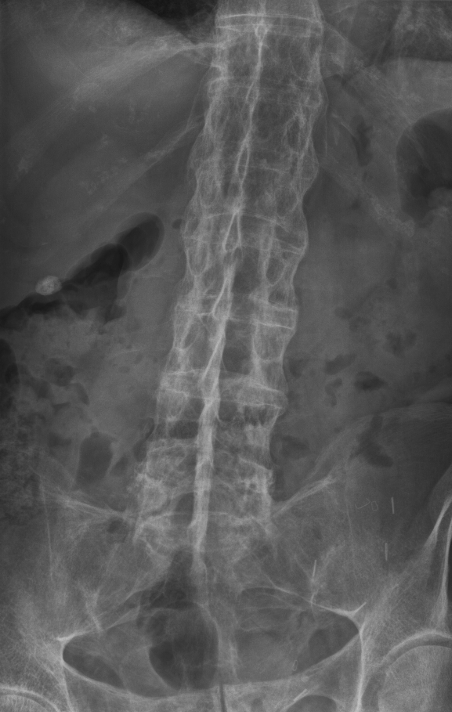
X-rayAnteroposterior lumbar spine radiograph: continuous ossification of the intervertebral ligaments forming a bamboo spine appearance, characteristic of advanced ankylosing spondylitis.
Source : Stevenfruitsmaak, Wikimedia Commons · CC BY-SA
The physical examination aims to document the loss of axial mobility and to look for signs of peripheral enthesitis.
Measures of spinal mobility (BASMI, Jenkinson 1994):4
- Occiput-to-wall (or tragus-to-wall) distance : thoracic kyphosis
- Cervical rotation in degrees
- Lumbar lateral flexion in cm
- Modified Schober test : lumbar flexion (normal > 5 cm of expansion)
- Intermalleolar distance : hip abduction
Chest expansion : the difference between inspiration and expiration at the 4th intercostal space (normal > 5 cm). It falls late, with costovertebral involvement.4
Investigations:
- HLA-B27 : present in 80-95 % of Caucasian AS patients, but also in 8-10 % of the general population. Its positive predictive value in isolation is modest; it is interpreted within a suggestive clinical context.5
- CRP / ESR : these can be raised, but a normal CRP does not exclude the diagnosis (about 40 % of patients with active axSpA have a normal CRP).5
- Anteroposterior pelvic radiographs : to look for sacroiliitis (modified NY stages 2-4). However, changes can take 5-10 years to appear: the radiograph is not sensitive at an early stage.1
- MRI of the sacroiliac joints : the reference investigation for early diagnosis. The STIR sequence detects bone marrow oedema (a sign of active inflammation). The updated ASAS/OMERACT definition (Lambert 2016) requires at least 2 lesions on 1 slice or 1 lesion on 2 consecutive slices.6
The ASAS decision algorithm for diagnosing axSpA
The logic of the ASAS 2009 criteria (Rudwaleit): two entry arms: imaging or clinical
After Rudwaleit M et al. Ann Rheum Dis 2009;68(6):777-83. Pooled sensitivity 73 %, specificity 88 % (Sepriano 2017 meta-analysis).
Should patients be classified, and what are the benefits?
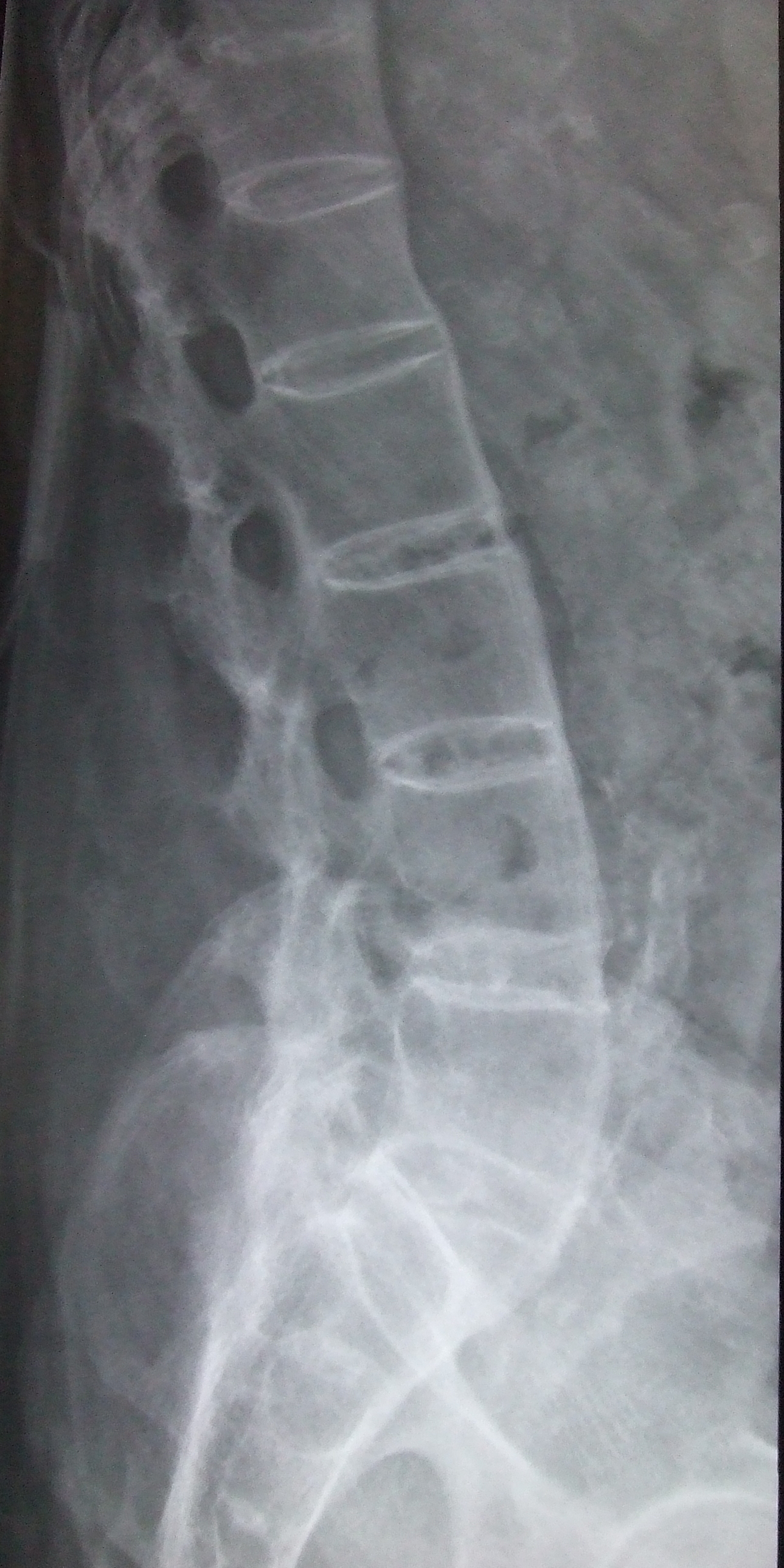
X-rayLateral lumbar spine radiograph: squaring of the vertebrae and intervertebral bony bridges (syndesmophytes), radiological signs of ankylosing spondylitis.
Source : Glitzy_queen00, Wikimedia Commons · Domaine public
The ASAS 2009 classification (Rudwaleit), distinct from the individual clinical diagnosis, makes populations homogeneous for research and for access to treatment.7 Its overall performance was assessed in the Sepriano 2017 meta-analysis (9 studies, 5,739 patients): sensitivity 73 %, specificity 88 %.7
Two subgroups within axSpA:
- r-axSpA (ankylosing spondylitis / AS): radiographic sacroiliitis under the modified NY criteria (van der Linden 1984).8
- nr-axSpA : no visible structural lesion but active sacroiliitis on MRI or the HLA-B27+ clinical arm.7
| Criterion / investigation | Sensitivity | Specificity | Level of evidence | Source PMID |
|---|---|---|---|---|
| Inflammatory back pain (IBP, Sieper) | ~70 % | ~80 % | High (1a) | 19147614 |
| HLA-B27 (Caucasian population) | ~85-95 % | ~90 % | High | 28110981 |
| Sacroiliac MRI (STIR oedema, ASAS 2016) | ~70-80 % | ~90 % | High (1a) | 26768408 |
| Sacroiliac radiograph (NY sacroiliitis) | ~30 % at an early stage | ~95 % | Moderate (changes appear late) | 6231933 |
| Modified Schober test | Variable | ~70 % | Moderate: poorly sensitive at an early stage | 7799351 |
| ASAS axSpA criteria (meta-analysis) | 73 % | 88 % | High (1a) | 28179264 |
| Sacroiliac provocation tests (cluster >= 3) | Variable | Variable | Low: high heterogeneity | 31391801 |
Critique and controversy: the grey areas of diagnosis
Several challenges persist. The first concerns using the ASAS criteria as a diagnostic tool in everyday clinical practice, when they were designed for classification in research.7 A patient can have axSpA without meeting the criteria and, conversely, someone who meets the clinical arm (HLA-B27+ with a few suggestive features) may have another condition.
The second point concerns interpreting the MRI : bone marrow oedema is not entirely specific. Similar lesions can be seen in athletes (repeated mechanical loading), in women postpartum, or in infectious spondylodiscitis, hence the imperative need for clinical correlation before making the diagnosis.6
Finally, the diagnostic delay of 7 years on average remains a major scourge, mainly due to poor recognition of the features of inflammatory back pain by front-line clinicians (general practitioners, physiotherapists) and to patients themselves treating it as ordinary.2
Key points
- Diagnosing axSpA is a clinical puzzle : IBP (Sieper 2009, 4/5 criteria) + HLA-B27 + CRP + sacroiliac MRI (STIR).
- MRI of the sacroiliac joints is the key early investigation: it reveals bone marrow oedema 5-10 years before the radiograph does.
- The ASAS 2009 criteria (sens. 73 %, spec. 88 %) distinguish r-axSpA (AS) from nr-axSpA, to standardise research.
- Median diagnostic delay ~7 years (Zhao 2021): recognising IBP is the major public health issue.
Bibliography
- Sieper J, Poddubnyy D. Axial spondyloarthritis. Lancet. 2017;390(10089):73-84. PMID 28110981.
- Zhao SS, Pittam B, Harrison NL, et al. Diagnostic delay in axial spondyloarthritis: a systematic review and meta-analysis. Rheumatology (Oxford). 2021;60(4):1620-1628. PMID 33428758.
- Sieper J, van der Heijde D, Landewe R, et al. New criteria for inflammatory back pain in patients with chronic back pain: a real patient exercise by experts from the Assessment of SpondyloArthritis international Society (ASAS). Ann Rheum Dis. 2009;68(6):784-788. PMID 19147614.
- Jenkinson TR, Mallorie PA, Whitelock HC, Kennedy LG, Garrett SL, Calin A. Defining spinal mobility in ankylosing spondylitis (AS). The Bath AS Metrology Index. J Rheumatol. 1994;21(9):1694-1698. PMID 7799351.
- Navarro-Compan V, Sepriano A, El-Zorkany B, van der Heijde D. Axial spondyloarthritis. Lancet. 2024;404(10470):2354-2367. doi:10.1016/S0140-6736(24)02263-3.
- Lambert RG, Bakker PA, van der Heijde D, et al. Defining active sacroiliitis on MRI for classification of axial spondyloarthritis: update by the ASAS MRI working group. Ann Rheum Dis. 2016;75(11):1958-1963. PMID 26768408.
- Sepriano A, Rubio R, Ramiro S, Landewe R, van der Heijde D. Performance of the ASAS classification criteria for axial and peripheral spondyloarthritis: a systematic literature review and meta-analysis. Ann Rheum Dis. 2017;76(5):886-890. PMID 28179264.
- van der Linden S, Valkenburg HA, Cats A. Evaluation of diagnostic criteria for ankylosing spondylitis: a proposal for modification of the New York criteria. Arthritis Rheum. 1984;27(4):361-368. PMID 6231933.
- Rudwaleit M, van der Heijde D, Landewe R, et al. The development of Assessment of SpondyloArthritis international Society classification criteria for axial spondyloarthritis (part II): validation and final selection. Ann Rheum Dis. 2009;68(6):777-783. PMID 19297344.
- Garrett S, Jenkinson T, Kennedy LG, Whitelock H, Gaisford P, Calin A. A new approach to defining disease status in ankylosing spondylitis: the Bath Ankylosing Spondylitis Disease Activity Index. J Rheumatol. 1994;21(12):2286-2291. PMID 7699630.
axSpA in women: why is it underdiagnosed, and how do we put that right?
For decades, ankylosing spondylitis was regarded as a “male” disease with an M/F ratio estimated at 2-3:1, based on the 1984 NY criteria, which require radiographic sacroiliitis.1 That view is now outdated: with the arrival of the ASAS 2009 criteria and the inclusion of non-radiographic forms, the ratio is close to 1:1 for nr-axSpA.2,3 This conceptual shift reveals a major problem of underdiagnosis in women.
What are the clinical and radiological differences between men and women?
Women with axSpA have a partly different clinical phenotype from men's, which contributes to the diagnostic delay:1,2
- More frequent peripheral involvement : arthritis, enthesitis (heel, plantar fascia), dactylitis.
- Earlier cervical involvement and more marked in advanced disease.
- More diffuse symptoms (neck, chest and peripheral pain), sometimes wrongly labelled “fibromyalgia”.
- Often more severe fatigue and a greater impact on quality of life at an equal level of disease activity.
- A higher BASDAI but a lower BASMI (less objective spinal stiffness).
- Slower radiographic structural progression : less syndesmophyte formation, so fewer patients cross over into r-axSpA.
- Sometimes a poorer response to anti-TNF (for example, lower retention rates in the Scandinavian registries), although the data are debated.
Clinical differences between women and men with axSpA
A synthesis after Rusman 2018, Wright 2020, Tournadre 2013
Sources: Rusman T et al. Curr Rheumatol Rep 2018;20(6):35; Tournadre A et al. Joint Bone Spine 2013;80(2):205-9. The diagnostic delay is on average 2-3 years longer in women, on data from the ASAS-COMOSPA and SCQM registries.
How do you reduce the diagnostic delay in women?
The Zhao 2021 meta-analysis (PMID 33982063) confirms that female sex is an independent risk factor for diagnostic delay in axSpA.4 Several strategies are validated or under evaluation:
- Raise awareness among front-line clinicians (general practitioners, physiotherapists, gynaecologists) of the female phenotype of axSpA: diffuse pain, fatigue, peripheral involvement, an underlying inflammatory back pain despite an “atypical” picture.2
- Avoid the trap differential diagnoses : fibromyalgia is over-diagnosed in women with axSpA. Any woman with chronic back pain before 45 + morning stiffness > 30 min + improvement with exercise should be assessed for axSpA.1
- Favour MRI of the sacroiliac joints in the symptomatic young woman: the radiograph is even less sensitive than in men (slower structural progression).5
- Interpret the MRI with caution postpartum : sacroiliac bone marrow oedema can be present in 30-60 % of women postpartum without inflammatory disease, settling within 6-12 months.6
- Use the screening tools : IBP questionnaires (Sieper 2009), with systematic referral for patients with IBP + HLA-B27+ or IBP + a family history.7
Specific points in women with axSpA
- Any woman < 45 years with chronic inflammatory back pain + morning stiffness > 30 min = a rheumatology assessment.
- A pregnant woman with axSpA : adapt the biologic therapy (certolizumab is compatible with pregnancy, per the CRIB data), with close follow-up (a flare is possible in the 1st-2nd trimester, with improvement in the 3rd).
- Postpartum : a high risk of flare (up to 60 % of women with axSpA), so monitor clinically.
- Do not confuse it with fibromyalgia : the two can coexist (~15-20 %), but IBP + MRI should guide the assessment.
Key points
- M/F ratio: 2-3:1 in r-axSpA (AS) but 1:1 in nr-axSpA. The historical under-representation of women stems from the radiographic NY 1984 criteria.
- The female phenotype: more peripheral involvement, less structural progression, more severe fatigue, a higher BASDAI.
- Diagnostic delay 2-3 years longer in women than in men: female sex = an independent risk factor (Zhao 2021).
- Strategies: raising awareness among front-line clinicians, early MRI, and care not to over-diagnose fibromyalgia.
Bibliography
- Rusman T, van Vollenhoven RF, van der Horst-Bruinsma IE. Gender Differences in Axial Spondyloarthritis: Women Are Not So Lucky. Curr Rheumatol Rep. 2018;20(6):35. PMID 29754330.
- Sieper J, Poddubnyy D. Axial spondyloarthritis. Lancet. 2017;390(10089):73-84. PMID 28110981.
- Tournadre A, Pereira B, Lhoste A, et al. Differences between women and men with recent-onset axial spondyloarthritis: results from a prospective multicentre French cohort. Arthritis Care Res (Hoboken). 2013;65(9):1482-1489. PMID 23463610.
- Zhao SS, Pittam B, Harrison NL, et al. Diagnostic delay in axial spondyloarthritis: a systematic review and meta-analysis. Rheumatology (Oxford). 2021;60(4):1620-1628. PMID 33428758.
- Navarro-Compan V, Sepriano A, El-Zorkany B, van der Heijde D. Axial spondyloarthritis. Lancet. 2024;404(10470):2354-2367. doi:10.1016/S0140-6736(24)02263-3.
- Eshed I, Miloh-Raz H, Dulitzki M, et al. Peripartum changes of the sacroiliac joints on MRI: increasing mechanical load correlating with signs of edema and inflammation kindling spondyloarthropathy in the genetically prone. Clin Rheumatol. 2015;34(8):1419-1426. PMID 26006255.
- Sieper J, van der Heijde D, Landewe R, et al. New criteria for inflammatory back pain in patients with chronic back pain: a real patient exercise by experts from the Assessment of SpondyloArthritis international Society (ASAS). Ann Rheum Dis. 2009;68(6):784-788. PMID 19147614.
Which treatment strategies are the most effective?
Where do you start? What is the recommended hierarchy of interventions?
The reference recommendations are the ASAS-EULAR 2022 update (Ramiro 2023, PMID 36270658)1 and ACR/SAA/SPARTAN 2019 (Ward 2019, PMID 31436026).2 The hierarchy is clear:
- Therapeutic education + regular exercise = the non-negotiable foundation for EVERY patient from diagnosis onwards.1
- NSAIDs as first-line pharmacotherapy (ibuprofen, naproxen, etoricoxib, and so on). The choice is guided by tolerance and by cardiovascular and gastric risk.1,2
- bDMARDs (biologic therapy) where disease activity persists despite optimal NSAIDs: anti-TNF (infliximab, etanercept, adalimumab, golimumab, certolizumab) or anti-IL-17 (secukinumab, ixekizumab, bimekizumab).1
- tsDMARDs (JAK inhibitors) : upadacitinib, tofacitinib. Approved since 2021-2022 in axSpA. Their place is still evolving.1
- csDMARDs (methotrexate, sulfasalazine) are effective only on peripheral involvement, not on axial disease.2
Physiotherapy comes in at the very first line, BEFORE any escalation of medication, and continues throughout the pathway.
The axSpA treatment pyramid: ASAS-EULAR 2023
Horizontal cards stacked from the foundation (a wide base) to the last-resort options
After Ramiro S et al. Ann Rheum Dis 2023;82(1):19-34; Ward MM et al. Arthritis Rheumatol 2019;71(10):1599-613. The width of each base reflects the percentage of patients concerned (foundation = 100 %, surgery = < 5 %).
What is the place of exercise, and is HIIT superior?
Physical exercise is not an option: it is a central therapeutic intervention. The Cochrane review Regnaux 2019 (14 RCTs, 1,579 participants) confirms that exercise programmes, whether supervised or in groups, are significantly superior to no intervention on physical function (BASFI), mobility (BASMI) and overall well-being (moderate-quality evidence).3
The major advance of the past 5 years concerns high-intensity interval training (HIIT). Several studies by Sveaas et al. have shown that it is:
- Safe in patients with stable axSpA: no rise in inflammatory markers and no induced flare.4,5,6
- Effective : it improves the ASDAS, the BASDAI, function (BASFI) and cardiorespiratory capacity (VO2max).4
- It also benefits non-spinal symptoms : fatigue, sleep, mood (secondary analysis, PT 2020, n=100).6
HIIT does not replace mobility and stretching exercises: it complements them. The ideal programme combines axial mobility, muscle strengthening (postural muscles in particular), cardiorespiratory exercise and HIIT 2-3 times a week.
Manual therapies, balneotherapy, TENS: how effective are they really?
Manual therapies (mobilisations, massage) are a useful short-term adjunct for pain and stiffness, never a stand-alone disease-modifying treatment.3 They can prepare the body for exercise or relieve flare episodes.
Meanwhile, balneotherapy and spa therapy (exercise in heated mineral water) has shown positive effects on pain and function, and is particularly valued for the buoyancy that makes movement easier.3
Finally, TENS lacks robust evidence specific to axSpA: its use is essentially empirical, for chronic pain.
How do you educate the patient and address psychological factors?
Structured therapeutic education is an essential component of treatment. A patient who understands their disease, the mechanisms of pain and the beneficial role of physical activity adheres better to treatment and achieves better functional outcomes.1
The prevalence of fatigue, anxiety and depression is high in axSpA (~25-35 % of patients). Screening for these psychological factors is crucial. Cognitive behavioural therapy (CBT) has shown efficacy in managing chronic pain, sleep and fatigue.7
Critique and controversy: the grey areas of management
Several challenges persist. The main one is the gap between the guidelines and everyday clinical practice : many patients still have no access to structured education or to supervised exercise programmes.1
The continued use of passive therapies with weak levels of evidence (TENS, ultrasound on its own) remains a problem. The science promotes an active, exercise-centred approach; yet passive modalities persist, sometimes out of clinician comfort or patient expectation.3
Finally, the place of the JAK inhibitors remains debated: efficacy is demonstrated, but there is a cardiovascular and neoplastic risk signal in patients over 65 or who smoke (EMA restrictions since 2023, by analogy with rheumatoid arthritis: the ORAL Surveillance study).1
Key points
- Foundation : education + regular supervised exercise = first line, for EVERY patient (ASAS-EULAR 2023, Ward 2019).
- HIIT = safe and effective in stable axSpA (Sveaas 2014/2020): combine it with mobility, strengthening and balneotherapy.
- NSAIDs = first-line pharmacotherapy. bDMARDs (anti-TNF or IL-17) where the ASDAS persists at >= 2.1. JAK inhibitors = third line (EMA restrictions).
- Manual therapies = a short-term adjunct only. TENS = no solid evidence in axSpA.
- Screen for fatigue, anxiety and depression. CBT = validated help for chronic pain.
Bibliography
- Ramiro S, Nikiphorou E, Sepriano A, et al. ASAS-EULAR recommendations for the management of axial spondyloarthritis: 2022 update. Ann Rheum Dis. 2023;82(1):19-34. PMID 36270658. doi:10.1136/ard-2022-223296.
- Ward MM, Deodhar A, Gensler LS, et al. 2019 Update of the American College of Rheumatology/Spondylitis Association of America/Spondyloarthritis Research and Treatment Network Recommendations for the Treatment of Ankylosing Spondylitis and Nonradiographic Axial Spondyloarthritis. Arthritis Rheumatol. 2019;71(10):1599-1613. PMID 31436026.
- Regnaux JP, Davergne T, Palazzo C, et al. Exercise programmes for ankylosing spondylitis. Cochrane Database Syst Rev. 2019;10(10):CD011321. PMID 31578051. doi:10.1002/14651858.CD011321.pub2.
- Sveaas SH, Berg IJ, Provan SA, et al. Efficacy of high intensity exercise on disease activity and cardiovascular risk in active axial spondyloarthritis: a randomized controlled pilot study. PLoS One. 2014;9(9):e108688. PMID 25268365.
- Sveaas SH, Bilberg A, Berg IJ, et al. High intensity exercise for 3 months reduces disease activity in axial spondyloarthritis (axSpA): a multicentre randomised trial of 100 patients. Br J Sports Med. 2020;54(5):292-297. PMID 30745314.
- Sveaas SH, Dagfinrud HS, Berg IJ, et al. High-Intensity Exercise Improves Fatigue, Sleep, and Mood in Patients With Axial Spondyloarthritis: Secondary Analysis of a Randomized Controlled Trial. Phys Ther. 2020;100(8):1323-1332. PMID 32367124.
- Sharip A, Kunz J. Understanding the Pathogenesis of Spondyloarthritis. Biomolecules. 2020;10(10):1461. PMID 33092023.
- Navarro-Compan V, Sepriano A, El-Zorkany B, van der Heijde D. Axial spondyloarthritis. Lancet. 2024;404(10470):2354-2367. doi:10.1016/S0140-6736(24)02263-3.
How do you secure lasting recovery and prevent flares?
How do you make the patient an active participant in their recovery?
Patient empowerment is an essential, non-negotiable component of axSpA management under the ASAS-EULAR 2023 recommendations.1 The shift from a passive approach to collaborative active management is the cornerstone of preventing flares.
The aim is to strengthen self-efficacy: the patient's confidence in their ability to manage their symptoms. Several strategies are validated:
- Structured therapeutic education : understanding the disease, the mechanisms of pain, the beneficial role of physical activity. The Zhao 2020 meta-analysis confirms that non-pharmacological interventions (education + exercise) produce significant improvements in pain, function and mobility.2
- Shared decision-making between patient and clinicians: it improves adherence and engagement.1
- Symptom tracking with PROMs : teaching the patient to complete the BASDAI (Garrett 1994) or the ASDAS so that flares are identified early and activities adjusted.3
- Digital health tools : mobile applications, online platforms. A recent systematic review (Druce 2023, npj Digit Med) showed that these tools can support symptom tracking, adherence to home exercise, and access to educational content.4
When and how should a safe return to sport be planned?
Exercise is not only a treatment, it is a fundamental prevention strategy. The return to physical activity should be encouraged, but in a structured way.
The “when” : there is no absolute contraindication to physical activity, even during active disease, provided it is adapted.5 The key principle: do not significantly exacerbate the pain, listen to the body, progress gradually.
The “how” : combine several modalities in a complete, personalised programme :
- Mobility and stretching : essential against stiffening (spine, hips). 5-10 min a day.
- Muscle strengthening : the postural muscles (abdominals, back extensors, glutes). 2-3 times a week.
- Cardiorespiratory activity : aim for the WHO recommendations (150 min of moderate activity a week, or 75 min of vigorous activity). Brisk walking, cycling, swimming.
- HIIT 2-3 times a week in patients with stable axSpA: it benefits the ASDAS, function and VO2max (Sveaas 2014/2020).6,7
The effect of 3 months of HIIT on disease activity in axSpA
Sveaas 2020 study (BJSM): a multicentre RCT, n=100 axSpA patients
After Sveaas SH et al. Br J Sports Med 2020;54(5):292-7 (PMID 30745314). Multicentre, n=100, PT-supervised for 3 months. ASDAS-CRP reduced by 0.5 (p<0.001), VO2max improved by 15 %, BASDAI improved (secondary analysis, PT 2020).
A notable advance: contrary to old fears, high-intensity exercise does not exacerbate the disease. On the contrary, it reduces activity. The key: adequate supervision and gradual progression.6,7
Critique and controversy
Long-term adherence to home programmes is notoriously poor (about 30-50 % at 6-12 months, depending on the study). The “intention-behaviour gap” is a major clinical reality that calls for active follow-up strategies (reminders, periodic contact, mobile apps).1
Separately, digital tools introduce a risk of a digital divide (older patients, disadvantaged groups who are less comfortable with them). The quality and scientific validation of health apps vary enormously.4
Finally, the return to competitive or high-impact sport lacks specific data. Most studies focus on general activity, which leaves elite athletes in a zone of uncertainty.
Key points
- Empowering the patient through education + shared decision-making = the basis of durable axSpA management.
- Exercise is non-negotiable : a programme combining mobility, strengthening and cardiorespiratory work. HIIT = safe and effective in stable axSpA.
- WHO recommendations : 150 min a week of moderate activity (brisk walking, cycling, swimming).
- The return to sport is progressive and guided by symptoms, not by the calendar.
- Digital tools are useful for tracking and motivation: check accessibility.
Bibliography
- Ramiro S, Nikiphorou E, Sepriano A, et al. ASAS-EULAR recommendations for the management of axial spondyloarthritis: 2022 update. Ann Rheum Dis. 2023;82(1):19-34. PMID 36270658.
- Regnaux JP, Davergne T, Palazzo C, et al. Exercise programmes for ankylosing spondylitis. Cochrane Database Syst Rev. 2019;10(10):CD011321. PMID 31578051.
- Garrett S, Jenkinson T, Kennedy LG, Whitelock H, Gaisford P, Calin A. A new approach to defining disease status in ankylosing spondylitis: the Bath Ankylosing Spondylitis Disease Activity Index. J Rheumatol. 1994;21(12):2286-2291. PMID 7699630.
- Telrandhe VU, Jawade S, Nimbalkar R. Effectiveness of Physiotherapy Exercises on Pain, Range of Motion, and Quality of Life in Patients With Ankylosing Spondylitis: A Case Report. Cureus. 2024;16(1):e53338. PMID 38435867.
- Ward MM, Deodhar A, Gensler LS, et al. 2019 Update of the ACR/SAA/SPARTAN Recommendations for the Treatment of Ankylosing Spondylitis and Nonradiographic Axial Spondyloarthritis. Arthritis Rheumatol. 2019;71(10):1599-1613. PMID 31436026.
- Sveaas SH, Berg IJ, Provan SA, et al. Efficacy of high intensity exercise on disease activity and cardiovascular risk in active axial spondyloarthritis: a randomized controlled pilot study. PLoS One. 2014;9(9):e108688. PMID 25268365.
- Sveaas SH, Bilberg A, Berg IJ, et al. High intensity exercise for 3 months reduces disease activity in axial spondyloarthritis (axSpA): a multicentre randomised trial of 100 patients. Br J Sports Med. 2020;54(5):292-297. PMID 30745314.
What do real clinical cases teach us?
A classic case: an evaluated physiotherapy protocol (Telrandhe 2024)
The clinical case reported by Telrandhe et al. (2024, Cureus, PMID 38435867) illustrates structured physiotherapy management in a patient with active AS.1 The 8-week protocol combined:
- Exercises for axial mobility (spinal rotations, stretching)
- Strengthening of the postural muscles (abdominals, paraspinals, glutes)
- Specific breathing exercises (chest expansion)
- Active stretching of the posterior and anterior muscle chains
- Progressive cardiorespiratory activity (walking, cycling)
The results reported at the end of the protocol:
- A reduction in pain (VAS scale)
- Improved spinal range of motion (BASMI)
- Improved function (BASFI) and quality of life (ASQoL)
This clinical case illustrates its consistency with the aggregated evidence: the Cochrane meta-analysis Regnaux 2019 (PMID 31578051) confirms that supervised exercise programmes significantly improve the BASDAI, the BASFI and the BASMI.2 However, a case report remains level 5 evidence (the weakest). It illustrates; it does not demonstrate.
The diagnostic challenge: when axSpA mimics another condition
One of the greatest challenges in axSpA remains the diagnostic delay, a median of about 7 years according to Zhao 2021.3 That delay often stems from atypical presentations that mimic more common musculoskeletal conditions:
- Recurrent sciatica : alternating buttock pain (typical of sacroiliitis) is frequently labelled “sciatica” and treated with short-course NSAIDs plus physiotherapy centred on the lumbar spine, with only partial relief. The distinguishing features: an inflammatory character (improvement with movement, waking at night), prolonged morning stiffness, a dramatic response to long-term NSAIDs. The lumbar MRI is normal, while STIR MRI of the sacroiliac joints reveals the bone marrow oedema that confirms sacroiliitis.4
- Plantar fasciitis / chronic heel pain : Achilles or plantar enthesitis is a cardinal manifestation of axSpA and can be the initial symptom. Any bilateral, recurrent plantar fasciitis in a young person with associated back pain should raise the possibility of spondyloarthritis.5
- Fibromyalgia : in women with axSpA, diffuse pain can be wrongly labelled fibromyalgia. The two can coexist, but assessment against the IBP criteria plus MRI should guide the decision.6
- Ordinary “mechanical” back pain : without systematically checking the IBP criteria (Sieper 2009), axSpA stays invisible.7
These presentations underline how important it is for front-line clinicians (physiotherapists in particular) to recognise the red flags of inflammatory back pain and to refer early to a rheumatologist.
A complex case: axSpA with extra-articular manifestations
axSpA is a systemic disease. The complex cases are often those in which extra-articular manifestations precede the spinal symptoms.
Acute anterior uveitis is the most frequent extra-articular manifestation (~23-26 % of axSpA patients, Stolwijk 2015).8 A typical case: a young woman or man presenting with a painful red eye and photophobia, treated in ophthalmology, then recurrences, then inflammatory back pain appearing several years later. Any recurrent acute anterior uveitis (particularly HLA-B27 positive) should prompt a search for axSpA.9
The association of axSpA + IBD (Crohn's disease or ulcerative colitis, ~6-7 % of axSpA patients) is another example of complexity. The two conditions share inflammatory (the IL-23/IL-17 axis) and genetic pathways. Management becomes multidisciplinary:8,10
- Choice of biologic therapy : monoclonal anti-TNF drugs (infliximab, adalimumab) are effective on both. Anti-IL-17 is to be AVOIDED in active IBD (a risk of worsening). Etanercept (a soluble receptor anti-TNF) is not effective in IBD.
- Adapting physiotherapy : take account of chronic fatigue (iron-deficiency anaemia), abdominal pain and episodes of bowel flare.
GRADE / Oxford CEBM pyramid: levels of evidence in axSpA physiotherapy
Strength of evidence decreasing from top to bottom; horizontal cards
A simplified GRADE / Oxford CEBM hierarchy. The length of the coloured bar illustrates the relative strength of evidence. Practical implication: where an appealing clinical case diverges from a meta-analysis, follow the meta-analysis. Clinical cases serve to generate hypotheses and to illustrate the approach, not to demonstrate efficacy.
Critique and controversy
Illuminating though clinical cases are, their low level of evidence calls for caution. An isolated case controls for neither placebo effect, nor regression to the mean, nor the natural history of the disease. Publication bias also favours the successes.
One persistent controversy: the intensity of exercise. HIIT programmes have shown superior benefits (Sveaas 2014/2020) but their clinical uptake remains timid, for fear of exacerbating inflammation. The data nonetheless confirm that with adequate supervision and gradual progression, HIIT is safe.5,6
Finally,MRI of the sacroiliac joints remains open to discussion: bone marrow oedema is possible in 30-60 % of women postpartum, in some athletes, in osteoarthritis, and so on.11 The diagnosis must rest on a constellation of clinical, laboratory and imaging arguments.
Key points
- The classic case : Telrandhe 2024 (Cureus, PMID 38435867) illustrates 8 weeks of multimodal physiotherapy improving pain, mobility, function and quality of life.
- The diagnostic challenge : axSpA mimics sciatica, plantar fasciitis and fibromyalgia. Inflammatory back pain = the red flag.
- Complexity : ~25 % acute anterior uveitis, ~10 % psoriasis, ~7 % IBD. A multidisciplinary approach (rheumatology + ophthalmology + gastroenterology + physiotherapy).
- Anti-TNF in axSpA + IBD : choose a monoclonal anti-TNF (infliximab, adalimumab). Avoid anti-IL-17 in active IBD.
- Level of evidence : a case report = level 5. It illustrates, it never demonstrates. Where they diverge, follow the meta-analyses (1a).
Bibliography
- Telrandhe VU, Jawade S, Nimbalkar R. Effectiveness of Physiotherapy Exercises on Pain, Range of Motion, and Quality of Life in Patients With Ankylosing Spondylitis: A Case Report. Cureus. 2024;16(1):e53338. PMID 38435867.
- Regnaux JP, Davergne T, Palazzo C, et al. Exercise programmes for ankylosing spondylitis. Cochrane Database Syst Rev. 2019;10(10):CD011321. PMID 31578051.
- Zhao SS, Pittam B, Harrison NL, et al. Diagnostic delay in axial spondyloarthritis: a systematic review and meta-analysis. Rheumatology (Oxford). 2021;60(4):1620-1628. PMID 33428758.
- Lambert RG, Bakker PA, van der Heijde D, et al. Defining active sacroiliitis on MRI for classification of axial spondyloarthritis: update by the ASAS MRI working group. Ann Rheum Dis. 2016;75(11):1958-1963. PMID 26768408.
- Schett G, Lories RJ, D'Agostino MA, Elewaut D, Kirkham B, Soriano ER, McGonagle D. Enthesitis: from pathophysiology to treatment. Nat Rev Rheumatol. 2017;13(12):731-741. PMID 29158573.
- Rusman T, van Vollenhoven RF, van der Horst-Bruinsma IE. Gender Differences in Axial Spondyloarthritis: Women Are Not So Lucky. Curr Rheumatol Rep. 2018;20(6):35. PMID 29754330.
- Sieper J, van der Heijde D, Landewe R, et al. New criteria for inflammatory back pain in patients with chronic back pain: a real patient exercise by experts from the Assessment of SpondyloArthritis international Society (ASAS). Ann Rheum Dis. 2009;68(6):784-788. PMID 19147614.
- Stolwijk C, van Tubergen A, Castillo-Ortiz JD, Boonen A. Prevalence of extra-articular manifestations in patients with ankylosing spondylitis: a systematic review and meta-analysis. Ann Rheum Dis. 2015;74(7):1373-1378. PMID 24013647.
- Sieper J, Poddubnyy D. Axial spondyloarthritis. Lancet. 2017;390(10089):73-84. PMID 28110981.
- Ward MM, Deodhar A, Gensler LS, et al. 2019 Update of the ACR/SAA/SPARTAN Recommendations for the Treatment of Ankylosing Spondylitis and Nonradiographic Axial Spondyloarthritis. Arthritis Rheumatol. 2019;71(10):1599-1613. PMID 31436026.
- Eshed I, Miloh-Raz H, Dulitzki M, et al. Peripartum changes of the sacroiliac joints on MRI: increasing mechanical load correlating with signs of edema and inflammation kindling spondyloarthropathy in the genetically prone. Clin Rheumatol. 2015;34(8):1419-1426. PMID 26006255.
How do you apply these recommendations concretely in your practice?
When and to which other professionals should you refer?
Referral to other professionals is a mark of competence. It is essential in several situations.
The first step is the rigorous identification of red flags. The international reference framework Finucane 2020 (PMID 32438853) stresses that red flags must be interpreted within the patient's overall context, never in isolation.1
Red flags specific to axSpA
- Spinal fracture in advanced AS : a high risk (secondary osteoporosis + ankylosis = a rigid, fragile spine). Any trauma, even minor, plus new persistent pain = urgent imaging.
- Aortic dissection : the risk is raised in advanced AS (aortic root involvement). Abrupt chest pain, tearing back pain = an emergency.
- Cauda equina syndrome : rare but possible (compression with cervical ankylosis). Saddle anaesthesia, sphincter disturbance, motor deficit = a neurosurgical emergency.
- Infectious spondylodiscitis : fever, rigors, acute or recent spinal pain, raised inflammatory markers = imaging + microbiological work-up.
- Acute anterior uveitis (a red eye, pain, photophobia): ophthalmology within 24-48 h to avoid lasting damage.
- Suspected IBD (chronic diarrhoea, blood in the stool, weight loss): gastroenterology.
- Unexplained weight loss + spinal pain : a work-up for neoplasia.
- Planned anaesthesia : warn the anaesthetist if there is cervical AS (difficult intubation, a risk of spinal cord injury).
Beyond the emergencies, assess the yellow flags (psychosocial factors: catastrophising, kinesiophobia, poor social support): predictors of chronic pain and disability. Collaboration with a psychologist specialising in pain is indicated where these factors are prominent.2
Essential interprofessional collaboration:
- Rheumatologist : the pivot for diagnosis and the biologic therapy decision.
- Ophthalmologist : follow-up for recurrent uveitis.
- Gastroenterologist : where IBD is associated.
- Dermatologist : where psoriasis is severe.
- Orthopaedic or spinal surgeon : disabling hip disease, severe kyphosis.
- Occupational physician : adapting the workstation (ergonomics, breaks, remote working).
- Occupational therapist : adapting the home and daily activities.
How do you measure outcomes and overcome barriers to implementation?
Putting the recommendations into practice is not enough: the effects have to be measured objectively. Systematic use of validated PROMs is fundamental in axSpA:
- BASDAI (Garrett 1994): disease activity on 0-10. A threshold above 4 = active disease.3
- ASDAS-CRP : a more modern composite score (BASDAI + neck/back pain + duration of stiffness + chest expansion + CRP). Recommended by ASAS.4
- BASFI : functional limitation on 0-10.
- BASMI (Jenkinson 1994): spinal mobility (5 clinical measures).5
- ASAS-HI : an axSpA-specific overall health index.
- BASDAI 50 or ASAS 20/40 : criteria for treatment response.
Implementing evidence-based practice (EBP) runs into documented obstacles:
- Lack of time in consultation
- Difficulty appraising the relevant scientific literature
- Lack of organisational support (access to databases, training)
- Divergent beliefs between patients and clinicians
The facilitators include: peer mentoring, journal clubs, pre-appraised evidence summaries, and the adoption of shared decision-making (integrating evidence + clinical expertise + the patient's values and preferences).6
Critique and controversy: the tension between the ideal and the real
The tyranny of averages : meta-analyses tell us the average effect of an intervention, whereas the clinician treats a unique individual. Applying a protocol rigidly can ignore what is specific to that person.
The implementation gap is systemic, not merely individual. Blaming the physiotherapist for a “lack of time” is reductive: reimbursement models reward the volume of procedures rather than quality, thinking time is not paid for, and access to publications remains costly.
Finally, the proliferation of “flags” can become counterproductive if it turns the clinician into a box-ticker at the expense of the therapeutic alliance. The real skill: taking a fine-grained history that takes in every dimension of the patient's experience.
Key points
- axSpA red flags : spinal fracture in AS, aortic dissection, cauda equina, spondylodiscitis, uveitis, active IBD. Refer promptly.
- Yellow flags : catastrophising, kinesiophobia, depression; collaborate with a psychologist.
- Multidisciplinary collaboration : rheumatologist, ophthalmologist, gastroenterologist, occupational therapist, occupational physician.
- The essential PROMs : BASDAI, ASDAS-CRP, BASFI, BASMI, ASAS-HI; track them regularly.
- EBP = evidence + expertise + patient values. Shared decision-making.
Bibliography
- Finucane LM, Downie A, Mercer C, et al. International Framework for Red Flags for Potential Serious Spinal Pathologies. J Orthop Sports Phys Ther. 2020;50(7):350-372. PMID 32438853. doi:10.2519/jospt.2020.9971.
- Garrett S, Jenkinson T, Kennedy LG, Whitelock H, Gaisford P, Calin A. A new approach to defining disease status in ankylosing spondylitis: the Bath Ankylosing Spondylitis Disease Activity Index. J Rheumatol. 1994;21(12):2286-2291. PMID 7699630.
- Ramiro S, Nikiphorou E, Sepriano A, et al. ASAS-EULAR recommendations for the management of axial spondyloarthritis: 2022 update. Ann Rheum Dis. 2023;82(1):19-34. PMID 36270658.
- Jenkinson TR, Mallorie PA, Whitelock HC, Kennedy LG, Garrett SL, Calin A. Defining spinal mobility in ankylosing spondylitis (AS). The Bath AS Metrology Index. J Rheumatol. 1994;21(9):1694-1698. PMID 7799351.
- Bernhardsson S, Larsson ME, Eggertsen R, et al. Evaluation of a tailored, multi-component intervention for implementation of evidence-based clinical practice guidelines in primary care physical therapy: a non-randomized controlled trial. BMC Health Serv Res. 2014;14:105. PMID 24589291.
- Navarro-Compan V, Sepriano A, El-Zorkany B, van der Heijde D. Axial spondyloarthritis. Lancet. 2024;404(10470):2354-2367. doi:10.1016/S0140-6736(24)02263-3.
And after this article?
This article is part of a collection of evidence-based clinical syntheses. A question, a comment, a correction to suggest? Contact us directly through the WhatsApp button at the bottom right of the screen.

