















In brief
Mechanical sacroiliac joint pain refers to pain generated by the sacroiliac joint and its peri-articular structures, and accounts for 15 to 30 % of chronic low back pain. Women predominate, with a peak during pregnancy (Pelvic Girdle Pain, 20 to 70 % of pregnancies). Diagnosis is clinical: unilateral pain strictly below L5, pinpointed by the Fortin Finger Test and aggravated by load transfers, confirmed by a cluster of provocation tests (at least 3 positive). Graded management starts with education, exercise and manual therapy; specific stabilisation exercises are effective for post-partum PGP, 90 % of which resolves by 3 months.
Clinical synthesis based on the most recent meta-analyses and international consensus statements: WIP / Pain Practice 2024 guideline, ASPN 2024, JOSPT cluster meta-analysis 2021, post lumbar fusion data 2023-2025.
Clinical synthesis
- Sacroiliac joint (SIJ) pain accounts for 15–30 % of chronic low back pain. Women, and pregnant women in particular (Pelvic Girdle Pain, PGP), are disproportionately affected. Lumbar fusion surgery and trauma complete the aetiological picture.
- The pathophysiology involves an imbalance between form stability (ligaments, joint congruence) and force stability (muscles: transversus abdominis, multifidus, pelvic floor, gluteals). Central sensitisation plays a part in chronicity.
- The course is often favourable after pregnancy (90 % resolution by 3 months post-partum), but a minority of women (~10 %) develop persistent pain beyond 3 months post-partum, sometimes for up to 2 years.
- Diagnosis is clinical: unilateral pain strictly below L5, pinpointed by the Fortin Finger Test, aggravated by load transfers (sit-to-stand, rolling over in bed).
- Use a cluster of provocation tests (Distraction, Thigh Thrust, Compression, Sacral Thrust, Gaenslen). At least 3 positive tests point towards an SIJ origin: the Saueressig 2021 meta-analysis (JOSPT) confirms that they are more useful to rule out than to confirm.
- Imaging is not useful for diagnosing mechanical pain but does rule out red flags (inflammatory sacroiliitis, fracture, tumour, infection).
- Systematically rule out the mimics: lumbar disc herniation, facet joint syndrome, hip osteoarthritis, piriformis syndrome, inflammatory sacroiliitis.
- Biomechanical classifications based on palpation of mobility have poor inter-examiner reliability and have not demonstrated clinical added value (Robinson 2007, Laslett 2008).
- The therapeutic approach is graded : education + exercise + manual therapy as first line; intra-articular injections or cooled RF neurotomy in documented refractory cases.
- The specific stabilisation exercises (Stuge 2004) are effective for post-partum PGP, with benefit maintained at 2 years. For non-pregnancy SIJ dysfunction, the Mapinduzi 2022 meta-analysis qualifies the effect on pain (similar to the comparator) but confirms a small benefit on disability.
- The manual therapy approach provides short-term benefits, above all when combined with exercise. It acts as a catalyst for engagement rather than as a stand-alone treatment.
- The biopsychosocial approach is essential: screening for yellow flags (kinesiophobia, catastrophising, FABQ), pain neurophysiology education, cognitive functional therapy (CFT, O'Sullivan 2018).
- Return to sport must be progressive and guided by functional criteria (rather than by the calendar): gluteal strength symmetry, Y-Balance Test, hop tests, sport-specific movement simulation under fatigue.
- Screen for red flags (Verhagen 2017, Cook 2018) so that medical referral is prompt. Interprofessional collaboration (rheumatologist, pain physician, midwife, psychologist) for complex cases.
- Use PROMs (VAS, ODI, Pelvic Girdle Questionnaire for PGP, FABQ for fear of movement) to objectify progress and support shared decision-making (Hoffmann 2014).
Contents
- What are the fundamentals to know about mechanical sacroiliac joint pain?
- How can mechanical sacroiliac joint pain be assessed and diagnosed with certainty?
- Which treatment strategies are the most effective for mechanical sacroiliac joint pain?
- Pregnancy and post-partum: how should Pelvic Girdle Pain be managed?
- How can lasting recovery be secured and recurrence of mechanical sacroiliac joint pain prevented?
- What do real-world case studies teach us about mechanical sacroiliac joint pain?
- How do you apply these recommendations concretely in your practice?
What are the fundamentals to know about mechanical sacroiliac joint pain?
How is this condition defined, who is affected and what are the risk factors?
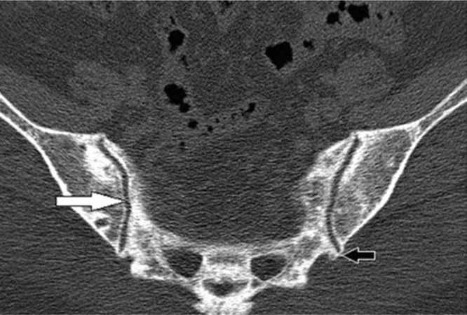
CT scanAxial CT of the sacroiliac joints: a normal-appearing joint (white arrow) and bridging calcification at the inferior joint margin (black arrow), in a 48-year-old woman with hypoparathyroidism.
Source : Jakkani et al., Radiology Case Reports, 2011, figure 6 · CC BY-NC-ND
- Trauma: a fall onto the buttocks, a road traffic accident, asymmetrical lifting: a frequent cause of acute dysfunction.¹
- Lumbar fusion surgery: a major risk factor for postoperative “new SIJ pain”. The Tetreault 2023 systematic review and meta-analysis (Neurochirurgie, 12 studies) reports a pooled incidence of 15.8 % of SIJ pain in the year following lumbar fusion, rising to 32.9 % when the fusion extends to the sacrum (versus 15.8 % without sacral extension).⁹ The later systematic reviews by Beaver 2024 (Acta Neurochirurgica) and the 2024 multicentre cohort confirm this trend and identify female sex, the number of fused levels and postoperative pelvic tilt as aggravating factors.¹⁰
- Leg length inequality: the landmark Friberg study (1983, Spine, n=798 chronic low back pain patients vs 359 controls) showed that 75.4 % of the low back pain patients had an LLD ≥ 5 mm against 43.5 % of the controls, and that correction with a heel lift brought relief in the majority.¹¹
- Repetitive sporting activities and occupations with asymmetrical loading can generate cumulative overload.
📊 Main aetiologies of mechanical sacroiliac joint pain
Estimates drawn from the landmark reviews (Cohen 2005, Buchanan 2021, Tetreault 2023)
Sources: Cohen SP. Anesth Analg. 2005;101(5):1440-53. Tetreault L et al. Neurochirurgie. 2023;69(2). Halliday H et al. Health Sci Rep. 2024.
What happens inside the body and how does mechanical sacroiliac joint pain evolve naturally?
🧬 The biomechanics of the SIJ was modelled by Vleeming, Snijders et al. in the concept of “self-bracing” resting on two complementary stability mechanisms.¹² Form stability (form closure) is provided by the congruence of the articular surfaces (roughness, ridges) and by the passive tension of the powerful interosseous, posterior and anterior sacroiliac ligaments. Force stability (force closure) is generated by the co-activation of the surrounding muscles: transversus abdominis, lumbar multifidus, pelvic floor, gluteus maximus and latissimus dorsi via the thoracolumbar fascia.¹²,¹³ Dysfunction emerges when this balance breaks down, either through hypermobility (post-traumatic, hormonal, post-partum) with irritation of the peri-articular structures, or through hypomobility (fibrosis, osteoarthritis, spasm) with compensatory loading.¹⁴ The SIJ also undergoes age-related degenerative changes (cartilage erosion, osteophytosis, fibrosis and sometimes ankylosis), but these imaging findings do not correlate closely with pain and are often incidental in asymptomatic individuals.⁴ Central sensitisation, a mechanism shared with other chronic pain conditions, can perpetuate pain independently of the local mechanical state, which justifies a biopsychosocial reading.¹⁵ The natural course depends on the aetiological context:- Pregnancy-related PGP: a largely favourable course, with ~90 % resolution by 3 months post-partum. The remaining 10 % may progress to chronic PGP persisting for 1 to 2 years (Halliday 2024).⁷,⁸
- Post-traumatic SIJ pain: a more uncertain course; early conservative treatment lowers the risk of chronification.
- Post-fusion SIJ pain: incidence rises over the 2 years following surgery. The mechanical profile (adjacent segment overload) partly explains this trajectory.⁹,¹⁰
- SIJ pain with no identified cause: a typically fluctuating course, alternating flare-ups and remissions. Without structured management, the risk of chronification is real.¹
Key points
- SIJ pain is a major cause of mechanical chronic low back pain (15–30 %, Cohen 2005, Buchanan 2021).
- The female predominance is marked, with a peak during pregnancy (PGP, 20-70 %, Wu 2004, Halliday 2024). After lumbar fusion, incidence reaches 32.9 % when the fusion extends to the sacrum (Tetreault 2023).
- Biomechanics: balance between form closure (ligaments) ↔ force closure (muscles: transversus abdominis, multifidus, pelvic floor, gluteals).
- Course: fluctuating or favourable, but a minority become chronic, hence the value of early, stratified management.
Bibliography
- Cohen SP. Sacroiliac joint pain: a comprehensive review of anatomy, diagnosis, and treatment. Anesth Analg. 2005;101(5):1440-1453. PMID 16244008.
- Buchanan P, Vodapally S, Lee DW, et al. Successful Diagnosis of Sacroiliac Joint Dysfunction. J Pain Res. 2021;14:3135-3143. PMID 34675642.
- Falowski S, Sayed D, Pope J, et al. A Review and Algorithm in the Diagnosis and Treatment of Sacroiliac Joint Pain. J Pain Res. 2020;13:3337-3348. PMID 33335420.
- Kiapour A, Joukar A, Elgafy H, et al. Biomechanics of the Sacroiliac Joint: Anatomy, Function, Biomechanics, Sexual Dimorphism, and Causes of Pain. Int J Spine Surg. 2020;14(s1):S3-S13. PMID 32123652.
- Slipman CW, Jackson HB, Lipetz JS, Chan KT, Lenrow D, Vresilovic EJ. Sacroiliac joint pain referral zones. Arch Phys Med Rehabil. 2000;81(3):334-338. PMID 10724079.
- Sembrano JN, Polly DW Jr. How often is low back pain not coming from the back? Spine (Phila Pa 1976). 2009;34(1):E27-E32. PMID 19127145.
- Wu WH, Meijer OG, Uegaki K, et al. Pregnancy-related pelvic girdle pain (PPP), I: Terminology, clinical presentation, and prevalence. Eur Spine J. 2004;13(7):575-589. PMID 15338362.
- Halliday H, Murphy J, Ní Bhriain S, et al. The prevalence of depression in women with pregnancy-related pelvic girdle pain: A systematic review and meta-analysis. Health Sci Rep. 2024;7(8):e2308. doi:10.1002/hsr2.2308.
- Shen J, Boudier-Reveret M, Majdalani C, et al. Incidence of sacroiliac joint pain after lumbosacral spine fusion: A systematic review. Neurochirurgie. 2023;69(2):101419. PMID 36754146.
- Xu C, Lin X, Zhou Y, et al. Incidence and risk factors of new-onset sacroiliac joint pain after spinal surgery: a systematic review and meta-analysis. PeerJ. 2024;12:e18083. PMID 39346039.
- Friberg O. Clinical symptoms and biomechanics of lumbar spine and hip joint in leg length inequality. Spine (Phila Pa 1976). 1983;8(6):643-651. PMID 6228021. (Founding article)
- Vleeming A, Schuenke MD, Masi AT, Carreiro JE, Danneels L, Willard FH. The sacroiliac joint: an overview of its anatomy, function and potential clinical implications. J Anat. 2012;221(6):537-567. PMID 22994881.
- Vleeming A, Mooney V, Stoeckart R. Movement, Stability & Lumbopelvic Pain: Integration of Research and Therapy. 2nd ed. Edinburgh: Churchill Livingstone; 2007. (Reference textbook: form closure / force closure concept)
- Sturesson B, Uden A, Vleeming A. A radiostereometric analysis of movements of the sacroiliac joints during the standing hip flexion test. Spine (Phila Pa 1976). 2000;25(3):364-368. PMID 10703111.
- Nijs J, Roussel N, Paul van Wilgen C, Köke A, Smeets R. Thinking beyond muscles and joints: therapists' and patients' attitudes and beliefs regarding chronic musculoskeletal pain are key to applying effective treatment. Man Ther. 2013;18(2):96-102. PMID 23273516.
How can mechanical sacroiliac joint pain be assessed and diagnosed with certainty?
Which questions should you ask to understand the patient and their history?
The history strongly points towards the SIJ hypothesis:- Location: pain around the PSIS (posterior superior iliac spine), often described as “a single point”. The Fortin Finger Test (the patient's ability to point to the pain with a single finger over the inferomedial area of the PSIS) is one of the most suggestive signs (Fortin & Falco 1997).³
- Description and radiation: pain that is most often unilateral, strictly below L5, and may radiate into the buttock, the groin, the posterior thigh and, more rarely, the calf.⁴ This distal radiation can mimic discogenic sciatica.⁵
- Typically asymmetrical aggravating factors:
- Sit-to-stand transfers
- Walking, stair climbing, running
- Rolling over in bed (a particularly specific sign)⁶
- Prolonged sitting, unilateral load carrying
- History: recent pregnancy, lumbar fusion (in particular down to the sacrum), trauma, unilateral impact sport, known leg length inequality.
Red flags to screen for from the history onwards
- Fever, chills, night sweats → infection (septic sacroiliitis, osteomyelitis)
- Unexplained weight loss, history of cancer, constant non-mechanical night pain → tumour
- Morning stiffness > 30 min, axial involvement in a young adult, inflammatory sacroiliitis on MRI → spondyloarthritis (HLA-B27)
- Bladder or bowel dysfunction, saddle anaesthesia, progressive motor deficit → cauda equina syndrome (emergency)
- High-energy trauma → fracture, major ligament rupture
Which clinical tests should be performed and which other conditions must be ruled out?
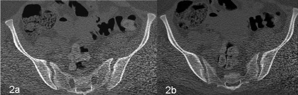
CT scanAxial CT of the pelvis: widening of both sacroiliac joints with slight subluxation of the left joint, in a context of pelvic ring disruption.
Source : Hou et al., Patient Safety in Surgery, 2011, figure 2 · CC BY
- Distraction Test : patient supine, posterolateral pressure on the ASIS.
- Thigh Thrust (Posterior Shear Test) : patient supine, hip and knee flexed to 90°, posterior axial pressure through the femur. Often considered the most sensitive individual test.
- Compression Test : patient side-lying, downward pressure on the iliac crest.
- Sacral Thrust : patient prone, firm anterior thrust on the sacrum.
- Gaenslen : patient supine at the edge of the couch, one knee drawn to the chest, the other leg extended off the couch (creating an SIJ torsion).
📊 Diagnostic performance of the cluster of provocation tests (Saueressig 2021 meta-analysis, JOSPT)
Likelihood ratios (LR+ and LR-) versus intra-articular anaesthetic block (gold standard)
Source: Saueressig T, Owen PJ, Diemer F, Zebisch J, Belavy DL. J Orthop Sports Phys Ther. 2021;51(9):422-431. PMID 34210160.
- Lumbar conditions: L5-S1 disc herniation, low lumbar facet joint syndrome, canal stenosis: a segmental neurological examination is essential.
- Hip conditions: hip osteoarthritis, femoroacetabular impingement (FAI), gluteal tendinopathy: FABER and FADIR tests, hip range of motion.
- Myofascial syndromes: piriformis, gluteal trigger points.
- Visceral conditions: endometriosis, interstitial cystitis, chronic prostatitis (referred pain).
- Inflammatory sacroiliitis: prolonged morning stiffness, axial involvement, young adult, HLA-B27: characteristic MRI.
| Individual test | Sensitivity | Specificity | Evidence level (CEBM) |
|---|---|---|---|
| Fortin Finger Test | 76 % | 47 % | 2b (Fortin 1997, n=54) |
| Thigh Thrust | ≈ 88 % | ≈ 69 % | 1a (Laslett SR) |
| Distraction | ≈ 60 % | ≈ 81 % | 1a (Laslett SR) |
| Compression | ≈ 69 % | ≈ 69 % | 1a (Laslett SR) |
| Sacral Thrust | ≈ 53 % | ≈ 90 % | 1a (Laslett SR) |
| Gaenslen | ≈ 50 % | ≈ 77 % | 1a (Laslett SR) |
| Cluster ≥ 3 positive | LR+ 2.13 | LR- 0.33 | 1a (Saueressig 2021 MA) |
| Palpation / mobility tests | Inter-examiner reliability poor | 2b (Robinson 2007) | |
Should patients with mechanical sacroiliac joint pain be classified, and with what benefits?
The idea of stratifying patients in order to tailor treatment is appealing. Several models have been proposed for the lumbopelvic region: O'Sullivan 2007 (BPS mechanical classification), MDT (McKenzie), classification by direction of provocation, “form/force closure” models.¹²,¹³ But the evidence for their clinical usefulness remains limited. The meta-analysis by Mapinduzi et al. 2022 (J Back Musculoskelet Rehabil, 12 RCTs, n=1407) showed that motor control exercises on their own are not superior to the comparator for pain in patients with PGP of sacroiliac origin; they are, however, moderately effective on disability.¹⁴ For the mobility classifications specific to the SIJ (palpating an “anterior ilium” or a “nutated sacrum”), inter-examiner reliability is poor to very poor (Robinson 2007), which invalidates their operational usefulness.⁷ The modern evidence-based approach therefore focuses on:- Precise clinical identification of SIJ involvement (cluster + Fortin)
- Assessment of the individual functional impairments (motor control, gluteal strength, ASLR for load transfer, FABQ for kinesiophobia)
- A personalised, multimodal and progressive treatment plan, rather than a rigid biomechanical category. ✨
Critique and controversy: the quest for relative certainty
The very notion of diagnostic “certainty” for mechanical SIJ pain remains a matter of debate. The placebo-controlled intra-articular anaesthetic block remains the research gold standard but is invasive, costly and hardly practicable. Clinical clusters are useful but imperfect proxies: the Saueressig 2021 meta-analysis should cool the initial enthusiasm for the high sensitivities and specificities reported.⁹ Moreover, the concept of sacroiliac “dysfunction” (asymmetrical positioning, palpated hypomobility) is not supported by acceptable reliability or validity data : SIJ movements are extremely small (1-3°), incompatible with reliable palpation.¹⁵ Focusing on the provocation of the familiar pain is the most scientifically defensible approach.Key points
- Diagnosis is clinical, by convergence: history + Fortin Finger Test + cluster of provocation tests. At least 3 positive tests point towards the SIJ.
- The Saueressig 2021 meta-analysis shows that the cluster is better at ruling out than at confirming (LR+ 2.13 / LR- 0.33). Keep a degree of diagnostic humility.
- Imaging does not diagnose mechanical SIJ pain. It rules out the red flags (sacroiliitis, fracture, tumour, infection).
- Systematically rule out lumbar, hip and myofascial conditions: the main mimics.
- Classifications based on palpation of mobility or position are unreliable and have not demonstrated clinical added value. Target the individual functional impairments rather than a biomechanical label.
Bibliography
- Cohen SP. Sacroiliac joint pain: a comprehensive review of anatomy, diagnosis, and treatment. Anesth Analg. 2005;101(5):1440-1453. PMID 16244008.
- Buchanan P, Vodapally S, Lee DW, et al. Successful Diagnosis of Sacroiliac Joint Dysfunction. J Pain Res. 2021;14:3135-3143. PMID 34675642.
- Fortin JD, Falco FJ. The Fortin finger test: an indicator of sacroiliac pain. Am J Orthop (Belle Mead NJ). 1997;26(7):477-480. PMID 9247654.
- Slipman CW, Jackson HB, Lipetz JS, Chan KT, Lenrow D, Vresilovic EJ. Sacroiliac joint pain referral zones. Arch Phys Med Rehabil. 2000;81(3):334-338. PMID 10724079.
- Visser LH, Woudenberg NP, de Bont J, et al. Treatment of the sacroiliac joint in patients with leg pain: a randomized-controlled trial. Eur Spine J. 2013;22(10):2310-2317. PMID 23720124.
- Dreyfuss P, Michaelsen M, Pauza K, McLarty J, Bogduk N. The value of medical history and physical examination in diagnosing sacroiliac joint pain. Spine (Phila Pa 1976). 1996;21(22):2594-2602. PMID 8961447.
- Robinson HS, Brox JI, Robinson R, Bjelland E, Solem S, Telje T. The reliability of selected motion- and pain provocation tests for the sacroiliac joint. Man Ther. 2007;12(1):72-79. PMID 16843031.
- Laslett M. Evidence-Based Diagnosis and Treatment of the Painful Sacroiliac Joint. J Man Manip Ther. 2008;16(3):142-152. PMID 19119403.
- Saueressig T, Owen PJ, Diemer F, Zebisch J, Belavy DL. Diagnostic Accuracy of Clusters of Pain Provocation Tests for Detecting Sacroiliac Joint Pain: Systematic Review With Meta-analysis. J Orthop Sports Phys Ther. 2021;51(9):422-431. PMID 34210160.
- Verhagen AP, Downie A, Maher CG, Koes BW. Most red flags for malignancy in low back pain guidelines lack empirical support: a systematic review. Pain. 2017;158(10):1860-1868. PMID 28708761.
- Cook CE, George SZ, Reiman MP. Red flag screening for low back pain: nothing to see here, move along: a narrative review. Br J Sports Med. 2018;52(8):493-496. PMID 28923844.
- O'Sullivan PB, Beales DJ. Diagnosis and classification of pelvic girdle pain disorders--Part 1: a mechanism based approach within a biopsychosocial framework. Man Ther. 2007;12(2):86-97. PMID 17449432.
- Vleeming A, Albert HB, Östgaard HC, Sturesson B, Stuge B. European guidelines for the diagnosis and treatment of pelvic girdle pain. Eur Spine J. 2008;17(6):794-819. PMID 18259783.
- Mapinduzi J, Ndacayisaba G, Mahaudens P, Hidalgo B. Effectiveness of motor control exercises versus other musculoskeletal therapies in patients with pelvic girdle pain of sacroiliac joint origin: A systematic review with meta-analysis of randomized controlled trials. J Back Musculoskelet Rehabil. 2022;35(4):713-731. doi:10.3233/BMR-210108.
- Sturesson B, Uden A, Vleeming A. A radiostereometric analysis of movements of the sacroiliac joints during the standing hip flexion test. Spine (Phila Pa 1976). 2000;25(3):364-368. PMID 10703111.
- Szadek KM, van der Wurff P, van Tulder MW, Zuurmond WW, Perez RS. Diagnostic validity of criteria for sacroiliac joint pain: a systematic review. J Pain. 2009;10(4):354-368. PMID 19101212.
- Mens JM, Vleeming A, Snijders CJ, Koes BW, Stam HJ. Validity of the active straight leg raise test for measuring disease severity in patients with posterior pelvic pain after pregnancy. Spine (Phila Pa 1976). 2002;27(2):196-200. PMID 11805667.
Which treatment strategies are the most effective for mechanical sacroiliac joint pain?
Where do you start? What is the recommended hierarchy of interventions?
The initial approach to the management of mechanical sacroiliac joint pain is conservative, multimodal and graded 🧐. Converging international guidelines (Vleeming/COST B13 2008 for PGP, ASPN 2024 for the SIJ, the Szadek 2024 chapter in Pain Practice) recommend a stepped model.¹,²,³🩺 Graded hierarchy of interventions (stepped care model)
Adapted from the ASPN 2024, Szadek 2024 and Vleeming 2008 guidelines
Stepped care model. No third or fourth line intervention should be undertaken without diagnostic confirmation by a controlled intra-articular anaesthetic block.
What is the role of exercise and is there a superior approach?
Therapeutic exercise is the cornerstone of conservative management. 💪 The most robust evidence concerns post-partum PGP. The landmark Stuge 2004 RCT (Spine, n=81, 2-year follow-up) showed that a specific stabilisation programme (transversus abdominis, multifidus, pelvic floor, gluteals, progressive and individualised) over 20 weeks was superior to conventional physiotherapy without specific exercises, with benefit maintained at 2 years.¹⁰ For non-pregnancy SIJ dysfunction, the Mapinduzi 2022 meta-analysis (12 RCTs, n=1407) adds nuance: motor control exercises on their own are not superior to other therapies for pain, but they are slightly superior for reducing disability.¹¹ The evidence supports a multimodal programme (exercise + education + manual therapy where indicated) rather than a single isolated protocol. The components of a modern programme are:- Reactivation of the deep stabilisers: transversus abdominis, lumbar multifidus, pelvic floor, with co-activation feedback.
- Gluteal strengthening: gluteus maximus, gluteus medius (crucial for load transfer through the SIJ, Sadler 2019).¹²
- Global strengthening: latissimus dorsi, hamstrings, quadriceps.
- Hip mobility: flexion, internal/external rotation.
- Progressive functional reintegration: sit-to-stand transfers, walking, stairs, lunges, sporting movements.
Manual therapies and technologies: how effective are they really?
The manual therapies (mobilisations, high-velocity low-amplitude (HVLA) manipulations, muscle energy techniques) are widely used. The evidence supports a short-term benefit (weeks), particularly when they are combined with exercise. No robust superiority of one technique over another has been demonstrated.¹⁴ The rationale is probably not mechanical (correcting a malposition) but rather neurophysiological (pain modulation, motor disinhibition), which facilitates engagement with the active programme. 🔬 On the technology side :- TENS, ultrasound, LLLT: weak and heterogeneous evidence specifically for the SIJ. Adjunctive use at best.
- Sacroiliac belt: external support useful in the acute phase of PGP, particularly in pregnant women.¹
- Cooled radiofrequency ablation (RFA): the most solid evidence base among interventional options, > 50 % pain reduction at 6-12 months in selected patients (see above).⁷,⁸
- Prolotherapy, PRP, stem cells: emerging but limited evidence; no recommendation for routine practice outside clinical trials.
Beyond the physical: how do you educate the patient and address psychological factors?
🧠 Modern pain management incorporates the biopsychosocial dimension. Cognitive and emotional factors, namely kinesiophobia, catastrophising (Pain Catastrophizing Scale), beliefs about fragility, anxiety and depression, are robust predictors of chronification and disability, as shown in the Wertli 2014 systematic review (Spine Journal) for non-specific low back pain.¹⁵ The therapeutic patient education (ETP) aims to:- Dismantle limiting beliefs : pain does not always mean tissue damage; movement is safe and beneficial.
- Restore self-efficacy : give concrete tools, namely a flare-up plan, pacing and self-mobilisations.
- Co-build functional goals with the patient, shared and attainable.
Critique and controversy: the grey areas of treatment
Three areas of uncertainty deserve a critical reading: 1. Diagnostic heterogeneity in research. RCTs on the SIJ often pool very different profiles (hypermobile vs hypomobile, post-partum vs post-traumatic vs post-fusion). This lack of stratification dilutes the effects observed. 2. Vague criteria for conservative failure. The move to second or third line (injections, RFA) varies widely between practitioners. There is no precise consensus on the timing (4, 6, 12 weeks?), the intensity or the minimum content of the conservative treatment that precedes it. The risk: premature recourse to interventions. 3. Placebo effect and surgery. For mini-invasive SIJ fusion, the INSITE RCT (Polly 2016) compares against standardised conservative treatment without blinding; sham-controlled trials are lacking. The size of the effect specific to surgery therefore remains to be confirmed.⁹Key points
- Management is graded : conservative care (education + exercise + manual therapy) as first line. Injections, cooled RF neurotomy and fusion are reserved for documented failures.
- The specific stabilisation exercises are effective for post-partum PGP at 2 years (Stuge 2004). For non-pregnancy SIJ dysfunction, the effect is more modest, mainly on disability (Mapinduzi 2022).
- The manual therapy approach provides short-term benefit, above all when combined with exercise. No robust superiority of one technique over another.
- The cooled radiofrequency (S1-S3 lateral branches) technique has the best evidence base among interventional options, > 50 % pain reduction at 6-12 months.
- The biopsychosocial approach (CFT, PIP, education) is essential for chronic cases or those with yellow flags.
Bibliography
- Vleeming A, Albert HB, Östgaard HC, Sturesson B, Stuge B. European guidelines for the diagnosis and treatment of pelvic girdle pain. Eur Spine J. 2008;17(6):794-819. PMID 18259783.
- Sayed D, Grider J, Strand N, et al. (ASPN). The American Society of Pain and Neuroscience (ASPN) Evidence-Based Clinical Guideline of Interventional Treatments for Low Back Pain. J Pain Res. 2022;15:3729-3832. PMID 36510616.
- Szadek K, Cohen SP, et al. Sacroiliac joint pain (chapter 5, evidence-based pain medicine guideline). Pain Pract. 2024;24(1):e13338. doi:10.1111/papr.13338.
- Falowski S, Sayed D, Pope J, et al. A Review and Algorithm in the Diagnosis and Treatment of Sacroiliac Joint Pain. J Pain Res. 2020;13:3337-3348. PMID 33335420.
- Simopoulos TT, Manchikanti L, Gupta S, et al. Systematic Review of the Diagnostic Accuracy and Therapeutic Effectiveness of Sacroiliac Joint Interventions. Pain Physician. 2015;18(5):E713-E756. PMID 26431129.
- Kennedy DJ, Engel A, Kreiner DS, Nampiaparampil D, Duszynski B, MacVicar J. Fluoroscopically Guided Diagnostic and Therapeutic Intra-Articular Sacroiliac Joint Injections: A Systematic Review. Pain Med. 2015;16(8):1500-1518. PMID 26178855.
- Cohen SP, Hurley RW, Buckenmaier CC, Kurihara C, Morlando B, Dragovich A. Randomized placebo-controlled study evaluating lateral branch radiofrequency denervation for sacroiliac joint pain. Anesthesiology. 2008;109(2):279-288. PMID 18648237.
- Maalouly J, Aouad D, Sebaaly A, et al. Cooled radiofrequency ablation of the sacroiliac joint: a retrospective case series. BMC Musculoskelet Disord. 2023;24(1):247. PMID 37016334.
- Polly DW, Cher DJ, Wine KD, et al. Randomized Controlled Trial of Minimally Invasive Sacroiliac Joint Fusion Using Triangular Titanium Implants vs Nonsurgical Management for Sacroiliac Joint Dysfunction: 12-Month Outcomes. Neurosurgery. 2015;77(5):674-690. PMID 26291338.
- Stuge B, Lærum E, Kirkesola G, Vøllestad NK. The efficacy of a treatment program focusing on specific stabilizing exercises for pelvic girdle pain after pregnancy: a randomized controlled trial. Spine (Phila Pa 1976). 2004;29(4):351-359. PMID 15094530.
- Mapinduzi J, Ndacayisaba G, Mahaudens P, Hidalgo B. Effectiveness of motor control exercises versus other musculoskeletal therapies in patients with pelvic girdle pain of sacroiliac joint origin: A systematic review with meta-analysis of randomized controlled trials. J Back Musculoskelet Rehabil. 2022;35(4):713-731. doi:10.3233/BMR-210108.
- Sadler S, Cassidy S, Peterson B, Spink M, Chuter V. Gluteus medius muscle function in people with and without low back pain: a systematic review. BMC Musculoskelet Disord. 2019;20(1):463. PMID 31638962.
- Lederman E. The myth of core stability. J Bodyw Mov Ther. 2010;14(1):84-98. PMID 20006294.
- Al-Subahi M, Alayat M, Alshehri MA, et al. The effectiveness of physiotherapy interventions for sacroiliac joint dysfunction: a systematic review. J Phys Ther Sci. 2017;29(9):1689-1694. PMID 28932014.
- Wertli MM, Rasmussen-Barr E, Weiser S, Bachmann LM, Brunner F. The role of fear-avoidance beliefs as a prognostic factor for outcome in patients with nonspecific low back pain: a systematic review. Spine J. 2014;14(5):816-836.e4. PMID 24412032.
- O'Sullivan PB, Caneiro JP, O'Keeffe M, et al. Cognitive Functional Therapy: An Integrated Behavioral Approach for the Targeted Management of Disabling Low Back Pain. Phys Ther. 2018;98(5):408-423. PMID 29669082.
Pregnancy and post-partum: how should Pelvic Girdle Pain be managed?
Before this chapter: this section deals with PGP as one presentation of the sacroiliac joint. The question that comes before it, whether a pregnant woman's complaint arises from the spine or from the pelvic girdle, is the subject of a dedicated article: lumbopelvic pain in pregnant women, which sets out the triage by posterior provocation tests and separates the treatment evidence according to the entity retained.
🤰 The Pelvic Girdle Pain (PGP) syndrome during pregnancy and post-partum is by far the most frequent clinical situation in which the sacroiliac joint is involved in women. It therefore forms a distinct clinical subgroup that deserves a dedicated strategy. PGP is defined as pain felt between the posterior iliac crest and the gluteal fold, particularly in the vicinity of the SIJs. The pain may radiate into the posterior thigh and coexist with pubic symphysis pain.¹What are the risk factors and the natural course of PGP?
The Halliday 2024 meta-analysis (Health Sci Rep) confirms a PGP prevalence of between 20 % and 70 % during pregnancy depending on the assessment method, with roughly 10 % persisting beyond 3 months post-partum.² A minority may keep symptoms for 1 to 2 years, and even up to 12 years in some cohorts.³ The risk factors identified (Bjelland 2010 SR, Wuytack 2018, 2020 scoping review) are:- Previous PGP in an earlier pregnancy (the most robust factor)
- Pre-pregnancy history of low back pain
- Multiparity
- Overweight or high pre-pregnancy BMI
- Physical work involving load carrying
- Psychological distress, antepartum depression (Halliday 2024 confirms a significant PGP/depression overlap)²
📈 Course of PGP from pregnancy to post-partum
After Halliday 2024, Wu 2004, and the Bergström 2014 and Bjelland 2010 prospective cohorts
The curve sketches PGP prevalence over time: acceleration in T2-T3, rapid decline over the first 3 months post-partum, plateau around 10 % in women with persistent PGP.
Which assessment and treatment protocol should be adapted for the pregnant or post-partum woman?
The specific assessment of PGP rests on:- Cluster of SIJ provocation tests (P4/Thigh Thrust is the best-performing individual test in pregnant women according to the Vleeming 2008 European guidelines).¹
- ASLR Test (Active Straight Leg Raise), Mens 2002 : a functional test of the ability to transfer load through the pelvis. The patient lies supine and is asked to raise one leg (knee straight) 20 cm above the couch. Difficulty or compensations are scored 0-5 on each side. Widely used in PGP, validated for severity and responsiveness.⁴
- Palpation of the long dorsal sacroiliac ligament and of the peri-articular structures.
- Pelvic Girdle Questionnaire (PGQ, Stuge 2011) : a PROM validated in French, measuring PGP-specific pain + disability.⁵
| Intervention | Evidence level (GRADE) | Recommendation |
|---|---|---|
| Education + reassurance (favourable prognosis, movement is safe) | High | Strong (first line) |
| Specific stabilisation exercises (Stuge 2004) | High | Strong for post-partum PGP |
| Pelvic belt (SI belt) | Moderate | Conditional (acute symptoms, support) |
| Manual therapy (gentle, mobilisation) | Moderate | Conditional (adjunct to exercise) |
| Acupuncture | Moderate | Conditional (alternative in pregnancy) |
| Hydrotherapy / aquatic exercise | Moderate | Conditional (pregnant women) |
| HVLA manipulations during pregnancy | Low | Reserved for the experienced practitioner |
| Injections / RFA during pregnancy | Relative contraindication (radiation, drugs) | |
- Favour gentle techniques and active rehabilitation (exercise, education, belt).
- Avoid unselected HVLA manipulations and positions carrying a risk of vena cava syndrome (prolonged supine lying after 20 weeks of gestation).
- No routine ionising imaging; MRI is possible without gadolinium.
- Close collaboration with the midwife and the obstetrician.
Red flags specific to PGP
- Major symphyseal diastasis after delivery (palpation, gap > 10 mm, loss of function) → imaging + surgeon
- Fever, chills, intense post-partum pain → suspected osteomyelitis, septic sacroiliitis → emergency
- Acute bladder or sphincter disturbance → neuro-urological emergency
- Severe depression, dark thoughts (PGP-depression overlap confirmed by Halliday 2024) → referral to psychologist/psychiatrist
Key points: PGP
- Prevalence 20-70 % during pregnancy, 10 % persisting at 3 months post-partum (Halliday 2024).
- Assessment: SIJ cluster + ASLR Test (Mens 2002) + Pelvic Girdle Questionnaire (Stuge 2011).
- Treatment: education + specific stabilisation exercises (Stuge 2004 protocol, benefit maintained at 2 years) + pelvic belt in the acute phase.
- The course is generally favourable. Screen for depression antepartum (significant overlap).
- Interprofessional collaboration: midwife, gynaecologist, physiotherapist specialised in pelvic health.
Bibliography
- Vleeming A, Albert HB, Östgaard HC, Sturesson B, Stuge B. European guidelines for the diagnosis and treatment of pelvic girdle pain. Eur Spine J. 2008;17(6):794-819. PMID 18259783.
- Halliday H, Murphy J, Ní Bhriain S, et al. The prevalence of depression in women with pregnancy-related pelvic girdle pain: A systematic review and meta-analysis. Health Sci Rep. 2024;7(8):e2308. doi:10.1002/hsr2.2308.
- Wu WH, Meijer OG, Uegaki K, et al. Pregnancy-related pelvic girdle pain (PPP), I: Terminology, clinical presentation, and prevalence. Eur Spine J. 2004;13(7):575-589. PMID 15338362.
- Mens JM, Vleeming A, Snijders CJ, Koes BW, Stam HJ. Validity of the active straight leg raise test for measuring disease severity in patients with posterior pelvic pain after pregnancy. Spine (Phila Pa 1976). 2002;27(2):196-200. PMID 11805667.
- Stuge B, Garratt A, Krogstad Jenssen H, Grotle M. The pelvic girdle questionnaire: a condition-specific instrument for assessing activity limitations and symptoms in people with pelvic girdle pain. Phys Ther. 2011;91(7):1096-1108. PMID 21596959.
- Stuge B, Lærum E, Kirkesola G, Vøllestad NK. The efficacy of a treatment program focusing on specific stabilizing exercises for pelvic girdle pain after pregnancy: a randomized controlled trial. Spine (Phila Pa 1976). 2004;29(4):351-359. PMID 15094530.
- Stuge B, Veierød MB, Lærum E, Vøllestad N. The efficacy of a treatment program focusing on specific stabilizing exercises for pelvic girdle pain after pregnancy: a two-year follow-up of a randomized clinical trial. Spine (Phila Pa 1976). 2004;29(10):E197-E203. PMID 15131454.
- Wuytack F, Daly D, Curtis E, Begley C. Prognostic factors for pregnancy-related pelvic girdle pain, a systematic review. Midwifery. 2018;66:70-78. PMID 30142609.
- Robinson HS, Vøllestad NK, Veierød MB. Clinical course of pelvic girdle pain postpartum - impact of clinical findings in late pregnancy. Man Ther. 2014;19(3):190-196. PMID 24508067.
- Kanakaris NK, Roberts CS, Giannoudis PV. Pregnancy-related pelvic girdle pain: an update. BMC Med. 2011;9:15. PMID 21324134.
- Bergström C, Persson M, Mogren I. Pregnancy-related low back pain and pelvic girdle pain approximately 14 months after pregnancy - pain status, self-rated health and family situation. BMC Pregnancy Childbirth. 2014;14:48. PMID 24460727.
How can lasting recovery be secured and recurrence of mechanical sacroiliac joint pain prevented?
How can patients be made active partners in their own recovery through self-management?
Self-management is the cornerstone of recurrence prevention. It aims to equip the patient with the knowledge, skills and confidence needed to manage their condition independently. 🧠 The Du 2020 Cochrane systematic review of self-management programmes in chronic musculoskeletal pain confirmed a modest but significant benefit on pain and disability, with a lasting effect.² The pillars of this approach are:- Pain neurophysiology education (PNE / TNE): dismantling the belief that “pain = damage”. Recent meta-analyses confirm an effect on kinesiophobia and catastrophising, more than on pain itself, with the effect potentiated when combined with exercise (Wood 2019 SR).³,⁴
- Home exercise programme (HEP): simple, individualised, progressive. Target the co-activation of the deep stabilisers (transversus abdominis, multifidus, pelvic floor, gluteals), then functional integration.⁵
- Pacing: modulating daily activities to avoid the overload peaks that trigger symptoms. Alternate periods of activity and relative rest. Increase exposure gradually.
- Flare-up action plan: relieving exercises (self-mobilisations, gentle stretches), relaxation techniques, and identification of the signs that call for a further consultation.
When and how should a safe return to sport and activities be planned?
Return to sport (RTS) or to high-load activities must be structured, progressive and guided by functional criteria rather than by a fixed calendar. 🏃♀️ A premature return is a major factor in recurrence.🏆 Phases of return to sport after sacroiliac joint pain
A progressive phase model with functional criteria for moving on
Criteria inspired by the RTS recommendations for low back pain in athletes and adapted to the SIJ.
- Gluteal strength symmetry (gluteus maximus, gluteus medius): essential for load transfer (Sadler 2019).⁶
- Y-Balance Test (Plisky 2006) : anterior reach difference < 4 cm between the two legs (beyond that: 2.5 × the risk of a lower limb incident).⁷
- Hop tests (single-leg forward, lateral and triple hop): Limb Symmetry Index > 90 %.
- Movement simulation under fatigue : reproducing the sport-specific movement after pre-fatigue.
- No recurrent pain 24-48 h after the session.
Critique and controversy: beyond stability, towards variability
The “core stability” paradigm long dominated lumbopelvic rehabilitation. The major critique formalised by Lederman 2010 (“The myth of core stability”) remains pertinent: an excessive focus on muscular rigidity can be counterproductive and impoverish the movement repertoire.⁸ Recent data suggest that a healthy back and pelvis is one that is able to move in many different ways, with confidence and without apprehension: movement variability is itself protective.⁹ Moreover, the boundary between “the SIJ” and “the lumbopelvic region as a whole” is artificial. Pain felt in the sacroiliac region is very often multifactorial (lumbar + hip + myofascial + SIJ), with a neurophysiological component (central sensitisation) that is often underestimated.¹⁰ An approach that is too joint-centred risks missing the psychosocial factors that powerfully modulate disability.Key points
- ✅ The prevention of recurrence rests on empowerment: pain education, home exercise programme, pacing, flare-up plan.
- 🧠 The biopsychosocial approach is essential: target beliefs, fear of movement, and antepartum depression in PGP.
- 💪 The motor control exercises targeting the deep stabilisers + gluteals are the foundation, integrated into progressive functional tasks.
- 🏆 The return to sport must be progressive and guided by functional criteria (Y-Balance, hop tests, strength symmetry), not by a fixed calendar.
- 🤔 Move on from the “core stability” dogma towards movement variability : a healthy pelvis is one that moves with confidence and in many different ways.
Bibliography
- Stuge B. Pelvic girdle pain: examination, treatment, and the development and implementation of the European guidelines. J Assoc Chartered Physiother Womens Health. 2012;111:8-18. (Note: editorial reference article)
- Du S, Yuan C, Xiao X, Chu J, Qiu Y, Qian H. Self-management programs for chronic musculoskeletal pain conditions: a systematic review and meta-analysis. Patient Educ Couns. 2011;85(3):e299-310. PMID 21458196.
- Wood L, Hendrick PA. A systematic review and meta-analysis of pain neuroscience education for chronic low back pain: Short- and long-term outcomes of pain and disability. Eur J Pain. 2019;23(2):234-249. PMID 30178503.
- Moseley GL, Butler DS. Fifteen Years of Explaining Pain: The Past, Present, and Future. J Pain. 2015;16(9):807-813. PMID 26051220.
- Richardson C, Jull G, Hodges P, Hides J. Therapeutic Exercise for Spinal Segmental Stabilization in Low Back Pain. Edinburgh: Churchill Livingstone; 1999. (Reference textbook)
- Sadler S, Cassidy S, Peterson B, Spink M, Chuter V. Gluteus medius muscle function in people with and without low back pain: a systematic review. BMC Musculoskelet Disord. 2019;20(1):463. PMID 31638962.
- Plisky PJ, Rauh MJ, Kaminski TW, Underwood FB. Star Excursion Balance Test as a predictor of lower extremity injury in high school basketball players. J Orthop Sports Phys Ther. 2006;36(12):911-919. PMID 17193868.
- Lederman E. The myth of core stability. J Bodyw Mov Ther. 2010;14(1):84-98. PMID 20006294.
- Hodges PW, van Dieën JH, Cholewicki J. Time to Reflect on the Role of Motor Control in Low Back Pain. J Orthop Sports Phys Ther. 2019;49(6):367-369. PMID 31151378.
- Nijs J, Roussel N, Paul van Wilgen C, Köke A, Smeets R. Thinking beyond muscles and joints: therapists' and patients' attitudes and beliefs regarding chronic musculoskeletal pain are key to applying effective treatment. Man Ther. 2013;18(2):96-102. PMID 23273516.
- Kamper SJ, Apeldoorn AT, Chiarotto A, et al. Multidisciplinary biopsychosocial rehabilitation for chronic low back pain: Cochrane systematic review and meta-analysis. BMJ. 2015;350:h444. PMID 25694111.
What do real-world case studies teach us about mechanical sacroiliac joint pain?
Illustrative case no. 1: the diagnostic challenge, an SIJ mimicking radiculopathy.
The illustrative case reported by Oliver et al. 2021 (J Neurosurg Case Lessons) describes the management of a patient presenting with leg radiculopathy alongside concomitant lumbar degeneration and sacroiliac joint dysfunction.¹ This case illustrates a frequent diagnostic challenge: SIJ pain can generate a pseudo-radicular pain pattern running down into the leg and perfectly mimic discogenic sciatica (“pseudo-sciatica”). 📌 Key lessons from this case:- Lumbar imaging (MRI) can reveal disc abnormalities that are incidental and are not the real source of the pain (Brinjikji 2015: 30 % of asymptomatic 30-year-olds have a disc herniation on MRI).²
- The structured sacroiliac clinical examination (Fortin Finger Test + cluster ≥ 3 positive) must be systematic in any patient whose “sciatica” resists standard treatment.
- The diagnostic intra-articular anaesthetic block remains the ultimate test when the clinical examination is ambiguous (Simopoulos 2015).³
- Management can combine treatment of the lumbar component and of the SIJ component: the two sources can coexist.¹
Illustrative case no. 2: severe post-partum PGP and a favourable trajectory with specific exercises.
The Stuge 2004 RCT (Spine, n=81 women with severe post-partum PGP) is the published reference for the specific PGP management model.⁴ The women in the intervention group received an individualised 20-week programme: Phase 1 (weeks 1-6) involves isolated activation of the deep stabilisers: transversus abdominis, lumbar multifidus, pelvic floor (breathing biofeedback, exercises in lying). Phase 2 (weeks 7-14) adds co-activation under load: progressive functional exercises (ball squats, lunges, asymmetrical transfers), with integration of gluteus maximus. Phase 3 (weeks 15-20) covers reintegration into everyday and working life: carrying the child, climbing stairs, resuming gentle sporting activity. 📊 Published results:- Significant reduction in pain (VAS) and disability (Oswestry, RMDQ) at 20 weeks compared with physiotherapy without specific exercises.
- Benefit maintained at 2 years (Stuge 2004 follow-up).⁵
- Return to near-normal function in the great majority of participants.
Illustrative case no. 3: a refractory post-fusion case and cooled RF neurotomy.
The retrospective series by Maalouly et al. 2023 (BMC Musculoskelet Disord, n=126 patients) assessed cooled radiofrequency (cooled RF) ablation of the L5 dorsal ramus and the S1-S3 lateral branches in patients with chronic SIJ pain refractory to conservative treatment and confirmed by anaesthetic block (≥ 50 % relief).⁶ Several patients in the cohort were in a post lumbar fusion situation. 🩻 Procedure: Targeted radiofrequency lesioning under fluoroscopy of the sensory lateral branches through the S1-S3 posterior sacral foramina, with cooling of the electrode allowing larger lesions and better anatomical coverage. 📈 Published results:- Pain reduction > 50 % in a significant proportion of patients at 6 and 12 months.
- Improvement in function and quality of life.
- Acceptable safety profile, minor transient complications.
Critique and controversy
📌 Three points call for caution when reading case studies: 1. Publication bias. Published cases tend to favour therapeutic successes. Failures and complications are under-represented in the literature, which biases clinical perception. 2. External validity. An individual case illustrates a possibility, not a norm. Generalising to other patients requires data from comparative trials. 3. Placebo effect of interventions. For procedures (injections, neurotomies), the effect specific to the technique versus the placebo or contextual effect can only be quantified precisely by sham-controlled RCTs. Initial enthusiasm must therefore be tempered by methodological caution.Key points: case studies
- The SIJ is a great mimic of sciatica (Oliver 2021). Always include it in the differential diagnosis of a resistant “sciatica”.
- The Stuge 2004 protocol remains the reference for severe post-partum PGP, with benefit maintained at 2 years.
- The cooled radiofrequency S1-S3 technique is effective in selected refractory cases (Maalouly 2023, Cohen 2008 RCT).
- Always confirm the diagnosis three times over (clinical + exclusion imaging + anaesthetic block) before third or fourth line interventions.
Bibliography
- Oliver JD, Goncalves S, Kerolus MG, et al. Radiculopathy with concomitant sacroiliac dysfunction and lumbosacral degenerative disease: illustrative case. J Neurosurg Case Lessons. 2021;2(12):CASE21102. PMID 35855407.
- Brinjikji W, Luetmer PH, Comstock B, et al. Systematic literature review of imaging features of spinal degeneration in asymptomatic populations. AJNR Am J Neuroradiol. 2015;36(4):811-816. PMID 25430861.
- Simopoulos TT, Manchikanti L, Gupta S, et al. Systematic Review of the Diagnostic Accuracy and Therapeutic Effectiveness of Sacroiliac Joint Interventions. Pain Physician. 2015;18(5):E713-E756. PMID 26431129.
- Stuge B, Lærum E, Kirkesola G, Vøllestad NK. The efficacy of a treatment program focusing on specific stabilizing exercises for pelvic girdle pain after pregnancy: a randomized controlled trial. Spine (Phila Pa 1976). 2004;29(4):351-359. PMID 15094530.
- Stuge B, Veierød MB, Lærum E, Vøllestad N. The efficacy of a treatment program focusing on specific stabilizing exercises for pelvic girdle pain after pregnancy: a two-year follow-up of a randomized clinical trial. Spine (Phila Pa 1976). 2004;29(10):E197-E203. PMID 15131454.
- Maalouly J, Aouad D, Sebaaly A, et al. Cooled radiofrequency ablation of the sacroiliac joint: a retrospective case series. BMC Musculoskelet Disord. 2023;24(1):247. PMID 37016334.
- Cohen SP, Hurley RW, Buckenmaier CC, Kurihara C, Morlando B, Dragovich A. Randomized placebo-controlled study evaluating lateral branch radiofrequency denervation for sacroiliac joint pain. Anesthesiology. 2008;109(2):279-288. PMID 18648237.
- Buchanan P, Vodapally S, Lee DW, et al. Successful Diagnosis of Sacroiliac Joint Dysfunction. J Pain Res. 2021;14:3135-3143. PMID 34675642.
- Falowski S, Sayed D, Pope J, et al. A Review and Algorithm in the Diagnosis and Treatment of Sacroiliac Joint Pain. J Pain Res. 2020;13:3337-3348. PMID 33335420.
- Kiapour A, Joukar A, Elgafy H, et al. Biomechanics of the Sacroiliac Joint: Anatomy, Function, Biomechanics, Sexual Dimorphism, and Causes of Pain. Int J Spine Surg. 2020;14(s1):S3-S13. PMID 32123652.
How do you apply these recommendations concretely in your practice?
When and to which other health professionals should patients be referred?
The physiotherapist working in direct access must take on a triage responsibility. Early identification of situations beyond their scope of practice is essential.¹ 🔴 Red flags (prompt medical referral):- Fever, chills, night sweats
- Unexplained weight loss, history of cancer, non-mechanical night pain
- Bladder or bowel dysfunction, saddle anaesthesia, progressive motor deficit (cauda equina: emergency)
- Morning stiffness > 30 min, axial involvement in a young adult (suspected spondyloarthritis)
- High-energy trauma, known osteoporosis (sacral insufficiency fracture)
- High catastrophising (Pain Catastrophizing Scale > 30)
- Marked kinesiophobia (FABQ-PA > 15 or Tampa Scale)
- Anxiety, depression (a particular overlap in PGP, Halliday 2024)
- Beliefs that the back or pelvis is fragile, persistent avoidance of activities
- General practitioner for coordination, prescribing imaging, analgesics and specialist referrals.
- Rheumatologist for suspected spondyloarthritis (sacroiliitis on MRI, HLA-B27), crystal arthropathies, associated fibromyalgia.
- Pain physician / pain specialist for the diagnostic anaesthetic block, injections and cooled RF neurotomy.
- Spinal surgeon / orthopaedic surgeon for refractory cases that are candidates for mini-invasive SIJ fusion.
- Midwife for pregnancy and post-partum PGP (a major collaboration).
- Clinical psychologist / CBT for emotional distress, severe kinesiophobia, anxiety and depression comorbidities.
- Podiatrist for leg length inequality with clinical consequences.
How do you measure outcomes and overcome the barriers to implementation?
📊 Systematic outcome measurement is indispensable in order to: objectify progress, adjust the plan, demonstrate the value of care and support shared decision-making. The use of PROMs (patient-reported outcome measures) that are standardised is recommended by the international guidelines.⁶ PROMs relevant to the SIJ / PGP:- VAS (Visual Analogue Scale) or NRS 0-10 for mean and peak pain. MCID ≈ 2 points.
- Oswestry Disability Index (ODI) for functional lumbar disability (10 items). MCID ≈ 10 points.
- Roland-Morris Disability Questionnaire (RMDQ) as an alternative to the ODI.
- Pelvic Girdle Questionnaire (PGQ, Stuge 2011) specific to PGP, validated in French.⁷
- FABQ (Fear-Avoidance Beliefs Questionnaire) with work and physical activity subscales.
- Patient-Specific Functional Scale (PSFS) for functional goals co-defined with the patient.
- EQ-5D-5L for health-related quality of life, useful for tracking the overall impact.
- Lack of time in clinical practice
- Lack of skills (searching, critical appraisal, integration)
- Limited access to databases and articles
- Organisational culture that is unsupportive (lack of managerial backing, conservative peer pressure)
- Clinical mentoring and journal clubs
- Integrating PROMs into the electronic patient record to automate follow-up
- Continuing education targeted at practical skills in appraising the literature
- Strong clinical leadership, institutional recognition of evidence-based practice
Critique and controversy: between scientific rigour and the reality of the field
🧠 Four pragmatic paradoxes are worth reflecting on: 1. The flag paradox. An excessive focus on red flags, whose prevalence is low and whose predictive value in isolation is limited, can produce a nocebo effect, raise anxiety and lead to needless, costly investigations (Cook 2018, Verhagen 2017).²,³ The balance between safety vigilance and preventing over-medicalisation is a delicate one. 2. PROMs: clinical tool or administrative paperwork? Without genuine integration into clinical reasoning and the dialogue with the patient, PROMs become a mere administrative chore. They have value only if they serve shared decision-making and therapeutic adjustment.⁶ 3. Interprofessional collaboration: ideal vs reality. Health systems often work in silos. Referral to another professional frequently amounts to a letter, with no genuine shared care plan. The Zwarenstein 2009 Cochrane review confirms that structured interventions (meetings, audits) do have an effect, but their routine implementation remains rare.⁸ 4. The “knowing-doing gap”. The challenge is no longer so much the lack of evidence as the inertia of practice. Passive dissemination of guidelines has almost no effect; changing practice demands an active organisational investment that is rarely granted.⁹ In conclusion: applying evidence-based recommendations for sacroiliac joint pain in concrete terms is less a matter of following a recipe than a complex skill of navigation. It requires the clinician to be at once a rigorous scientist, an empathic communicator, a pragmatic agent of change and a thoughtful critic of their own system of practice.Key points: practice
- Systematically screen for red flags (Verhagen 2017, Cook 2018) with clinical discernment, avoiding over-medicalisation.
- Assess the yellow flags (kinesiophobia, catastrophising, depression in PGP); refer to a psychologist if they dominate.
- Build an interprofessional network : doctor, rheumatologist, pain physician, midwife, surgeon, psychologist.
- Systematically use suitable PROMs (VAS, ODI, PGQ for PGP, FABQ) to objectify progress and support shared decision-making.
- Implementing evidence-based practice demands an organisational investment (mentoring, journal clubs, PROMs integrated into the electronic record, clinical leadership).
Bibliography
- Finucane LM, Downie A, Mercer C, et al. International Framework for Red Flags for Potential Serious Spinal Pathologies. J Orthop Sports Phys Ther. 2020;50(7):350-372. PMID 32438853.
- Verhagen AP, Downie A, Maher CG, Koes BW. Most red flags for malignancy in low back pain guidelines lack empirical support: a systematic review. Pain. 2017;158(10):1860-1868. PMID 28708761.
- Cook CE, George SZ, Reiman MP. Red flag screening for low back pain: nothing to see here, move along: a narrative review. Br J Sports Med. 2018;52(8):493-496. PMID 28923844.
- Main CJ, George SZ. Psychologically informed practice for management of low back pain: future directions in practice and research. Phys Ther. 2011;91(5):820-824. PMID 21451091. (Founding article)
- Hoffmann TC, Montori VM, Del Mar C. The connection between evidence-based medicine and shared decision making. JAMA. 2014;312(13):1295-1296. PMID 25268434.
- Kyte DG, Calvert M, van der Wees PJ, et al. An introduction to patient-reported outcome measures (PROMs) in physiotherapy. Physiotherapy. 2015;101(2):119-125. PMID 25620440.
- Stuge B, Garratt A, Krogstad Jenssen H, Grotle M. The pelvic girdle questionnaire: a condition-specific instrument for assessing activity limitations and symptoms in people with pelvic girdle pain. Phys Ther. 2011;91(7):1096-1108. PMID 21596959.
- Zwarenstein M, Goldman J, Reeves S. Interprofessional collaboration: effects of practice-based interventions on professional practice and healthcare outcomes. Cochrane Database Syst Rev. 2009;(3):CD000072. PMID 19588316.
- Lizarondo L, Grimmer K, Kumar S. Assisting allied health in performance evaluation: a systematic review. BMC Health Serv Res. 2014;14:572. PMID 25394559.
- Vleeming A, Albert HB, Östgaard HC, Sturesson B, Stuge B. European guidelines for the diagnosis and treatment of pelvic girdle pain. Eur Spine J. 2008;17(6):794-819. PMID 18259783.
- Hoy D, March L, Brooks P, et al. The global burden of low back pain: estimates from the Global Burden of Disease 2010 study. Ann Rheum Dis. 2014;73(6):968-974. PMID 24665116.
And after this read?
This article is part of a collection of evidence-based clinical syntheses. A question, some feedback, a correction to suggest? Contact us directly via the WhatsApp button at the bottom right of the screen.

