














On-siteDPCFIFPL

Pelvic health rehabilitation
The basics of postnatal abdominal and pelvic floor rehabilitation: helping patients take charge
Julie Cantournet
Go further
A woman seven months pregnant points to her lower back and says it hurts. The gesture is the same in every patient; what it points to is not.

Pregnancy-related low back pain and pelvic girdle pain are not the same thing: they are two distinct entities, sorted by provocation tests.
45%of pregnant women affected by lumbopelvic pain
What follows covers each of these points in detail, with sources. It is there if you need it.
This topic is taught in a course: The basics of postnatal abdominal and pelvic floor rehabilitation: helping patients take charge, on-site with Julie Cantournet.
1 / 11
10 article chapters · 59 min in total
In brief◔ 4 min
PPGP and PLBP are distinct entities, with about half pelvic pain, a third low back pain.
Two entities◔ 6 min
PPGP and PLBP are two distinct entities, proposed as such by Wu 2004.
Examining◔ 8 min
Seven tests are recommended: four provocation, two palpation, one functional.
Risk and course◔ 6 min
The risk factors retained are a history of low back pain and pelvic trauma.
Treating◔ 10 min
The Cochrane review splits its results by entity: an effect on low back pain, not on isolated pelvic pain.
Manipulation◔ 4 min
Spinal manipulation is rated inconclusive, even in a chiropractic review.
Postpartum◔ 6 min
62.5 % of women are no longer in pain one month after delivery.
Symphysis◔ 4 min
Symphyseal separation typically shows itself within 48 hours of delivery.
In practice◔ 4 min
The assessment follows an order: red flags, history, spine, pelvic tests, neurological, distress.
Questions◔ 7 min
The P4 was built exactly to tell apart low back pain and pelvic girdle pain.
Train on this
Pelvic health rehabilitation
Julie Cantournet
Next comes practice: the course that teaches this topic, with Julie Cantournet.
Our courses on this topic may be covered by DPC or FIFPL, the French continuing-education funding schemes, depending on your profession and situation.
Pregnancy and postpartum · Pelvic girdle · Clinical synthesis
A woman seven months pregnant points to her lower back and says it hurts. The gesture is the same in every patient; what it points to is not. Roughly one time in two, it is pelvic girdle pain, an entity that research separated from low back pain twenty years ago and that has neither the same examination, nor the same prognosis, nor the same treatment. Confusing the two means applying to one the evidence accumulated for the other.
These are not two shades of one and the same complaint. The systematic review by Wu et al. proposed separating pregnancy-related pelvic girdle pain (PPGP) from pregnancy-related low back pain (PLBP), and showed that these are distinct entities. Among the women concerned, about half have pelvic pain, a third low back pain, a sixth both (PMID 15338362).
Sorting is done on examination, not on the history alone. The European guidelines retain four provocation tests (P4 or thigh thrust, Patrick-FABER, Gaenslen, modified Trendelenburg), two palpation tests (long dorsal sacroiliac ligament, symphysis) and one functional test, the active straight leg raise. They explicitly rule out palpatory mobility tests, radiography, CT and bone scintigraphy (PMID 18259783).
The P4 was created for this precise question. Östgaard built it in 1994 to tell apart low back pain and posterior pelvic pain in the pregnant woman: highly specific, with a high positive predictive value for pelvic pain and a high negative predictive value for low back pain (PMID 7866847). The full classification into three syndromes reaches a kappa of 0.79 between examiners (PMID 19632883).
Prognosis depends on the syndrome. In Albert's Danish cohort, 62.5 % of the women no longer have pain one month after delivery and 8.6 % still have pain at two years. But none of the women classified as having symphysiolysis had pain at six months, against 21 % of those with a full pelvic girdle syndrome, still in pain at two years (PMID 11380285). Announcing a prognosis without having classified is drawing lots.
The treatment evidence splits along the same line. In the Cochrane review, land-based exercise reduces pregnancy-related low back pain (SMD -0.64; 95 % CI -1.03 to -0.25), but group exercise does not change the number of women reporting pelvic pain (RR 0.97; 95 % CI 0.77 to 1.23). On the combined complaint, an eight to twelve week programme lowers the number of women in pain (RR 0.66; 95 % CI 0.45 to 0.97) and sick leave (RR 0.76; 95 % CI 0.62 to 0.94). The authors conclude that research needs a classification system in order to tailor treatment (PMID 26422811).
What has trials, and what those trials really show. Individualised supervised exercise does better than a leaflet at one month postpartum (ODI, mean difference 5.4; 95 % CI 3.3 to 7.6), but the gap is no longer demonstrated at three months, and the belt adds nothing to exercise (PMID 41612579). The most recent meta-analysis on pelvic belts gives a pain reduction whose confidence interval contains zero, with a low to very low level of evidence (PMID 40716213). Water gymnastics has a favourable randomised trial on pain intensity and sick leave, with no excess infectious risk (PMID 10078577).
Manipulation, for its part, does not have that. The systematic review that was a priori the most favourable, published in a chiropractic journal, rates spinal manipulation as inconclusive, unclear strength for pregnancy-related low back pain as much as for pelvic pain (PMID 32900544). The osteopathy meta-analysis that does show an effect covers only low back pain, never the pelvic girdle (PMID 29037623).
The immediate postpartum is a window of its own. Postnatally, trunk strengthening exercise reduces the severity of lumbopelvic pain by 2.21 points out of 10 (95 % CI -3.33 to -1.08) and disability with a large effect size (SMD -1.17; 95 % CI -1.92 to -0.43), with moderate certainty (PMID 39922568).
A red flag is looked for at the symphysis. Peripartum symphyseal separation typically appears within the 48 hours following delivery: suprapubic pain radiating towards the posterior pelvis, inability to walk or to climb stairs. Conservative treatment with a pelvic belt and physiotherapy settles most cases within six weeks, sometimes up to six months; persistence calls for surgery (PMID 25816704).
This article deals with sorting between low back pain and pelvic girdle pain in the pregnant woman. For the sacroiliac joint as a source, see the dedicated article.
The mechanical sacroiliac pain article starts from a joint and asks whom that joint makes suffer, pregnancy included: there you will find Stuge's specific stabilisation protocol in detail, the Pelvic Girdle Questionnaire and the management of refractory forms outside pregnancy. What you are reading here starts from the complaint and asks what it stems from, one step earlier: it is an article about sorting, not about a joint. For the expected course, see how long sacroiliac pain lasts ; for low back pain outside pregnancy, about which none of the data in this article allows an inference, see non-specific low back pain.
Contents
In this chapter: where the separation comes from, what each entity weighs, why a blurred vocabulary long made studies impossible to compare, and what the distinction changes from the very first consultation.
For a long time the literature named the same complaint with a dozen terms: pelvic insufficiency, symphysiolysis, peripartum pelvic pain, posterior pelvic pain, pregnancy-related low back pain. Wu et al., in a 2004 systematic review, sorted that vocabulary out and proposed two terms only: pregnancy-related pelvic girdle pain (PPGP) and pregnancy-related low back pain (PLBP). Their contribution is not terminological: they bring the arguments showing that the two add up to form lumbopelvic pain, and that they are distinct entities, even if the underlying mechanisms may overlap.1
The practical consequence is immediate. A study that recruits “pregnant women with low back pain” without classifying them mixes populations whose responses to treatment differ. That is precisely what the Cochrane review would hold against the literature eleven years later, when it recommended adopting a classification system so that treatment could be tailored.2
Prevalence figures vary enormously from one study to another, and Wu et al. used a quantitative analysis to explain that spread: it comes down to definitions, to collection methods and to whether or not high-risk subgroups are included. Their overall estimate: about 45 % of pregnant women and 25 % of postpartum women have PPGP or PLBP. These values fall by about 20 % if mild complaints are excluded.1
The European guidelines, which apply a strict definition of pelvic girdle pain, retain a point prevalence of about 20 % in the pregnant woman.3 The two figures do not contradict each other: the first counts the lumbopelvic complaint as a whole, the second only the pelvic entity at a given moment.
What the lumbopelvic complaint of pregnancy covers
Distribution among symptomatic patients, after the Wu 2004 systematic review
Source: Wu WH et al., Eur Spine J 2004;13(7):575-589 (PMID 15338362). The proportions are given by the authors as fractions; they are not percentages measured to the decimal place.
These proportions justify the sorting approach on their own. If half of the patients who point to their lower back belong to a pelvic entity, then the reflex of treating “pregnancy-related low back pain” starts from the wrong place one time in two.
The same review documents intensity: mean pain during pregnancy is 50 mm on a visual analogue scale of 100 mm, and it is lower postpartum. Pain described as serious occurs in about 25 % of patients, and severe disability in about 8 % during pregnancy; after pregnancy, problems remain serious in about 7 %.1
Four figures that frame the problem
Each comes from a distinct source, named under the box
Sources: Vleeming A et al., Eur Spine J 2008 (PMID 18259783); Wu WH et al., Eur Spine J 2004 (PMID 15338362).
Those 7 to 8 % are the population that justifies all the rest of this article. Most women will do well without us; the clinical work consists in spotting those who will not, and in not making them lose time on management pitched at the wrong entity.
The European guidelines give an operational definition of it, taken up since in most of the work: pain felt between the posterior iliac crest and the gluteal fold, in particular in the vicinity of the sacroiliac joints, which may radiate to the posterior thigh, and which may coexist with pain at the pubic symphysis. They add that the capacity to bear load is diminished, and that endurance for standing, walking and sitting is reduced.3
The point to retain from this definition is what it does not say: it mentions neither subluxation, nor blockage, nor asymmetry in the height of the iliac spines. The diagnosis is functional and provocative, not positional.
| Item | Pregnancy-related low back pain (PLBP) | Pelvic girdle pain (PPGP) |
|---|---|---|
| Site pointed to | Lumbar region, above the sacrum | Between the posterior iliac crest and the gluteal fold, often below L5; symphysis possible |
| Radiation | Depending on the lumbar mechanism involved | Posterior thigh, usually stopping at the knee |
| What provokes it | Flexion, prolonged sitting, spinal movements | Load transfers: sit-to-stand, turning over in bed, stairs, walking |
| The examination that decides | Mechanical assessment of the lumbar spine, repeated end-range movements | Pelvic provocation tests (P4, Patrick-FABER, Gaenslen), palpation, ASLR |
| Exercise evidence | Documented pain reduction (SMD -0.64)2 | No difference demonstrated for group exercise (RR 0.97)2 |
| Postpartum prognosis | Generally favourable | Depends on the syndrome: 0 % to 21 % persistence according to the form4 |
In this chapter: the seven tests the European guidelines retain and those they rule out, what each is worth in figures, the test that was built for this precise question, and the measured reliability of the complete approach.
The recommended approach is not a single test but a sequence. The European guidelines distinguish three families: tests of pain provocation, tests of palpation, and one functional test. They also state, which is rarer and more useful, what should not be done.1
| Family | Test | What it looks for |
|---|---|---|
| Provocation | P4 or thigh thrust | Reproduction of deep posterior pelvic pain on the side tested |
| Patrick-FABER | Hip flexion, abduction and external rotation; posterior pelvic or inguinal pain | |
| Gaenslen | One hip stressed in extension and the other in flexion, off the edge of the couch | |
| Modified Trendelenburg | Single-leg stance, contralateral hip and knee flexed to 90 degrees; symphyseal pain | |
| Palpation | Long dorsal sacroiliac ligament | Tenderness directly caudal to the posterior superior iliac spine |
| Pubic symphysis | Tenderness persisting more than five seconds after the pressure is released | |
| Functional | Active straight leg raise (ASLR) | Capacity to transfer load through the pelvis |
| Explicitly not recommended: palpatory mobility tests, radiography, CT, bone scintigraphy, diagnostic injections and diagnostic external pelvic fixation. MRI keeps its place to rule out ankylosing spondylitis and when red flags are positive.1 | ||
The bottom line is worth pausing on. Palpatory mobility tests, the ones that claim to feel a sacroiliac joint out of line or locked, are not merely of little use: they are ruled out by the consensus text that is the reference in the field. The consequence is direct for the report handed to the patient: speaking of a pelvis that is “out of line” or of a sacroiliac joint that is “stuck” amounts to making a diagnosis with a tool the guidelines reject.
The posterior pelvic pain provocation test is not a generic test applied to pregnancy: it was created for it. Östgaard, Zetherström and Roos-Hansson published it in 1994 with an explicit aim, to differentiate low back pain from posterior pelvic pain in the pregnant woman. The test was evaluated in 72 pregnant women at various stages, by two physiotherapists, in an ordinary antenatal clinic.2
Their results: a strong correlation between a positive test and a history of posterior pelvic pain (p < 0.01), no adverse effects, a test easy to learn, to perform and to interpret, and applicable at every stage of pregnancy. The authors describe it as highly specific, with a high positive predictive value for posterior pelvic pain and a high negative predictive value for low back pain.2
That last wording is the one that counts in practice. A negative P4 points towards the spine; a positive P4 points towards the pelvis. That is exactly the sorting this article argues for, and it has existed for thirty years.
What the two central sorting tests are worth
Published figures, each in its own study population; they do not come from a head-to-head comparison on a single sample
Sources: Mens JM et al., Spine 2001;26(10):1167-1171 (PMID 11413432); Östgaard HC et al., Eur Spine J 1994;3(5):258-260 (PMID 7866847).
The active straight leg raise is performed in supine, legs straight, feet about twenty centimetres apart. The patient is asked to lift one heel a few centimetres off the couch without bending the knee, and to rate the difficulty she feels from 0 (no difficulty) to 5 (impossible), for each side. The total score therefore runs from 0 to 10.
Mens et al. assessed its reliability in 50 women with lumbopelvic pain of varied aetiology and severity, its sensitivity in 200 patients and its specificity in 50 healthy women. Test-retest at one week gives a Pearson correlation coefficient of 0.87 (ICC 0.83); agreement between the patient's score and that of a blinded assessor reaches 0.78 (ICC 0.77). In the patient group, the score runs from 0 to 10; in the control group, from 0 to 2. At the chosen threshold (1 to 10 positive, 0 negative), the sensitivity is 0.87 and the specificity 0.94. The authors note that the sensitivity of the ASLR exceeds that of the P4.3
One methodological point deserves mention: specificity was measured against healthy women, not against women with low back pain. The figure of 0.94 therefore says that the test separates a patient in pain from a woman with no complaint; it does not say, on its own, that it separates the pelvic girdle from the spine. It is the P4 that was built for that, and that is why the two complement each other instead of replacing each other.
Sorting sequence in pregnancy-related lumbopelvic pain
Tree built from the standardised approach whose inter-examiner reliability was measured by Gutke 2010
Source: Gutke A, Kjellby-Wendt G, Öberg B, Man Ther 2010;15(1):13-18 (PMID 19632883). The list of tests is that of the European guidelines (PMID 18259783).
One objection comes up often: classifying is all very well, but will two clinicians classify alike? Gutke, Kjellby-Wendt and Öberg answered by measuring. Thirty-one consecutive pregnant women with non-specific lumbopelvic pain were assessed by two examiners and classified as low back pain, PGP or combined form. The approach comprised a standardised history of positions and activities of daily living, a mechanical assessment of the lumbar spine with repeated end-range movements, the pelvic provocation tests, a hip rotation mobility test, the ASLR and a neurological examination.4
Agreement between the two examiners on the three syndromes reaches 87 % (27 cases out of 31), with a kappa of 0.79 (95 % CI 0.60 to 0.98), which corresponds to substantial agreement. The authors note that the procedure could be carried out at every stage of pregnancy.4
This figure must be read for what it is: thirty-one patients, two examiners, a wide confidence interval that drops as low as 0.60. It is a small reliability study, not a large-scale validation. It is nevertheless enough to dispose of the main objection, namely that such sorting would be purely subjective.
In this chapter: the risk factors the consensus retains and, more useful still, those it rules out; Albert's four syndromes and their very unequal prognoses; what predicts persistence; and why a prognosis is not given before the patient has been classified.
The European guidelines are unusually clear on this point, and their negative list is worth as much as the positive one. The risk factors for developing pelvic girdle pain during pregnancy are, very probably, a history of low back pain and a history of pelvic trauma. By contrast, there is agreement that the following are not risk factors: oral contraception, the interval since the last pregnancy, height, weight, smoking, and very probably age.1
That negative list is a clinical tool in its own right. It lets you answer a patient who wonders whether it is because she was on the pill for a long time, or because her pregnancies came close together, or because she put on weight. The documented answer is no, and it lifts a guilt that feeds the distress which, as the next chapter shows, weighs on the prognosis.
The explanation by hormonally driven ligament laxity is the one most widely offered to patients, and the most convenient. Its flaw is to suggest that nothing can be changed, since nothing can be controlled. The European guidelines do not retain the hormonal profile among the established risk factors, and do retain a history of low back pain and pelvic trauma, which are not hormonal.1 Telling a patient that her hormones have softened her ligaments and that she must wait for delivery is therefore neither accurate nor useful.
The Danish cohort of Albert, Godskesen and Westergaard remains the prognostic reference. Of 1,789 pregnant women followed at Odense University Hospital, those whose daily pelvic pain could be objectively confirmed were divided into five subgroups (n = 405): four classification groups, pelvic girdle syndrome (pain in all three pelvic joints), symphysiolysis, unilateral sacroiliac syndrome and bilateral sacroiliac syndrome, plus a miscellaneous group. They were re-examined at regular intervals for two years after delivery, or until the symptoms disappeared; 341 took part in the postpartum follow-up.2
The overall results are reassuring: 62.5 % of the women in the four classification groups have no more pain in the month following delivery, and 8.6 % are still in pain two years later. But the average hides the essential, and the authors say so: persistence varies significantly from one group to another. None of the women initially classified as having symphysiolysis had pain six months after delivery, against 21 % of those with pelvic girdle syndrome, still in pain at two years.2
Prognosis depends on the syndrome, not on the initial complaint
Odense cohort: 1,789 women followed, 405 classified, 341 reviewed up to two years after delivery
Source: Albert H, Godskesen M, Westergaard J, Acta Obstet Gynecol Scand 2001;80(6):505-510 (PMID 11380285). The two boxed figures are not measured at the same time point: six months for symphysiolysis, two years for pelvic girdle syndrome. They are reported as the authors publish them.
The authors identify two predictors of the highest relative risk of long-term pain: a high number of positive tests and a low mobility index.2 The first is directly usable in the clinic: the more painful sites the examination provokes, the more guarded the prognosis becomes. That is one more reason to carry out the complete examination rather than stopping at the first positive test.
Emotional distress has been studied in large numbers in the Norwegian MoBa cohort. Bjelland et al. examined its effect on the persistence of pelvic girdle pain after delivery in a longitudinal population study,3 and also followed 10,603 women eighteen months after delivery to study the relationship between breastfeeding and pelvic girdle pain.4 A systematic review of prospective cohorts published in 2026 was devoted specifically to the psychosocial predictors of persistent postpartum pelvic girdle pain.5
This work converges with what Gutke et al. documented about the impact: postpartum lumbopelvic pain comes with disability, impaired health-related quality of life, a lower activity level, kinesiophobia and depressive symptoms.6 The practical consequence is simple: screening for distress is part of the assessment, in the same way as the P4. A patient who is reassured about the prognosis, and shown that movement remains safe, receives an active intervention, not a courtesy.
Norén et al. ran a consecutive prospective cohort study over three years in women who had had back pain during an index pregnancy, explicitly separating low back pain, posterior pelvic pain and the combined form. They stress that the relative incidence of the three types of pain and the degree of disability associated with each had never been reported before, nor had a physical examination carried out three years after delivery with that focus on the type of pain.7 Östgaard et al. had already described the regression of back pain and posterior pelvic pain after pregnancy in an earlier cohort.8
In this chapter: what the Cochrane review finds, and above all how its results split by entity; the most recent trial on supervised exercise; the contradictory state of the evidence on pelvic belts; water; and prevention.
This is where the distinction drawn in chapter 1 stops being theoretical. The Cochrane review by Liddle and Pennick included 34 randomised trials, 15 of them on low back pain (1,847 participants), 6 on pelvic pain (889 participants) and 13 on the combined form (2,385 participants). All the interventions were added to usual antenatal care and compared, unless stated otherwise, with that care alone. The authors point out that diagnoses ranged from self-reported symptoms to a clinician's interpretation of specific tests, and that the quality of the evidence runs from moderate to low.1
The same treatment, three results depending on what has been diagnosed
Meta-analyses from the 2015 Cochrane review, exercise versus usual antenatal care
Source: Liddle SD, Pennick V, Cochrane Database Syst Rev 2015;(9):CD001139 (PMID 26422811). The three lines do not measure the same outcome: a standardised mean difference on pain for the first, a relative risk on the number of women in pain for the other two. They therefore cannot be compared with one another.
Reading this chart calls for a precaution the review itself imposes: the three lines do not measure the same thing, and the absence of a demonstrated effect on isolated pelvic pain rests on two trials and 374 participants only, with a low quality of evidence for methodological limitations and imprecision.1 This is therefore not proof of ineffectiveness: it is an absence of proof of effectiveness, and the distinction is crucial before announcing anything to a patient.
What the table does allow, by contrast, is to refuse the reverse extrapolation. The SMD of -0.64 obtained on low back pain cannot be presented as the benefit to be expected in a patient whose P4 is positive and whose spinal examination is negative. That is the heart of the problem this article addresses.
Oishi et al. published in 2026 a pragmatic multicentre cluster trial, with allocation by site, in women 28 weeks pregnant or more with lumbopelvic pain. Three groups: leaflet alone, exercise, exercise plus belt. All received the leaflet; the two active groups received three supervised 30-minute face-to-face sessions, in late pregnancy, then at 5 days and at 1 month postpartum. The primary outcome was the Oswestry Disability Index. 127 participants were included, 107 followed to the end.2
At one month postpartum, the ODI is lower in the exercise group (mean difference 5.4; 95 % CI 3.3 to 7.6) and in the exercise plus belt group (4.9; 95 % CI 2.6 to 7.2) than in the leaflet group. At three months postpartum, the mean differences are 4.4 (95 % CI -7.1 to 15.9) and 3.7 (95 % CI -7.9 to 15.2): the confidence intervals contain zero. No clear difference is observed between exercise alone and exercise plus belt.2
Three reservations, which do not cancel the result but set its scope. The trial is not randomised, allocation being made by site, which exposes it to confounding by the characteristics of the centres. The sample is modest. And the benefit demonstrated at one month is no longer demonstrated at three months. The honest reading is therefore this: three supervised sessions bring something measurable in the short term after delivery, and nothing allows one to state that the effect persists into the following quarter.
This is the point where the literature turned around, and where citing a single source would give a false picture.
In 2015, the systematic review by Gutke et al. on physiotherapy modalities concluded to a strong level of evidence for a positive effect of acupuncture and of pelvic belts, and to a weak level of evidence for exercise in general as well as for specific stabilisation exercises. No meta-analysis could be performed because of the heterogeneity of the studies, and the authors called for caution regarding the other interventions.3
In 2025, the meta-analysis by Lauridsen et al., which applies GRADE and pools seven studies, arrives at a mean pain reduction of -20.17 (95 % CI -40.73 to 0.4) and at a disability reduction of -4.65 (95 % CI -16.57 to 7.26). The level of evidence is judged low to very low. The authors conclude that belts bring a modest reduction in pain and a minimal improvement in disability, and that the level of evidence does not allow a clinical recommendation to be made.4
The interval has to be read: it contains zero. The point estimate is favourable, the precision does not allow a conclusion. An earlier randomised pilot trial had compared two types of belt in pregnancy-related symphyseal pain, on adherence, tolerance and effectiveness.5
The defensible position in 2026: a pelvic belt is an inexpensive trial, with no documented risk, whose measured mean effect is modest and imprecise. It can be offered, in particular in symphyseal forms and in the acute phase, while saying that it relieves some patients and not others, and while reassessing it. What can no longer be done is to announce it as strongly supported by the evidence: the only GRADE meta-analysis available says the opposite.4 And the most recent trial shows that, added to supervised exercise, it brings no measurable additional benefit.2
Kihlstrand et al. randomised 258 pregnant women, 129 to weekly water gymnastics during the second half of pregnancy and 129 to a control group. Participants rated the intensity of their back pain daily from week 18 until delivery.6
Results: pain intensity rises over the course of pregnancy in both groups, but the women in the water group report a lower intensity. The total number of days of sick leave for back pain is 982 in the water group (124 women) against 1,484 in the control group (120 women). After weeks 32 and 33, seven women in the water group were on sick leave for back pain, against seventeen in the control group. The authors report no excess risk of urinary or vaginal infection, which is the first question patients ask.6
The scope of this trial must be made clear: the outcome is back pain, with no classification of the entity. It therefore supports pool activity as a general measure, not as a specific treatment for the pelvic girdle. The review by Gutke et al. in fact rates the evidence on water gymnastics as very limited.3
Santos et al. brought together six randomised trials totalling 2,231 participants, in pregnant women without lumbopelvic pain, with a GRADE analysis. Moderate quality evidence that exercise alone is acceptable for pregnant women (RR 0.60; 95 % CI 0.42 to 0.84) and that it prevents long-term episodes of low back pain (RR 0.92; 95 % CI 0.85 to 0.99). For the other interventions, the evidence, of moderate to very low quality, shows no preventive effectiveness in the short or the long term.7
The RR of 0.92 deserves a sober reading: the upper bound of the interval is 0.99, the effect is real but small. Advising physical activity as prevention remains justified; promising that it will prevent pain is not.
Modalities and level of evidence in pregnancy-related lumbopelvic pain
Cards ordered from best supported to least supported, each with its source
Sources, in the order of the cards: PMID 26422811, PMID 39922568, PMID 26422811, PMID 41612579, PMID 10078577 and PMID 26018758, PMID 40716213, PMID 32900544. The level-of-evidence labels are those used by each source; they do not come from a single GRADE rating redone for this article.
In this chapter: what the a priori most favourable systematic review says, why the osteopathy meta-analysis does not answer the question asked here, and what remains defensible in practice.
The question is asked without imputing motives: manual techniques are widely used in pregnant women, and it is worth knowing what stands behind them.
Weis et al. published in 2020, in the Journal of Manipulative and Physiological Therapeutics, a systematic review of chiropractic care options for pregnancy-related low back pain, pelvic girdle pain and the combined form. Fifty articles were retained, drawn from 18 systematic reviews, 30 randomised trials and 2 cohort studies. The strength of the evidence was rated using an adapted version of the US Preventive Services Task Force criteria.1
Their conclusions, by subgroup:
| Entity | Modality | Reported strength of evidence |
|---|---|---|
| Pregnancy-related low back pain 7 reviews, 12 trials | Electrotherapy, osteopathy | Moderate, favourable |
| Chiropractic care, exercise, support devices | Inconclusive, favourable | |
| Spinal manipulation | Inconclusive, unclear strength | |
| Pelvic girdle pain 4 reviews, 4 trials | Exercise | Inconclusive, favourable |
| Education, information, support devices | Inconclusive, unclear strength | |
| Low back pain or pelvic girdle pain 13 reviews, 12 trials | Complementary medicine, exercise | Moderate, unclear strength |
| Multimodal care, education, physiotherapy | Inconclusive, favourable | |
| Spinal manipulation, osteopathy, support devices | Inconclusive, unclear strength |
One methodological point gives this review particular weight. It is published in a chiropractic journal, by authors affiliated with chiropractic institutions, and it was designed to serve the practitioners and students of that discipline. If a bias exists, it pushes towards a favourable conclusion. The fact that it nevertheless rates spinal manipulation as inconclusive, unclear strength, for pregnancy-related low back pain as much as for the combined low back pain or pelvic girdle pain group, makes that conclusion all the more solid. The authors sum it up themselves: although conclusive evidence is lacking, many interventions have moderate or uncertain but favourable evidence.1
The meta-analysis by Franke et al. is regularly cited in support of osteopathic manipulative treatment in pregnant women. What exactly it covers has to be read. Of 102 studies examined, five dealt with osteopathic treatment of low back pain during pregnancy and three with postpartum low back pain. Moderate quality evidence suggests a significant medium-sized effect on pain (MD -16.65) and on functional status (SMD -0.50) in pregnant women with low back pain. Low quality evidence suggests a significant moderate-sized effect postpartum (MD -38.00; SMD -2.12).2
The title of the article mentions “ low back and pelvic girdle pain ”, but the studies included deal with low back pain. That result therefore does not carry over to a patient whose P4 is positive and whose spinal examination is negative: it is precisely the extrapolation that chapter 1 forbids. The authors themselves call for larger randomised trials, of better quality and with robust comparison groups.2
The Cochrane review adds a nuance that it would be dishonest to leave out. It reports, from individual studies and with moderate quality evidence, that osteopathic manipulative therapy significantly reduces low back pain and functional disability, and that acupuncture or craniosacral therapy improve pelvic pain more than usual antenatal care does. It also indicates, with largely low quality evidence, that a multimodal intervention combining manual therapy, exercise and education reduces pain and disability, but not sick leave. The adverse effects reported were minor and transient.3
These results come from individual studies that were not pooled, because clinical heterogeneity prevented pooling. The authors report substantial statistical heterogeneity in all but three of the meta-analyses, not improved by sensitivity analyses, and conclude that publication bias and selective reporting cannot be ruled out.3
The tenable clinical position is therefore this. Gentle manual therapy carries no documented danger in this population, it can relieve in the short term, and it has its place as an adjunct that eases engagement in exercise. What is not supported is presenting it as a mainstay treatment for pelvic girdle pain, or building management on repeated high-velocity manipulation. None of the sources gathered here allows that.
In this chapter: what delivery changes in the complaint, the evidence specific to the postpartum period, the window of the first weeks, and what belongs to another article.
The postpartum period is not the continuation of pregnancy: it is a distinct clinical situation, with a literature of its own. Most women see their pain disappear quickly, 62.5 % within the month according to Albert's cohort,1 but those who remain in pain enter a phase where intervention has clearer evidence than during pregnancy.
Ruchat et al. published in 2025, in the British Journal of Sports Medicine a systematic review with meta-analysis covering 37 studies and 3,769 participants, from 15 countries, in women in the first year following delivery.2
Results: evidence of moderate certainty shows that exercise-only interventions, including various strengthening exercises targeting the trunk muscles, are associated with a greater reduction in the severity of lumbopelvic pain symptoms (4 randomised trials, n = 210; mean difference -2.21 points on a 0 to 10 VAS; 95 % CI -3.33 to -1.08) and in the associated disability (6 randomised trials, n = 296; SMD -1.17; 95 % CI -1.92 to -0.43, a large effect size), compared with no exercise. Similar results are found for bodily pain (2 trials, n = 318). The evidence remains limited and inconclusive regarding the effect on kinesiophobia.2
Two points deserve emphasis. First, the effect size on disability is large, which is rare in this field. Second, it is the interventions with exercise alone that carry the result, not the combinations with electrotherapy, infrared or ultrasound. That guides the content of the sessions.
On a 0 to 10 scale, a reduction of 2.21 points exceeds the minimal clinically important difference thresholds usually retained for musculoskeletal pain, which sit around 1.5 to 2 points. The confidence interval, from -3.33 to -1.08, stays clear of zero along its whole length. This is therefore a result that is both statistically solid and clinically legible, which is not the case for most of the figures cited in this article.
The postpartum period, from delivery to the second quarter
What the natural course does on its own, and where trials have measured an effect
Sources: Albert H et al., Acta Obstet Gynecol Scand 2001 (PMID 11380285); Oishi K et al., BJOG 2026 (PMID 41612579); Ruchat SM et al., Br J Sports Med 2025 (PMID 39922568); Herren C et al., Injury 2015 (PMID 25816704).
The trial by Oishi et al. is the only one in the file to intervene explicitly in the immediate postpartum period: its supervised sessions take place in late pregnancy, then at 5 days and to 1 month after delivery. It is at one month that the difference on the ODI is clearest (5.4; 95 % CI 3.3 to 7.6), and it is no longer demonstrated at three months.3
One possible reading, to be presented as a hypothesis and not as an established fact: the effect observed at one month could stem from an acceleration of recovery rather than from a change in the final trajectory, the favourable natural course catching the control group up afterwards. The protocol cannot settle it, and the authors do not settle it.
In practice, that does not diminish the value of intervening early. Speeding up the recovery of a woman who lifts a newborn several times a day, who sleeps badly and who has to get up at night has a value of its own, even if the gap fades in the following quarter.
Three elements are added to those of pregnancy.
The symphysis must be examined systematically in the first 48 hours and the days that follow, for the reason set out in the next chapter: that is the window in which a symphyseal separation shows itself.4
Distress and kinesiophobia are screened for, postpartum lumbopelvic pain coming with disability, impaired quality of life, a reduced activity level, kinesiophobia and depressive symptoms.5 A systematic review of prospective cohorts has specifically examined the psychosocial predictors of persistence.6
The pelvic floor and the abdominal wall belong to a separate examination, and to separate articles. What you are reading here deals with lumbopelvic pain. For continence, see stress urinary incontinence, which details pelvic floor testing and the rationale for perineal strengthening. For the abdominal wall, see diastasis recti, which deals with measuring the inter-recti distance and postpartum abdominal rehabilitation. These three subjects often coexist in the same patient without merging into one another, and none of them is treated with the evidence for another.
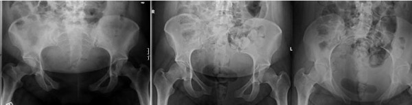
X-raySeries of three postpartum pelvic X-rays showing severe symphyseal diastasis, initially 8.8 cm and reduced to 6.8 cm after one week of bed rest.
Source : Hou et al., Patient Safety in Surgery, 2011, figure 1 · CC BY
In this chapter: the clinical picture of peripartum symphyseal separation, its window of onset, what distinguishes it from ordinary symphyseal pain, and what to do up to the point of a surgical indication.
Pubic symphysis pain is common and most often benign: it is part of the picture of pelvic girdle pain, and symphysiolysis is even, in Albert's cohort, the syndrome with the best prognosis, no patient being in pain at six months.1 There is, however, a form that does not belong to that frame and that has to be recognised: peripartum symphyseal separation.
Herren et al. synthesised it from the literature and from two cases. The characteristic symptoms, suprapubic pain and tenderness radiating towards the posterior pelvic girdle or the lower back, may be noted 48 hours after delivery. Pain on movement, in particular on walking or climbing stairs, is often present.2
The authors sum up how it presents: postpartum symphyseal rupture may show itself through the rare occurrence of pelvic pain after delivery, with sciatica or low back pain and reduced mobility. The diagnosis rests on clinical data as well as on radiographs of the pelvic girdle.2
Immediate action: do not continue mobilisation, contact the obstetric team, request imaging of the pelvic girdle. The diagnosis is clinical and radiographic.2
The prognosis is broadly good, provided that management is put in place. Conservative treatments, such as a pelvic belt combined with physiotherapy and local interventions such as injection, are effective in most cases. A reduction in symptoms within six weeks is the commonest course, but it can take up to six months in some cases.2
Surgery is necessary in cases of persistent separation. Anterior plate fixation is put forward as a known and safe procedure; minimally invasive sacroiliac screw fixation is required when there are associated injuries of the posterior pelvic girdle. The authors conclude that conservative treatment with a pelvic belt is the reference treatment in cases of symphyseal dysfunction, before as well as after delivery.2
That nuance is worth retaining, because it reconciles two statements that seem to contradict each other in this article. In run-of-the-mill pelvic girdle pain, the most recent GRADE meta-analysis does not allow the belt to be recommended.3 In established symphyseal separation, the reference review makes it the reference conservative treatment.2 These are not the same patients, and that is why the sorting in chapter 2 also governs the treatment decision.
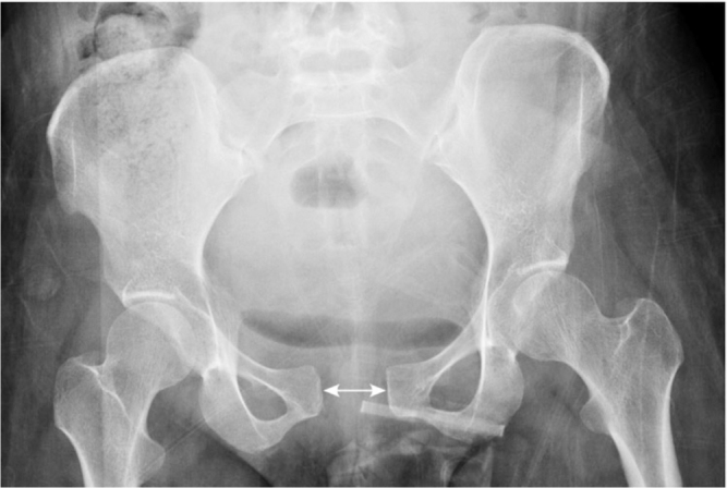
X-rayAnteroposterior pelvic X-ray in a postpartum woman: widening of the pubic symphysis measured with a double-headed arrow, indicating symphyseal diastasis.
Source : Khorashadi et al., Radiology case reports, 2011, figure 1 · CC BY-NC-ND
In this chapter: the assessment session minute by minute, what is said to the patient, how the programme is built according to the class, and when to refer.
Red flags first. Fever, neurological deficit, recent sphincter disturbance, pain at rest unrelated to position, obstetric signs. A lumbopelvic complaint in a pregnant woman may be obstetric before it is musculoskeletal.
History of positions and activities. This is the step the validated approach places before any physical examination: flexion, sitting, standing, walking, lying down, and above all load transfers, sit-to-stand, turning over in bed, stairs.1 Pain triggered by transfers points towards the pelvic girdle; pain linked to flexion and prolonged sitting points towards the spine.
Mechanical assessment of the lumbar spine, with repeated end-range movements, to test the lumbar lead.1
Pelvic provocation tests. P4, Patrick-FABER, Gaenslen, modified Trendelenburg, then palpation of the long dorsal sacroiliac ligament and of the symphysis.2 Count the positive tests: their number is a prognostic predictor.3
ASLR, rated 0 to 5 per side, total score out of 10.4
Neurological examination, which is part of the standardised approach.1
Screening for distress and kinesiophobia.5
| Class | Main content | What is announced |
|---|---|---|
| Pregnancy-related low back pain | Land-based exercise, on the model of the trials that showed an effect on pain and disability6 | Expected benefit of exercise documented, low quality of evidence |
| Pelvic girdle | Education, management of load transfers, tailored exercise; belt on trial in the acute or symphyseal phase | Say honestly that group exercise has not proved an effect on this form6, and that the prognosis depends on the syndrome3 |
| Combined form | Structured 8 to 12 week programme, the best supported modality in the file6 | Expected reduction in the number of women in pain and in sick leave, moderate quality6 |
| Postpartum | Trunk strengthening, exercise alone rather than combined with physical agents7 | Best evidence in the file: -2.21/10 on severity, large effect size on disability7 |
| Symphyseal separation | Pelvic belt and physiotherapy, the reference treatment; obstetric opinion and imaging8 | Regression within six weeks most often, up to six months sometimes8 |
We say that most women get better quickly after delivery, with the figure to back it up.3 That movement is safe. That neither the pill, nor the interval between pregnancies, nor weight, nor smoking figures among the established risk factors.2 That the pain does not signal a lesion that is getting worse.
We do not say that a pelvis is out of line or locked: the palpatory mobility tests on which such a statement would rest are explicitly not recommended.2 We do not promise that a belt will bring relief, the measured mean effect being modest and imprecise.9 We do not transfer to the pelvic girdle the results obtained on low back pain.
By what triggers the pain and by one test. Pain woken by load transfers, sit-to-stand, turning over in bed, climbing stairs, points towards the pelvic girdle; pain linked to flexion and prolonged sitting points towards the spine. The posterior pelvic pain provocation test, or P4, was built exactly for that separation: it is highly specific, with a high negative predictive value for low back pain.1 A complete examination remains necessary, and the full classification reaches a kappa of 0.79 between two examiners.2
That depends on the situation. In an established symphyseal separation, the belt combined with physiotherapy is described as the reference conservative treatment.3 In run-of-the-mill pelvic girdle pain, the most recent meta-analysis with GRADE rating gives a pain reduction whose confidence interval contains zero, with a low to very low level of evidence.4 It can therefore be offered on trial, with no documented risk and at low cost, but without promising a result, and with reassessment. The most recent trial also shows that, added to supervised exercise, it brings no measurable additional benefit.5
A randomised trial in 258 women showed that weekly water gymnastics during the second half of pregnancy reduces the intensity of back pain and the number of women on sick leave. The authors explicitly report no excess risk of urinary or vaginal infection.6 An important reservation: the outcome was back pain with no classification of the entity, and a systematic review rates the evidence on water gymnastics as very limited.7 It is therefore a good general measure, not a specific treatment for the pelvic girdle.
The question is not one of contraindication but of evidence. None of the sources gathered here reports any particular risk, and the adverse effects reported in the Cochrane review were minor and transient.8 By contrast, the systematic review published in a chiropractic journal, and therefore the one best disposed to find an effect, rates spinal manipulation as inconclusive evidence of unclear strength, for pregnancy-related low back pain as much as for pelvic girdle pain.9 Gentle manual therapy keeps its place as an adjunct that eases engagement in exercise; it does not have the data to be the mainstay treatment.
It is the most widespread explanation, and it is not retained by the European consensus among the established risk factors. That text retains as very probable risk factors a history of low back pain and a history of pelvic trauma, and rules out by agreement oral contraception, the interval since the last pregnancy, height, weight, smoking and very probably age.10 Explaining the pain by hormones that soften the ligaments suggests that there is nothing to be done until delivery, which is false and demoralising.
Probably not, but the answer depends on the class. In the reference cohort, 62.5 % of women have no more pain in the month following delivery and 8.6 % still have pain at two years. None of the women classified as having symphysiolysis was in pain at six months, against 21 % of those with pelvic girdle syndrome, still in pain at two years. A high number of positive tests and a low mobility index are the two predictors of the highest risk of long-term pain.11
Not routinely. The European guidelines explicitly rule out radiography, CT and bone scintigraphy from the diagnostic work-up of pelvic girdle pain, together with palpatory mobility tests, diagnostic injections and diagnostic external pelvic fixation. MRI keeps its place to rule out ankylosing spondylitis and when red flags are positive.10 One important exception: when a postpartum symphyseal separation is suspected, the diagnosis rests on the clinical picture and on radiographs of the pelvic girdle.3
Partly. A meta-analysis of six randomised trials totalling 2,231 participants, with GRADE rating, shows with moderate quality evidence that exercise alone is acceptable for pregnant women and that it prevents long-term episodes of low back pain, with a relative risk of 0.92 and a confidence interval of 0.85 to 0.99. The other interventions have shown no preventive effectiveness.12 The effect is real but small: advising physical activity is justified, promising the absence of pain is not.
When intense suprapubic pain appears within the 48 hours following delivery, radiating towards the posterior pelvis or the lower back, with disproportionate incapacity: the inability to walk or to climb a step. That picture should raise the suspicion of peripartum symphyseal separation and lead to an obstetric opinion and imaging of the pelvic girdle.3 Any associated fever, any neurological deficit and any recent sphincter disturbance call for emergency referral.
This article deals with sorting between low back pain and pelvic girdle pain in the pregnant woman. The neighbouring subjects are the subject of dedicated syntheses.
Behind this article
An author who explains, a reviewer who checks.
How we write and check our content

Physiotherapist · co-founder of Physio Learning
Marked for life by his first four-hour lecture without a single image, he took a master’s in instructional design so that it would never happen to anyone again. He hunts down publication bias and unreadable slides with the same intransigence.

Head of scientific content
Physiotherapist specialising in neuro-musculoskeletal practice and holder of a master’s in public health. He checks the methodological rigour of every article: primary sources, levels of evidence, no exceptions.
