















Clinical synthesis on postnatal diastasis recti abdominis (DRA): measurement, rehabilitation and controversies, based on recent studies. Every reference has been verified on PubMed.
📝 In brief: clinical summary
- Diastasis is the rule, not the exception, in the peripartum period. In a prospective cohort of 300 primiparous women, prevalence (palpation, separation ≥ 2 fingerbreadths) was 33.1 % at the 21st week of pregnancy, 60,0 % at 6 weeks postnatally, 45,4 % at 6 months and 32,6 % at 12 months: spontaneous resolution is substantial, but one woman in three is still affected a year after giving birth 1.
- Diastasis is not the cause of low back pain or of pelvic floor disorders. At 12 months postnatally, there was no difference in lumbopelvic pain between women with and without diastasis (p = 0.10) 1. Nor was there any difference in pelvic floor strength, urinary incontinence or prolapse at 6 weeks, 6 months and 12 months 26, including when the IRD threshold was varied from 20 to 50 mm 27. The real impact is read in reported symptoms, abdominal strength 29, quality of life and body image 1731.
- Measure in the right place, with the right tool. Ultrasound and callipers are the adequate methods; the fingerbreadth method offers only moderate inter-rater agreement (63 %, weighted Kappa = 0.53) 6. Ultrasound reliability is excellent above the umbilicus (ICC 0,91 at rest and 0,96 on head lift, 5 cm above) but falls at the umbilicus itself (0,72 and 0,63): favour standardised supra-umbilical sites 7. Reference value in healthy nulliparous women: IRD < 10 mm à toutes les localisations (≈ 8,77 mm ombilical, 7,22 mm épigastrique, 4,09 mm sous-ombilical) 11.
- Spontaneous recovery is early, real… and incomplete. Most of it happens over the first 2 months, but at 12 months the rectus abdominis is still thinner and wider, and the IRD still larger, than in nulliparous women (p < 0,0001 à tous les temps) 21. In primiparous women, the IRD can be considered “normal” at wider values than in nulliparous women 12. Methodological consequence: most trials recruit within the first 6 months, where spontaneous reduction is confounded with the treatment effect 36.
- Exercise reduces the gap, with no demonstrated functional translation. Meta-analysis of 9 RCTs (450 women): significant reduction in IRD (MD −8.05 mm; 95 % CI −10.43 to −5.68), greater when the intervention starts before 3 months postnatally (−10.2 mm), with no difference between training types (p = 0.32), but no benefit on the Oswestry Disability Index (MD 0.82; 95 % CI −2.75 to 4.38) 38. GRADE certainty “low” 42. Antenatally, exercise reduced the presence of diastasis by 35 % (RR 0,65; 95 % CI 0,46–0,92), on low-quality evidence 40 ; postnatally, conservative interventions do not produce a clinically significant reduction in IRD (16 trials, 698 women) 41.
- The crunch is not the enemy. RCT (PEDro 8/10): a 12-week home programme including curl-ups in women with diastasis at 6–12 months postnatally neither worsened nor improved the IRD (mean difference 1 mm; 95 % CI −1 to 4), while increasing abdominal strength and thickness 32. The crunch narrows the supra-umbilical IRD, whereas drawing-in widens it below the umbilicus 24 ; the curl-up narrows the IRD but distorts the linea alba more, whereas pre-activation of the transversus distorts it less 25. Instagram lists of “forbidden” exercises are unfounded in 61 % of cases and generate confusion and a nocebo effect 37.
- Physiotherapy first, surgery second, and screen for hernia. Physiotherapy is the first step; if symptoms persist after 2 to 6 months, surgical referral can be discussed, ideally beyond 1 year postnatally 43. No physiotherapy programme has shown complete resolution of the diastasis at rest 39 : the candidate subgroup is that of persistent pain despite effective strengthening 4546. Red flag: an associated umbilical or epigastric hernia raises the recurrence rate after simple suture from 8,3 % to 31,2 % (p < 0,001) 5.
🤰 What are the fundamentals to know about diastasis recti abdominis?
🤰 Diastasis resolves on its own, but not in everyone
Prevalence of diastasis through pregnancy and the postnatal period: the peak is just after birth, spontaneous recovery is substantial up to 6 months, then it plateaus.
Prospective cohort of 300 primiparous women (diastasis = palpated separation ≥ 2 fingerbreadths). One woman in three is still affected a year after giving birth. Source: Sperstad et al., 2016 (PMID 27324871).
Diastasis recti abdominis is probably one of the topics where the gap between everyday clinical talk and the available data is widest. It is readily presented as a lesion to be closed, a cause of low back pain, a factor in urinary leakage, and its management comes bundled with lists of forbidden exercises. Yet the literature says something else: it is an extremely common adaptation to pregnancy, largely self-resolving, whose link with lumbopelvic pain and pelvic floor dysfunction is not established, but whose impact on body image and quality of life is real and underestimated. This section sets out the essential reference points before any rehabilitation decision: definition, measurement, epidemiology, anatomy of the linea alba, and the mechanisms of distension.
Definition: a measured widening of the linea alba, to be distinguished from hernia
Diastasis recti abdominis (DRA, diastasis recti abdominis) is defined by measurement: it is the increase in the distance separating the two rectus abdominis muscles either side of the linea alba: the inter-recti distance, or IRD (inter-recti distance). All the landmark studies diagnose it this way, by a palpated separation 1 or by an ultrasound IRD exceeding a threshold 243. This is a point of vocabulary with direct consequences in clinic: a patient who hears “separation” or “gap” often understands “tear”, whereas what is measured is a distance, and diastasis is, as we shall see, the usual situation at the end of pregnancy.
In the landmark study in this field, the Norwegian prospective cohort of 300 primiparous women followed from pregnancy to one year postnatally, diastasis is defined as a palpated separation of at least 2 fingerbreadths, assessed at three levels: 4.5 cm above the umbilicus, at the umbilicus, and 4.5 cm below, with the patient supine performing an abdominal crunch 1. Note this triple location straight away: measurement is not taken at a single point, but above and below the umbilicus: a diastasis may be present at only one level.
Diastasis must be distinguished from hernia of the linea alba, which is a different entity and a frequent comorbidity. In a multicentre retrospective study of 1,294 cases of diastasis followed over 10 years, 35.8 % of women and 56.1 % of men had one or more hernias (p < 0.001), umbilical hernia being the most prominent type; in women, age (OR 1.51 [1.33–1.72]; p = 0.000) and smoking (OR 1.66 [1.13–2.44]; p = 0.010) were risk factors for an associated hernia 4. This screening is not cosmetic: in 231 patients operated on for a small umbilical or epigastric hernia (< 2 cm) by simple suture, the recurrence rate reached 31,2 % (29/93) when an associated diastasis was present, versus 8,3 % (9/108) in its absence (p < 0,001), leading the authors to strongly recommend systematic preoperative screening for diastasis 5. A painful or irreducible midline bulge therefore calls for medical and surgical advice, not for watchful waiting in rehabilitation.
Strong evidence Finally, the definition must include from the outset what diastasis is not. In the Sperstad cohort, the presence of a diastasis at 12 months postnatally was not associated with more lumbopelvic pain: no significant difference between women with and without diastasis (p = 0.10) 1. Diastasis should therefore not be presented straight away as the cause of postnatal low back pain.
Key points: the definition that matters for the physiotherapist
- Diastasis is a measured widening of the linea alba (increased IRD): what is diagnosed is a distance.
- Historical reference threshold: ≥ 2 fingerbreadths on palpation, measured 4.5 cm above, at, and 4.5 cm below the umbilicus, during a crunch 1.
- Measurement is multi-site : supra-umbilical and infra-umbilical. A single point is not enough.
- Systematically screen for an associated umbilical or epigastric hernia : present in 35.8 % of women with diastasis 4 ; a painful or irreducible bulge = surgical referral.
- At 12 months postnatally, diastasis is not associated with more lumbopelvic pain 1.
Measuring the IRD: is the finger enough? And where should you measure?
Measurement is not a methodological detail: it determines the diagnosis, the follow-up, and above all the critical reading of the literature. A systematic review of methods for measuring diastasis (13 studies evaluating fingerbreadth, tape measure, callipers, ultrasound, CT and MRI) concludes that ultrasound and callipers are the adequate methods for assessing diastasis, with low measurement error above the umbilicus; the fingerbreadth method, by contrast, offers only moderate inter-rater agreement: 63 %, weighted Kappa = 0.53 6.
Moderate evidence The most counter-intuitive point concerns the location of the measurement. Ultrasound reliability depends strongly on the site: excellent above the umbilicus, poor at the umbilical level itself. At rest, the ICC reaches 0.91 at 5 cm above the umbilicus but falls to 0.72 at the upper edge of the umbilicus ; on head lift, it goes from 0.96 (5 cm above) to 0.63 at the umbilical level. The authors' practical recommendation: favour standardised supra-umbilical measurement sites 7. A clinical paradox to take on board: it is precisely at the umbilicus that the gap is widest, the maximum separation was located there in women with diastasis (4,59 ± 1,14 cm) 27, and that is where the measurement is least reliable.
This is consistent with the reproducibility data for 2D ultrasound: good to very good test-retest reliability (ICC between 0.74 and 0.90), with the single exception of the measurement 2 cm below the umbilicus during the crunch (ICC 0.50) ; intra-observer reproducibility on the same images exceeds 0.90 9. In other words: ultrasound is reliable, but not everywhere and not in every position.
| Method | Reported performance | Reasonable use in practice |
|---|---|---|
| Fingerbreadth (palpation) | Inter-rater agreement moderate : 63 %, weighted Kappa 0.53 6 | Quick screening, communication; insufficient for fine quantitative follow-up |
| Callipers | Judged adequate, low measurement error above the umbilicus 6 | Credible field alternative, used in cohort studies 10 |
| Ultrasound: 5 cm supra-umbilical | ICC 0.91 at rest, 0.96 on head lift 7 | Reference site to favour for follow-up |
| Ultrasound: umbilical level | ICC 0.72 at rest, 0.63 on head lift 7 | Widest site but the least reliable: interpret with caution |
| Ultrasound: 2 cm infra-umbilical during a crunch | ICC 0.50 9 | Avoid making it a stand-alone decision criterion |
What reference values? The single 2 cm threshold does not hold
To interpret an IRD, you first need to know what a “normal” linea alba is. In healthy nulliparous women, a meta-analysis establishes that the maximum IRD reported on ultrasound is below 10 mm at every location, with pooled values of approximately 8.77 mm in the umbilical zone, 7.22 mm in the epigastric zone and 4.09 mm in the infra-umbilical zone 11. The normal value is therefore not a single figure: it varies twofold depending on the level measured.
< 10 mmMaximum IRD reported in healthy nulliparous women, at every location 11
Moderate evidence In primiparous women the reference shifts. In the longitudinal ultrasound follow-up of 84 primiparous women, the 20th and 80th percentiles of the IRD at 6 months postnatally were 9–21 mm at 2 cm below the umbilicus, 17–28 mm at 2 cm above and 12–24 mm at 5 cm above. The authors draw two directly operational conclusions: the linea alba is widest 2 cm above the umbilicus, during pregnancy as well as postnatally; and in primiparous women, the IRD can be considered “normal” at wider values than in nulliparous women 12. This is a strong argument against using a single 2 cm threshold applied at every level and to every woman.
How sensitive the figure is to the chosen threshold can also be read in the long-term prevalence data: in 1,000 women assessed by ultrasound, prevalence was 36 %, 31 %, 22 %, 26 % and 30 % at 3, 5, 10, 20 and 30 years postnatally with the criterion IRD > 2 cm, but only 13 %, 8 %, 6 %, 8 % and 10 % with the criterion IRD > 3 cm 3. One centimetre of threshold divides prevalence by three: any reading of the literature requires checking the definition used.
Postnatal epidemiology: very common, largely self-resolving, never quite resolved
Strong evidence The most cited figure in this field comes from Sperstad's prospective cohort of 300 primiparous women, with diastasis defined by palpation (≥ 2 fingerbreadths): prevalence was 33.1 % at 21 weeks of pregnancy, 60.0 % at 6 weeks postnatally, 45.4 % at 6 months and 32.6 % at 12 months postnatally 1. Two readings coexist in these four figures, and both are true: spontaneous resolution is substantial (from 60 % to 32.6 % in one year, with no intervention), and one woman in three is still affected a year after giving birth.
33.1 %at 21 weeks of pregnancy 1
60.0 %at 6 weeks postnatally 1
45.4 %at 6 months postnatally 1
32.6 %at 12 months postnatally 1
Mota's longitudinal ultrasound follow-up shows the same trajectory with an even more striking amplitude: in 84 primiparous women measured at week 35 of gestation then at 6–8, 12–14 and 24–26 weeks postnatally, the prevalence of diastasis falls from 100 % at the end of pregnancy to 39 % at 6 months postnatally 24. Every woman has a diastasis at term (diastasis in pregnancy is the rule, not the exception) and four in ten still do at the sixth month, spontaneously.
The figures nevertheless vary widely with the population and the method. A Chinese observational study of 534 women, with diastasis measured by palpation, reported a prevalence of 78.1 % (417/534) 13. The message is not that one figure is wrong: it is that prevalence only means something when it comes with its definition, its postnatal time point and its measurement method.
And in the very long term?
Moderate evidence Diastasis does not disappear: it stabilises, then rises again. In the cross-sectional ultrasound study of 1,000 women assessed at 3, 5, 10, 20 and 30 years postnatally, prevalence (IRD > 2 cm) was 36 %, 31 %, 22 %, 26 % and 30 % ; with the criterion IRD > 3 cm, 13 %, 8 %, 6 %, 8 % and 10 % 3. The decline stops at around 10 years postnatally, then prevalence rises again, and about one woman in ten still has a diastasis > 3 cm thirty years after giving birth.
Risk factors: what holds up, and what does not
Not all the “obvious” risk factors survive analysis. In 84 primiparous women followed by ultrasound, no statistically significant difference was found at 6 months postnatally in pre-pregnancy BMI, gestational weight gain, the baby's birth weight or abdominal circumference between women with and without diastasis 24In another cohort, each additional year of maternal age increased the risk by 10 % (OR 1,10; 95 % CI 1,04–1,16; p = 0,002) and the number of births doubled it (OR 2,09; 95 % CI 1,30–3,37; p = 0,002), but fetal weight, pre-pregnancy BMI and postnatal BMI were not significant factors in the final model (p > 0.05) 14.
| Factor | Data | Interpretation |
|---|---|---|
| Multiparity / number of births | OR 2.67 (1.30–5.45) vs nulliparity, cohort of 4,426 women at 6 weeks postnatally 15 ; OR 2,09 (1,30–3,37) 14 | The most consistent factor. The more pregnancies a woman has had, the greater the risk 16 |
| Caesarean section | Independent risk factor, OR 2,297 (1,327–3,978); p = 0.003 13. Conversely, vaginal birth is associated with a reduced risk: OR 0.45 (0.40–0.51) 15. Factor for persistence of diastasis, p = 0,05 17 | Consistent but nuanced signal: caesarean section “appears to be a risk factor only in women who have given birth twice” 16 |
| Pelvic floor strength | Type I strength of grade II to IV is associated with a reduced risk of diastasis 15 | A direct argument in favour of pelvic floor work postnatally |
| Maternal age | OR 1.10 per year (1.04–1.16); p = 0.002 14. Risk factor for an associated hernia in women: OR 1,51 4 | Contradictory results in the literature: not significant in Mota and Sperstad, a risk factor in Spitznagle, protective in Wu 16 |
| BMI | Not significant at 6 months 2414. But a risk factor at 10 years postnatally (p = 0,000) 3 ; retained among the most plausible 16 | Depends on the time horizon: not discriminating in the short term, associated in the long term |
| Diabetes | Risk factor at 20 and 30 years postnatally (p = 0.000 and 0.004) 3 ; retained among the most plausible 16 | A late signal, consistent with impaired connective tissue |
| Twin pregnancy | Risk factor at 3 years postnatally (p = 0.001) 3 | Consistent with the role played by the extent of the distension |
| Fetal weight, weight gain, abdominal circumference | Not significant 2414 | Not to be presented to the patient as established causes |
The most honest summary remains that of Cavalli's review: among all the risk factors proposed, only the number of pregnancies, BMI and diabetes emerge as the most plausible 16.
Anatomy: the linea alba is not a simple fibrous band
Understanding diastasis means understanding the structure that widens. The linea alba is an aponeurotic interweaving organised into two families of fibres with distinct mechanical roles : the transverse fibres resist intra-abdominal pressure, whereas the oblique fibres are mainly loaded during trunk movements 18. This architecture directly illuminates the respective roles of the transversus and the obliques: they are not interchangeable synergists: they tension the linea alba along different vectors.
Axer's morphological study also reports sex-related differences that are not incidental to the topic at hand: in women, the infra-umbilical linea alba is thinner but wider than in men, with a higher proportion of transverse fibres relative to oblique fibres. The mean diameter of the fibril bundles was also smaller in the supra-umbilical region than in the infra-umbilical region. The authors honestly state that whether these morphological differences reflect an adaptation to the biomechanical stress of pregnancy remains to be demonstrated 18.
The histological organisation of the collagen completes the picture: transverse fibres on the dorsal side, oblique fibres on the ventral side. Under tension, the oblique fibres of the ventral layer reorient towards the axis of traction, which progressively increases the stiffness of the tissue and produces a non-linear stress-strain relationship. Elastin, for its part, forms a superficial layer that covers the collagen network and follows its movements, which suggests that it provides its elastic recoil: the return to the original shape after deformation 19. Put simply for the patient: collagen takes the load, elastin brings the tissue back to its shape.
An anisotropic tissue: it does not deform the same way in every direction
Moderate evidence The linea alba is anisotropic. Under a physiological abdominal pressure of about 20 kPa, its Young's modulus is approximately 50 kPa in the transverse direction against about 20 kPa in the longitudinal direction, for strains of around 6 % in both axes, that is a compliance ratio of about 2:1 between longitudinal and transverse. The corresponding biaxial forces are 3.4 N/mm transversely and 1.5 N/mm longitudinally. The overall behaviour is non-linear elastic, typical of soft biological tissues 20.
What this means in practice: the wall is roughly twice as stiff transversely as longitudinally. It is therefore structurally built to resist the transverse tension imposed by intra-abdominal pressure: exactly what pregnancy will load, right up to its functional breaking point.
Pathophysiology of the distension: mechanics and tissue quality
Distension in pregnancy is massive and quantifiable. In 84 primiparous women followed by ultrasound, at the end of pregnancy (weeks 35–41) the IRD ranged between the 20th and 80th percentiles at 49–79 mm at 2 cm below the umbilicus, 54–86 mm at 2 cm above, and 44–79 mm at 5 cm above 12. In other words, the supra-umbilical IRD can exceed 8 cm at term, against less than 10 mm in nulliparous women 11 : a factor of 8 to 9.
The spontaneous narrowing that follows is considerable (the same values fall back to 9–21 mm, 17–28 mm and 12–24 mm at 6 months postnatally 12), but it is early and incomplete. The clearest demonstration of this comes from Coldron's study, which followed 115 postnatal women by ultrasound against 69 age-matched nulliparous controls: the rectus abdominis is significantly thinner, wider, and the IRD significantly larger than in the controls at all measurement time points: day 1, 2 months, 6 months and 12 months (p < 0.0001 for the IRD at all four time points). Most of the recovery takes place over the first two months: the muscle thickens (p = 0.0003), width and IRD decrease (p < 0.0001 and p = 0.0002), but the values do not return to those of the controls at 12 months 21.
The same plateau is found for function. Liaw followed 40 postnatal women (measurements at 4–8 weeks then 6–8 months, ultrasound IRD at 4 sites around the umbilicus) compared with 20 matched nulliparous women: the IRD decreases significantly 2.5 cm above the umbilical ring (p = 0.013) and at its upper margin (p = 0.002), but neither the IRD nor abdominal muscle function had returned to normal values at 6 months 22. Spontaneous improvement does not mean cure.
It is not just stretching: the connective tissue is altered
Moderate evidence Reducing diastasis to passive mechanical distension would be a modelling error. In a case-control study of 18 women with diastasis and 18 without (midline fascia samples, immunohistochemistry), both type I and type III collagen were significantly less abundant in women with diastasis (p < 0.001). Supra-umbilically, type I collagen measured 244.5 ± 73.5 kpixels against 381.1 ± 101.1 in the controls; infra-umbilically, 217.1 ± 58.8 against 397.4 ± 82.9. Type III collagen had also collapsed (54.3 ± 33.1 vs 154.9 ± 59.4 supra-umbilically; 58.5 ± 36.9 vs 152.0 ± 68.0 infra-umbilically). The authors conclude that a low level of type I and III collagen in the midline could play a key role in the development of diastasis 23.
This carries clinical weight: it suggests that part of the diastasis stems from a tissue quality on which exercise has no direct purchase, and it offers a plausible explanation for the absence of complete anatomical resolution described in the rehabilitation studies.
A reorganised wall, not merely a stretched one
Limited evidence Diastasis also alters the mechanical properties of the muscles themselves, and does so non-uniformly. On ShearWave elastography, in 36 postnatal women with diastasis against 24 healthy nulliparous women, shear wave velocity (a marker of stiffness) was significantly lower in the rectus abdominis (p = 0.003) and in the external oblique (1.65 ± 0.15 vs 1.79 ± 0.14; p = 0.001), but significantly higher in the transversus abdominis (p < 0.001). The maximum separation was located at the umbilicus (4.59 ± 1.14 cm) 27.
This contrast (rectus abdominis and external oblique softened, transversus stiffened), suggests a reorganisation of the wall rather than simple passive stretching. The authors do, however, explicitly urge caution in interpreting the correlation between wave velocity and IRD in the rectus abdominis 27 : the signal is interesting, the sample remains small and the mechanism is not established.
The biomechanical corollary: not every exercise pulls in the same direction
The anisotropy of the linea alba and the specialisation of its fibres translate directly into experimental observations. In 38 primiparous women, the IRD above the umbilicus was significantly reduced during the abdominal crunch, whereas the IRD below the umbilicus was significantly wider during the abdominal drawing-in exercise (drawing-in, transversus activation) than at rest 24. The same exercise therefore does not produce the same effect at the two levels.
More importantly, visibly reducing the gap is not necessarily the functional objective. In women with diastasis, the automatic curl-up narrows the IRD but distorts the linea alba more, whereas pre-activation of the transversus reduces the IRD less while distorting the linea alba less. The authors stress that this smaller narrowing of the IRD by the transversus, advised against until now, could on the contrary favour abdominal mechanics through tension transfer 25. This is the conceptual turning point of the whole subject: “closing the gap” is not synonymous with “transmitting forces well”.
What diastasis does, and does not, cause
Strong evidence Contrary to a stubborn belief in rehabilitation, the Norwegian prospective cohort found no link between diastasis and pelvic floor dysfunction : women with diastasis had neither a weaker pelvic floor, nor more urinary incontinence, nor more prolapse, at 6 weeks, 6 months and 12 months postnatally. Counter-intuitively, at 6 weeks postnatally prolapse was even significantly more frequent in women without diastasis (15.9 % vs 4.1 %; p = 0.001) 26.
This result withstands the severity test. In a retrospective cohort of 229 women during the first year postnatally, no difference in urinary incontinence or prolapse was observed between women with and without diastasis, and this held whatever IRD threshold was used (20, 30, 40 or 50 mm) 27. A wider diastasis does not come with a more symptomatic pelvic floor: a strong argument against the idea that “closing the gap” protects the pelvic floor.
Moderate evidence When an association is found, it is weak. In a prospective study of 150 women from 6 weeks to 6 months postnatally, the correlation between diastasis and stress urinary incontinence symptoms was only r = 0.283 ; those with maximum voluntary contraction of the pelvic floor (r = −0.278) and contraction duration (r = −0.274) were also weak, and no correlation was found with the area of the urogenital hiatus 28.
The impact is better read elsewhere: in reported symptoms and abdominal strength. In primiparous women assessed postnatally, diastasis was not correlated with morphological changes of the pelvic floor on imaging, but was associated with a significantly higher PFDI-20 urinary symptom score (12.5 ± 22.8 without diastasis vs 26.8 ± 18.2 with; p = 0.01). And extensive diastases (grades 2–3) showed significantly reduced abdominal strength and endurance : dynamic endurance test 13.4 ± 11.8 vs 6.46 ± 4.59 repetitions (p = 0.025); manual muscle testing 4 vs 3 (p = 0.04) 29.
Finally, quality of life emerges as a more relevant outcome measure than pelvic floor symptoms. In 253 patients followed after giving birth, severe diastasis had a significant impact on quality of life (HerQLes score) at birth (p = 0.001) and at 3–6 months (p = 0.01), but no effect on pelvic floor symptoms measured with the FPFQ 17. A mixed-methods study confirms a negative effect on quality of life, functional capacity and urogynaecological and digestive health, along with an emotional impact involving altered body image and poor perceived health: the authors describing feelings of abandonment by health institutions, shame, sadness, helplessness, a lack of self-esteem, resignation and social pressure 30.
The scale of the aesthetic concern is, moreover, widely underestimated in clinic. In a cross-sectional study of 460 primiparous women at 6–8 months postnatally, 73.3 % reported having worried during pregnancy about the appearance of their abdomen after giving birth, the mean degree of concern about their current abdominal appearance was 5.5/10 (SD 2.4), and nearly 80 % felt their abdominal muscles were weaker than before pregnancy. Women reporting a midline protrusion (20.9 %) described themselves as weakened significantly more often than those without protrusion 31.
Key points: the fundamentals in 8 points
- Definition : measured widening of the linea alba (IRD). Historical threshold ≥ 2 fingerbreadths at 4.5 cm above, at, and 4.5 cm below the umbilicus, during a crunch 1.
- Epidemiology : 33.1 % at 21 weeks of gestation, 60.0 % at 6 weeks postnatally, 45.4 % at 6 months, 32.6 % at 12 months 1 ; 100 % at the end of pregnancy → 39 % at 6 months 24. It is the peripartum norm, not an abnormality.
- Measurement : ultrasound and callipers are adequate; the fingerbreadth method has only moderate agreement (63 %, Kappa 0.53) 6. Favour standardised supra-umbilical sites : ICC 0.91–0.96 at 5 cm above vs 0.63–0.72 at the umbilicus 7.
- Reference values : < 10 mm in nulliparous women at every location (≈ 8.77 mm umbilical, 7.22 epigastric, 4.09 infra-umbilical) 11 ; but in primiparous women, the IRD can be “normal” at wider values 12. No single 2 cm threshold.
- Anatomy : transverse fibres = resistance to intra-abdominal pressure; oblique fibres = trunk movements 18. Anisotropic tissue, ~2× stiffer transversely 20. Collagen = load, elastin = elastic recoil 19.
- Pathophysiology : massive distension (supra-umbilical IRD up to > 8 cm at term), considerable spontaneous narrowing but early and incomplete, rectus abdominis still thinner, wider and IRD still larger than in nulliparous women at 12 months 211222.
- It is not just stretching : type I and III collagen significantly less abundant in the midline (p < 0.001) 23 ; rectus abdominis and external oblique softened, transversus stiffened on elastography 27.
- To put into perspective : no association with lumbopelvic pain at 12 months 1, nor with pelvic floor strength, urinary incontinence or prolapse, whatever the threshold 2627. The real impact is on abdominal strength 29, on quality of life 17 and body image 3130.
📏 How do you measure and diagnose a diastasis?
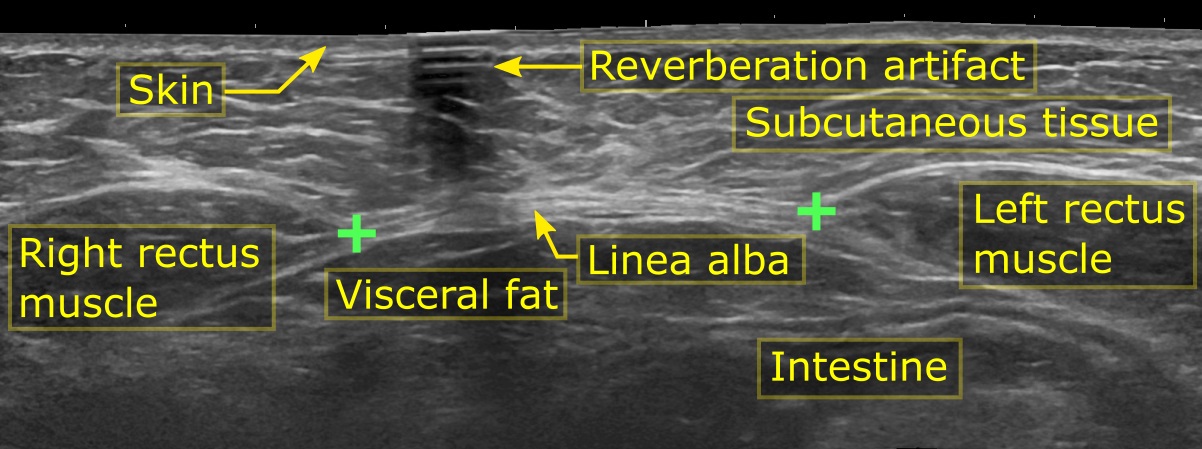
UltrasoundAnnotated axial abdominal ultrasound, between the xiphoid process and the navel: distance of 3.5 cm between the rectus abdominis muscles on either side of the linea alba, without associated hernia.
Source : Häggström, Wikimedia Commons · CC0
🎯 Measure above the umbilicus, not on it
Reliability (ICC) of ultrasound measurement of the IRD by site: excellent 5 cm above the umbilicus, poor at the umbilical level.
An ICC close to 1 indicates a reproducible measurement. Palpation in fingerbreadths, by contrast, offers only moderate inter-rater agreement (63 %; weighted Kappa 0.53): ultrasound and callipers are the adequate methods. Sources: Keshwani et al., 2016 (PMID 27909371); van de Water & Benjamin, 2016 (PMID 26474542).
📏 From what point is it abnormal?
Inter-recti distance (IRD) measured by ultrasound in healthy nulliparous women: the reference values for placing a pathological threshold.
In healthy nulliparous women, the maximum IRD reported is below 10 mm at every location. The normal value therefore depends on the measurement site: the same number of millimetres is not judged the same way above and below the umbilicus. Source: Wang & Wang, 2023 (PMID 35001809).
Diagnosing diastasis recti abdominis looks simple: you part the muscles, you measure, you compare with a threshold. In practice, each of these three steps is booby-trapped. The choice of tool changes inter-rater agreement, the measurement site changes reliability, and the chosen threshold changes prevalence to a considerable degree. In one and the same cross-sectional cohort of 1,000 women assessed by ultrasound, prevalence falls from 36 % to 13 % at 3 years postnatally depending on whether the criterion is an inter-recti distance (IRD) greater than 2 cm or greater than 3 cm 3. In other words: the figure you announce to your patient depends as much on your method as on her abdomen.
What exactly is measured: the inter-recti distance (IRD)
The reference variable is the inter-recti distance : the distance separating the medial borders of the two rectus abdominis muscles, that is, the width of the interposed linea alba. It is neither a measure of a “hole”, nor a measure of hernia, nor a measure of tone: it is a transverse distance, measured at a given point, in a given position, at a given moment of the contractile cycle.
This precision is not methodological pedantry. The linea alba is not a homogeneous tissue: its transverse fibres oppose intra-abdominal pressure while its oblique fibres are mainly loaded during trunk movements, and its structure differs by level, in women, the infra-umbilical portion is thinner but wider than in men, with a higher proportion of transverse fibres relative to oblique fibres 18. Mechanically, this tissue is anisotropic: under a physiological abdominal pressure of about 20 kPa, its Young's modulus is around 50 kPa in the transverse direction against about 20 kPa in the longitudinal direction, a compliance ratio of roughly 2:1 20. A structure that does not deform in the same way along different axes or at different levels cannot honestly be summed up by a single figure.
And diastasis does not come down to passive distension: in a case-control study of 18 women with diastasis and 18 without, both type I and type III collagen were significantly less abundant in the midline fascia samples of women with diastasis (p < 0.001): 244.5 ± 73.5 kpixels against 381.1 ± 101.1 for type I collagen supra-umbilically 23. What you measure with a tape or an ultrasound probe is therefore the visible symptom of a qualitative alteration of the connective tissue, not merely a geometric gap.
Finger palpation: accessible, standardisable, but imperfect
The digital method remains the clinical entry point, and it is the one used by the landmark prospective cohort of 300 primiparous women: diastasis there was defined as a palpated separation of at least 2 fingerbreadths, assessed at three levels: 4.5 cm above the umbilicus, at the umbilicus, and 4.5 cm below, with the patient supine performing an abdominal crunch 1. It is with this protocol that the classic prevalence figures were established: 33.1 % at 21 weeks of pregnancy, 60.0 % at 6 weeks postnatally, 45.4 % at 6 months and 32.6 % at 12 months 1.
Two immediate practical lessons. First, measurement is not taken at a single point : the protocol explicitly requires going above and below the umbilicus. Second, the test is done in contraction (crunch), not at rest, which is not neutral, as we shall see.
The limitation is documented. The systematic review of measurement methods (13 studies evaluating fingerbreadth, tape measure, callipers, ultrasound, CT and MRI) concludes that the fingerbreadth method offers only moderate inter-rater agreement: 63 %, weighted Kappa = 0.53 6. Clinical translation: two physiotherapists assessing the same patient will agree about two times out of three. For longitudinal follow-up, “is it getting better?”, that is structurally insufficient, all the more so because fingerbreadth varies from one examiner to another and has no standardised metric value.
| Method | What the data say | Level of evidence | Reasonable use |
|---|---|---|---|
| Fingerbreadth (palpation) | Moderate inter-rater agreement: 63 %, weighted Kappa 0.53 6 | Moderate | Screening, first appointment, settings without an ultrasound machine |
| Callipers | Retained as an adequate method, low measurement error above the umbilicus 6 ; used in a prospective cohort of 504 women 10 | High | Quantitative follow-up in the clinic, pragmatic alternative to ultrasound |
| 2D ultrasound | Adequate method 6 ; test-retest ICC 0,74–0,90, intra-observer > 0,90 9 ; ICC up to 0,96 at 5 cm above the umbilicus on head lift 7 | High | Reference method for objective measurement and follow-up |
| Tape measure | Evaluated in the review of methods without being retained among the adequate methods 6 | Low | Not recommended for quantification |
| CT / MRI | Evaluated in the review of methods; ultrasound and callipers are the supported methods 6 | Moderate | No first-line indication in rehabilitation |
Ultrasound: the reference method, provided you know where to place the probe
The systematic review of measurement methods is unambiguous: “ultrasound and callipers are the adequate methods for assessing diastasis, with low measurement error above the umbilicus” 6. The reproducibility of 2D ultrasound is good to very good, with test-retest intraclass correlation coefficients between 0,74 and 0,90, and intra-observer reproducibility on the same images exceeding 0,90 9.
But this quality is not uniform, and this is probably the most underestimated point of the whole subject. Reliability depends massively on the site. In Keshwani 7, at rest, the ICC reaches 0,91 at 5 cm above the umbilicus but falls to 0,72 at the upper edge of the umbilicus; on head lift, it goes from 0,96 at 5 cm above to 0.63 at the umbilical level. The same signal in Mota 9 : the only exception to good reliability is the measurement 2 cm below the umbilicus during the crunch, with an ICC collapsed to 0,50.
The practical recommendation that follows is explicit: favour standardised supra-umbilical measurement sites 7. If you have to follow a patient over time, always measure at the same place, above the umbilicus, in the same position, and write it down. An operational protocol documented in sports practice uses four ultrasound sites: xiphoid, supra-umbilical, umbilical and infra-umbilical 33. The seminal palpation study uses three: 4,5 cm above, at, and 4,5 cm below 1. The exact number matters less than consistency: your protocol, written down, reproduced identically.
Which threshold? The worst-posed question of the subject
Three families of reference points must be distinguished, and not confused.
1. Values in healthy nulliparous women. The meta-analysis of ultrasound reference values establishes that the maximum inter-recti distance reported in healthy nulliparous women is below 10 mm at every location, with pooled values of approximately 8.77 mm in the umbilical zone, 7.22 mm in the epigastric zone and 4.09 mm in the infra-umbilical zone 11. This is the “ideal” anatomical reference point, and that is precisely why it should not serve as a therapeutic goal postnatally.
2. Normal values in primiparous women. They are not the same, and this has been demonstrated. In the longitudinal ultrasound follow-up of 84 primiparous women, at 6 months postnatally, the 20th and 80th percentiles corresponded to 9–21 mm at 2 cm below the umbilicus, 17–28 mm at 2 cm above and 12–24 mm at 5 cm above 12. The authors explicitly conclude that “in primiparous women, the IRD can be considered normal up to wider values than in nulliparous women”, and that the linea alba is widest 2 cm above the umbilicus, during pregnancy as well as postnatally 12. This is a head-on argument against using a single 2 cm threshold at every level.
3. The operational thresholds used in studies. They vary, and you need to know this to read the literature: ≥ 2 fingerbreadths on palpation 1, IRD ≥ 16 mm at 2 cm below the umbilicus 2, IRD ≥ 28 mm at rest and/or a visible protrusion as an inclusion criterion in pregnancy 34, IRD ≥ 2,8 cm 35, or else > 2 cm versus > 3 cm in epidemiology 3. A retrospective cohort of 229 women even tested 20, 30, 40 and 50 mm in turn as diagnostic thresholds 27: a sign that the scientific community itself has no consensus value.
To put the scale of the pregnancy phenomenon in context: at the end of pregnancy (weeks 35–41), the 20th and 80th percentiles of the IRD ranged between 49–79 mm at 2 cm below the umbilicus, 54–86 mm at 2 cm above and 44–79 mm at 5 cm above 12. At term, a supra-umbilical IRD greater than 8 cm falls within the normal range. Measuring an “enormous” diastasis at 38 weeks has no prognostic value in itself.
Position and instruction: you are not measuring the same thing depending on what you ask for
This is the methodological trap least well mastered in the clinic. The motor instruction given during the measurement changes the measured value, and not in the same direction at every level.
In 38 primiparous women, the IRD above the umbilicus was significantly reduced during the abdominal crunch, whereas the IRD below the umbilicus was significantly wider during the abdominal drawing-in exercise (drawing-in / transversus activation) than at rest 24. The longitudinal study in 38 pregnant women with a diastasis ≥ 2,8 cm quantifies each instruction: pelvic floor contraction increases the IRD by 2 mm (95 % CI: 2 to 3), drawing-in by 4 mm (95 % CI: 3 to 5), the two combined by 5 mm (95 % CI: 4 to 6); conversely the head lift decreases it by 3 mm (95 % CI: -4 to -2), the curl-up by 3 mm (95 % CI: -4 to -2), the diagonal curl-up by 4 mm (95 % CI: -5 to -3) 35.
In other words: depending on whether you say “pull your tummy in” or “lift your head”, you can shift the measurement by nearly a centimetre on the same patient, within the same minute. Any comparison between two assessments must therefore fix three parameters: the site, the position, the instruction.
And above all: the measured narrowing is not necessarily the objective. In women with diastasis, the automatic curl-up narrows the IRD but distorts the linea alba more, whereas pre-activation of the transversus reduces the IRD less while distorting the linea alba less; the authors stress that this smaller narrowing by the transversus, advised against until now, could on the contrary favour abdominal mechanics through tension transfer 25. A “smaller gap” on the screen is not “better function”.
The functional assessment: what the ruler does not measure
This is where the physiotherapist adds value, because the literature is clear on one point: the width of the gap correlates poorly with what makes patients suffer.
Do not try to explain low back pain by the diastasis. In the prospective cohort of 300 primiparous women, the presence of a diastasis at 12 months postnatally was not associated with more lumbopelvic pain: no significant difference between women with and without diastasis (p = 0.10) 1.
Nor should you present it as the cause of a pelvic floor disorder. The Norwegian cohort finds no significant difference in pelvic floor variables at 6 weeks, 6 months and 12 months postnatally, nor in the prevalence of urinary incontinence; at 6 weeks, prolapse was even significantly more frequent in women without diastasis (15.9 % vs 4.1 %, p = 0.001) 26. This absence of a link withstands the severity test: in a retrospective cohort of 229 women, no difference in urinary incontinence or prolapse whatever the threshold used: 20, 30, 40 or 50 mm 27. When an association is found, it is weak: r = 0,283 with stress urinary incontinence symptoms, r = -0,278 with maximum voluntary contraction of the pelvic floor, and no correlation with the area of the urogenital hiatus 28.
Look instead at strength, endurance and lived experience. That is where the signal is. In primiparous women assessed postnatally, diastasis was not correlated with morphological changes of the pelvic floor on imaging, but extensive diastases (grades 2–3) showed significantly reduced abdominal strength and endurance: 13.4 ± 11.8 repetitions on the dynamic endurance test against 6.46 ± 4.59 (p = 0.025), and manual muscle testing of 4 against 3 (p = 0.04); the PFDI-20 urinary symptom score was also higher (12.5 ± 22.8 without diastasis vs 26.8 ± 18.2 with, p = 0.01) 29. Your assessment must therefore include manual muscle testing of the abdominals and an endurance test, not just a ruler.
And ask about body image, every time. In a cross-sectional study of 460 primiparous women at 6–8 months postnatally, 73.3 % reported having worried during pregnancy about the appearance of their abdomen after giving birth, the mean degree of concern about their current abdominal appearance was 5.5/10 (SD 2.4), nearly 80 % felt their abdominal muscles were weaker than before pregnancy, and 20.9 % reported a midline protrusion: the latter describing themselves as weakened significantly more often than women without protrusion 31. Severe diastasis has a significant impact on quality of life measured with the HerQLes score, at birth (p = 0,001) and at 3–6 months (p = 0,01), with no effect on pelvic floor symptoms on the FPFQ 17. A mixed-methods study describes an emotional impact involving altered body image, feelings of abandonment by health institutions, shame, sadness, helplessness, lack of self-esteem, resignation and social pressure 30. The complaint is almost never “3 cm”. It is “I do not recognise myself”.
What the assessment must screen for: hernia
This is the one part of the assessment where the issue is not nuance but safety. Hernia of the linea alba is a frequent comorbidity: in a multicentre retrospective study over 10 years covering 1,294 cases of diastasis, 35.8 % of the women had one or more hernias, umbilical hernia being the most prominent type; in women, age (OR 1.51 [1.33–1.72]; p = 0.000) and smoking (OR 1.66 [1.13–2.44]; p = 0.010) were risk factors for an associated hernia 4.
The stakes are surgical, and heavy. In 231 patients operated on for a small umbilical or epigastric hernia (< 2 cm) by simple suture without mesh, the recurrence rate was 31.2 % (29/93) in those with an associated diastasis, against 8.3 % (9/108) without diastasis (p < 0.001); the authors strongly recommend screening for diastasis preoperatively and using a prosthetic reinforcement when it is present 5. A painful or irreducible midline bulge calls for medical and surgical referral, with no delay for physiotherapy. In real-world practice the proportion is not negligible: in a pilot study of 37 sportswomen, 6 were referred for surgery: 2 for hernia, 4 for a diastasis greater than 4 cm 33.
The chronological trap: spontaneous resolution contaminates your reading of the result
A diagnosis has a date. Without that date, it cannot be interpreted.
Spontaneous recovery is real but early and incomplete. In the ultrasound follow-up of 115 postnatal women compared with 69 age-matched nulliparous women, most of the recovery takes place over the first 2 months: the rectus abdominis thickens (p = 0.0003), its width and the IRD decrease (p < 0.0001 and p = 0.0002), but the values do not return to those of the controls at 12 months : at every measurement time point (day 1, 2 months, 6 months, 12 months), the rectus abdominis remains significantly thinner and wider, and the IRD significantly larger (p < 0.0001 for the IRD at all four time points) 21. The same signal in Liaw 22 : in 40 women followed from 4–8 weeks to 6–8 months, the IRD decreases significantly 2,5 cm above the umbilical ring (p = 0,013) and at its upper margin (p = 0,002), but normal values are not reached at 6 months. Prevalence, for its part, falls from 100 % at the end of pregnancy to 39 % at 6 months postnatally, with no intervention 2.
A direct methodological consequence, and it applies to your own practice too: Skoura's scoping review 36 stresses that most studies included women within the first 6 months postnatally, “a period during which the spontaneous reduction in inter-recti distance may be confounded with the effect of the intervention”. A “good result” from a protocol started at 6 weeks is therefore, in part, natural history. Do not take all the credit for it, and do not conclude too quickly that a woman seen late has failed.
In the long term, finally, the phenomenon does not disappear: prevalence declines until around 10 years postnatally then rises again (36 %, 31 %, 22 %, 26 % and 30 % at 3, 5, 10, 20 and 30 years with the > 2 cm criterion), and about one woman in ten still has a diastasis > 3 cm thirty years on 3.
A word on what you will find in front of you
Your patients often arrive with a diagnosis already made by social media, and a list of prohibitions. A mixed-methods study screened 1,000 Instagram posts on diastasis, of which 28 were included: 61 % (n = 17) offered recommendations that were not evidence-based and only 7.1 % (n = 2) cited a scientific source, quality being rated poor on the QUEST tool; the twenty women with diastasis who were interviewed reported confusion, misinformation and nocebo effects when faced with the ubiquitous prescriptive “do / do not” lists, while recommendations consistent with the literature influenced only 20 % of them positively 37. The diagnostic appointment is therefore also, and sometimes first of all, an appointment for dismantling beliefs.
Key points: measuring and diagnosing a diastasis
- Ultrasound and callipers are the methods supported by the review of measurement methods, with low error above the umbilicus; the fingerbreadth method offers only moderate inter-rater agreement (63 %, weighted Kappa 0.53) 6.
- Measure above the umbilicus, never at the umbilical level alone. ICC 0.91 at 5 cm above against 0.72 at the upper edge of the umbilicus at rest; 0.96 against 0.63 on head lift 7. 2D ultrasound is otherwise reliable (ICC 0,74–0,90), except 2 cm below the umbilicus during the crunch (ICC 0,50) 9.
- The reference palpation protocol : ≥ 2 fingerbreadths, at 4.5 cm above, at, and 4.5 cm below the umbilicus, supine with a crunch 1.
- No single threshold. In healthy nulliparous women, the maximum IRD is < 10 mm at every location (≈ 8.77 mm umbilical, 7.22 mm epigastric, 4.09 mm infra-umbilical) 11 ; in primiparous women, it “can be considered normal up to wider values”, the linea alba being widest 2 cm above the umbilicus 12.
- Fix the site + the position + the instruction. Drawing-in widens the IRD (+4 mm), the curl-up narrows it (-3 mm) in the same patient 35 ; and the narrowing on curl-up comes with increased distortion of the linea alba 25.
- The gap does not explain the complaint. No link with lumbopelvic pain 1, no link with pelvic floor strength, incontinence or prolapse 2627, weak correlations where they exist 28.
- Test function : abdominal strength and endurance are significantly reduced in extensive diastases (13.4 vs 6.46 repetitions, p = 0.025; MMT 4 vs 3, p = 0.04) 29.
- Screen for hernia every time : 35.8 % of women with diastasis have one 4, and an associated diastasis multiplies the risk of recurrence after simple suture by almost 4 (31,2 % vs 8,3 %) 5. Painful or irreducible bulge = referral.
- Date the diagnosis : spontaneous resolution is substantial before 6 months (100 % → 39 %) 2 and can be confounded with the effect of your intervention 36, but it plateaus, and the anatomy does not become nulliparous again at 12 months 2122.
⚠️ What are the risk factors and the natural course?
⚠️ The risk factors, and one surprise
Odds ratios for diastasis. Above 1: the factor increases the risk. Below: it decreases it, and vaginal birth is protective.
Contrary to intuition, it is caesarean section that is associated with the risk 13 and vaginal birth that is protective 15. Age weighs with every year (OR 1,10; 95 % CI 1,04–1,16), while fetal weight and BMI did not come out in this model 14. ⚠️ These odds ratios come from different cohorts and are not directly comparable with one another. Sources: Wan et al., 2026 (PMID 41659165); Zhu et al., 2024 (PMID 38313599); He et al., 2025 (PMID 41221207).
Two questions dominate the first postnatal appointment: “why me?” and “will it close on its own?”. The literature answers both, but not in the expected way. The “obvious” risk factors (the baby's weight, weight gain) hold up poorly in multivariate models, while spontaneous recovery is at once substantial, early and incomplete. And the reflex link between diastasis and low back pain does not hold.
The risk factors that survive multivariate analysis
The first observation is that pregnancy itself is the risk factor, and that the number of pregnancies is the most robust determinant. A literature review dedicated to risk factors concludes that, among all the candidates proposed, only the number of pregnancies, BMI and diabetes emerge as the most plausible: the more pregnancies a woman has had, the greater the risk of diastasis Moderate evidence 16.
Recent cohorts quantify this dose effect. In a cohort of 4,426 women assessed at 6 weeks postnatally, multiparous women had a risk of diastasis more than twice as high as nulliparous women (OR 2.67; 95 % CI 1.30–5.45) 15. Another regression model finds the same order of magnitude: the number of births doubles the risk (OR 2,09; 95 % CI 1,30–3,37; p = 0,002), and each additional year of maternal age increases it by 10 % (OR 1,10; 95 % CI 1,04–1,16; p = 0,002) 14.
OR 1.10 per year of age, that is, +10 % risk per additional year of maternal age 14
A caveat on age, though: Cavalli's review points out that the results are contradictory across cohorts, not significant in Mota and Sperstad, a risk factor in Spitznagle, but protective in Wu 16. Age is therefore not a reliable triage marker at the individual level.
Mode of birth: caesarean section, a risk factor to be qualified
This is one of the rare points where the data converge, in mirror image. In the cohort of 4,426 women, vaginal birth is associated with a reduced risk of diastasis (OR 0.45; 95 % CI 0.40–0.51), as is type I pelvic floor strength of grade II to IV: a direct argument in favour of pelvic floor work in postnatal rehabilitation 15. An observational study of 534 Chinese women (palpated diastasis, prevalence 78,1 %) confirms the symmetrical reading: caesarean section is an independent risk factor, with an incidence 2,297 times higher (OR 2,297; 95 % CI 1,327–3,978; p = 0.003) 13. In 253 patients followed after giving birth, caesarean section (p = 0,05) and multiparity (p = 0,04) also appeared as factors for persistence of the diastasis 17.
The qualification comes from Cavalli's review: caesarean section would appear to be a risk factor only in women who have given birth twice 16. The clinical message is therefore not “caesarean section causes diastasis”, but: mode of birth alters the risk, in interaction with parity.
| Factor | Reported effect | Source | Level |
|---|---|---|---|
| Multiparity | OR 2.67 (1.30–5.45) vs nulliparity | Wan 2026 (n = 4,426) | High |
| Number of births | OR 2.09 (1.30–3.37); p = 0.002 | He 2025 | Moderate |
| Maternal age | OR 1.10 (1.04–1.16) per year; p = 0.002 (contradictory results elsewhere) | He 2025; Cavalli 2021 | Low / discordant |
| Caesarean section | OR 2,297 (1,327–3,978); p = 0.003: independent factor | Zhu 2024 | Moderate |
| Vaginal birth | OR 0.45 (0.40–0.51): protective | Wan 2026 | High |
| Pelvic floor strength (type I, grade II–IV) | Associated with a lower risk | Wan 2026 | High |
| BMI, diabetes, twin pregnancy | Independent associations, varying with the postnatal time point | Cavalli 2021; Lin 2024 | Moderate |
| Fetal weight, pre-pregnancy BMI, postnatal BMI | Not significant in the final model (p > 0.05) | He 2025 | Moderate |
The false risk factors: what the assessment must not over-interpret
This is probably the most counter-intuitive point of this section. In He's final model, fetal weight, pre-pregnancy BMI and postnatal BMI were not significant factors (p > 0.05) 14. And Fernandes da Mota's prospective ultrasound study of 84 primiparous women points the same way: at 6 months postnatally, no statistically significant difference was found in pre-pregnancy BMI, gestational weight gain, the baby's birth weight or abdominal circumference between women with and without diastasis 2.
This is not a definitive refutation: in the long term, a higher BMI is associated with diastasis. Lin's cross-sectional ultrasound study of 1,000 women assessed at 3, 5, 10, 20 and 30 years postnatally shows that the independent factors change with the postnatal time point : at 3 years, they are the number of deliveries and twin pregnancies (p = 0.000 and 0.001); at 10 years, BMI and the number of deliveries (p = 0.000); at 20 and 30 years, diabetes (p = 0.000 and 0.004) 3.
Gestational weight gain and the baby's weight, the two explanations patients most often give themselves, do not come out of the models at 6 months. What the assessment retains is parity, metabolic background and mode of birth.
The natural course: substantial resolution, but not cure
The landmark cohort is Norwegian: 300 primiparous women followed prospectively, diastasis measured by palpation (separation ≥ 2 fingerbreadths, tested at three levels, 4.5 cm above, at, and 4.5 cm below the umbilicus, with the patient supine performing an abdominal crunch). Prevalence was 33.1 % at the 21st week of pregnancy, 60,0 % at 6 weeks postnatally, 45,4 % at 6 months and 32,6 % at 12 months 1 High-quality evidence.
Prevalence of diastasis at 6 weeks, 6 months and 12 months postnatally 1
Substantial spontaneous resolution, but one woman in three still affected a year later.
Fernandes da Mota's longitudinal ultrasound follow-up (84 primiparous women, measurements at week 35 of gestation then at 6–8, 12–14 and 24–26 weeks postnatally) gives a comparable slope, with more extreme figures owing to the method and the threshold: diastasis falls from 100 % of women at the end of pregnancy to 39 % at 6 months postnatally 2. In other words: about 4 women in 10 still have a diastasis at the 6th month, with no intervention at all.
The scale of the distension of pregnancy and of its retreat can be quantified. In these same 84 primiparous women, at the end of pregnancy (weeks 35–41), the 20th–80th percentiles of the inter-recti distance (IRD) were 49–79 mm at 2 cm below the umbilicus, 54–86 mm at 2 cm above and 44–79 mm at 5 cm above. At 6 months postnatally, these same values fall back respectively to 9–21 mm, 17–28 mm and 12–24 mm 12. The linea alba can therefore exceed an 8 cm gap at term supra-umbilically, and retract by more than 60 mm in six months: spontaneously.
The plateau: why “it will keep closing” is a misuse of language
This is where reassurance has to become precise. Recovery is real, but early and incomplete.
Coldron's study (115 postnatal women followed by ultrasound against 69 age-matched nulliparous women, measurements at day 1, 2 months, 6 months and 12 months) is the clearest demonstration. Most of the recovery takes place over the first two months : the rectus abdominis thickens (p = 0.0003), its width and the IRD decrease (p < 0.0001 and p = 0.0002). But at every measurement time point, including 12 months, the rectus abdominis remains significantly thinner (p < 0.0001 from day 1 to 6 months; p < 0.0478 at 12 months) and wider (p < 0.0001 then p = 0.0326 at 12 months) than in the controls, and the IRD remains significantly larger at all four time points (p < 0.0001) 21.
Liaw finds exactly this plateau for function as well as for anatomy: in 40 women followed from 4–8 weeks to 6–8 months (ultrasound IRD at 4 sites around the umbilicus) compared with 20 matched nulliparous women, the IRD decreases significantly 2.5 cm above the umbilical ring (p = 0.013) and at its upper margin (p = 0.002), but normal values are not reached at 6 months: neither for the IRD nor for abdominal muscle function 22.
“Spontaneous improvement does not mean cure.” Recovery plateaus at around 6 to 12 months, and the postnatal anatomy does not become nulliparous again.
This conclusion has a major normative corollary: in primiparous women, the IRD can be considered “normal” at wider values than in nulliparous women 12, whereas the meta-analysis of reference values in healthy nulliparous women places the maximum IRD below 10 mm at every location (about 8.77 mm in the umbilical zone, 7.22 mm in the epigastric zone, 4.09 mm in the infra-umbilical zone) 11. Comparing a woman 6 months after her first birth with the nulliparous norm is manufacturing a pathology.
And after 12 months? Diastasis stabilises, then rises again
The natural history does not stop at the first year. Lin's cross-sectional ultrasound study of 1,000 women shows that the decline stops at around 10 years postnatally, then prevalence rises again :
| Time postnatally | 3 years | 5 years | 10 years | 20 years | 30 years |
|---|---|---|---|---|---|
| Criterion IRD > 2 cm | 36 % | 31 % | 22 % | 26 % | 30 % |
| Criterion IRD > 3 cm | 13 % | 8 % | 6 % | 8 % | 10 % |
About one woman in ten keeps a diastasis > 3 cm thirty years later 3. The nadir is at 10 years, then the effect of age takes over again. Diastasis is therefore not an episode that closes: it is a trajectory that plateaus, then reopens.
Diastasis and lumbopelvic pain: the link that does not exist
This is one of the most important, and most poorly transmitted, messages of this literature. In the prospective cohort of 300 primiparous women, at 12 months postnatally there was no difference in reported lumbopelvic pain between women with and without diastasis (p = 0.10) 1 High-quality evidence.
The same group tested the neighbouring hypothesis, diastasis = weakened pelvic floor, and refuted it as well: no significant difference in pelvic floor variables at 6 weeks, 6 months and 12 months postnatally, no excess of urinary incontinence, no excess of prolapse. Frankly counter-intuitive: at 6 weeks, prolapse was even significantly more frequent in women WITHOUT diastasis (15.9 % vs 4.1 %; p = 0.001) 26.
The absence of a link withstands the severity test. In a retrospective cohort of 229 women during the first year postnatally, no difference in urinary incontinence or prolapse was observed between women with and without diastasis, whatever IRD threshold was used (20, 30, 40 or 50 mm) 27. A wider diastasis does not come with a more symptomatic pelvic floor, which destroys the argument that “closing the gap” protects the pelvic floor.
When an association is found, it is weak. In a prospective study of 150 women from 6 weeks to 6 months postnatally, the correlation between diastasis and stress urinary incontinence symptoms was only r = 0.283, and those with maximum voluntary contraction of the pelvic floor (r = −0.278) and with contraction duration (r = −0.274) were also weak; no correlation with the area of the urogenital hiatus 28.
So where is the real impact to be read?
Not in low back pain, not in pelvic floor anatomy, but in abdominal strength, in reported symptoms and quality of life.
- Strength and endurance. In primiparous women assessed postnatally, diastasis was not correlated with morphological changes of the pelvic floor (imaging), but extensive diastases (grades 2–3) showed significantly reduced abdominal strength and endurance: dynamic endurance test 13.4 ± 11.8 vs 6.46 ± 4.59 repetitions (p = 0.025); manual muscle testing 4 vs 3 (p = 0.04). The PFDI-20 urinary symptom score was, by contrast, higher in the diastasis group (12.5 ± 22.8 without vs 26.8 ± 18.2 with; p = 0.01) 29.
- Quality of life. In 253 patients followed after giving birth, severe diastasis had a significant impact on quality of life (HerQLes score) at birth (p = 0.001) and at 3–6 months (p = 0.01), but no effect on pelvic floor symptoms measured with the FPFQ 17.
- Body image. In a cross-sectional study of 460 primiparous women at 6–8 months postnatally, 73.3 % reported having worried during pregnancy about the appearance of their abdomen after giving birth; the mean degree of concern about their current abdominal appearance was 5.5/10 (SD 2.4); nearly 80 % felt their abdominal muscles were weaker than before pregnancy; and the 20.9 % reporting a midline protrusion described themselves as weakened significantly more often than those without protrusion 31.
- Lived experience. A mixed-methods study reports a negative effect on quality of life, functional capacity and urogynaecological and digestive health, along with an emotional impact: altered body image, poor perceived health, feelings of abandonment by health institutions, shame, sadness, helplessness, lack of self-esteem, resignation and social pressure 30.
The methodological trap: natural history contaminates the trials
Understanding the natural course is not only a matter of prognosis: it is the key to reading the whole rehabilitation literature. Skoura's scoping review (28 studies) stresses that most studies included women within the first 6 months postnatally, a period during which the spontaneous reduction in IRD may be confounded with the effect of the intervention 36. A “good result” displayed by a protocol started at 6 weeks is therefore, to an unknown extent, natural history.
This bias also throws light on a result often cited the other way round: the meta-analysis of 9 RCTs (450 participants) shows that interventions started before 3 months postnatally achieve a greater reduction in IRD (MD −10.2 mm; 95 % CI −14.94 to −5.46) than late starts, which is exactly the window in which spontaneous resolution is strongest 38.
A word on measurement, since it determines all these figures
The prevalence figures above are not interchangeable, because the methods are not. The systematic review of measurement methods (13 studies: fingerbreadth, tape measure, callipers, ultrasound, CT, MRI) retains ultrasound and callipers as the adequate methods, with low measurement error above the umbilicus; the fingerbreadth method offers only moderate inter-rater agreement (63 %; weighted Kappa = 0.53) 6.
Above all, reliability depends on the site. At rest, the ultrasound ICC reaches 0.91 at 5 cm above the umbilicus but falls to 0.72 at the upper edge of the umbilicus; on head lift, it goes from 0.96 (5 cm above) to 0.63 at the umbilical level 7. Practical recommendation: favour standardised supra-umbilical measurement sites. 2D ultrasound otherwise remains reliable on test-retest (ICC 0.74–0.90), with the notable exception of the measurement 2 cm below the umbilicus during the crunch (ICC 0.50); intra-observer reproducibility on the same images exceeds 0.90 9.
Key points
- Confirmed risk factors: parity 1514, caesarean section 1317, BMI and diabetes in the long term 163. And a vaginal birth is protective (OR 0.45), as is type I pelvic floor strength of grade II–IV 15.
- False culprits: fetal weight, pre-pregnancy BMI and postnatal BMI are not significant in the final model 14 ; neither BMI, nor weight gain, nor birth weight differed at 6 months between women with and without diastasis 2. Age gives contradictory results across cohorts 16.
- Natural course: 60.0 % at 6 weeks → 45.4 % at 6 months → 32.6 % at 12 months 1 ; 100 % at the end of pregnancy → 39 % at 6 months on ultrasound 2. The supra-umbilical IRD goes from 54–86 mm at term to 17–28 mm at 6 months 12.
- The plateau is real: most of it happens over the first 2 months, then it levels off; at 12 months the rectus abdominis is still thinner and wider and the IRD still larger than in nulliparous women 21 ; at 6 months normal values are still not reached 22. At 30 years postnatally, ~10 % keep a diastasis > 3 cm, with a nadir at 10 years then a rise 3.
- No link with lumbopelvic pain: no difference at 12 months between women with and without diastasis 1. Nor with the pelvic floor: no weakness, no more incontinence, no more prolapse 26, including when the threshold is varied from 20 to 50 mm 27. The associations found elsewhere are weak 28.
- What really suffers: abdominal strength and endurance in extensive diastases 29, quality of life 17, body image 31 and emotional experience 30.
- Practical consequence: measure at a standardised supra-umbilical site (ICC 0.91–0.96) rather than at the umbilical level (ICC 0.63–0.72) 7, with callipers or ultrasound rather than with the fingers 6 ; and never compare a woman 6 months after her first birth with nulliparous norms 11, since “normal” in primiparous women corresponds to wider values 12.
- Reading the trials: the spontaneous resolution of the first 6 months is confounded with the effect of the interventions in most studies 36, hence the caution to keep when faced with the results of protocols started early 38.
💪 What rehabilitation for diastasis?
🏥 What to say before raising surgery
Complications and recurrence reported after diastasis surgery, in 931 operated patients (follow-up from 3 weeks to 20 years).
Reported satisfaction is high overall, and this systematic review does not allow the techniques to be ranked. There is, moreover, no consensus on the surgical indications 47: information to be passed on honestly to the patient. Source: Van Kerckhoven et al., 2021 (PMID 33612425).
💪 Exercise reduces the gap, and the earlier it starts, the better
Reduction in inter-recti distance achieved by a structured exercise programme (mean difference vs control).
Meta-analysis of 9 randomised trials (450 participants): −8.05 mm (95 % CI −10.43 to −5.68; p < 0,05), et −10,2 mm quand l'intervention débute avant 3 mois post-partum (IC 95 % −14,94 à −5,46), sans différence significative entre types d'entraînement (p = 0,32). Two major caveats : this anatomical gain does not systematically translate into a functional gain, and a meta-analysis of 16 trials (698 women) concludes that conservative interventions do not reduce the IRD in a clinically meaningful way. Early spontaneous recovery also contaminates the interpretation of trials started early. Sources: Capoccia Giovannini et al., 2026 (PMID 41995887); Benjamin et al., 2023 (PMID 36934466); Skoura et al., 2024 (PMID 38340172).
This is where the gap between everyday practice and the data is widest. Rehabilitation for diastasis was built around an implicit objective, close the gap, and an equally implicit prohibition, never do a crunch. Both deserve re-examination. Not in order to conclude that “physiotherapy is useless”, but to redeploy the intervention onto what it really changes: strength, function, lived experience, body image, and to identify the subgroup that belongs to another therapeutic tier.
The frame of reference is set by a systematic review of 20 articles (1,691 patients, 100 of them in physiotherapy): no physiotherapy programme has shown complete resolution of diastasis measured at rest ; physiotherapy achieves a limited reduction in inter-recti distance (IRD) during muscle contraction, whose impact on satisfaction, appearance and function remains uncertain 39. This is not a failure: it is a redefinition of the target.
What rehabilitation changes, and what it does not
Three levels of evidence, three messages that do not say quite the same thing. They must be held together.
Low-quality evidence The seminal review on exercise and diastasis (8 studies, 336 women) concludes that non-specific exercise “may or may not” help prevent or reduce diastasis in the peri- and postnatal periods, because of the poor methodological quality of the included studies. A favourable signal exists antenatally: a 35 % reduction in the presence of diastasis (RR 0.65; 95 % CI 0.46–0.92), but it is to be interpreted with caution 40.
Moderate evidence Nine years later, the update with meta-analysis (16 trials, 698 women) is sharper: conservative interventions do not produce a clinically significant reduction in IRD in postnatal women, but abdominal exercises “may provide other physical and psychosocial benefits” in the management of diastasis 41. The sentence deserves to be read twice: the benefit exists, it simply is not where it was being looked for.
Moderate evidence The most recent meta-analysis (9 RCTs, 450 participants) qualifies this further: structured exercise programmes significantly reduce the IRD, by −8.05 mm (95 % CI: −10.43 to −5.68; p < 0.05). But the same work immediately delivers the counterpoint: on function (Oswestry Disability Index, 3 comparisons from 2 RCTs, n = 115), no difference between groups (MD: 0.82; 95 % CI: −2.75 to 4.38; p = 0.75; I² = 0 %). The anatomical gain does not translate into superior functional recovery 38.
Low-quality evidence (GRADE) Finally, the systematic review in the British Journal of Sports Medicine (eight databases searched up to 12 January 2024) concludes that “low” certainty of evidence supports a greater reduction in IRD, measured at rest and during a head lift, after abdominal muscle training compared with no exercise 42. In other words: resuming abdominal work is supported by the data, but with low certainty, which argues for individualised prescription rather than rigid protocols.
Why the gap does not close: what the tissue imposes
This anatomical limit is not a protocol flaw, it is written into the tissue. Diastasis does not come down to passive mechanical distension: it comes with a qualitative alteration of the connective tissue. In a case-control study (18 women with diastasis, 18 without, midline fascia samples, immunohistochemistry), type I collagen and type III collagen are both significantly less abundant in women with diastasis (p < 0.001): supra-umbilically, type I collagen at 244.5 ± 73.5 kpixels against 381.1 ± 101.1 in the controls; type III collagen at 54.3 ± 33.1 against 154.9 ± 59.4. The authors conclude that a low level of type I and III collagen in the midline could play a key role in the development of diastasis 23. You cannot re-tighten a collagen-depleted tissue with exercises.
The wall itself reorganises, and not uniformly. On ShearWave elastography (36 postnatal women with diastasis vs 24 healthy nulliparous women), shear wave velocity, a marker of stiffness, is significantly lower in the rectus abdominis (p = 0.003) and in the external oblique (1.65 ± 0.15 vs 1.79 ± 0.14; p = 0.001), but significantly higher in the transversus (p < 0.001). The maximum separation was located at the umbilicus (4.59 ± 1.14 cm). This contrast (recti and external oblique softened, transversus stiffened), suggests a reorganisation of the wall, not simple passive stretching 27. The authors nevertheless urge caution in interpreting the correlation between wave velocity and IRD in the rectus abdominis.
Finally, the linea alba is an anisotropic tissue: under a physiological abdominal pressure of about 20 kPa, its Young's modulus is around 50 kPa in the transverse direction against about 20 kPa longitudinally, for strains of around 6 % in both axes: a compliance ratio of roughly 2:1 between longitudinal and transverse (biaxial forces: 3.4 N/mm transversely, 1.5 N/mm longitudinally). The behaviour is non-linear elastic, typical of soft tissues 20. Histologically, the collagen is organised in two layers (transverse fibres on the dorsal side, oblique fibres on the ventral side) and under tension, the ventral oblique fibres reorient towards the axis of traction, which progressively increases stiffness and produces the non-linear stress-strain relationship; the elastin fibres form a superficial layer that follows the movement of the collagen network, suggesting that they provide its elastic recoil 19. Collagen takes the load, elastin brings the tissue back to its shape.
Transversus and obliques: two vectors, not two interchangeable synergists
“Transversus work” is the historical foundation of rehabilitation for diastasis. Functional anatomy gives this intuition a real basis, but not the one usually assumed. The two families of fibres in the linea alba have distinct mechanical roles: the transverse fibres resist intra-abdominal pressure, whereas the oblique fibres are mainly loaded during trunk movements. The authors also note sex-related differences: in women, the infra-umbilical linea alba is thinner but wider than in men, with a higher proportion of transverse fibres relative to oblique fibres 18. Clinical translation: transversus and obliques are not interchangeable synergists, they tension the linea alba along different vectors.
But, and this is the awkward part, contraction of the transversus does not have the effect of closing the gap. In 38 primiparous women assessed by ultrasound, the IRD below the umbilicus was significantly wider during the abdominal drawing-in exercise (drawing-in, transversus activation) than at rest, whereas the IRD above the umbilicus was significantly reduced during the abdominal crunch exercise 24. The result is reproduced in pregnant women: in an experimental longitudinal study in 38 women with a diastasis ≥ 2,8 cm (ultrasound at weeks 27 and 37), pelvic floor contraction increased the IRD by 2 mm (95 % CI: 2 to 3), drawing-in by 4 mm (95 % CI: 3 to 5) and the two combined by 5 mm (95 % CI: 4 to 6); conversely the head lift decreased it by 3 mm (95 % CI: −4 to −2), the curl-up by 3 mm (95 % CI: −4 to −2) and the diagonal curl-up by 4 mm (95 % CI: −5 to −3). The distance also increased naturally by 8 mm on average (95 % CI: 6 to 9) between weeks 27 and 37 35.
If narrowing of the IRD were taken as the quality criterion for an exercise, we would therefore have to ban the transversus and prescribe the crunch. That is exactly the opposite of the dominant discourse, and it is precisely what shows that narrowing of the IRD is a poor criterion.
The crunch controversy: what the gap does not tell you
Lee and Hodges' work reframes the question. In women with diastasis, the automatic curl-up narrows the IRD but distorts the linea alba more, whereas pre-activation of the transversus reduces the IRD less while distorting the linea alba less. The authors explicitly stress that this smaller narrowing of the IRD by the transversus, advised against until then for that very reason, could on the contrary favour abdominal mechanics through tension transfer 25. A narrower gap with a linea alba that buckles is not a better result than a wider gap with a linea alba under tension.
High-quality evidence That leaves the practical question: does the crunch make diastasis worse? A methodologically high-quality randomised controlled trial (PEDro score 8/10) tested a 12-week home programme containing head lifts (curl-ups) in women with diastasis 6–12 months postnatally. Result: the programme neither worsened nor improved the IRD (mean difference 1 mm at rest 2 cm above the umbilicus; 95 % CI: −1 to 4), did not change the severity of pelvic floor disorders or low back or pelvic girdle pain, while increasing the strength and thickness of the abdominal muscles 32. The classic contraindication to the crunch in moderate diastasis is therefore not supported by this trial.
| Exercise | Acute effect on IRD | Effect on the linea alba | Source |
|---|---|---|---|
| Crunch / curl-up | Reduces the supra-umbilical IRD; −3 mm in pregnant women with DRA (95 % CI: −4 to −2) | A more marked distortion of the linea alba (automatic curl-up) | Sancho 2015; Theodorsen 2023; Lee 2016 |
| Head lift | −3 mm (95 % CI: −4 to −2) | Not reported | Theodorsen 2023 |
| Diagonal curl-up | −4 mm (95 % CI: −5 to −3) | Not reported | Theodorsen 2023 |
| Drawing-in (transversus) | Increases the infra-umbilical IRD; +4 mm in pregnant women (95 % CI: 3 to 5) | TrA pre-activation: smaller distortion of the linea alba | Sancho 2015; Theodorsen 2023; Lee 2016 |
| Pelvic floor contraction | +2 mm (95 % CI: 2 to 3) | Not reported | Theodorsen 2023 |
| PFM + drawing-in combined | +5 mm (95 % CI: 4 to 6) | Not reported | Theodorsen 2023 |
Consequence for measurement: if you assess the IRD, the exercise performed during the measurement determines the figure. Remember that ultrasound reliability is excellent above the umbilicus and poor at the umbilical level itself: ICC 0.91 at 5 cm above the umbilicus against 0.72 at the upper edge of the umbilicus at rest, and 0.96 against 0.63 on head lift 7, which argues for standardised supra-umbilical measurement sites. In 2D, test-retest reproducibility is good to very good (ICC 0,74 to 0,90), with the sole exception of the measurement 2 cm below the umbilicus during the crunch (ICC 0.50); intra-observer reproducibility on the same images exceeds 0.90 9. And overall, ultrasound and callipers are the adequate methods, finger palpation offering only moderate inter-rater agreement (63 %; weighted Kappa = 0,53) 6.
Timing: start early, and the natural-history trap
The 2026 meta-analysis provides the most operational argument on timing: interventions started before 3 months postnatally achieve a greater reduction in IRD (MD: −10.2 mm; 95 % CI: −14.94 to −5.46) than late starts, and no significant difference appears between types of training (p = 0.32): it is the structure of the programme, not its recipe, that makes the difference compared with no intervention or standard care 38. A reduction of nearly 1 cm is not trivial: it can tip a “moderate” diastasis into a “mild” one.
But this early timing is also the main interpretation bias of the whole literature. Skoura's scoping review (28 studies) stresses that most studies included women within the first 6 months postnatally, a period during which the spontaneous reduction in IRD may be confounded with the effect of the intervention 36. And this spontaneous resolution is substantial. In 84 primiparous women followed by ultrasound, the IRD (20th–80th percentiles) goes from 49–79 mm at 2 cm below the umbilicus, 54–86 mm at 2 cm above and 44–79 mm at 5 cm above at the end of pregnancy (weeks 35–41), to 9–21 mm, 17–28 mm and 12–24 mm respectively at 6 months postnatally 12. Prevalence, in this same cohort, falls from 100 % at the end of pregnancy to 39 % at 6 months postnatally, with no intervention 2. And on palpation, in 300 primiparous women: 33,1 % at 21 weeks of pregnancy, 60,0 % at 6 weeks postnatally, 45,4 % at 6 months, 32,6 % at 12 months 1.
A “good result” from a protocol started at 6 weeks is therefore, in part, natural history. This does not disqualify early rehabilitation: it requires you not to take all the credit for it in front of the patient.
Symmetrically, this spontaneous recovery plateaus and remains incomplete. In 115 postnatal women followed by ultrasound against 69 age-matched nulliparous women, the rectus abdominis remains significantly thinner and wider, and the IRD significantly larger than in the controls at every time point (p < 0.0001 for the IRD at day 1, 2, 6 and 12 months); most of the recovery takes place over the first 2 months (the muscle thickens, p = 0.0003; width and IRD decrease, p < 0.0001 and p = 0.0002), without a return to the control values at 12 months 21. Liaw finds the same trajectory in 40 women: the IRD decreases significantly 2,5 cm above the umbilical ring (p = 0,013) and at its upper margin (p = 0,002), but neither the IRD nor abdominal muscle function reaches normal values at 6 months 22. Spontaneous improvement does not mean cure, and at 30 years postnatally, about one woman in ten still keeps a diastasis > 3 cm, the decline stopping at around 10 years before rising again 3.
During pregnancy: neither a clear preventive benefit, nor a risk of worsening
High-quality evidence A randomised trial published in the Journal of Physiotherapy included 96 pregnant women (≥ 18 years, 24 weeks of gestation, IRD ≥ 28 mm at rest and/or visible protrusion), allocated to a 12-week programme of abdominal and pelvic floor exercises, or to a control group with no intervention. The effect of the intervention was 2 mm (95 % CI: −2 to 7) immediately after the programme: negligible, both at the end of the intervention and at 6 weeks postnatally 34. The title of the article says the essential: pregnant women can train the abdominals and the pelvic floor without increasing diastasis.
This neutral result sits alongside Benjamin's antenatal signal: a 35 % reduction in the presence of diastasis (RR 0.65; 95 % CI 0.46–0.92), whose scope the authors themselves temper, concluding that non-specific exercise “may or may not” help 40. Message for the antenatal appointment: prevention is not promised, but nothing is restricted either.
The pelvic floor impact: an overestimated association
This is probably the most counter-intuitive point of the whole file, and the one that should most change what is said to patients.
Moderate evidence In Bø's Norwegian prospective cohort, women with diastasis had neither a weaker pelvic floor, nor more urinary incontinence, nor more prolapse, at 6 weeks, 6 months and 12 months postnatally, no statistically significant difference on any pelvic floor variable. Counter-intuitively, at 6 weeks postnatally prolapse was even significantly more frequent in women without diastasis (15.9 % vs 4.1 %; p = 0.001) 26.
The absence of an association withstands the severity test: in 229 women followed during the first year postnatally, no difference in urinary incontinence or prolapse between women with and without diastasis, whatever IRD threshold was used (20, 30, 40 or 50 mm) 27. A wider diastasis does not come with a more symptomatic pelvic floor: a head-on argument against the idea that “closing the gap” protects the pelvic floor. And when an association is found, it is weak: in 150 women from 6 weeks to 6 months postnatally, the correlation between diastasis and stress urinary incontinence symptoms was only r = 0,283, that with maximum voluntary contraction of the pelvic floor r = −0,278 and that with contraction duration r = −0,274; no correlation with the area of the urogenital hiatus 28.
A useful qualification: the impact is better read in reported symptoms than in pelvic floor anatomy. In primiparous women, diastasis was not correlated with morphological changes of the pelvic floor on imaging, but was associated with a significantly higher PFDI-20 urinary symptom score (12.5 ± 22.8 without diastasis vs 26.8 ± 18.2 with; p = 0.01); extensive diastases (grades 2–3) showed significantly reduced abdominal strength and endurance: dynamic endurance test: 13.4 ± 11.8 vs 6.46 ± 4.59 repetitions (p = 0.025); manual muscle testing: 4 vs 3 (p = 0.04) 29. And in a cohort of 253 patients, severe diastasis had a significant impact on quality of life (HerQLes score) at birth (p = 0,001) and at 3–6 months (p = 0,01), but no effect on pelvic floor symptoms measured with the FPFQ 17.
Does that mean pelvic floor work should be abandoned? No, but for other reasons. In a cohort of 4,426 women at 6 weeks postnatally, type I pelvic floor strength of grade II to IV was associated with a reduced risk of diastasis, as was vaginal birth (OR 0.45; 95 % CI 0.40–0.51) 15. The pelvic floor appears here as a protective factor against diastasis: the argument runs in the opposite direction to the one usually invoked. Conversely, pelvic floor contraction increases the IRD acutely (+2 mm; 95 % CI: 2 to 3), and more still when combined with drawing-in (+5 mm; 95 % CI: 4 to 6) 35: a further demonstration that the instantaneous IRD is not an indicator of effectiveness.
Finally, a point of vigilance that has nothing to do with the pelvic floor: low back pain. In the cohort of 300 primiparous women, the presence of a diastasis at 12 months postnatally was not associated with more lumbopelvic pain (p = 0.10) 1. Diastasis should not be presented straight away as the cause of postnatal low back pain.
Key points: rehabilitation for diastasis
- The target is not the gap. No physiotherapy programme has shown complete resolution of diastasis measured at rest; physiotherapy achieves a limited reduction in IRD during contraction, of uncertain impact on satisfaction, appearance and function 39. Conservative interventions do not produce a clinically significant reduction in IRD, but they may provide other physical and psychosocial benefits 41.
- Structured exercise reduces the IRD (−8.05 mm), with no superior functional gain (ODI: p = 0.75). Starting before 3 months postnatally gives −10.2 mm vs late starts; no difference between types of training (p = 0.32) 38. GRADE certainty “low” 42 → individualised prescription, no rigid protocol.
- The crunch is not contraindicated in moderate diastasis. RCT PEDro 8/10: 12 weeks of home curl-ups at 6–12 months postnatally neither worsened nor improved the IRD (1 mm; 95 % CI: −1 to 4), without changing pelvic floor disorders or low back and pelvic girdle pain, and while increasing abdominal strength and thickness 32.
- The transversus does not close the gap, and that may be a good thing. Drawing-in widens the infra-umbilical IRD 2435, but TrA pre-activation distorts the linea alba less, which “could favour abdominal mechanics” 25.
- The diastasis ↔ pelvic floor link is overestimated : no difference in pelvic floor strength, incontinence or prolapse 26, including at IRD 20/30/40/50 mm 27 ; weak correlations where they exist 28. No link with low back pain either 1.
- Beware the natural-history bias : most studies include women within the first 6 months, when spontaneous reduction is confounded with the effect of the intervention 36.
- Surgical referral : persistent symptoms after 2 to 6 months of conservative care, waiting if possible until ≥ 1 year postnatally 43 ; associated hernia or a painful/irreducible midline bulge = referral with no delay.
Sport, impact and return to activity
Diastasis resolves whatever the level of activity resumed: including high impact. In a longitudinal prospective cohort of 504 women followed at 3, 6, 9 and 12 months postnatally, grouped by their activity at 3 months into non-exercisers (n = 105), minimal low impact (n = 249), regular low impact (n = 117) and high impact (n = 32), with diastasis measured using callipers: the sensation of vaginal heaviness and the diastasis decreased in every group. Non-exercisers, by contrast, reported more severe pelvic girdle pain and had a weaker pelvic floor; stress urinary incontinence increased in them while it remained stable or improved in the active groups 10. The authors recommend encouraging a return to low-impact activity early after pregnancy.
In sportswomen, a dedicated protocol is feasible and compatible with an early return. A prospective observational pilot study in 37 sportswomen (training ≥ 3 times/week with competitions), IRD measured by ultrasound at 4 sites (xiphoid, supra-umbilical, umbilical, infra-umbilical) at inclusion (≈ 3 months postnatally), at 2 months and at 4 months, with 3 weekly sessions (1 supervised + 2 at home): the patients who followed the protocol improved on all 4 measurements and returned to sport early 33. Above all, this work provides an observed triage algorithm : 31 patients benefited from conservative care, 6 were referred for surgery, 2 for hernia, 4 for a diastasis > 4 cm.
A contextual marker: in elite athletes, real-world practice runs well ahead of the classic reference points. In a comparative study including 34 Norwegian elite athletes (mean age 33.1 years) and 34 active controls (31.5 years), the majority of the athletes and one control in three returned to sport or exercise between 0 and 6 weeks postnatally ; four athletes had stress fractures postnatally, and the athletes described themselves as dissatisfied with the advice received on strength training and nutrition during pregnancy 44. Caution: small sample, limited generalisability, and this study did not measure diastasis: it documents the gap between practice and support, not an effect on the linea alba.
Lists of “forbidden” exercises: an iatrogenic problem
The physiotherapist often inherits a patient who is already informed: badly. A mixed-methods study screened 1,000 Instagram posts on diastasis, of which 28 were included: 61 % (n = 17) offered recommendations that were not evidence-based and only 7.1 % (n = 2) cited a scientific source, quality being rated poor on the QUEST tool. Twenty women with diastasis were interviewed: the prescriptive “do / do not” lists were ubiquitous and the women reported confusion, misinformation and nocebo effects, while recommendations consistent with the literature influenced only 20 % of them positively (n = 4). The authors conclude that Instagram carries significant risks of misinformation regarding diastasis and participation in exercise 37.
This informational context is not clinically neutral, because the psychological burden of diastasis is already high. In 460 primiparous women at 6–8 months postnatally, 73.3 % reported having worried during pregnancy about the appearance of their abdomen after giving birth, and the mean degree of concern about their current abdominal appearance was 5.5/10 (SD 2.4), with nearly 80 % feeling their abdominal muscles were weaker than before pregnancy; women reporting a midline protrusion (20.9 %) described themselves as weakened significantly more often than those without protrusion 31. A mixed-methods study also reports a negative effect of diastasis on quality of life, functional capacity and urogynaecological and digestive health, along with an emotional impact involving altered body image and poor perceived health, with feelings of abandonment by health institutions, shame, sadness, helplessness, a lack of self-esteem, resignation and social pressure 30. These dimensions belong to the physiotherapy interview just as much as the analytical assessment does.
When to refer on: surgical indications and triage criteria
Physiotherapy is the first therapeutic tier for symptomatic diastasis ; surgery comes only second-line. The proposed sequence is explicit: when a patient has a symptomatic diastasis with no associated ventral hernia, the first option is physiotherapy; if she comes back after 2 to 6 months of conservative treatment with persistent symptoms, referral to a surgeon may be considered ; and where possible, surgery should wait until at least 1 year postnatally, since some patients experience spontaneous resolution during the first year 43. The authors note in passing that evidence is lacking on which physiotherapy regimen to use and that success rates are not established.
The red flag that cannot wait: hernia
Hernia of the linea alba is a frequent comorbidity of diastasis, to be screened for systematically. In a multicentre retrospective series over 10 years covering 1,294 cases of diastasis, 35.8 % of the women and 56.1 % of the men had one or more hernias (p < 0.001), umbilical hernia being the most prominent type; in women, age (OR 1.51 [1.33–1.72]; p = 0.000) and smoking (OR 1.66 [1.13–2.44]; p = 0.010) were risk factors for an associated hernia 4.
The stakes are surgical and they are major. In 231 patients operated on for a small umbilical or epigastric hernia (< 2 cm) by simple suture without mesh, the recurrence rate was 31.2 % (29/93) in those with an associated diastasis, against 8.3 % (9/108) without diastasis (p < 0.001); the authors strongly recommend screening for diastasis preoperatively and using a prosthetic reinforcement when it is present (absorbable sutures did worse still: 28.9 % vs 10.8 %; p = 0.001) 5. A painful or irreducible midline bulge calls for medical and surgical referral, with no delay for physiotherapy.
The profile of the “training-resistant diastasis”
The only randomised trial comparing surgery with muscle training sheds precise light on the referral criterion. 86 patients: 29 retromuscular mesh, 27 double-row plication (Quill), 32 on a 3-month training programme. At 1 year, a significant muscle gain was achieved in every group, including the training group, but the perceived strength gain (VAS) was significantly greater in the two operated groups, and the patients in the training group continued to have bodily pain at follow-up 45. Physiotherapy therefore genuinely strengthens the wall; it is the persistence of pain and discomfort despite that strengthening which marks out the candidate subgroup.
This subgroup has been described in its own right. A cohort of 60 postnatal women with diastasis and abdominal canister dysfunctions resistant to training underwent plication of the linea alba: at 3 years, every parameter of the Disability Rating Index was improved (p < 0.001), with gains in canister strength and stability (p < 0.001), in back muscle strength (p < 0.001) and in abdominal strength (p = 0.002); urinary incontinence (UDI-6, p < 0.001; IIQ-7, p = 0.004) and quality of life (SF-36) were also improved, stably between 1 and 3 years 46. This is the best available argument for not letting a patient who is not responding drift.
What must be said honestly
Three pieces of information must be conveyed without embellishment.
First, there is no surgical consensus on the indications. A PRISMA systematic review (37 studies, 45 techniques) explicitly notes the lack of consensus on repair indications and on the optimal technique. After adjustment for concomitant hernia repair, no significant difference in complications (p = 0.165) or recurrences (p = 0.133) between open and laparoscopic approaches; double-layer suture closure is associated with a significantly lower complication rate (p = 0.002), and suture absorbability affects neither complications nor recurrences 47.
Second, the outcome and risk figures. In 931 patients operated on for diastasis (18–70 years, follow-up from 3 weeks to 20 years): recurrence in 5 % of patients; the most frequent complication is seroma (7 %), then abdominal hypoaesthesia (6 %) and surgical site infection (2 %); chronic pain in 4 %. Satisfaction, assessed subjectively in the majority, is high overall, but this review does not allow simple plication and mesh reinforcement to be ranked 48.
Third, a qualification about plication performed during an abdominoplasty: the functional benefit is more consistent than the strength gain. Systematic review of 7 articles (497 patients, mean age 44.5 years, 94.4 % women): improvement in the SF-36 physical function subscale in the 4 studies that used it, but only 2 of the 3 studies measuring abdominal strength show a significant improvement; overall complication rate of 17.0 % 49.
Summary of clinical triage
| Situation | Documented course of action | Source |
|---|---|---|
| Symptomatic diastasis without ventral hernia | Physiotherapy first-line | Jessen 2019 |
| Persistent symptoms after 2 to 6 months of conservative care | Referral to a surgeon may be considered | Jessen 2019 |
| Postnatal < 1 year | Defer surgery if possible (spontaneous resolution possible in the 1st year) | Jessen 2019 |
| Associated umbilical or epigastric hernia | Referral; an associated diastasis multiplies recurrence after simple suture (31.2 % vs 8.3 %) → prosthetic reinforcement recommended | Köhler 2015; Yuan 2021 |
| Strengthening achieved but pain/discomfort persist | “Training-resistant” profile: surgical functional benefit documented at 3 years | Emanuelsson 2016; Olsson 2021 |
| Sportswoman, diastasis > 4 cm or hernia | Surgical referral observed (6/37 in the cohort: 2 hernias, 4 > 4 cm) | Vita 2025 |
| Explicit morphological goal | Make explicit that no complete resolution at rest is described after physiotherapy | Mommers 2017 |
In practice, the reasoning shifts: the success of rehabilitation is no longer judged by the width of a gap. It is judged by abdominal strength and endurance (significantly compromised in extensive diastases 29, and improvable including with curl-ups 32), by quality of life (an outcome on which severe diastasis genuinely weighs, unlike pelvic floor symptoms 17), by body image 3130, and by the return to activity 1033. And when strength returns without the pain giving way, you do not dig in: you refer 4546.
🔄 Recovery and return to activity
🪞 The most common concern is not the one we measure
What 460 primiparous women report at 6-8 months postnatally: the appearance of the abdomen and the feeling of weakness dominate their experience.
The mean degree of concern about current abdominal appearance is 5.5/10 (standard deviation 2.4). Body image is a reason for consulting in its own right, widely underestimated, and quality of life is a more relevant outcome measure than pelvic floor symptoms alone 17. Source: Gluppe et al., 2022 (PMID 36324105).
This is the part of care where most is expected of the physiotherapist, and the one where they are most at risk of taking credit for natural history. Diastasis recti abdominis largely closes on its own, very fast, then stops. Understanding when the spontaneous window opens and closes, what exercise really adds on top of it, and on what criteria to refer on, is the core of postnatal clinical reasoning.
The timeline: a substantial, early trajectory, then a plateau
Strong evidence The epidemiological reference remains the Norwegian prospective cohort of 300 primiparous women followed by palpation: the prevalence of diastasis (palpated separation ≥ 2 fingerbreadths) was 33.1 % at the 21st week of pregnancy, 60,0 % at 6 weeks postnatally, 45,4 % at 6 months and 32,6 % at 12 months postnatally 1. In other words: six women in ten on leaving the maternity unit, and still one in three a year later.
The longitudinal ultrasound follow-up of primiparous women gives the same trajectory with finer resolution: diastasis falls from 100 % of women at the end of pregnancy to 39 % at 6 months postnatally 2. So four women in ten still have a diastasis at the 6th month: spontaneously.
The distension of pregnancy and its retreat have been quantified in the same population of 84 primiparous women 12. The orders of magnitude are worth knowing, because they completely reframe what a “large” diastasis is:
| Measurement site (ultrasound) | End of pregnancy (weeks 35-41) 20th–80th percentiles |
6 months postnatally 20th–80th percentiles |
|---|---|---|
| 2 cm below the umbilicus | 49–79 mm | 9–21 mm |
| 2 cm above the umbilicus | 54–86 mm | 17–28 mm |
| 5 cm above the umbilicus | 44–79 mm | 12–24 mm |
Two lessons. First, the supra-umbilical IRD can exceed 8 cm at term: the wall must open, and that is physiological. Second, the linea alba is widest 2 cm above the umbilicus, during pregnancy as well as afterwards, and in primiparous women the IRD can be considered normal at wider values than in nulliparous women 12. A single 2 cm threshold applied at every level, in every woman, therefore makes no physiological sense. For the record, in healthy nulliparous women the maximum inter-recti distance is below 10 mm at every location, with pooled values of about 8,77 mm in the umbilical zone, 7,22 mm in the epigastric zone and 4,09 mm in the infra-umbilical zone 11.
What spontaneous recovery does not do
Moderate evidence The point that is poorly conveyed to patients: the wall does not become nulliparous again. In the ultrasound follow-up of 115 postnatal women compared with 69 age-matched nulliparous women (day 1, 2 months, 6 months, 12 months), the rectus abdominis remains significantly thinner and wider, and the IRD significantly larger than in the controls at EVERY measurement time point (p < 0.0001 for the IRD at all four time points; p < 0.0478 and p = 0.0326 at 12 months for thickness and width). Most of the repair takes place over the first two months: the muscle thickens (p = 0.0003), width and IRD decrease (p < 0.0001 and p = 0.0002), but the values do not catch up with those of the controls at 12 months 21.
The same plateau is seen for function. In 40 women followed from 4-8 weeks to 6-8 months postnatally and compared with 20 matched nulliparous women, the IRD decreases significantly 2.5 cm above the umbilical ring (p = 0.013) and at its upper margin (p = 0.002), but neither the IRD nor abdominal muscle function had returned to normal values at 6 months 22. “It is better” does not mean “it is repaired”.
And in the very long term, the decline stops then reverses. In 1,000 women assessed by ultrasound at 3, 5, 10, 20 and 30 years postnatally, the prevalence of diastasis (criterion IRD > 2 cm) is 36 %, 31 %, 22 %, 26 % and 30 % ; with the criterion IRD > 3 cm, 13 %, 8 %, 6 %, 8 % and 10 % 3. The curve reaches its minimum at around 10 years postnatally then rises again. About one woman in ten keeps a diastasis > 3 cm thirty years later.
Key points: the timeline to give in clinic
- 0 to 2 months : this is where most of the spontaneous repair happens, thickening of the rectus abdominis, reduction in width and in IRD 21.
- 2 to 6 months : the resolution continues but slows; at 6 months, 39 % of primiparous women still have a diastasis 2 and normal values are not reached 22.
- 12 months : 32.6 % prevalence 1 ; the anatomy remains significantly different from that of nulliparous women 21.
- Beyond 10 years : plateau, then a rise in prevalence with age 3.
- Consequence : promising a “return to how it was” is a documented lie. Promising functional improvement is honest.
The bias this timeline imposes on any reading of a trial
This early spontaneous window is not just information for the patient: it contaminates the interpretation of the whole rehabilitation literature. Skoura's scoping review 36 says so explicitly: most studies included women within the first 6 months postnatally, a period during which the spontaneous reduction in IRD may be confounded with the effect of the intervention 36. An “excellent result” displayed by a protocol started at 6 weeks is, to an extent impossible to isolate, natural history.
This bias must be kept in mind when reading the next section, which is precisely the one where the temptation to overclaim is greatest.
Progression: when to start, and what to expect from exercise
Moderate evidence Starting early does better on anatomy. The most recent meta-analysis (9 RCTs, 450 participants, searched up to August 2025) shows that structured exercise programmes significantly reduce the IRD (mean difference −8.05 mm ; 95 % CI: −10.43 to −5.68; p < 0.05), and that interventions started before 3 months postnatally achieve a greater reduction (−10.2 mm; 95 % CI: −14.94 to −5.46) than late starts 38. A reduction of nearly 1 cm is not cosmetic: it can tip a “moderate” diastasis into a “mild” one.
Low-quality evidence But the anatomical gain does not translate into a functional gain. In the same meta-analysis, on the Oswestry Disability Index (3 comparisons from 2 RCTs, n = 115), no difference between groups (MD: 0.82; 95 % CI: −2.75 to 4.38; p = 0.75; I² = 0 %) 38. The message is uncomfortable but it has to be held: the gap closes a little, women are not made more functional as a result, within the time frames observed.
This dissociation is consistent with the rest of the corpus:
| Source | What is measured | Result | Level |
|---|---|---|---|
| Capoccia Giovannini 2026 Meta-analysis, 9 RCTs, 450 women |
IRD; ODI | IRD −8.05 mm (p < 0.05); ODI no difference (p = 0.75). No difference between types of training (p = 0.32) | Moderate |
| Beamish 2025 Systematic review + meta-analysis, BJSM |
IRD at rest and on head lift | Greater reduction after abdominal training vs no exercise: certainty GRADE “low” | Low |
| Benjamin 2023 Systematic review + meta-analysis, 16 trials, 698 women |
IRD, associated benefits | Conservative interventions do not produce a clinically significant reduction in IRD ; abdominal exercises may provide other physical and psychosocial benefits | Moderate |
| Mommers 2017 Systematic review, 1,691 patients (100 physiotherapy) |
Anatomical resolution | No complete resolution of diastasis measured at rest is described after a physiotherapy programme; limited reduction in IRD during contraction, of uncertain impact on satisfaction, appearance and function | Moderate |
| Benjamin 2014 Systematic review, 8 studies, 336 women |
Peri- and postnatal prevention / reduction | Non-specific exercise may or may not help; favourable antenatal signal (RR 0.65; 95 % CI 0.46–0.92) | Low |
The practical conclusion is not “physiotherapy is useless”. It is: physiotherapy works on function, strength and lived experience, not on anatomical restoration of the linea alba 3941. That is exactly what must be spelled out with a patient whose goal is morphological, on pain of selling a result the literature does not document.
Which exercises, and the end of the crunch trial
Moderate evidence The most useful point of the recent meta-analysis almost went unnoticed: no significant difference between types of training (p = 0.32), with a consistent advantage of structured exercise over no intervention or standard care 38. In other words, the “transversus vs crunch” turf war is not settled by the data, what counts is training at all.
On the crunch specifically, the cleanest randomised controlled trial (PEDro score 8/10) tested a 12-week home programme containing head lifts in women with diastasis at 6-12 months postnatally: it neither worsened nor improved the IRD (mean difference of 1 mm at rest 2 cm above the umbilicus; 95 % CI: −1 to 4), without changing the severity of pelvic floor disorders or low back and pelvic girdle pain, while increasing the strength and thickness of the abdominal muscles 32. The classic contraindication to the crunch in moderate diastasis therefore does not stand up to the data.
This does not mean that every exercise does the same thing to the linea alba: they even do opposite things, and counter-intuitively so:
- In primiparous postnatal women (n = 38): the IRD above the umbilicus is significantly reduced during the abdominal crunch, whereas the IRD below the umbilicus is significantly wider during drawing-in (pulling the tummy in / transversus activation) than at rest 24.
- In pregnant women with a diastasis ≥ 2.8 cm (n = 38, ultrasound at weeks 27 and 37): pelvic floor contraction increases the IRD by 2 mm (95 % CI: 2 to 3), drawing-in by 4 mm (3 to 5), the two combined by 5 mm (4 to 6); conversely the head lift decreases it by 3 mm (−4 to −2), the curl-up by 3 mm (−4 to −2), the diagonal curl-up by 4 mm (−5 to −3) 35.
And above all: narrowing the gap is not the functional objective. In women with diastasis, the automatic curl-up narrows the IRD but distorts the linea alba more, whereas pre-activation of the transversus reduces the IRD less while distorting the linea alba less; the authors stress that this smaller narrowing by the transversus, advised against until then, could on the contrary favour abdominal mechanics through tension transfer 25. Clinical translation: the physiotherapist who judges an exercise by eye, on the “gap closing”, is choosing the wrong criterion.
Return to sport: what is known, and what is not
Low-quality evidence Diastasis resolves whatever the level of activity resumed: including high impact. In a longitudinal prospective cohort of 504 women followed at 3, 6, 9 and 12 months postnatally (diastasis measured with callipers), grouped by their activity at 3 months, non-exercisers (n = 105), minimal low impact (n = 249), regular low impact (n = 117), high impact (n = 32), the sensation of vaginal heaviness and the diastasis decreased in every group. Non-exercisers, by contrast, reported more severe pelvic girdle pain, had a weaker pelvic floor, and their stress urinary incontinence increased while it remained stable or improved in the active groups 10. The authors explicitly invite physiotherapists to encourage an early return to low-impact activity.
Low-quality evidence A dedicated protocol allows an early return to sport in sportswomen. A prospective observational pilot study in 37 sportswomen (training ≥ 3 times/week with amateur or competitive events), IRD measured by ultrasound at 4 sites (xiphoid, supra-umbilical, umbilical, infra-umbilical) at inclusion (≈ 3 months postnatally), at 2 months and at 4 months; protocol of 3 weekly sessions (1 supervised + 2 at home). The patients improve on all 4 measurements and return to sport early 33. A crucial point for triage: of the 37, 6 were referred for surgery, 2 hernias and 4 diastases > 4 cm, and 31 benefited from conservative treatment.
In elite athletes, the actual return is far earlier than any rehabilitation reference point: in a comparative study of 34 Norwegian elite athletes (mean age 33.1 years) and 34 active controls (31.5 years), the majority of the athletes and one control in three returned to sport or exercise between 0 and 6 weeks postnatally ; four athletes had stress fractures postnatally, and the athletes described themselves as dissatisfied with the advice received on strength training and nutrition during pregnancy 44. Caution: small sample, limited generalisability, and this study did not measure diastasis: it documents a practice, not a safety margin.
Moderate evidence The lists of “forbidden” exercises circulating on social media, by contrast, are actively harmful. A mixed-methods study screened 1,000 Instagram posts on diastasis, of which 28 were included: 61 % (n = 17) offered recommendations that were not evidence-based and only 7.1 % (n = 2) cited a scientific source, quality being rated poor on the QUEST tool. The twenty women with diastasis who were interviewed described the ubiquity of the prescriptive “do / do not” lists, and reported confusion, misinformation and nocebo effects ; recommendations consistent with the literature influenced only 20 % of them positively (n = 4) 37. Part of the rehabilitation work therefore consists literally of defusing what the patient has read before arriving.
Key points: framing the return to sport
- Diastasis decreases in every activity group, including high impact; it is the non-exercisers who do worst on pelvic girdle pain, pelvic floor strength and stress incontinence 10.
- Encouraging an early return to low impact is the message supported by the data 10.
- A structured protocol (3 sessions/week, 1 supervised + 2 at home) allows an early return to sport 33.
- Two reasons for referral identified in the sporting field: hernia and diastasis > 4 cm 33.
- Instagram's lists of forbidden exercises produce nocebo effects and are not based on evidence 37.
Prevention during pregnancy: neither a promise nor a prohibition
Moderate evidence The question patients ask is twofold: “can I avoid diastasis?” and “do I risk making it worse by moving?”. Both answers are now fairly clear.
Worsening: no. A randomised trial included 96 pregnant women (≥ 18 years, at 24 weeks of gestation, with an IRD ≥ 28 mm at rest and/or a visible protrusion) allocated to a 12-week programme of abdominal and pelvic floor exercises, or to a control group with no intervention. The effect of the intervention was 2 mm (95 % CI: −2 to 7) immediately after the programme: negligible, both at the end of the intervention and at 6 weeks postnatally 34. The very title of the trial is the message: pregnant women can train the abdominals and the pelvic floor without increasing diastasis.
Prevention: weak signal. The landmark systematic review (8 studies, 336 women) finds a favourable antenatal signal: a 35 % reduction in the presence of diastasis (RR 0.65; 95 % CI: 0.46–0.92), but concludes cautiously that non-specific exercise “may or may not” help prevent or reduce diastasis in the peri- and postnatal periods, given the poor methodological quality of the included studies 40.
Finally, a trajectory marker to give to women worried about seeing the gap grow during the 3rd trimester: in pregnant women with diastasis, the IRD increases naturally by 8 mm on average (95 % CI: 6 to 9) between weeks 27 and 37 35. That is not a failure of the programme, it is pregnancy.
Key points: the right antenatal message
- “Training your abdominals and your pelvic floor will not make your diastasis worse”, effect of 2 mm, 95 % CI: −2 to 7 34.
- “It may prevent it a little”, RR 0.65, but low-quality evidence 40.
- “The gap will keep increasing until term, and that is normal”, +8 mm between weeks 27 and 37 35.
- Neither a promise of prevention nor a ban on movement: the only tenable position.
The next pregnancy: the best-established risk factor
Moderate evidence If a single variable had to be retained from the whole literature on risk factors, it would be the number of pregnancies. A literature review concludes that among all the factors proposed, only the number of pregnancies, BMI and diabetes emerge as the most plausible, and that the more pregnancies a woman has had, the greater the risk of diastasis 16. The cohorts confirm this:
| Factor | Reported effect | Source |
|---|---|---|
| Multiparity vs nulliparity | OR 2.67 (95 % CI: 1.30–5.45) at 6 weeks postnatally | Wan 2026 (cohort, 4,426 women) |
| Number of births | OR 2.09 (95 % CI: 1.30–3.37; p = 0.002) | He 2025 |
| Maternal age | OR 1.10 per year (95 % CI: 1.04–1.16; p = 0.002) | He 2025 |
| Caesarean section | Independent risk factor: OR 2,297 (95 % CI: 1,327–3,978; p = 0.003) | Zhu 2024 (534 women, palpation) |
| Vaginal birth | Risk reduced : OR 0.45 (95 % CI: 0.40–0.51) | Wan 2026 |
| Type I pelvic floor strength grade II-IV | Associated with a reduced risk of diastasis | Wan 2026 |
| Persistence of diastasis at 3-6 months | Caesarean section (p = 0.05) and multiparity (p = 0.04) | Cardaillac 2020 (253 patients) |
Two qualifications not to be glossed over. First, caesarean section is not an unequivocal factor : Cavalli's review notes that it “appears to be a risk factor only in women who have given birth twice” 16, whereas two cohorts find it an independent factor 13 or as the mirror image of the protective effect of vaginal birth 15. Second, on age, the results are contradictory: not significant in Mota and Sperstad, a risk factor in Spitznagle but protective in Wu 16, while He 2025 finds it significant. So we do not conclude in place of the data.
And the “obvious” factors are not confirmed. In the ultrasound follow-up of 84 primiparous women, at 6 months postnatally, no statistically significant difference in pre-pregnancy BMI, gestational weight gain, the baby's birth weight or abdominal circumference between women with and without diastasis 2. The same finding in He 2025: fetal weight, pre-pregnancy BMI and postnatal BMI not significant in the final model (p > 0.05). In other words, the patient who blames herself for having “put on too much” is very probably wrong.
In the long term, by contrast, parity and metabolic background do come out: in 1,000 women, at 3 years the number of deliveries and twin pregnancies are risk factors (p = 0.000 and 0.001); at 10 years, BMI and the number of deliveries (p = 0.000); at 20 and 30 years, diabetes (p = 0.000 and 0.004) 3.
What to do with this clinically. You do not tell a woman that a new pregnancy will “ruin” her abdomen: you tell her that parity is the best-documented factor 161514, that the diastasis will again largely resolve 12, that pelvic floor strength appears protective 15, and that nothing in the data indicates she should stop moving: neither during 34, nor after 10. If a surgical plan exists, the question of obstetric timing must be raised beforehand, with the surgeon.
Postnatal support: defusing without trivialising
This is probably the aspect where the physiotherapist adds the most value, and the one least measured by trials.
What should be defused, because the data allow it
Strong evidence Diastasis is not the cause of postnatal low back pain. In the cohort of 300 primiparous women, at 12 months postnatally, no difference in lumbopelvic pain between women with and without diastasis (p = 0.10) 1. Presenting it straight away as the culprit behind back pain is a documented error.
Strong evidence Diastasis is not a marker of pelvic floor dysfunction. The Norwegian prospective cohort finds no significant difference in pelvic floor variables at 6 weeks, 6 months and 12 months postnatally between women with and without diastasis, nor in urinary incontinence at any time point; counter-intuitively, at 6 weeks prolapse was significantly more frequent in women WITHOUT diastasis (15.9 % vs 4.1 %; p = 0.001) 26. The result withstands the severity test: in a retrospective cohort of 229 women during the first year postnatally, no difference in urinary incontinence or prolapse, whatever IRD threshold was used (20, 30, 40 or 50 mm) 27. When an association is found, it is weak: r = 0,283 with stress urinary incontinence symptoms, r = −0,278 with maximum voluntary contraction, r = −0,274 with its duration, and no correlation with the area of the urogenital hiatus 28.
What should not be trivialised, because the data show that too
Moderate evidence The impact is to be read somewhere other than the pelvic floor. In primiparous women assessed postnatally, diastasis was not correlated with morphological changes of the pelvic floor (imaging), but was associated with a significantly higher PFDI-20 urinary symptom score (12.5 ± 22.8 without diastasis vs 26.8 ± 18.2 with; p = 0.01); and extensive diastases (grades 2-3) had significantly reduced abdominal strength and endurance: dynamic endurance test 13.4 ± 11.8 vs 6.46 ± 4.59 repetitions (p = 0.025), manual muscle testing 4 vs 3 (p = 0.04) 29. An extensive diastasis really does come with weak abdominals.
On quality of life, the outcome is more relevant than pelvic floor symptoms: in 253 patients, severe diastasis had a significant impact on the HerQLes score at birth (p = 0.001) and at 3-6 months (p = 0.01), but no effect on pelvic floor symptoms measured with the FPFQ 17.
Moderate evidence Body image is massively underestimated in clinic. Cross-sectional study of 460 primiparous women at 6-8 months postnatally: 73.3 % reported having worried during pregnancy about the appearance of their abdomen after giving birth ; the mean degree of concern about their current abdominal appearance was 5.5/10 (SD 2.4) ; nearly 80 % felt their abdominal muscles were weaker than before pregnancy ; and among the 20.9 % (n = 96) reporting a midline protrusion, significantly more women described themselves as weakened than those without protrusion 31.
And the lived experience goes far beyond the musculoskeletal field. A mixed-methods study reports a negative effect of diastasis on quality of life, functional capacity and urogynaecological and digestive health, along with an emotional impact involving altered body image and poor perceived health: the authors describe feelings of abandonment by health institutions, shame, sadness, helplessness, a lack of self-esteem, resignation and social pressure 30. These dimensions belong to the physiotherapy interview; ignoring them repeats the abandonment described.
How to measure, so as not to manufacture false progress
Moderate evidence If you follow a patient over twelve months, the measurement method must be stable, otherwise you are following noise. The systematic review of methods (13 studies) supports ultrasound and callipers as the adequate methods, with low measurement error above the umbilicus; the fingerbreadth method offers only moderate inter-rater agreement (63 %; weighted Kappa = 0.53) 6. 2D ultrasound has good to very good test-retest reproducibility (ICC 0,74 to 0,90), the only exception being the measurement 2 cm below the umbilicus during the crunch (ICC 0,50); intra-observer reproducibility on the same images exceeds 0,90 9.
The measurement site counts as much as the tool: at rest, the ICC reaches 0.91 at 5 cm above the umbilicus but falls to 0.72 at the upper edge of the umbilicus; on head lift, it goes from 0.96 (5 cm above) to 0.63 at the umbilical level, hence the recommendation to favour standardised supra-umbilical measurement sites 7. And remember that the reference definition measures at three levels: 4.5 cm above, at, and 4.5 cm below the umbilicus, supine with a crunch 1 : measurement is not taken at a single point.
When recovery does not come: the referral threshold
Moderate evidence Physiotherapy is the first therapeutic tier for symptomatic diastasis; surgery comes only second-line. In practice: if the patient comes back after 2 to 6 months of conservative treatment with persistent symptoms, referral to a surgeon may be considered, and, where possible, surgery should wait until at least 1 year postnatally, since some patients experience spontaneous resolution during the first year 43. This one-year wait is not stalling: it is the direct translation of the Sperstad 2016 curve.
The profile that tips over is not “the widest diastasis”, it is the training-resistant diastasis. The only randomised trial comparing surgery with muscle training (86 patients: 29 retromuscular mesh, 27 double-row plication, 32 on a 3-month training programme) shows at 1 year a significant muscle gain in EVERY group, including the training group, but the perceived strength gain was significantly greater in the two operated groups, and the patients in the training group continued to have bodily pain at follow-up 45. Physiotherapy genuinely strengthens the wall; it is the persistence of pain and discomfort despite that strengthening which marks out the candidate subgroup. This profile is precisely the one documented in the cohort of 60 postnatal women operated on for “training-resistant canister dysfunctions”, in whom plication improved, at 3 years, every parameter of the Disability Rating Index (p < 0.001), canister strength and stability (p < 0.001), back muscle strength (p < 0.001) and abdominal strength (p = 0.002), urinary incontinence (UDI-6 p < 0.001; IIQ-7 p = 0.004) and quality of life (SF-36) 46.
Key points: the flags that cut short any watchful waiting
- Associated hernia (umbilical or epigastric) : in 231 patients operated on for a small hernia < 2 cm by simple suture without mesh, the recurrence rate was 31.2 % (29/93) with a diastasis present against 8.3 % (9/108) without (p < 0.001); the authors strongly recommend screening for diastasis preoperatively and reinforcing with a prosthesis 5. A painful or irreducible midline bulge calls for medical and surgical referral.
- Comorbidity to screen for systematically : in 1,294 cases of diastasis, 35.8 % of the women had one or more hernias (56.1 % of men; p < 0.001), umbilical hernia being the main type; in women, age (OR 1.51 [1.33–1.72]; p = 0.000) and smoking (OR 1.66 [1.13–2.44]; p = 0.010) are risk factors for an associated hernia 4.
- Diastasis > 4 cm in a sportswoman : a referral reason used in practice 33.
- Persistent symptoms after 2-6 months of conservative care : surgical opinion may be considered, surgery ideally after 1 year postnatally 43.
Finally, what must be said honestly to a patient considering surgery: there is no consensus on the surgical indications or on the optimal technique 47. In 931 operated patients (18-70 years, follow-up from 3 weeks to 20 years), recurrence is reported in 5 %, the most frequent complication being seroma (7 %), followed by abdominal hypoaesthesia (6 %) and surgical site infection (2 %); chronic pain is reported in 4 % and satisfaction is high overall 48. On plication performed during an abdominoplasty, the functional benefit is more consistent than the strength gain: in 497 patients (mean age 44,5 years; 94,4 % women), the 4 studies that used the SF-36 show an improvement in the physical function subscale, but only 2 of the 3 studies measuring abdominal strength find a significant improvement, for an overall complication rate of 17.0 % 49.
Key points: the “recovery and return to activity” summary in ten lines
- The spontaneous window plays out mainly over the first 2 months, continues to 6-12 months, then plateaus without returning to nulliparous values 21221.
- Starting before 3 months postnatally gives the best IRD reduction 38, but part of that gain is natural history 36.
- The anatomical gain does not become a functional gain 38 and no programme completely closes the linea alba at rest 3941.
- The type of exercise is not discriminating 38 ; the crunch does not worsen the IRD at 12 weeks 32 ; the visible narrowing of the gap is a poor criterion 25.
- Return to sport : diastasis resolves in every activity group; it is the non-exercisers who do worst 10. Encourage an early return to low impact.
- During pregnancy : training does not make things worse 34 ; a preventive effect is possible but the evidence is weak 40 ; the IRD increases by 8 mm between weeks 27 and 37, normally 35.
- Next pregnancy : parity is the best-established factor 161514 ; weight and BMI are not confirmed in the short term 214.
- Defuse : no more low back pain 1, no more pelvic floor dysfunction, whatever the severity 262728.
- Without trivialising : impaired quality of life 17, body image 31, a heavy emotional experience 30, genuinely weaker abdominals if the diastasis is extensive 29.
- Refer on in the face of a hernia 54, a diastasis > 4 cm in a sportswoman 33, or persistent symptoms after 2-6 months of conservative care: surgery after 1 year postnatally if possible 43.
📋 What do concrete case studies teach us?
Warning: the two cases presented below are entirely fictional. They are neither real patients nor vignettes taken from a published series. They are teaching constructions, assembled solely from the confirmed data presented earlier in this article: the prevalence figures, measurement protocols, normative percentiles and intervention effects cited all come from referenced studies, but their arrangement into a “patient story” is illustration. No clinical conclusion should be drawn from the cases themselves: they serve to make a piece of reasoning visible, what is measured, where, with what tool, what is said to the patient, and how the load is progressed.
Why go via a case? Because diastasis is probably one of the presentations in perinatal rehabilitation where the gap is greatest between what the literature says and what the patient arrives with. A mixed-methods analysis of 1,000 Instagram posts on the subject (28 included) showed that 61 % of the posts offered recommendations that were not evidence-based and that only 7.1 % cited a scientific source, with quality rated poor on the QUEST tool; the women interviewed reported confusion, misinformation and nocebo effects, while recommendations consistent with the literature influenced only 20 % of them positively 37. Clinical reasoning therefore begins, very concretely, with a piece of defusing work.
Illustrative case no. 1: Léa, 32 years old, primiparous, 4 months postnatally
The reason for consulting
Léa (fictional case) gave birth vaginally 4 months ago. She is consulting for what she describes as “a hole above my navel” and a belly that “points into a triangle” when she gets up from lying down. She stopped all abdominal activity after reading that she must “never, ever do a crunch”. She also attributes to her diastasis the low back pain that started at the end of pregnancy. When asked, she rates her aesthetic concern at 7/10.
This profile is in no way exceptional. In a cross-sectional study of 460 primiparous women at 6-8 months postnatally, 73.3 % reported having worried already during pregnancy about the appearance of their abdomen after giving birth; the mean degree of concern about their current abdominal appearance was 5.5/10 (SD 2.4), and nearly 80 % felt their abdominal muscles were weaker than before pregnancy. The 20.9 % of women reporting a midline protrusion described themselves as weakened significantly more often than those without protrusion 31.
What is measured, and above all where
The first reflex is not to measure at a single point. The reference protocol of the Norwegian prospective cohort defines diastasis as a palpated separation of at least 2 fingerbreadths, assessed at three levels : 4.5 cm above the umbilicus, at the umbilicus, and 4.5 cm below, with the patient supine performing an abdominal crunch 1. Measuring only “at the navel” means ignoring two thirds of the problem.
The second reflex is to know the worth of your own tool. Moderate evidence A systematic review of measurement methods (13 studies: fingerbreadth, tape measure, callipers, ultrasound, CT, MRI) concludes that ultrasound and callipers are the adequate methods, with low measurement error above the umbilicus; the fingerbreadth method offers only moderate inter-rater agreement: 63 %, weighted Kappa = 0.53 6. In other words: the fingers are for screening, not for fine longitudinal follow-up.
The third reflex is topographical. Strong evidence The reliability of ultrasound measurement depends massively on the site: at rest, the ICC reaches 0.91 at 5 cm above the umbilicus but falls to 0.72 at the upper edge of the umbilicus; on head lift, it goes from 0.96 (5 cm above) to 0.63 at the umbilical level 7. Another series finds good to very good test-retest reproducibility (ICC 0,74 to 0,90), with one exception : the measurement 2 cm below the umbilicus during the crunch, whose ICC collapses to 0.50 9. Practical conclusion: standardise supra-umbilical sites, note the exact position, and be wary of umbilical and infra-umbilical measurements in contraction.
Léa's (fictional) assessment: palpation: 2 to 3 fingerbreadths at 4.5 cm above the umbilicus, 2 at the umbilical level, fewer than 2 infra-umbilically. 2D ultrasound: 26 mm at 2 cm above the umbilicus at rest, 19 mm at 5 cm above, 14 mm at 2 cm below. Search for a painful or irreducible midline bulge: negative. Abdominal testing: dynamic endurance at 7 repetitions, manual muscle testing at 3.
Interpreting: against what?
This is where most of the reasoning happens. Comparing Léa with a nulliparous woman would be a mistake. Moderate evidence The normative values in healthy nulliparous women place the maximum inter-recti distance below 10 mm at every location, with pooled values of about 8.77 mm in the umbilical zone, 7.22 mm in the epigastric zone and 4.09 mm infra-umbilically 11. But in primiparous women, the longitudinal ultrasound follow-up of 84 women gives, at 6 months postnatally, 20th-80th percentiles of 9-21 mm at 2 cm below the umbilicus, 17-28 mm at 2 cm above and 12-24 mm at 5 cm above: the authors explicitly conclude that in primiparous women the inter-recti distance “can be considered normal at wider values than in nulliparous women”, and that the linea alba is widest 2 cm above the umbilicus 12. Léa's 26 mm at 2 cm above the umbilicus therefore fall within the 20-80 range for primiparous women. That reframing alone changes the appointment.
What is explained to the patient
- “It is common, and it largely resolves on its own.” In the cohort of 300 primiparous women, prevalence was 33.1 % at 21 weeks of pregnancy, 60.0 % at 6 weeks postnatally, 45.4 % at 6 months and 32.6 % at 12 months 1. An ultrasound follow-up of primiparous women finds a fall from 100 % at the end of pregnancy to 39 % at 6 months 2. The scale of the retreat is spectacular: at the end of pregnancy (weeks 35-41), the 20th-80th percentiles ranged from 54-86 mm at 2 cm above the umbilicus, against 17-28 mm at 6 months postnatally 12.
- “But spontaneous recovery plateaus, and it is incomplete.” In 115 women followed by ultrasound against 69 age-matched nulliparous women, the rectus abdominis remains significantly thinner and wider, and the inter-recti distance significantly larger, at every time point: day 1, 2 months, 6 months and 12 months (p<0,0001 pour l'IRD aux quatre temps); l'essentiel de la récupération se fait sur les 2 premiers mois, et les valeurs ne rejoignent pas celles des témoins à 12 mois 21. Another follow-up shows a significant decrease in IRD 2,5 cm above the umbilical ring (P = 0,013) and at its upper margin (P = 0,002), without a return to normal values at 6 months 22. The message is not “it will close”, it is “it is improving, and the wall will not become nulliparous again”.
- “Your diastasis is not the cause of your low back pain.” In the same cohort, at 12 months postnatally, no significant difference in lumbopelvic pain was found between women with and without diastasis (p = 0.10) 1.
- “Nor does it put your pelvic floor at risk.” Women with diastasis had neither a weaker pelvic floor, nor more urinary incontinence, nor more prolapse at 6 weeks, 6 months and 12 months; at 6 weeks, prolapse was even significantly more frequent in women without diastasis (15.9 % vs 4.1 %, p = 0.001) 26. This result withstands the severity test: in 229 women, no difference in incontinence or prolapse whatever the threshold used: 20, 30, 40 or 50 mm 27. And when an association is found, it is weak (r = 0,283 with stress incontinence symptoms; r = -0,278 with maximum voluntary contraction) 28.
- “The crunch is not forbidden.” Strong evidence A randomised controlled trial with a PEDro score of 8/10, in women with diastasis between 6 and 12 months postnatally, shows that a 12-week home programme containing head lifts neither worsened nor improved the inter-recti distance (mean difference 1 mm at rest 2 cm above the umbilicus, 95 % CI -1 to 4), while increasing the strength and thickness of the abdominal muscles 32.
How progression works
Three principles guide Léa's programming (fictional case).
1. You load, and you do not delay. Moderate evidence A meta-analysis of 9 RCTs (450 participants) shows that structured exercise programmes significantly reduce the inter-recti distance (mean difference -8.05 mm; 95 % CI -10.43 to -5.68; p < 0,05), avec un effet supérieur quand l'intervention débute before 3 months postnatally (-10.2 mm; 95 % CI -14.94 to -5.46) and, a crucial point for programming, no significant difference between types of training (p = 0.32) 38. A systematic review with meta-analysis in the BJSM, searching eight databases up to 12 January 2024, concludes in the same direction with “low” GRADE certainty: abdominal training reduces the inter-recti distance compared with no exercise 42. Low-quality evidence
2. You know what each exercise does, and it is not uniform. In 38 primiparous women, the IRD above the umbilicus was significantly reduced during the crunch, whereas the IRD below the umbilicus was significantly wider during drawing-in than at rest 24. In pregnant women with diastasis, the acute effects are quantified: pelvic floor contraction +2 mm (95 % CI 2 to 3), drawing-in +4 mm (3 to 5), the two combined +5 mm (4 to 6); conversely head lift -3 mm (-4 to -2), curl-up -3 mm (-4 to -2), diagonal curl-up -4 mm (-5 to -3) 35. But narrowing the gap is not the objective: the automatic curl-up narrows the IRD while distorting the linea alba more, whereas pre-activation of the transversus reduces the IRD less while distorting the linea alba less, and the authors suggest that this smaller narrowing, advised against until then, could on the contrary favour abdominal mechanics through tension transfer 25.
3. You do not forget the pelvic floor, not in order to “close”, but because it is protective. In a large cohort, vaginal birth is associated with a reduced risk of diastasis (OR 0.45; 95 % CI 0.40-0.51), as is type I pelvic floor strength of grade II to IV 15.
(Fictional) follow-up at 12 weeks: IRD at 2 cm above the umbilicus: 21 mm (against 26). Dynamic endurance: 14 repetitions (against 7). Aesthetic concern: 3/10. Running resumed.
And an indispensable honesty in interpreting this “success”. A scoping review (28 studies) stresses that most studies included women within the first 6 months postnatally, a period during which the spontaneous reduction in IRD may be confounded with the effect of the intervention 36. Part of Léa's result is natural history. Saying so is a clinical act, not false modesty: it protects the patient from believing that one particular protocol “worked”, and therefore from believing that another would have made things worse.
Illustrative case no. 2: Sonia, 39 years old, second caesarean section, 14 months postnatally
The reason for consulting
Sonia (fictional case) gave birth to her second child 14 months ago, by caesarean section (both of them). An amateur triathlete, she has gone back to swimming but is stuck on running and strength work. She describes a permanently visible midline bulge, a sense of trunk instability on exertion, and discomfort when carrying her older child. She did “the exercises” found online for 8 months, with no visible change. She says she feels “abandoned” and avoids the changing rooms.
This dimension is not an optional extra. A mixed-methods study reports a negative effect of diastasis on quality of life, functional capacity and urogynaecological and digestive health, along with an emotional impact involving altered body image, feelings of abandonment by health institutions, shame, sadness, helplessness, lack of self-esteem, resignation and social pressure 30. In 253 patients followed after giving birth, severe diastasis had a significant impact on quality of life (HerQLes score) at birth (p = 0,001) and at 3-6 months (p = 0,01), but no effect on pelvic floor symptoms measured with the FPFQ 17.
What is measured, and what the risk profile says
Sonia's profile methodically ticks the documented factors:
- Multiparity : more than twice the risk compared with nulliparity (OR 2.67; 95 % CI 1.30-5.45) 15 ; the number of births doubles the risk (OR 2,09; 95 % CI 1,30-3,37; p = 0,002) 14. A literature review moreover retains the number of pregnancies, BMI and diabetes as the most plausible factors 16.
- Caesarean section : independent risk factor, incidence 2,297 times higher (OR 2,297; 95 % CI 1,327-3,978; p = 0.003) in 534 women 13 ; in mirror image, vaginal birth is protective (OR 0,45) 15. Caesarean section (p = 0,05) and multiparity (p = 0,04) are among the factors for persistence of the diastasis 17. A qualification: one review notes that caesarean section “appears to be a risk factor only in women who have given birth twice” 16, which is precisely Sonia's case.
- Age : each additional year increases the risk by 10 % (OR 1.10; 95 % CI 1.04-1.16; p = 0.002) 14. To be qualified: the results are contradictory across cohorts 16.
- What is NOT a factor, contrary to the prevailing discourse : in He 2025's final model, fetal weight, pre-pregnancy BMI and postnatal BMI were not significant (p > 0.05). At 6 months postnatally, no significant difference in pre-pregnancy BMI, gestational weight gain, birth weight or abdominal circumference between women with and without diastasis 2. Do not place on the patient a weight-related responsibility the data do not support.
Sonia's (fictional) assessment: palpation: 4 fingerbreadths at the umbilical level, 3 at 4.5 cm above, 3 below. Ultrasound: 42 mm at the umbilical level, 38 mm at 2 cm above. This topography is consistent with ShearWave elastography of 36 postnatal women with diastasis, in whom the maximum separation was located at the umbilicus (4.59 ± 1.14 cm) 27. Testing: dynamic endurance at 6 repetitions, manual muscle testing at 3.
These strength figures are not incidental: in primiparous women, extensive diastases (grades 2-3) showed significantly reduced abdominal strength and endurance: dynamic endurance test 13.4 ± 11.8 vs 6.46 ± 4.59 repetitions (p = 0.025); manual muscle testing 4 vs 3 (p = 0.04), and a significantly higher PFDI-20 urinary symptom score (12.5 ± 22.8 without diastasis vs 26.8 ± 18.2 with, p = 0.01), with no correlation with morphological changes of the pelvic floor on imaging 29. With Sonia, then, you follow strength and symptoms, not the IRD alone.
The flag not to miss
Before any programming: look for a hernia. Strong evidence In a multicentre retrospective study over 10 years covering 1,294 cases of diastasis, 35.8 % of the women had one or more hernias, umbilical hernia being the most prominent type; in women, age (OR 1.51 [1.33-1.72]; p = 0.000) and smoking (OR 1.66 [1.13-2.44]; p = 0.010) were risk factors for an associated hernia 4. Sonia is 39: she is in the target group.
Why this changes everything: in 231 patients operated on for a small umbilical or epigastric hernia (<2 cm) par simple suture sans filet, le taux de récidive était de 31.2 % (29/93) with an associated diastasis against 8.3 % (9/108) without (p < 0.001); the authors strongly recommend looking for a diastasis before surgery and using a prosthetic reinforcement where one is found 5. A painful or irreducible midline bulge calls for medical and surgical referral with no delay for physiotherapy. In Sonia's (fictional) case, the bulge is reducible and painless: we continue.
What is explained, and why physiotherapy does not “close” it
Sonia asks why 8 months of exercises changed nothing about her belly. The answer lies in the nature of the tissue.
Diastasis is not only a mechanical distension: it is also a qualitative alteration of the connective tissue. In a case-control study (18 women with diastasis, 18 without, midline fascia samples, immunohistochemistry), type I and type III collagen were both significantly less abundant (p < 0,001) — collagène I sus-ombilical 244,5 ± 73,5 kpixels vs 381,1 ± 101,1 chez les témoins; collagène III sus-ombilical 54,3 ± 33,1 vs 154,9 ± 59,4 23. The linea alba is also anisotropic: under a physiological abdominal pressure of about 20 kPa, its Young's modulus is around 50 kPa transversely against 20 kPa longitudinally, a compliance ratio of roughly 2:1, with non-linear elastic behaviour 20. Its structure combines transverse fibres, which “counterbalance” intra-abdominal pressure, and oblique fibres, mainly loaded during trunk movements 18 ; under tension, the ventral oblique fibres reorient towards the axis of traction, progressively increasing stiffness, while elastin, in a superficial layer, provides the elastic recoil 19. And the wall itself reorganises: on elastography, stiffness is significantly lower in the rectus abdominis (p = 0.003) and the external oblique (1.65 ± 0.15 vs 1.79 ± 0.14, p = 0.001) but significantly higher in the transversus (p < 0,001) chez les femmes avec diastasis 27.
Hence the central message, to be given without hedging to a patient whose goal is morphological: Moderate evidence a systematic review of 20 articles (1,691 patients: 1,591 surgery / 100 physiotherapy) concludes that no physiotherapy programme has shown complete resolution of diastasis measured at rest ; physiotherapy achieves a limited reduction in IRD during muscle contraction, whose impact on satisfaction, appearance and function remains uncertain 39. A systematic review with meta-analysis of 16 trials (698 women) points the same way: conservative interventions do not produce a clinically significant reduction in IRD, but abdominal exercises may provide other physical and psychosocial benefits 41. And even when the anatomical gain exists (-8,05 mm), it does not translate into superior functional recovery: on the Oswestry Disability Index, no difference between groups (MD 0,82; 95 % CI -2,75 to 4,38; p = 0,75; I² = 0 %) 38.
How progression works, and when to refer
The sequence is clear in the literature: physiotherapy is the first tier for a symptomatic diastasis with no associated hernia; if the patient comes back after 2 to 6 months of conservative treatment with persistent symptoms, referral to a surgeon may be considered, and, where possible, surgery should wait until at least 1 year postnatally, since some patients experience spontaneous resolution during the first year 43. Sonia is at 14 months: the spontaneous resolution window is well past, but a structured conservative trial remains the prerequisite: all the more so because her 8 months of self-administered online exercises do not constitute an assessable therapeutic trial.
She is therefore offered a structured protocol. In a prospective observational pilot study in 37 sportswomen (training ≥ 3 times/week with competition), the IRD was measured by ultrasound at 4 sites (xiphoid, supra-umbilical, umbilical, infra-umbilical) at inclusion (≈ 3 months postnatally), at 2 months and at 4 months, with 3 weekly sessions (1 supervised + 2 at home) ; the patients improved on all 4 measurements and returned to sport early. A point directly transposable to Sonia's triage: of these 37 women, 6 were referred for surgery: 2 for hernia, 4 for a gap greater than 4 cm 33. Sonia's 42 mm place her in that subgroup.
(Fictional) follow-up at 4 months: umbilical IRD: 36 mm. Dynamic endurance: 15 repetitions. Running resumed to 40 minutes. But : the midline bulge is unchanged, discomfort persists when carrying loads, quality of life is still impaired.
This picture (improved strength, persistent pain) is exactly the one documented by the only randomised trial comparing surgery with muscle training (86 patients: 29 retromuscular mesh, 27 double-row plication, 32 on a 3-month training programme). At 1 year, a significant muscle gain was achieved in every group, including the training group; but the perceived strength gain was significantly greater in the two operated groups, and the patients in the training group continued to have bodily pain at follow-up 45. The subgroup that is a candidate for surgery is therefore not “the one with the big gap”: it is the one whose pain and discomfort persist despite effective strengthening. A cohort of 60 postnatal women with diastasis and “training-resistant abdominal canister dysfunctions”, operated on by plication of the linea alba, shows at 3 years an improvement in every parameter of the Disability Rating Index (p < 0,001), de la force et stabilité du caisson (p < 0,001), de la force des muscles du dos (p < 0,001) et des abdominaux (p = 0,002), de l'incontinence urinaire (UDI-6 p < 0,001; IIQ-7 p = 0,004) et de la qualité de vie (SF-36), de façon stable entre 1 et 3 ans 46.
The fair information to give before referring. There is no surgical consensus: a PRISMA systematic review (37 studies, 45 techniques) explicitly notes the absence of consensus on repair indications and on the optimal technique, with no significant difference in complications (p = 0.165) or recurrences (p = 0.133) between open and laparoscopic approaches after adjustment for concomitant hernia repair 47. In 931 operated patients (18-70 years, follow-up from 3 weeks to 20 years), recurrence is reported in 5 %; the most frequent complication is seroma (7 %), followed by abdominal hypoaesthesia (6 %) and surgical site infection (2 %); chronic pain is reported in 4 % 48. On plication performed during an abdominoplasty (7 articles, 497 patients, 94,4 % women), the SF-36 physical function subscale improves in the 4 studies that used it, but only 2 of the 3 studies measuring abdominal strength show a significant improvement, for an overall complication rate of 17,0 % 49.
What these two trajectories have in common
| Step of the reasoning | Case 1: Léa (fictional) | Case 2: Sonia (fictional) |
|---|---|---|
| Risk profile | Primiparous, vaginal birth, 32 years old: baseline risk | Multiparous, 2 caesarean sections, 39 years old: OR 2.67 15, OR 2,297 13, OR 1,10/year 14 |
| Measurement sites | 3 levels on palpation 1 + standardised supra-umbilical ultrasound 7 | Same + 4 ultrasound sites 33 ; umbilical maximum 27 |
| Comparison reference | Primiparous percentiles at 6 months: 17-28 mm at 2 cm above 12 → within the normal range | 42 mm, above the referral threshold set at > 4 cm 33 |
| Hernia screening | Negative | Negative, but at 39: OR 1.51 for the age factor 4 |
| Key message | Defuse: no link with low back pain 1 or with the pelvic floor 2627 ; the crunch does not make it worse 32 | Be straight: physiotherapy does not restore the anatomy at rest 3941 |
| Follow-up criterion | Strength, endurance, aesthetic concern, not the IRD alone | Strength and pain: it is persistent pain despite the strength gain that does the sorting 45 |
| Outcome | Physiotherapy continued; the share of natural history acknowledged 36 | Structured conservative trial of 2-6 months, then surgical discussion 4346 |
Three constants emerge from these two constructions.
First, you never measure “the” diastasis. You measure a distance, at a given site, with a tool whose ICC you know, in a given position, and you compare it with a reference matched to the patient's parity. The difference between an ICC of 0.96 and one of 0.63 for 5 cm of transducer movement 7 is larger than most published intervention effects.
Second, you systematically decouple anatomy from function and lived experience. The anatomical gain of -8.05 mm produces no gain on the ODI 38 ; the IRD does not correlate with morphological changes of the pelvic floor but does correlate with PFDI-20 urinary symptoms 29 ; severe diastasis degrades the HerQLes without touching the FPFQ 17. Three outcomes, three trajectories.
Finally, you move. In 504 women followed at 3, 6, 9 and 12 months postnatally (diastasis measured with callipers), grouped into non-exercisers (n = 105), minimal low impact (n = 249), regular low impact (n = 117) and high impact (n = 32), the sensation of vaginal heaviness and the diastasis decreased in every group; non-exercisers, by contrast, reported more severe pelvic girdle pain, had a weaker pelvic floor, and saw their stress urinary incontinence increase while it remained stable or improved in the active women 10. During pregnancy, a randomised trial in 96 women (IRD ≥ 28 mm at rest and/or visible protrusion) shows that a 12-week programme of abdominal and pelvic floor exercises has a negligible effect, 2 mm (95 % CI -2 to 7), that is, neither a demonstrated preventive benefit nor any worsening 34 ; a moderate preventive signal nevertheless exists antenatally (a 35 % reduction in the presence of diastasis, RR 0,65; 95 % CI 0,46-0,92), on low-quality evidence and with a cautious conclusion: non-specific exercise “may or may not” help 40. Low-quality evidence
Key points: the reasoning, not the recipe
- These two cases are fictional. They illustrate a decision-making logic; they constitute no data whatsoever.
- Measure at three levels minimum 1, and favour standardised supra-umbilical sites : ICC 0.91-0.96 at 5 cm above against 0.63-0.72 at the umbilical level 7. Fingers screen (inter-rater agreement 63 %, Kappa 0,53), ultrasound and callipers follow up 6.
- Compare with the right reference : < 10 mm in nulliparous women 11, but 17-28 mm at 2 cm above the umbilicus for the 20th-80th percentiles in primiparous women at 6 months 12. A single 2 cm threshold at every level does not hold.
- Defuse using data : no difference in lumbopelvic pain at 12 months 1 ; no weaker pelvic floor, no more incontinence, no more prolapse 26, whatever the IRD threshold 27.
- Do not ban the crunch on principle : RCT PEDro 8/10, 12 weeks at home, IRD unchanged (MD 1 mm; 95 % CI -1 to 4) and strength + thickness increased 32.
- Screen for hernia at every assessment : 35.8 % of women with diastasis 4 ; in the case of simple suture of a hernia < 2 cm, récidive 31,2 % avec diastasis vs 8,3 % sans 5. Painful or irreducible bulge = referral, no watchful waiting.
- Load early and structure it : -8.05 mm of IRD, -10.2 mm if started before 3 months, with no superiority of any type of training 38 ; low GRADE certainty 42 ; early low-impact exercise reduces the severity of pelvic girdle pain 10.
- Tell the truth about the ceiling : no complete resolution at rest with physiotherapy 39, no clinically significant reduction in IRD 41, and the anatomical gain does not translate into a functional gain on the ODI 38.
- The referral criterion is not the millimetre, it is resistant pain : after 2 to 6 months of conservative care, persistent symptoms → surgical opinion, ≥ 1 year postnatally if possible 43 ; the training group gains strength but keeps its pain 45, and it is this “training-resistant” profile that benefits lastingly from plication 46. Then inform about the risks: recurrence 5 %, seroma 7 %, chronic pain 4 % 48, complications 17,0 % for plication during abdominoplasty 49, absence of consensus on the indications 47.
- Treat body image as a goal in its own right : 73.3 % worried during pregnancy, mean concern 5.5/10 31 ; shame, resignation, a sense of abandonment 30 ; and actively counter the “forbidden” exercise lists from social media, unfounded in 61 % of cases and generating nocebo effects 37.
🧭 How do you apply this in practice?
The whole clinical difficulty of diastasis recti abdominis lies in a gap: between what the patient comes looking for (“close the hole”) and what the literature allows us to promise (improving function, lived experience and strength, without anatomical restoration). The role of the physiotherapist and the midwife is therefore as much to reframe the goal as to deliver an exercise. This section offers a decision algorithm, the messages to hold to in session, the most frequent pitfalls, and the objective criteria for referral.
1. The decision algorithm, in five steps
The sequence proposed below follows the logic of the available data: first date it, then measure it properly, then attribute (or not) the symptoms to the diastasis, and finally treat and reassess.
| Step | Question to settle | What guides the decision | Output |
|---|---|---|---|
| ① Date it | How many weeks/months postnatally are we? | Prevalence falls spontaneously from 60.0 % at 6 weeks to 45.4 % at 6 months and 32.6 % at 12 months postnatally 1. Most of the recovery happens over the first 2 months 21. | Before 6 months: never draw a conclusion about “response to treatment” without factoring in natural history. |
| ② Measure | What is the inter-recti distance (IRD), and at which levels? | Measure at several levels, supra- and infra-umbilical 1 ; ultrasound or callipers rather than fingers 6 ; standardised supra-umbilical sites 7. | One IRD per site, at rest and on head lift. |
| ③ Attribute | Are the symptoms attributable to the diastasis? | No association with lumbopelvic pain 1, nor with pelvic floor strength, incontinence or prolapse 2627. By contrast: a real impact on quality of life 17 and on abdominal strength in extensive forms 29. | Reframe the reason for consulting: function, strength, body image, not “the cause of my back pain”. |
| ④ Treat | What content, at what dose, from when? | Structured exercise: IRD −8.05 mm (95 % CI −10.43 to −5.68), and −10.2 mm if started before 3 months postnatally 38, low GRADE certainty 42. | An early, structured, individualised programme, with no list of prohibitions. |
| ⑤ Reassess / refer on | After 2 to 6 months of conservative care, where are we? | Physiotherapy is the first tier; if symptoms persist after 2–6 months, a surgical opinion may be considered, and surgery should if possible wait until 1 year postnatally 43. | Continue, or make a reasoned medical and surgical referral. |
2. Measuring: where, how, and what it is worth
The reference technique
The diagnostic criterion used in the landmark cohort is a palpated separation of at least 2 fingerbreadths, assessed at three levels: 4.5 cm above the umbilicus, at the umbilicus, and 4.5 cm below, with the patient supine performing an abdominal crunch 1. The essential practical point: measurement is not taken at a single point. An abdomen that is “normal” at the umbilicus may be diastasic 4.5 cm higher up, and vice versa.
Choosing your tool (and knowing its limits)
| Method | What the data say | Level |
|---|---|---|
| Fingerbreadth | Only moderate inter-rater agreement: 63 %, weighted Kappa = 0.53 6. Usable for screening, insufficient for tracking change. | Low-quality evidence |
| Callipers | Retained as an adequate assessment method, with low measurement error above the umbilicus 6. Used in a large cohort 10. | Moderate evidence |
| 2D ultrasound | Adequate method 6 ; good to very good test-retest reproducibility (ICC 0,74–0,90), intra-observer > 0.90 9. | High-quality evidence |
The location trap
Ultrasound reliability is not uniform along the linea alba. At rest, the ICC reaches 0.91 at 5 cm above the umbilicus but falls to 0.72 at the upper edge of the umbilicus ; on head lift, it goes from 0.96 to 0.63 7. The same signal in Mota 9 : the only exception to good reproducibility is the measurement 2 cm below the umbilicus during the crunch (ICC 0,50).
Practical recommendation: favour standardised supra-umbilical measurement sites, and do not base a patient's follow-up on a measurement taken right at the umbilical level during a crunch.
What should the figure be compared with?
Three reference points, not to be confused:
- In healthy nulliparous women, the maximum IRD reported on ultrasound is below 10 mm at every location, with pooled values of about 8.77 mm in the umbilical zone, 7.22 mm in the epigastric zone and 4.09 mm infra-umbilically 11.
- In primiparous women at 6 months postnatally, the 20th–80th percentiles are 9–21 mm at 2 cm below the umbilicus, 17–28 mm at 2 cm above and 12–24 mm at 5 cm above 12. The authors explicitly conclude that in primiparous women the IRD can be considered normal at wider values than in nulliparous women, and that the linea alba is widest 2 cm above the umbilicus, during pregnancy as well as postnatally.
- At the end of pregnancy, these same percentiles are 49–79 mm, 54–86 mm and 44–79 mm 12 : the distension is massive, and its spontaneous retreat considerable.
3. The key messages to deliver in session
These messages are not “communication”: they are interventions in their own right, in a condition where the nocebo effect of social media is documented 37.
Message 1: “Your diastasis is probably not the cause of your back pain”
In the prospective cohort of 300 primiparous women, no difference in lumbopelvic pain was observed at 12 months between women with and without diastasis (p = 0.10) 1. Defusing here is not cheap reassurance: it is correcting a mistaken causal attribution that misdirects the whole of care.
Message 2: “Closing the gap will not protect your pelvic floor”
The Norwegian cohort finds no difference in pelvic floor variables, urinary incontinence or prolapse between women with and without diastasis, at 6 weeks, 6 months and 12 months, and, counter-intuitively, at 6 weeks prolapse was even significantly more frequent in women without diastasis (15.9 % vs 4.1 %, p = 0.001) 26. The absence of a link withstands the severity test: in a cohort of 229 women, no difference in incontinence or prolapse whatever the threshold used (20, 30, 40 or 50 mm) 27. When an association is found, it is weak : r = 0.283 with stress urinary incontinence symptoms, r = −0.278 with maximum voluntary contraction 28.
A qualification not to be erased : reported urinary symptoms (PFDI-20) were higher in the diastasis group (26.8 ± 18.2 vs 12.5 ± 22.8, p = 0.01), with no correlation with morphological changes of the pelvic floor on imaging 29. And type I pelvic floor strength of grade II to IV is associated with a reduced risk of diastasis 15: an argument for working the pelvic floor, but for its own sake.
Message 3: “Part of the journey happens without us, and part of it will not happen at all”
Spontaneous recovery is real but early and incomplete. The rectus abdominis thickens (p = 0.0003) and the IRD decreases (p = 0.0002) over the first 2 months, but none of these values catches up with those of nulliparous controls at 12 months: the rectus abdominis remains thinner and wider, and the IRD larger, at day 1, 2, 6 and 12 months 21. The same conclusion in Liaw 22 : the IRD decreases significantly 2,5 cm above the umbilical ring (p = 0,013) and at its upper margin (p = 0,002), without reaching normal values at 6 months.
Message 4: “The goal is function, not closure”
No physiotherapy programme has shown complete resolution of diastasis measured at rest; physiotherapy achieves a limited reduction in IRD during contraction, whose impact on satisfaction, appearance and function remains uncertain 39. The meta-analysis of 16 trials (698 women) concludes that conservative interventions do not produce a clinically significant reduction in IRD, but that abdominal exercises may provide other physical and psychosocial benefits 41. And even when an anatomical gain is obtained (−8,05 mm), it does not translate into superior functional recovery on the ODI (MD 0,82; 95 % CI −2,75 to 4,38; p = 0,75) 38.
Message 5: “What you are feeling is documented”
In 460 primiparous women at 6–8 months postnatally, 73.3 % reported having worried during pregnancy about the appearance of their abdomen, the mean degree of concern about their current appearance was 5.5/10 (SD 2.4), and nearly 80 % felt their abdominal muscles were weaker than before pregnancy; women reporting a midline protrusion (20.9 %) described themselves as weakened significantly more often 31. Vicente-Campos' mixed-methods study 30 describes an emotional impact with feelings of abandonment by health institutions, shame, sadness, helplessness, lack of self-esteem, resignation and social pressure. Severe diastasis significantly affects quality of life (HerQLes) at birth (p = 0,001) and at 3–6 months (p = 0,01), with no effect on pelvic floor symptoms measured with the FPFQ 17.
4. What goes into the session
The crunch is not the enemy
This is probably the most counter-intuitive point in the file. A methodologically high-quality randomised trial (PEDro 8/10) in women with diastasis at 6–12 months postnatally shows that a 12-week home programme containing curl-ups neither worsened nor improved the IRD (mean difference 1 mm at rest 2 cm above the umbilicus, 95 % CI −1 to 4), while increasing the strength and thickness of the abdominal muscles 32. The classic contraindication to the crunch in moderate diastasis is therefore to be put into perspective.
Exercises do not all pull in the same direction
In 38 primiparous women, the IRD above the umbilicus was significantly reduced during a crunch, whereas the IRD below the umbilicus was significantly wider during an abdominal drawing-in exercise (drawing-in / transversus) than at rest 24. In pregnant women with a diastasis ≥ 2,8 cm, the acute effects are quantified 35 :
| Exercise | Acute effect on the IRD |
|---|---|
| Pelvic floor contraction | +2 mm (95 % CI 2 to 3) |
| Drawing-in | +4 mm (95 % CI 3 to 5) |
| Pelvic floor + drawing-in | +5 mm (95 % CI 4 to 6) |
| Head lift | −3 mm (95 % CI −4 to −2) |
| Curl-up | −3 mm (95 % CI −4 to −2) |
| Diagonal curl-up | −4 mm (95 % CI −5 to −3) |
| For reference: the IRD increases naturally by 8 mm (95 % CI 6 to 9) between weeks 27 and 37 of gestation. | |
But “smaller IRD” ≠ “better”
This is the argument that stops the table above from being turned into a protocol. In women with diastasis, the automatic curl-up narrows the IRD but distorts the linea alba more, whereas pre-activation of the transversus reduces the IRD less while distorting the linea alba less; the authors stress that this smaller narrowing by the transversus, advised against until now, could on the contrary favour abdominal mechanics through tension transfer 25. Anatomy supports this reading: the transverse fibres of the linea alba resist intra-abdominal pressure, whereas the oblique fibres are mainly loaded during trunk movements 18: transversus and obliques are not interchangeable synergists. And the linea alba is an anisotropic tissue: its Young's modulus is about 50 kPa transversely against 20 kPa longitudinally under physiological pressure (~20 kPa), a compliance ratio of roughly 2:1 20.
The most profitable parameter: timing
Structured programmes started before 3 months postnatally achieve a greater IRD reduction (−10.2 mm; 95 % CI −14.94 to −5.46) than late starts, with no significant difference between types of training (p = 0.32) 38. In other words: when you start weighs more than what you choose. One format tested in 37 sportswomen: 3 weekly sessions, 1 supervised + 2 at home, started at around 3 months postnatally 33.
And during pregnancy?
A preventive signal exists antenatally: a 35 % reduction in the presence of diastasis (RR 0.65; 95 % CI 0.46–0.92), but the authors conclude that non-specific exercise “may or may not” help, because of the poor methodological quality 40. Low-quality evidence Theodorsen's randomised trial 34 in 96 pregnant women at least settles the question of safety: the effect of a 12-week programme of abdominal and pelvic floor exercises was 2 mm (95 % CI −2 to 7), negligible both at the end of the intervention and at 6 weeks postnatally. Neither a demonstrated benefit nor any worsening: pregnant women can be left to train.
5. Common mistakes
Mistake no. 1: Measuring at the umbilicus, with the fingers, and tracking progress with that
That combines the two least reliable methods: inter-rater agreement of 63 % for the fingers 6, and an ICC that collapses to 0,63 at the umbilical level on head lift 7. An “improvement” measured that way may be nothing but noise.
Mistake no. 2: Crediting your protocol with your protocol's results
Skoura's scoping review 36 stresses that most studies included women within the first 6 months postnatally, a period during which the spontaneous reduction in IRD may be confounded with the effect of the intervention. Prevalence goes from 100 % at the end of pregnancy to 39 % at 6 months with no intervention 24. A “fine result” at 6 weeks–6 months is partly natural history: this applies to reading trials just as much as to appraising your own practice.
Mistake no. 3: Banning exercises
Of 1,000 Instagram posts screened, 28 were included: 61 % offered recommendations that were not evidence-based and only 7.1 % cited a scientific source, quality being rated poor (QUEST tool). The prescriptive “do / do not” lists were ubiquitous, and the women interviewed reported confusion, misinformation and nocebo effects ; recommendations consistent with the literature influenced only 20 % of them positively 37. Reproducing that list in the clinic means importing the problem.
Mistake no. 4: Discouraging a return to activity
In a prospective cohort of 504 women followed from 3 to 12 months postnatally (diastasis measured with callipers), the sensation of vaginal heaviness and the diastasis decreased in every group, including in the high-impact women. Conversely, non-exercisers reported more severe pelvic girdle pain, had a weaker pelvic floor, and their stress urinary incontinence increased while it remained stable or improved in the active women 10. The authors explicitly invite physiotherapists to encourage an early return to low impact.
Mistake no. 5: Getting the risk factors wrong
The “obvious” factors do not always hold. At 6 months postnatally, no significant difference in pre-pregnancy BMI, gestational weight gain, birth weight or abdominal circumference between women with and without diastasis 24 ; in He's final model 14, fetal weight, pre-pregnancy BMI and postnatal BMI were not significant (p > 0.05) whereas maternal age (OR 1.10 per year; 95 % CI 1.04–1.16) and the number of births (OR 2.09; 95 % CI 1.30–3.37) were. Cavalli's review 16 retains parity, BMI and diabetes as the most plausible, notes that caesarean section appears to be a risk factor only in women who have given birth twice, and reports contradictory results for age. On mode of birth, the data partly converge: vaginal birth associated with a reduced risk (OR 0,45; 95 % CI 0,40–0,51) 15, caesarean section an independent risk factor (OR 2,297; 95 % CI 1,327–3,978; p = 0,003) 13, and a factor for persistence (p = 0,05) 17. In the long term, BMI, parity, twin pregnancies and diabetes come out depending on the postnatal time point 3.
Mistake no. 6: Promising anatomy
Diastasis is not just a reversible mechanical distension. Type I and type III collagen are significantly less abundant in the midline fascia of women with diastasis (p < 0,001; ex. collagène I sus-ombilical 244,5 ± 73,5 vs 381,1 ± 101,1 kpixels) 23. ShearWave elastography shows a wall that is reorganised and not merely stretched: stiffness lowered in the rectus abdominis (p = 0.003) and the external oblique (1.65 ± 0.15 vs 1.79 ± 0.14, p = 0.001), but increased in the transversus (p < 0,001), avec un écartement maximal siégeant au niveau de l'ombilic (4,59 ± 1,14 cm) 27.
6. When to refer on, and to whom
Referral without delay (before any conservative trial)
A painful or irreducible midline bulge, or any suspicion of an associated umbilical or epigastric hernia. The comorbidity is frequent: in 1,294 cases of diastasis, 35.8 % of the women had one or more hernias (p < 0,001 vs hommes), la hernie ombilicale étant le type le plus important; chez les femmes, l'âge (OR 1,51; IC 95 % 1,33–1,72; p = 0,000) et le tabagisme (OR 1,66; IC 95 % 1,13–2,44; p = 0,010) étaient des facteurs de risque de hernie associée 4. The stakes are surgical, not rehabilitative: in 231 patients operated on for a small umbilical or epigastric hernia (< 2 cm) par simple suture sans filet, la récidive a été de 31.2 % (29/93) with an associated diastasis against 8.3 % (9/108) without (p < 0.001), with the authors strongly recommending that a diastasis be looked for before surgery 5. In Vita's series of sportswomen 33, 6 patients out of 37 were referred for surgery: 2 for hernia, 4 for a diastasis > 4 cm.
Referral after a conservative trial
Physiotherapy is the first therapeutic tier for symptomatic diastasis; surgery comes only second-line. If the patient comes back after 2 to 6 months of conservative treatment with persistent symptoms, referral to a surgeon may be considered, and, where possible, surgery should wait until at least 1 year postnatally, since some patients experience spontaneous resolution during the first year 43.
Which profile benefits?
The documented subgroup is not “the largest IRD”, it is the training-resistant diastasis. In the only randomised trial comparing surgery with training (86 patients: 29 retromuscular mesh, 27 double-row plication, 32 on a 3-month programme), a significant muscle gain was achieved in every group, training included, but the perceived strength gain was significantly greater in the two operated groups, and the patients in the training group continued to have bodily pain at follow-up 45. Olsson's cohort 46 concerns precisely 60 postnatal women with diastasis and “training-resistant canister dysfunctions”, operated on by plication: at 3 years, every parameter of the Disability Rating Index was improved (p < 0,001), avec gain de force et stabilité du caisson (p < 0,001), force des muscles du dos (p < 0,001) et des abdominaux (p = 0,002), et amélioration de l'incontinence urinaire (UDI-6 p < 0,001; IIQ-7 p = 0,004), stable entre 1 et 3 ans.
What must be said honestly before referring
- There is no surgical consensus on the repair indications or on the optimal technique (PRISMA review, 37 studies, 45 techniques); no significant difference in complications (p = 0.165) or recurrences (p = 0.133) between open and laparoscopic approaches after adjustment for concomitant hernia repair; double-layer suture closure is associated with a significantly lower complication rate (p = 0.002) 47.
- The figures to give : in 931 operated patients (18–70 years, follow-up from 3 weeks to 20 years), recurrence 5 %, seroma 7 %, abdominal hypoaesthesia 6 %, surgical site infection 2 %, chronic pain 4 %; satisfaction high overall but assessed subjectively in the majority 48.
- Plication during an abdominoplasty : in 497 patients (mean age 44.5 years, 94.4 % women), improvement in the SF-36 physical function subscale in the 4 studies that used it, but only 2 studies out of the 3 measuring abdominal strength show a significant improvement; complications 17.0 % 49. The strength improvements are less consistent than the functional results.
A special case: the sportswoman
The classic rehabilitation reference points are widely overtaken by real-world practice: in 34 Norwegian elite athletes and 34 active controls, the majority of the athletes and one control in three returned to sport or exercise between 0 and 6 weeks postnatally; four athletes had stress fractures postnatally, and the athletes described themselves as dissatisfied with the advice received on strength training and nutrition during pregnancy 44. The operational message is less “slow down” than “support them, or they will go back without you”.
Key points
- Time works for you: up to a point. Prevalence goes from 60.0 % at 6 weeks to 45.4 % at 6 months and 32.6 % at 12 months postnatally 1, with most of it happening over the first 2 months 21. But the wall does not become nulliparous again: IRD, thickness and width of the rectus abdominis remain significantly altered at 12 months 2122.
- Measure in the right place, with the right tool. Ultrasound or callipers rather than fingers 6 ; standardised supra-umbilical sites 7 ; several levels, above and below 1. Reference points: < 10 mm partout chez la nullipare 11 ; 17–28 mm at 2 cm above the umbilicus in primiparous women at 6 months 12.
- Do not make diastasis carry what does not belong to it. No association with lumbopelvic pain 1, nor with pelvic floor strength, incontinence or prolapse 26, including at thresholds from 20 to 50 mm 27. When an association exists, it is weak 28.
- What is genuinely affected: quality of life in severe forms 17, abdominal strength and endurance in extensive forms 29, and body image 3130.
- The crunch is not contraindicated in moderate diastasis. 12 weeks of a home programme containing curl-ups: IRD unchanged (MD 1 mm, 95 % CI −1 to 4), abdominal strength and thickness increased, PEDro 8/10 32. And a “narrower IRD” is not the goal: the curl-up narrows the IRD but distorts the linea alba more than pre-activation of the transversus does 25.
- Start early, structure it, do not ban things. Structured exercise: IRD −8.05 mm, and −10.2 mm if started before 3 months postnatally, with no superiority of any type of training (p = 0.32) 38: low GRADE certainty 42. During pregnancy, training does not make diastasis worse 34. Instagram's lists of prohibitions are unfounded in 61 % of cases and generate a nocebo effect 37.
- State the expected result: no complete resolution of diastasis at rest is described after physiotherapy 39 ; no clinically significant reduction in IRD with conservative care 41 ; and the anatomical gain does not translate into a functional gain on the ODI 38.
- Refer without delay in the face of a painful or irreducible midline bulge / an associated hernia 54. Refer after 2 to 6 months of conservative care if symptoms persist, with surgery waiting if possible until 1 year postnatally 43. The profile that benefits = a training-resistant diastasis with persistent pain despite effective strengthening 4546.
- Be wary of your own results. Before 6 months postnatally, the spontaneous reduction in IRD is confounded with the effect of the intervention 36 : prevalence falls from 100 % at the end of pregnancy to 39 % at 6 months without doing anything 24.
Bibliography
Every reference verified individually on PubMed (clickable PMID). 49 sources. Click a superscript note marker in the text: the bibliography opens and highlights the source.
- Sperstad JB, Tennfjord MK, Hilde G, Ellström-Engh M, Bø K (2016). British Journal of Sports Medicine. PMID 27324871. doi:10.1136/bjsports-2016-096065.
- Fernandes da Mota PG, Pascoal AGBA, Carita AIAD, Bø K (2015). Manual Therapy. PMID 25282439.
- Lin S, Lu J, Wang L, Zhang Y, Zhu C, Qian S, Xu H, Gu Y (2024). Scientific Reports. PMID 39465305. doi:10.1038/s41598-024-76974-x.
- Yuan S, Wang H, Zhou J (2021). Frontiers in Surgery 8:730875. PMID 34604296. doi:10.3389/fsurg.2021.730875.
- Köhler G, Luketina RR, Emmanuel K (2015). World Journal of Surgery. PMID 25217109. doi:10.1007/s00268-014-2765-y.
- van de Water ATM, Benjamin DR (2016). Manual Therapy. PMID 26474542. doi:10.1016/j.math.2015.09.013.
- Keshwani N, Hills N, McLean L (2016). Physiotherapy Canada. PMID 27909371. doi:10.3138/ptc.2015-36.
- He K, Zhou X, Zhu Y, Wang B, Fu X, Yao Q, Chen H, Wang X (2021). Insights into Imaging. PMID 34185190. doi:10.1186/s13244-021-01021-6.
- Mota P, Pascoal AG, Sancho F, Bø K (2012). Journal of Orthopaedic & Sports Physical Therapy. PMID 22810966. doi:10.2519/jospt.2012.4115.
- Vesting S, Gutke A, Fagevik Olsén M, Rembeck G, Larsson MEH (2024). Physical Therapy. PMID 38109793. doi:10.1093/ptj/pzad171.
- Wang Y, Wang H (2023). Journal of Plastic Surgery and Hand Surgery. PMID 35001809. doi:10.1080/2000656X.2021.2024555.
- Mota P, Pascoal AG, Carita AI, Bø K (2018). Musculoskeletal Science and Practice. PMID 29494833. doi:10.1016/j.msksp.2018.02.004.
- Zhu Y, Jiang L, Ye T, Liu Y, Sun L, Xiao L, Chen J, Wang T, Ren Y, Xu H, Zhao H, Mao X, Shen T (2024). International Journal of Women's Health. PMID 38313599.
- He X, Lin Y, He S, Lin J (2025). PeerJ. PMID 41221207.
- Wan D, Guo L, Cheng S, Ren L, Qin T, Zhang X, Wang H, Zheng Z, Gan X, Liu T, Lin Y (2026). Frontiers in Global Women's Health. PMID 41659165.
- Cavalli M, Aiolfi A, Bruni PG, Manfredini L, Lombardo F, Bonfanti MT, Bona D, Campanelli G (2021). Hernia. PMID 34363190. doi:10.1007/s10029-021-02468-8.
- Cardaillac C, Vieillefosse S, Oppenheimer A, Joueidi Y, Thubert T, Deffieux X (2020). European Journal of Obstetrics & Gynecology and Reproductive Biology 252:228-232. PMID 32623254. doi:10.1016/j.ejogrb.2020.06.038.
- Axer H, von Keyserlingk DG, Prescher A (2001). Journal of Surgical Research. PMID 11266279. doi:10.1006/jsre.2000.6071.
- Levillain A, Orhant M, Turquier F, Hoc T (2016). Journal of the Mechanical Behavior of Biomedical Materials. PMID 27100469. doi:10.1016/j.jmbbm.2016.04.006.
- Förstemann T, Trzewik J, Holste J, Batke B, Konerding MA, Wolloscheck T, Hartung C (2011). Journal of Biomechanics. PMID 21130459. doi:10.1016/j.jbiomech.2010.11.021.
- Coldron Y, Stokes MJ, Newham DJ, Cook K (2008). Manual Therapy. PMID 17208034. doi:10.1016/j.math.2006.10.001.
- Liaw LJ, Hsu MJ, Liao CF, Liu MF, Hsu AT (2011). Journal of Orthopaedic & Sports Physical Therapy. PMID 21289454. doi:10.2519/jospt.2011.3507.
- Blotta RM, Costa SDS, Trindade EN, Meurer L, Maciel-Trindade MR (2018). Clinics (Sao Paulo). PMID 29898006. doi:10.6061/clinics/2018/e319.
- Sancho MF, Pascoal AG, Mota P, Bø K (2015). Physiotherapy. PMID 26094117. doi:10.1016/j.physio.2015.04.004.
- Lee D, Hodges PW (2016). Journal of Orthopaedic & Sports Physical Therapy. PMID 27363572. doi:10.2519/jospt.2016.6536.
- Bø K, Hilde G, Tennfjord MK, Sperstad JB, Engh ME (2017). Neurourology and Urodynamics 36(3):716-721. PMID 27037746. doi:10.1002/nau.23005.
- Fei H, Liu Y, Li M, He J, Liu L, Li J, Wan Y, Li T (2021). BMC Women's Health 21(1). PMID 33588826. doi:10.1186/s12905-021-01194-8.
- Hagovská M, Dudič R, Švihra J, Urdzík P (2024). European Journal of Obstetrics & Gynecology and Reproductive Biology 301:222-226. PMID 39154519. doi:10.1016/j.ejogrb.2024.08.006.
- Eisenberg VH, Sela L, Weisman A, Masharawi Y (2021). International Urogynecology Journal. PMID 33416967. doi:10.1007/s00192-020-04619-4.
- Vicente-Campos V, Fuentes-Aparicio L, Rejano-Campo M (2023). Rehabilitación (Madrid). PMID 35760642. doi:10.1016/j.rh.2022.03.006.
- Gluppe S, Engh ME, Bø K (2022). BMC Women's Health. PMID 36324105. doi:10.1186/s12905-022-02009-0.
- Gluppe SB, Ellström Engh M, Bø K (2023). Journal of Physiotherapy. PMID 37286390. doi:10.1016/j.jphys.2023.05.017.
- Vita F, Donati D, Amouso V, Stella SM, Fantini M, Tedeschi R, Miceli M, Galletti S, Faldini C (2025). Journal of Sport Rehabilitation. PMID 39826531. doi:10.1123/jsr.2024-0252.
- Theodorsen NM, Bø K, Fersum KV, Haukenes I, Moe-Nilssen R (2024). Journal of Physiotherapy. PMID 38472049.
- Theodorsen NM, Moe-Nilssen R, Bø K, Haukenes I (2023). Physiotherapy. PMID 37812848. doi:10.1016/j.physio.2023.08.001.
- Skoura A, Billis E, Papanikolaou DT, Xergia S, Tsarbou C, Tsekoura M, Kortianou E, Maroulis I (2024). International Urogynecology Journal. PMID 38340172. doi:10.1007/s00192-024-05727-1.
- Giagio S, Rial-Rebullido T, Salvioli S, Innocenti T, Pillastrini P, Moore I, Donnelly G (2025). BMC Women's Health. PMID 40618057. doi:10.1186/s12905-025-03885-y.
- Capoccia Giovannini S, Hoffmann H, Bracale U, Cavallaro G, Iacone BM, Camerini G, Stabilini C (2026). Hernia. PMID 41995887. doi:10.1007/s10029-026-03671-1.
- Mommers EHH, Ponten JEH, Al Omar AK, de Vries Reilingh TS, Bouvy ND, Nienhuijs SW (2017). Surgical Endoscopy. PMID 28597282. doi:10.1007/s00464-017-5607-9.
- Benjamin DR, van de Water ATM, Peiris CL (2014). Physiotherapy. PMID 24268942. doi:10.1016/j.physio.2013.08.005.
- Benjamin DR, Frawley HC, Shields N, van de Water ATM, Taylor NF (2023). Physiotherapy. PMID 36934466. doi:10.1016/j.physio.2023.02.002.
- Beamish NF, Davenport MH, Ali MU, Gervais MJ, Sjwed TN, Bains G, Sivak A, Deering RE, Ruchat SM (2025). British Journal of Sports Medicine. PMID 39694630. doi:10.1136/bjsports-2024-108619.
- Jessen ML, Öberg S, Rosenberg J (2019). Frontiers in Surgery. PMID 31803753. doi:10.3389/fsurg.2019.00065.
- Sundgot-Borgen J, Sundgot-Borgen C, Myklebust G, Sølvberg N, Torstveit MK (2019). BMJ Open Sport & Exercise Medicine. PMID 31803497.
- Emanuelsson P, Gunnarsson U, Dahlstrand U, Strigård K, Stark B (2016). Surgery. PMID 27475817. doi:10.1016/j.surg.2016.05.035.
- Olsson A, Kiwanuka O, Wilhelmsson S, Sandblom G, Stackelberg O (2021). BJS Open. PMID 34518875. doi:10.1093/bjsopen/zrab085.
- ElHawary H, Abdelhamid K, Meng F, Janis JE (2020). Plastic and Reconstructive Surgery. PMID 33136963. doi:10.1097/PRS.0000000000007252.
- Van Kerckhoven L, Nevens T, Van De Winkel N, Miserez M, Vranckx JJ, Segers K (2021). Journal of Plastic, Reconstructive & Aesthetic Surgery. PMID 33612425. doi:10.1016/j.bjps.2021.01.004.
- Gormley J, Copeland A, Augustine H, Axelrod C, McRae M (2020). Cureus. PMID 33520552. doi:10.7759/cureus.12358.
❓ Frequently asked questions
What is diastasis recti abdominis, and from what gap do we call it a diastasis?
Diastasis is a widening of the rectus abdominis muscles either side of the linea alba. There is no universal threshold: the landmark study uses a palpated separation of at least 2 fingerbreadths, measured at three levels: 4.5 cm above, at, and 4.5 cm below the umbilicus, with the patient supine performing a crunch 1. On ultrasound, the maximum inter-recti distance in healthy nulliparous women is below 10 mm at every location (≈ 8,77 mm in the umbilical zone, 7,22 mm in the epigastric zone, 4,09 mm infra-umbilically) 11. But in primiparous women, the IRD can be considered normal at wider values than in nulliparous women, and the linea alba is widest 2 cm above the umbilicus: a single threshold applied at every level makes no sense 12.
Does diastasis disappear on its own after giving birth?
Largely, but not completely. Prevalence goes from 60.0 % at 6 weeks postnatally to 45.4 % at 6 months and 32.6 % at 12 months 1. In 84 primiparous women, the IRD 2 cm above the umbilicus goes from 20th–80th percentiles of 54–86 mm at the end of pregnancy to 17–28 mm at 6 months 12. Most of the recovery happens over the first 2 months, then it plateaus: at 12 months, the rectus abdominis is still significantly thinner and wider and the IRD still larger than in age-matched nulliparous women 2122. In the very long term, the decline stops at around 10 years postnatally then prevalence rises again: with the criterion IRD > 3 cm, it is 13 %, 8 %, 6 %, 8 % and 10 % at 3, 5, 10, 20 and 30 years 3.
Does diastasis cause low back pain or urinary leakage?
The data do not confirm it. At 12 months postnatally, no difference in lumbopelvic pain between women with and without diastasis (p = 0.10) 1. Women with diastasis had neither a weaker pelvic floor, nor more urinary incontinence, nor more prolapse at 6 weeks, 6 months and 12 months, at 6 weeks, prolapse was even more frequent in women without diastasis (15,9 % vs 4,1 %; p = 0,001) 26. This absence of a link holds even when the IRD threshold is varied from 20 to 50 mm 27. When an association is found, it is weak (r = 0,283 with stress incontinence symptoms) 28. Diastasis is, on the other hand, associated with a higher PFDI-20 urinary symptom score (26,8 ± 18,2 vs 12,5 ± 22,8; p = 0,01) and, in its extensive forms, with significantly reduced abdominal strength and endurance 29.
Are crunches and planks forbidden with a diastasis?
No. A methodologically high-quality RCT (PEDro 8/10) shows that a 12-week home programme containing curl-ups, in women with diastasis at 6–12 months postnatally, neither worsened nor improved the IRD (mean difference 1 mm at rest 2 cm above the umbilicus; 95 % CI −1 to 4), while increasing abdominal strength and thickness 32. Nor do exercises all act in the same direction: the crunch reduces the supra-umbilical IRD whereas drawing-in widens it below the umbilicus 24 ; the curl-up narrows the IRD but distorts the linea alba more, whereas pre-activation of the transversus distorts it less 25. During pregnancy, pelvic floor contraction increases the IRD by 2 mm and drawing-in by 4 mm, while the head lift and the curl-up decrease it by 3 mm 35. The “do / do not” lists that are so widespread on Instagram are not evidence-based in 61 % of cases and generate confusion, misinformation and a nocebo effect 37.
Do exercises really close a diastasis?
Partly, and the anatomical gain does not translate into a functional gain. A meta-analysis of 9 RCTs (450 participants) shows a significant reduction in IRD with a structured exercise programme (MD −8.05 mm; 95 % CI −10.43 to −5.68; p < 0,05), supérieure quand l'intervention débute avant 3 mois post-partum (−10,2 mm; IC 95 % −14,94 à −5,46), sans différence entre types d'entraînement (p = 0,32) — mais sans différence sur l'Oswestry Disability Index (DM 0,82; IC 95 % −2,75 à 4,38; p = 0,75) 38. The certainty of evidence is rated “low” 42, and a meta-analysis of 16 trials (698 women) concludes that conservative interventions do not produce a clinically significant reduction in IRD, while acknowledging other physical and psychosocial benefits 41. No physiotherapy programme has shown complete resolution of diastasis measured at rest 39. Antenatally, exercise reduced the presence of diastasis by 35 % (RR 0,65; 95 % CI 0,46–0,92), on low-quality evidence 40, and 12 weeks of abdominal and pelvic floor training during pregnancy has a negligible effect on the IRD (2 mm; 95 % CI −2 to 7) without making it worse 34.
When should surgery be considered, and what can be expected from it?
Physiotherapy is the first therapeutic tier; if the patient comes back after 2 to 6 months of conservative treatment with persistent symptoms, surgical referral may be considered, and surgery should if possible wait until at least 1 year postnatally, since some patients experience spontaneous resolution during the first year 43. The documented profile is the “training-resistant” diastasis: in the only RCT comparing surgery with training (86 patients), a significant muscle gain was achieved in every group, including the training group, but the non-operated patients continued to have bodily pain at follow-up 45 ; after plication, 60 postnatal women showed an improvement in every parameter of the Disability Rating Index at 3 years (p < 0,001) 46. To be stated honestly: in 931 operated patients, recurrence is 5 %, seroma 7 %, abdominal hypoaesthesia 6 %, surgical site infection 2 % and chronic pain 4 % 48, with no consensus on the indications or the optimal technique 47. Finally, an associated umbilical or epigastric hernia calls for referral: after simple suture without mesh, recurrence reaches 31,2 % when a diastasis is present against 8,3 % without (p < 0,001) 5.
Also worth reading in the review

