















In brief
Sciatica, or radicular pain from a lumbar disc herniation, is a radicular pain of the lower limb, most often caused by an L4-L5 or L5-S1 herniation compressing the L5 or S1 nerve roots. Its pathophysiology is twofold, combining mechanical compression of the root with intense neurochemical inflammation; its natural history is very favourable, with 60 to 90% improvement within six to twelve weeks and spontaneous resorption of the herniation in around 66% of cases. First-line treatment rests on education, reassurance and staying active, since bed rest is harmful. The cauda equina syndrome is an absolute surgical emergency.
Clinical synthesis based on the NASS 2014 (Kreiner), Danish 2018 (Stochkendahl), Jensen BMJ 2019, Qaseem ACP 2017 and IFOMPT Finucane 2020 guidelines, the Cochrane meta-analysis by van der Windt 2010 and the JOSPT 2025 network meta-analysis (Liu).
Clinical synthesis
- Sciatica is a radicular pain of the lower limb, most often due to an L4-L5 or L5-S1 disc herniation affecting the L5/S1 nerve roots (Jensen BMJ 2019).
- Point prevalence ~5%, lifetime cumulative prevalence estimated at 13-40 % depending on the definitions used; peak incidence at 40-50 years (Stafford BJA 2007, Konstantinou ATLAS 2018).
- Risk factors: heavy lifting, whole-body vibration, constrained postures, smoking, high BMI, psychosocial factors (Cook 2014 Physiother Res Int, Shiri 2010).
- The pathophysiology is twofold : mechanical compression of the nerve root + intense neurochemical inflammation (TNF-α, IL-1β, IL-6) caused by contact with the nucleus pulposus (Dower World Neurosurg 2019).
- Natural history very favourable : 60-90% of patients improve within 6-12 weeks with conservative treatment (Jensen BMJ 2019, Peul NEJM 2007).
- Spontaneous resorption of the herniation observed in 66% of patients on imaging (Zhong meta-analysis Pain Physician 2017); paradoxically more frequent for large extruded or sequestrated herniations.
- Diagnosis essentially clinical ; imaging is not recommended as a first-line investigation unless red flags are present or symptoms persist at 6-12 weeks (Stochkendahl 2018, Jensen BMJ 2019).
- The SLR (Lasègue) is sensitive (pooled 0.92) but poorly specific (0.28), Cochrane van der Windt 2010; the crossed SLR is highly specific but poorly sensitive.
- Combining tests (positive SLR + consistent neurological deficit + compatible history) markedly increases the post-test probability (Al Nezari Spine J 2013).
- The cauda equina syndrome (saddle anaesthesia, sphincter disturbance, bilateral weakness) is an absolute surgical emergency requiring decompression within 24-48 h (Ahn Spine 2000, Finucane 2020).
- The centralisation phenomenon (McKenzie method), the retreat of distal pain towards the spine under repeated movements, is an excellent prognostic indicator (May & Aina Man Ther 2012).
- First-line treatment: education + reassurance + staying active ; conversely, strict bed rest is harmful (Dahm Cochrane 2010, Qaseem ACP 2017).
- Exercise is effective but no specific type is superior (Liu JOSPT NMA 2025, Hayden Cochrane 2021, Owen BJSM 2020). Patient adherence is what matters most.
- Manual therapy = a short-term adjunct; traction is ineffective (Wegner Cochrane 2013); epidural injections: short-term relief only (Bhatia 2016).
- Surgery (discectomy) is considered after 6-12 weeks of failed conservative care, severe progressive motor deficit or cauda equina syndrome; outcomes at 1-2 years are equivalent to prolonged conservative treatment (Peul NEJM 2007).
- Preventing recurrence: regular exercise reduces the risk by 33% (Shiri Am J Epidemiol 2018); managing yellow flags (kinesiophobia, catastrophising: Wertli Spine J 2014) prevents chronicity.
- Return to activity is based on functional criteria (absence of pain, neurological normalisation, mobility, motor control) rather than on a timeline (Ardern BJSM Bern 2016).
Contents
- What are the fundamentals to know about sciatica from disc herniation?
- How do you assess and diagnose sciatica with certainty?
- Which treatment strategies are most effective for sciatica?
- How do you secure lasting recovery and prevent flares?
- Cauda equina syndrome and motor deficits: when sciatica becomes an emergency
- What do real case studies teach us about sciatica?
- How do you apply these recommendations concretely in your practice?
What are the fundamentals to know about sciatica from disc herniation?
How is this condition defined, who is affected and what are the risk factors?
The term sciatica (or lumbosacral radicular pain) is defined as pain radiating into the dermatomal territory of one or more lumbar or sacral nerve roots, most often associated with paraesthesia, sensory disturbance or motor deficit in the territory concerned.⁵,⁶ The most frequent cause is compression of the nerve root by a lumbar disc herniation; other aetiologies (foraminal stenosis, facet synovial cyst, tumour, abscess) are rarer but must be considered systematically.⁵ Epidemiologically, the point prevalence of sciatica in the general population is estimated at around 5 %, and the lifetime cumulative prevalence is reported to lie between 13% and 40% depending on the clinical definitions used and the data-collection methods (Stafford et al., British Journal of Anaesthesia 2007).²,³ The nerve roots most often involved are, in order of frequency, L5 then S1, which corresponds to disc herniations at the L4-L5 and L5-S1 levels.⁴,⁵📊 Risk factors for sciatica: pooled odds ratios
Synthesis of the systematic reviews Cook 2014, Shiri 2010, Heuch 2007: modifiable factors (green) vs non-modifiable factors (grey)
Indicative pooled estimates. Sources: Cook CE et al. Physiother Res Int. 2014;19(2):65-78 (PMID 24327326); Shiri R et al. Am J Med. 2010;123(1):87.e7-35 (PMID 20102998, smoking/LBP); Shiri R et al. Cardiovascular and lifestyle risk factors in lumbar radicular pain or clinically defined sciatica. Eur Spine J. 2007;16(11):1853-64 (PMID 17525856). Caveat: substantial heterogeneity in the definitions of sciatica and in the methodologies used across studies (case-control vs cohort).
- Occupational physical demands 🏗️: repetitive heavy lifting, whole-body vibration (prolonged occupational driving), constrained postures in trunk flexion or rotation.²,⁸
- Modifiable individual factors 🚬 : current smoking (impaired disc microcirculation and disc nutrition), obesity (increased mechanical load + systemic inflammatory component), sedentary behaviour.⁹
- Non-modifiable individual factors : age (fourth and fifth decades), previous low back pain or sciatica, tall stature (a longer spinal column with greater mechanical levers).²,⁸
- Psychosocial factors 🧠: chronic stress, job dissatisfaction, depression, recognised as major contributors to the development and the perceived intensity of symptoms and, above all, to the chronicity of symptoms.⁸,¹⁰
What happens in the body and how does it evolve spontaneously?
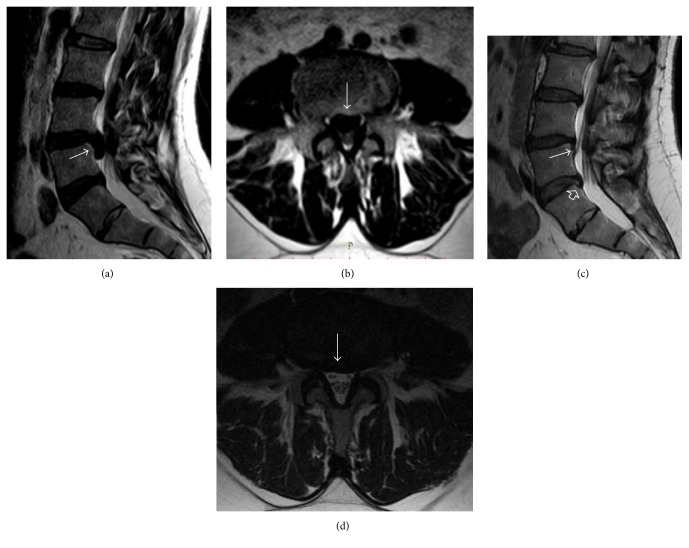
MRISagittal and axial lumbar MRI, T2-weighted: large extruded and migrated disc herniation at L4-L5, arrows on the herniation, then complete regression of the herniation three years later on the same views.
Source : Hakan et al., Case Reports in Orthopedics, 2016, figure 2 · CC BY
- Direct mechanical compression ⚙️: the disc herniation exerts pressure on the nerve root and its dorsal root ganglion, disrupting intraneural blood flow (focal ischaemia) and axonal transport. This mechanical component produces a motor or sensory deficit when it is severe and prolonged.¹¹
- Intense neurochemical inflammation 🔥: extruded nucleus pulposus material is richly loaded with pro-inflammatory substances: TNF-α, IL-1β, IL-6, IL-8, phospholipase A2. These mediators trigger a peri-radicular inflammatory cascade that chemically sensitises the nerve root, even in the absence of severe mechanical compression.¹¹,¹²
🧬 The twofold pathophysiological mechanism of radicular pain
Synthesis of Dower 2019 and of the literature on the peri-radicular inflammatory cascade
Summary diagram based on Dower A, Davies MA, Ghahreman A. Pathologic Basis of Lumbar Radicular Pain. World Neurosurg. 2019;128:114-121 (PMID 31028982) and Zhong M et al. Pain Physician. 2017;20(1):E45-E52 (PMID 28072796).
Key points
- Sciatica is a radicular pain most often due to a disc herniation affecting the L5 or S1 nerve roots (L4-L5 and L5-S1 levels).
- Point prevalence ~5%, lifetime cumulative prevalence 13-40%, peak incidence at 40-50 years.
- Key modifiable risk factors: lifting, smoking, obesity, vibration, constrained working postures; psychosocial factors for chronicity.
- Pathophysiology twofold : mechanical compression + intense chemical inflammation (TNF-α, IL-1β, IL-6).
- Natural history very favourable : 60-90% improvement within 6-12 weeks, spontaneous resorption of the herniation in 66 % of patients (Zhong 2017).
References
- Alexander CE, Varacallo M. Lumbosacral Radiculopathy. In: StatPearls. Treasure Island (FL): StatPearls Publishing; 2024. NBK430837.
- Stafford MA, Peng P, Hill DA. Sciatica: a review of history, epidemiology, pathogenesis, and the role of epidural steroid injection in management. Br J Anaesth. 2007;99(4):461-473. PMID 17704089.
- Konstantinou K, Hider SL, Jordan JL, Lewis M, Dunn KM, Hay EM. The impact of low back-related leg pain on outcomes as compared with low back pain alone: a systematic review of the literature. Clin J Pain. 2013;29(7):644-654. PMID 23328336.
- Kreiner DS, Hwang SW, Easa JE, et al. An evidence-based clinical guideline for the diagnosis and treatment of lumbar disc herniation with radiculopathy (NASS). Spine J. 2014;14(1):180-191. PMID 24239490.
- Jensen RK, Kongsted A, Kjaer P, Koes B. Diagnosis and treatment of sciatica. BMJ. 2019;367:l6273. doi:10.1136/bmj.l6273.
- Stochkendahl MJ, Kjaer P, Hartvigsen J, et al. National Clinical Guidelines for non-surgical treatment of patients with recent onset low back pain or lumbar radiculopathy. Eur Spine J. 2018;27(1):60-75. PMID 28429142.
- Zhu Z, Schouten T, Strijkers R, Koes B, Chiarotto A, Gerger H. Effectiveness of Nonsurgical Interventions for Patients With Acute and Subacute Sciatica: A Systematic Review With Network Meta-Analysis. J Orthop Sports Phys Ther. 2025;55(6):1-12. PMID 40434940.
- Cook CE, Taylor J, Wright A, Milosavljevic S, Goode A, Whitford M. Risk factors for first time incidence sciatica: a systematic review. Physiother Res Int. 2014;19(2):65-78. PMID 24327326.
- Shiri R, Karppinen J, Leino-Arjas P, Solovieva S, Viikari-Juntura E. The association between smoking and low back pain: a meta-analysis. Am J Med. 2010;123(1):87.e7-35. PMID 20102998.
- Foster NE, Anema JR, Cherkin D, et al. Prevention and treatment of low back pain: evidence, challenges, and promising directions. Lancet. 2018;391(10137):2368-2383. PMID 29573872.
- Dower A, Davies MA, Ghahreman A. Pathologic Basis of Lumbar Radicular Pain. World Neurosurg. 2019;128:114-121. PMID 31028982.
- Cunha C, Silva AJ, Pereira P, Vaz R, Gonçalves RM, Barbosa MA. The inflammatory response in the regression of lumbar disc herniation. Arthritis Res Ther. 2018;20(1):251. PMID 30400975.
- Brinjikji W, Luetmer PH, Comstock B, et al. Systematic literature review of imaging features of spinal degeneration in asymptomatic populations. AJNR Am J Neuroradiol. 2015;36(4):811-816. PMID 25430861.
- Zhong M, Liu JT, Jiang H, Mo W, Yu PF, Li XC, Xue RR. Incidence of Spontaneous Resorption of Lumbar Disc Herniation: A Meta-Analysis. Pain Physician. 2017;20(1):E45-E52. PMID 28072796.
- Chiu CC, Chuang TY, Chang KH, Wu CH, Lin PW, Hsu WY. The probability of spontaneous regression of lumbar herniated disc: a systematic review. Clin Rehabil. 2015;29(2):184-195. PMID 25009200.
How do you assess and diagnose sciatica with certainty?
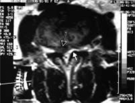
MRIAxial lumbar MRI: lateralised disc herniation (black arrow) associated with contralateral hypertrophy of the ligamentum flavum (white arrow), causing canal narrowing.
Source : Karabekir et al., Archives of Medical Science, 2010, figure 2 · CC BY-NC-ND
Which questions should you ask to understand the patient and their history?
Questioning the patient is the cornerstone of diagnosis.¹ It must be systematic and aim to characterise the pain, identify risk factors and, above all, screen for warning signs (red flags). A well-conducted history allows a solid diagnostic hypothesis to be formed even before the physical examination.² The essential questions include:- Location and distribution of the pain 📍: does the pain radiate into the lower limb? Does it follow a precise distribution (dermatome)? Pain travelling below the knee is more suggestive of true radicular pain than of referred facet or muscular pain.²,⁴
- Characteristics of the pain ⚡: is it electric, shooting, burning? These descriptors point towards neuropathic pain consistent with nerve root involvement.⁵
- Modifying factors : manoeuvres that increase intradiscal pressure (coughing, sneezing, straining at stool, prolonged sitting) typically exacerbate the symptoms. Standing or walking may relieve some patients.²
- Neurological symptoms : paraesthesia (pins and needles), dysaesthesia, muscle weakness in the leg or foot? Any progressive motor deficit must raise the alarm.⁴
- Screening for red flags 🚨, a step that is non-negotiable according to the IFOMPT framework (Finucane JOSPT 2020).⁶
⚠ Red flags in sciatica: urgent medical referral
- Cauda equina syndrome (absolute surgical emergency, 24-48 h): saddle anaesthesia, sphincter disturbance (urinary or faecal retention or incontinence), progressive bilateral motor weakness, recent-onset sexual dysfunction.⁶,⁷
- Severe or rapidly progressive motor deficit : acute foot drop (L5 root), weakness > 3/5 on great toe extension or plantar flexion, progressive loss of strength.⁸
- Suspected malignancy : history of cancer, unexplained weight loss (> 5% in 6 months), non-mechanical night pain, age > 50 years with no previous history and a first episode.⁹
- Suspected infection : fever, immunosuppression, intravenous drug use, recent infection, febrile low back pain with no improvement.⁶,⁹
- Suspected fracture : significant trauma, known osteoporosis, long-term corticosteroid therapy, age > 70 years.⁹
Which clinical tests should you perform and which other conditions must be ruled out?
The physical examination aims to reproduce the patient's radicular pain and to demonstrate a neurological deficit objectively. 🧠 No test is perfect: combining several tests significantly increases diagnostic certainty.¹⁰,¹¹Neurodynamic tests
These tests place the lumbosacral nerve roots under tension in order to reproduce the radiating pain.- Straight leg raise (SLR/Lasègue) : the best-known test. The Cochrane meta-analysis by van der Windt 2010 (16 studies) reports a pooled sensitivity of 0.92 (CI 0.87-0.95) in surgical populations but a specificity of 0.28 (CI 0.18-0.40).¹⁰ A negative test reasonably rules out nerve root compression; a positive test does not confirm it with certainty.¹⁰
- Crossed SLR : raising the non-painful leg reproduces the symptoms in the painful leg. High specificity (~0.90) but low sensitivity (~0.28-0.30). A very strong diagnostic indicator when it is positive.¹⁰
- Slump test : a combination of spinal flexion + cervical flexion + knee extension + ankle dorsiflexion. Sensitivity and specificity comparable to the SLR (Majlesi 2008: sensitivity 0.84, specificity 0.83 in that sample).¹²
- Femoral nerve stretch test (prone knee bend) : used for high lumbar radicular pain (L2, L3, L4: femoral neuralgia).¹
Structured neurological examination
The meta-analysis by Al Nezari et al. (Spine J 2013, 14 studies, n=2168) reports that sensory, motor and reflex deficits taken in isolation have low sensitivity but moderate specificity ; combining them improves diagnostic accuracy.¹¹| Nerve root | Reflex | Motor test (myotome) | Dermatome |
|---|---|---|---|
| L4 | Patellar | Knee extension, foot dorsiflexion | Medial aspect of the leg + medial instep |
| L5 | (None reliable) | Great toe extension, foot dorsiflexion | Lateral aspect of the leg + dorsum of the foot + great toe |
| S1 | Achilles | Plantar flexion (walking on tiptoes) | Posterior aspect of the leg + lateral border of the foot + fifth toe |
Differential diagnosis
Several conditions can mimic discogenic sciatica and must be considered systematically:- Degenerative lumbar stenosis : gradual onset of pain, neurogenic claudication (brought on by walking, relieved by trunk flexion or sitting).¹³
- Piriformis syndrome : buttock pain, reproduced by stretching or contracting the muscle, without consistent objective neurological signs (Hopayian Eur Spine J 2010: controversial, often a diagnosis of exclusion).¹⁴
- Hip pathology (hip osteoarthritis, femoroacetabular impingement): groin and trochanteric pain, restricted range of motion in internal rotation.
- Lumbar facet syndrome : axial pain, sometimes radiating as far as the buttock but rarely below the knee.
- Sacroiliac dysfunction : peri-sacral pain, reproduced by specific provocation tests (compression, distraction, FABER, Gaenslen, thigh thrust).
- Non-musculoskeletal causes : extraspinal tumour, vascular disease (aortic aneurysm), gynaecological or urological causes.
📊 Diagnostic performance of the main clinical tests in sciatica
Sensitivity (green) vs specificity (burgundy), from Cochrane van der Windt 2010 and Majlesi 2008
Pooled data: van der Windt DA et al. Cochrane CD007431 (PMID 20166095); Majlesi J et al. J Clin Rheumatol. 2008;14(2):87-91 (PMID 18391677) ; Al Nezari NH et al. Spine J. 2013;13(6):657-674 (PMID 23499340). Caveat: performance measured mainly in surgical populations with a high prevalence of herniation: values in primary care are probably more modest.
Should these patients be classified, and what are the benefits?
The classification (subgrouping) is an approach increasingly recommended for optimising management.¹⁵,¹⁶ The aim: to move from "one size fits all" treatment to a personalised approach by identifying subgroups likely to respond preferentially to specific interventions. One of the most studied systems in physiotherapy is Mechanical Diagnosis and Therapy (MDT / McKenzie method). Patients are classified according to their symptomatic and mechanical response to repeated movements. For radicular pain, the key phenomenon sought is centralisation : regression of the most distal pain (from the foot towards the thigh, from the thigh towards the spine) in response to a specific direction of movement.¹⁵,¹⁷ The benefits of this classification:- Better prognosis : centralisation is an extremely favourable prognostic indicator, associated with better outcomes and a reduced likelihood of surgery.¹⁵
- Treatment direction : identifying a directional preference directly guides the choice of exercises.¹⁷
- Patient empowerment : simple targeted exercises, a strengthened sense of self-efficacy.
Key points
- The diagnosis is essentially clinical : rigorous history-taking + structured physical examination; imaging is not required as a first-line investigation unless there are red flags or conservative treatment fails.
- The search for red flags (cauda equina, severe motor deficit, malignancy, infection, fracture) is a step that is non-negotiable (Finucane IFOMPT 2020).
- SLR is sensitive (0.92) but poorly specific (0.28); Crossed SLR is highly specific but poorly sensitive: combined, they frame the diagnosis well.
- The Slump test has diagnostic accuracy equivalent to the SLR (Majlesi 2008).
- No single test is sufficient: the combination of a positive SLR + a consistent neurological deficit + a compatible history markedly increases the post-test probability.
- The centralisation phenomenon under repeated movements (McKenzie/MDT method) is a favourable prognostic indicator and guides treatment.
References
- Jensen RK, Kongsted A, Kjaer P, Koes B. Diagnosis and treatment of sciatica. BMJ. 2019;367:l6273. doi:10.1136/bmj.l6273.
- Vroomen PC, de Krom MC, Knottnerus JA. Diagnostic value of history and physical examination in patients suspected of sciatica due to disc herniation: a systematic review. J Neurol. 1999;246(10):899-906. PMID 10552236.
- Stochkendahl MJ, Kjaer P, Hartvigsen J, et al. National Clinical Guidelines for non-surgical treatment of patients with recent onset low back pain or lumbar radiculopathy. Eur Spine J. 2018;27(1):60-75. PMID 28429142.
- Alexander CE, Varacallo M. Lumbosacral Radiculopathy. In: StatPearls. StatPearls Publishing; 2024. NBK430837.
- Scholz J, Finnerup NB, Attal N, et al. The IASP classification of chronic pain for ICD-11: chronic neuropathic pain. Pain. 2019;160(1):53-59. PMID 30586071.
- Finucane LM, Downie A, Mercer C, et al. International Framework for Red Flags for Potential Serious Spinal Pathologies. J Orthop Sports Phys Ther. 2020;50(7):350-372. doi:10.2519/jospt.2020.9971.
- Ahn UM, Ahn NU, Buchowski JM, Garrett ES, Sieber AN, Kostuik JP. Cauda equina syndrome secondary to lumbar disc herniation: a meta-analysis of surgical outcomes. Spine. 2000;25(12):1515-1522. PMID 10851100.
- Henschke N, Maher CG, Refshauge KM, et al. Prevalence of and screening for serious spinal pathology in patients presenting to primary care settings with acute low back pain. Arthritis Rheum. 2009;60(10):3072-3080. PMID 19790051.
- Downie A, Williams CM, Henschke N, et al. Red flags to screen for malignancy and fracture in patients with low back pain: systematic review. BMJ. 2013;347:f7095. PMID 24335669.
- van der Windt DA, Simons E, Riphagen II, et al. Physical examination for lumbar radiculopathy due to disc herniation in patients with low-back pain. Cochrane Database Syst Rev. 2010;(2):CD007431. PMID 20166095.
- Al Nezari NH, Schneiders AG, Hendrick PA. Neurological examination of the peripheral nervous system to diagnose lumbar spinal disc herniation with suspected radiculopathy: a systematic review and meta-analysis. Spine J. 2013;13(6):657-674. PMID 23499340.
- Majlesi J, Togay H, Unalan H, Toprak S. The sensitivity and specificity of the Slump and the Straight Leg Raising tests in patients with lumbar disc herniation. J Clin Rheumatol. 2008;14(2):87-91. PMID 18391677.
- Lurie J, Tomkins-Lane C. Management of lumbar spinal stenosis. BMJ. 2016;352:h6234. PMID 26727925.
- Hopayian K, Song F, Riera R, Sambandan S. The clinical features of the piriformis syndrome: a systematic review. Eur Spine J. 2010;19(12):2095-2109. PMID 20596735.
- May S, Aina A. Centralization and directional preference: a systematic review. Man Ther. 2012;17(6):497-506. PMID 22695365.
- Hill JC, Whitehurst DG, Lewis M, et al. Comparison of stratified primary care management for low back pain with current best practice (STarT Back): a randomised controlled trial. Lancet. 2011;378(9802):1560-1571. PMID 21963002.
- Werneke MW, Hart DL, Cutrone G, et al. Association between directional preference and centralization in patients with low back pain. J Orthop Sports Phys Ther. 2011;41(1):22-31. PMID 20972343.
- Konstantinou K, Dunn KM, Ogollah R, et al. Prognosis of sciatica and back-related leg pain in primary care: the ATLAS cohort. Spine J. 2018;18(6):1030-1040. PMID 29174459.
- Foster NE, Konstantinou K, Lewis M, et al. Stratified versus usual care for the management of primary care patients with sciatica: the SCOPiC RCT. Health Technol Assess. 2020;24(49):1-130. PMID 33043881.
Which treatment strategies are most effective for sciatica?
Where do you start? What is the recommended hierarchy of interventions?
Initial management follows a stepped approach, favouring the least invasive interventions. International guidelines agree on a clear hierarchy (NASS 2014, Stochkendahl 2018, Qaseem ACP 2017, Jensen BMJ 2019).¹,²,³,⁴ First line: education, reassurance, activity :- Explain the nature of the condition and its largely positive prognosis (60-90% improvement within 6-12 weeks), and deconstruct mistaken beliefs about pain and imaging.⁴,⁵
- Stay as active as possible by adapting activities to avoid severe flare-ups. Conversely, strict bed rest is harmful (Cochrane Dahm 2010, n=10 RCTs, moderate level of evidence).⁶
- Simple analgesics (paracetamol) or NSAIDs: modest effect on radicular pain ; to be used in a supervised way and for a limited time.¹,²
- The epidural corticosteroid injections can provide short-term pain relief (1-3 months) to facilitate active rehabilitation, but their long-term benefits are limited (Bhatia 2016 meta-analysis, n=12 RCTs).⁷
| Modality | Short-term effect | Long-term effect | Level of evidence (GRADE) | Key source |
|---|---|---|---|---|
| Education + activity | ✓✓ | ✓✓ | HIGH | Stochkendahl 2018, Foster Lancet 2018 |
| Exercise (all types) | ✓✓ | ✓✓ | HIGH | Liu JOSPT NMA 2025, Hayden Cochrane 2021 |
| Manual therapy | ✓ | ~ | MODERATE | Qaseem ACP 2017 |
| Epidural injections | ✓ | ~ | MODERATE | Bhatia 2016 |
| NSAIDs | ~ | — | MODERATE | NASS 2014, Stochkendahl 2018 |
| Lumbar traction | — | — | LOW | Cochrane Wegner 2013 |
| Strict bed rest | ✗ (harmful) | ✗ | MODERATE | Cochrane Dahm 2010 |
| Surgery (failure at 6-12 wks) | ✓✓ (faster) | = conservative | HIGH | Peul NEJM 2007 (5 years) |
What is the place of exercise, and is there a superior approach?
Exercise is a cornerstone of conservative treatment. 🏋️ The network meta-analysis by Liu et al. (JOSPT 2025, n=RCTs in adults with acute or subacute sciatica < 3 months, literature search to June 2024) confirms the effectiveness of exercise on lower limb pain and physical function in the short term, with no specific protocol standing out clearly.⁸ The Cochrane review by Hayden 2021 (>200 RCTs, NMA, chronic low back pain) reaches the same conclusion: exercise is effective, but no type of exercise is consistently superior to the others.⁹📊 Comparative effectiveness of exercise approaches for sciatica
No clear superiority of one type over the others: Hayden Cochrane 2021 (n>200 RCTs) and Liu JOSPT NMA 2025
Indicative pooled effect values vs advice alone, 0-100 scale. Differences between modalities are not clinically significant (all < MCID 15 points). Sources: Hayden JA et al. Cochrane Database Syst Rev. 2021;CD009790 (PMID 34580864); Liu C et al. J Orthop Sports Phys Ther. 2025 (PMID 40434940); Owen PJ et al. Br J Sports Med. 2020;54(21):1279-1287 (PMID 31666220).
Manual therapies, traction, injections: what is their real effectiveness?
- Spinal manipulation / mobilisation 🖐️: can provide short-term relief, possibly through improved segmental mobility and neurophysiological modulation of pain. Level of evidence low to moderate ; it must be regarded as an adjunct to exercise, never a primary treatment (Qaseem ACP 2017: conditional recommendation for chronic low back pain).³
- Lumbar traction ❌: the Cochrane review by Wegner 2013 (32 RCTs, n=2762) concludes that traction, whether mechanical or manual, provides no benefit compared with placebo or other interventions for pain, function or return to work. Not recommended.¹¹
- Epidural corticosteroid injections 💉: short-term pain relief (1-3 months) in patients with discogenic sciatica (Bhatia 2016, 12 RCTs). The effect on disability is more modest. No long-term effect on the natural history or on the need for surgery.⁷
- Electrophysical modalities (ultrasound, laser, interferential currents, TENS): very low quality evidence; they must not replace active approaches.¹
Beyond the physical: how do you educate the patient and address psychological factors?
The biopsychosocial approach is fundamental. 🧠 Ignoring psychological and social factors is one of the major causes of treatment failure and chronicity.¹² Therapeutic patient education (TPE) must go beyond anatomical information:- Modify the patient's perception of pain and reduce kinesiophobia (fear of movement) and increase the sense of self-efficacy.¹²
- Explaining that "pain does not always mean more damage" is a key message.
- Imaging (MRI, CT) must be used judiciously : finding degenerative abnormalities, which are almost universal after the age of 50 in asymptomatic people (Brinjikji 2015), can increase anxiety and catastrophising without changing the initial strategy.¹³
- Negative beliefs about pain (catastrophising, mistaken structural beliefs);
- Behaviours of avoidance / fear of movement (Wertli Spine J 2014: major predictors of disability at 12 months, far more so than imaging findings);¹⁴
- Low mood, anxiety, low perceived social support;
- Unfavourable occupational context (job dissatisfaction, conflict, work injury).
Critique and controversies: surgical timing
The optimal timing of a discectomy remains a point of debate. The landmark trial by Peul et al. (NEJM 2007, n=283 patients with severe sciatica of 6-12 weeks, randomised to early surgery vs prolonged conservative care) showed that:- The early-surgery route provides faster relief (pain reduction and earlier return to activities at 4-8 weeks);
- The results at 1 year, 2 years and 5 years are equivalent between the two strategies;
- About 40% of patients in the conservative group were ultimately operated on within the year.¹⁵
Key points
- ✅ First line: education + reassurance + staying active. Note that strict bed rest is harmful.
- 🤸 Exercise is effective but no specific type is superior (Liu JOSPT 2025, Hayden Cochrane 2021). Adherence matters most.
- ✋ Manual therapies = a short-term adjunct ; traction is ineffective (Wegner Cochrane 2013); epidural injections: short-term relief only.
- 🧠 Identifying and managing yellow flags (kinesiophobia, catastrophising, fear-avoidance) is crucial to prevent chronicity (Wertli 2014).
- 📉 Surgery is considered after 6-12 weeks of failed conservative care or a severe/progressive neurological deficit. Outcomes at 1-2 years are equivalent to the prolonged conservative strategy (Peul NEJM 2007).
References
- Kreiner DS, Hwang SW, Easa JE, et al. An evidence-based clinical guideline for the diagnosis and treatment of lumbar disc herniation with radiculopathy (NASS). Spine J. 2014;14(1):180-191. PMID 24239490.
- Stochkendahl MJ, Kjaer P, Hartvigsen J, et al. National Clinical Guidelines for non-surgical treatment of patients with recent onset low back pain or lumbar radiculopathy. Eur Spine J. 2018;27(1):60-75. PMID 28429142.
- Qaseem A, Wilt TJ, McLean RM, Forciea MA. Noninvasive Treatments for Acute, Subacute, and Chronic Low Back Pain: A Clinical Practice Guideline From the American College of Physicians. Ann Intern Med. 2017;166(7):514-530. PMID 28192789.
- Jensen RK, Kongsted A, Kjaer P, Koes B. Diagnosis and treatment of sciatica. BMJ. 2019;367:l6273. doi:10.1136/bmj.l6273.
- Traeger AC, Hübscher M, Henschke N, Moseley GL, Lee H, McAuley JH. Effect of primary care-based education on reassurance in patients with acute low back pain: systematic review and meta-analysis. JAMA Intern Med. 2015;175(5):733-743. PMID 25799308.
- Dahm KT, Brurberg KG, Jamtvedt G, Hagen KB. Advice to rest in bed versus advice to stay active for acute low-back pain and sciatica. Cochrane Database Syst Rev. 2010;(6):CD007612. PMID 20556780.
- Bhatia A, Flamer D, Shah PS, Cohen SP. Transforaminal Epidural Steroid Injections for Treating Lumbosacral Radicular Pain from Herniated Intervertebral Discs: A Systematic Review and Meta-Analysis. Anesth Analg. 2016;122(3):857-870. PMID 26891397.
- Zhu Z, Schouten T, Strijkers R, Koes B, Chiarotto A, Gerger H. Effectiveness of Nonsurgical Interventions for Patients With Acute and Subacute Sciatica: A Systematic Review With Network Meta-Analysis. J Orthop Sports Phys Ther. 2025;55(6):1-12. PMID 40434940.
- Hayden JA, Ellis J, Ogilvie R, Stewart SA, Bagg MK, Stanojevic S, et al. Some types of exercise are more effective than others in people with chronic low back pain: a network meta-analysis. J Physiother. 2021;67(4):252-262. PMID 34538747.
- May S, Aina A. Centralization and directional preference: a systematic review. Man Ther. 2012;17(6):497-506. PMID 22695365.
- Wegner I, Widyahening IS, van Tulder MW, et al. Traction for low-back pain with or without sciatica. Cochrane Database Syst Rev. 2013;(8):CD003010. PMID 23959683.
- Foster NE, Anema JR, Cherkin D, et al. Prevention and treatment of low back pain: evidence, challenges, and promising directions. Lancet. 2018;391(10137):2368-2383. PMID 29573872.
- Brinjikji W, Luetmer PH, Comstock B, et al. Systematic literature review of imaging features of spinal degeneration in asymptomatic populations. AJNR Am J Neuroradiol. 2015;36(4):811-816. PMID 25430861.
- Wertli MM, Rasmussen-Barr E, Weiser S, Bachmann LM, Brunner F. The role of fear avoidance beliefs as a prognostic factor for outcome in patients with nonspecific low back pain: a systematic review. Spine J. 2014;14(5):816-836.e4. PMID 24412032.
- Peul WC, van Houwelingen HC, van den Hout WB, et al. Surgery versus prolonged conservative treatment for sciatica. N Engl J Med. 2007;356(22):2245-2256. PMID 17538084.
How do you secure lasting recovery and prevent flares?
How do you make the patient an active participant in their recovery through self-management?
The empowerment of the patient 💪 is the cornerstone of preventing recurrence. Moving from a passive role as "recipient of care" to an active role as "manager of one's own health" is a major prognostic factor for positive long-term outcomes.Therapeutic education: understanding in order to act better
Modern therapeutic education has moved beyond the simple biomedical model to adopt a biopsychosocial model.² The aim is to give the patient tools to:- Deconstruct limiting beliefs : fear-avoidance beliefs (high FABQ) are stronger predictors of chronic disability than imaging findings (Wertli Spine J 2014, meta-analysis of 17 studies).³
- Understand that pain does not mean damage : a key message for restoring confidence in movement and effort.⁴
- Manage modifiable risk factors : smoking (impaired disc microcirculation: Shiri 2010), excess weight, sedentary behaviour, chronic stress.⁵
- Recognise warning signals : distinguish "normal" exertion-related pain from a genuine inflammatory flare or a neurological red flag.
Physical exercise: the best preventive treatment
🧘 Exercise is the best-validated intervention for preventing recurrence of low back pain and sciatica. The meta-analysis by Shiri (Am J Epidemiol 2018, 13 RCTs, n=20,868) shows that exercise alone reduces the risk of recurrence by about 33%, and the combination of exercise + education is the most effective preventive strategy.⁶ A crucial fact confirmed by recent meta-analyses: no type of exercise is consistently superior (Hayden Cochrane 2021).⁷ The most important predictor of success is the patient's long-term adherence to the programme.⁸ An effective programme must be:- Individualised : adapted to the patient's preferences, capacities and goals in order to maximise adherence.
- Progressive : load, complexity and volume must increase gradually.
- Functional : exercises must approximate the movements and demands of daily and working life.
When and how should you plan a safe return to activity and sport?
Return to sport (RTS) or to intense physical activity after sciatica is a delicate step that must not be dictated by the calendar, but by the achievement of precise functional criteria.⁹A criteria-based approach, not a time-based one
The Bern 2016 international consensus (Ardern et al., BJSM) on return to sport sets out a decision framework based on assessing several domains before full resumption.⁹ For sciatica, the patient must have met:- Pain control : the ability to carry out activities of daily living without significant pain and without regular analgesics.
- Absence of neurological deficit signs : any loss of strength, sensation or reflexes must have completely resolved or stabilised and be compatible with participation.
- Full functional mobility : symmetrical lumbar spine and hip ranges of motion, without pain through the functional arcs.
- Restored neuromuscular control and strength : quantified functional tests (Shirado-Ito test for the flexors, plank test, McGill core endurance battery).
🏃 Progressive phases of return to sport in sciatica
Criteria-based model (Ardern BJSM Bern 2016, Creighton CJSM 2010): adapted to discogenic radiculopathy
Model adapted from Ardern CL et al. 2016 Consensus statement on return to sport. Br J Sports Med. 2016;50(14):853-864 (PMID 27226389) and Creighton DW et al. Return-to-play in sport: a decision-based model. Clin J Sport Med. 2010;20(5):379-385 (PMID 20818198).
Critique and controversies: beyond the certainties
Several grey areas persist. The "myth of the perfect exercise" continues to influence practice: meta-analyses confirm that no type of exercise is superior (Hayden 2021, Liu 2025), yet the search for a universal protocol remains active, sometimes at the expense of individualisation and adherence, which are the real levers of success. The radiological-clinical discordance remains a major challenge: a patient can be asymptomatic with a large residual herniation on MRI (Brinjikji 2015). This reality can be a source of anxiety if it is poorly communicated, and argues for relying almost exclusively on functional criteria and clinical criteria when deciding on resumption.¹⁰ Finally, the very definition of "recurrence" is heterogeneous across studies (a new consultation? A new period of sick leave? Reappearance of the radicular pain?), which hampers the standardisation of preventive strategies.¹Key points
- ✅ Empowerment is the key : a patient educated within a biopsychosocial model, who understands their symptoms and deconstructs their fear of movement, has better chances of lasting recovery.
- 🏃 Adherence matters more than the type of exercise : the regularity of an individualised, progressive programme is the best safeguard against recurrence (Shiri 2018: -33% recurrence with exercise).
- 🚦 Return to sport is based on criteria, not on a calendar: resolution of pain, neurological normalisation, mobility, passing functional tests.
- 📈 Progression is non-negotiable : a gradual move from general reconditioning to sport-specific movements before full return to competition.
References
- da Silva T, Mills K, Brown BT, Herbert RD, Maher CG, Hancock MJ. Risk of recurrence of low back pain: a systematic review. J Orthop Sports Phys Ther. 2017;47(5):305-313. PMID 28355981.
- O'Sullivan PB, Caneiro JP, O'Sullivan K, et al. Back to basics: 10 facts every person should know about back pain. Br J Sports Med. 2020;54(12):698-699. PMID 31892534.
- Wertli MM, Rasmussen-Barr E, Weiser S, Bachmann LM, Brunner F. The role of fear avoidance beliefs as a prognostic factor for outcome in patients with nonspecific low back pain: a systematic review. Spine J. 2014;14(5):816-836.e4. PMID 24412032.
- Traeger AC, Hübscher M, Henschke N, et al. Effect of primary care-based education on reassurance in patients with acute low back pain: systematic review and meta-analysis. JAMA Intern Med. 2015;175(5):733-743. PMID 25799308.
- Shiri R, Karppinen J, Leino-Arjas P, Solovieva S, Viikari-Juntura E. The association between smoking and low back pain: a meta-analysis. Am J Med. 2010;123(1):87.e7-35. PMID 20102998.
- Shiri R, Coggon D, Falah-Hassani K. Exercise for the Prevention of Low Back Pain: Systematic Review and Meta-Analysis of Controlled Trials. Am J Epidemiol. 2018;187(5):1093-1101. PMID 29053873.
- Hayden JA, Ellis J, Ogilvie R, et al. Some types of exercise are more effective than others in people with chronic low back pain: a network meta-analysis. J Physiother. 2021;67(4):252-262. PMID 34538747.
- Owen PJ, Miller CT, Mundell NL, et al. Which specific modes of exercise training are most effective for treating low back pain? Network meta-analysis. Br J Sports Med. 2020;54(21):1279-1287. PMID 31666220.
- Ardern CL, Glasgow P, Schneiders A, et al. 2016 Consensus statement on return to sport from the First World Congress in Sports Physical Therapy, Bern. Br J Sports Med. 2016;50(14):853-864. PMID 27226389.
- Brinjikji W, Luetmer PH, Comstock B, et al. Systematic literature review of imaging features of spinal degeneration in asymptomatic populations. AJNR Am J Neuroradiol. 2015;36(4):811-816. PMID 25430861.
- Creighton DW, Shrier I, Shultz R, Meeuwisse WH, Matheson GO. Return-to-play in sport: a decision-based model. Clin J Sport Med. 2010;20(5):379-385. PMID 20818198.
Cauda equina syndrome and motor deficits: when sciatica becomes an emergency
Why does this subgroup deserve a dedicated section?
The cauda equina syndrome (CES) 🚨 is a severe acute compression of the lumbosacral nerve roots (below the conus medullaris, L1-L2), most often caused by a massive central disc herniation (but also tumour, abscess or haematoma).²,³ This is an absolute neurosurgical emergency whose neurological prognosis depends directly on the time to decompression.² The cardinal symptoms to screen for systematically:- Saddle anaesthesia : loss of sensation in the perineum, the genitals and the inner buttocks, the territory of the S2-S5 sacral roots.²,³
- Sphincter disturbance : urinary retention (high post-void residual volume, catheterisation sometimes necessary), overflow incontinence, faecal incontinence, loss of voluntary control.²,³
- Bilateral motor weakness and/or progressive weakness of the lower limbs.²
- Sexual dysfunction of recent onset.²
- Often bilateral radicular pain or alternating right/left, with intense low back pain of recent onset.²,³
🚨 Decision algorithm: suspected cauda equina syndrome
Adapted from the IFOMPT framework (Finucane JOSPT 2020) and the Cauda Equina Society recommendations
Algorithme adapté de Finucane LM et al. International Framework for Red Flags for Potential Serious Spinal Pathologies. J Orthop Sports Phys Ther. 2020;50(7):350-372 (DOI 10.2519/jospt.2020.9971) and Todd NV. Cauda equina syndrome: the timing of surgery. Br J Neurosurg. 2017;31(5):500-501 (PMID 28859562).
What surgical timing applies to cauda equina and acute foot drop?
Cauda equina syndrome: early decompression
The seminal meta-analysis by Ahn et al. (Spine 2000, n=322 patients) remains the reference: decompression within 48 hours of symptom onset is associated with better urinary and sphincter outcomes than later surgery.² A delay of < 24 h brings no additional demonstrated benefit over 24-48 h in this meta-analysis, but the guiding principle remains: "the sooner the better". A more recent meta-analysis (Srikandarajah 2019, n=464 patients) confirms this trend: surgery within 48 hours significantly improves bladder recovery compared with a longer delay.⁴Acute foot drop: an ongoing debate
The foot drop (inability to dorsiflex the foot, caused by severe involvement of the L5 root or, more rarely, L4) is a sign of severity that changes management. The therapeutic dilemma:- The early-surgery option (within 7-14 days of the deficit appearing) appears to offer better chances of motor recovery than late surgery or conservative treatment alone, according to several surgical series and reviews.,
- However, spontaneous recovery is possible with conservative treatment in a non-negligible proportion of cases, although its quality and timing are less predictable.
- The high-level evidence (RCTs) is lacking in this area: recommendations rest mainly on observational studies and case series, so the decision remains a shared one, strongly individualised according to the severity of the deficit, its change over time and the patient's preferences.,
Key points
- 🚨 The cauda equina syndrome is an absolute neurosurgical emergency : decompression ideally < 48 hours (Ahn Spine 2000).
- 🔍 Cardinal symptoms to screen for at every consultation: saddle anaesthesia, sphincter disturbance, progressive bilateral weakness, recent-onset sexual dysfunction.
- ⚠ The acute foot drop (L5 deficit) warrants a prompt neurosurgical opinion (24-48 h) to discuss early decompression: moderate-level evidence, shared decision.
- 📋 Use the IFOMPT framework systematically (Finucane JOSPT 2020) to frame red flag screening and referral decisions.
- 💡 The front-line physiotherapist plays a pivotal role in early triage: simple questions and rigorous screening save neurological function.
References
- Jensen RK, Kongsted A, Kjaer P, Koes B. Diagnosis and treatment of sciatica. BMJ. 2019;367:l6273. doi:10.1136/bmj.l6273.
- Ahn UM, Ahn NU, Buchowski JM, Garrett ES, Sieber AN, Kostuik JP. Cauda equina syndrome secondary to lumbar disc herniation: a meta-analysis of surgical outcomes. Spine. 2000;25(12):1515-1522. PMID 10851100.
- Fraser S, Roberts L, Murphy E. Cauda equina syndrome: a literature review of its definition and clinical presentation. Arch Phys Med Rehabil. 2009;90(11):1964-1968. PMID 19887225.
- Srikandarajah N, Boissaud-Cooke MA, Clark S, Wilby MJ. Does early surgical decompression in cauda equina syndrome improve bladder outcome?. Spine (Phila Pa 1976). 2015;40(8):580-583. PMID 25646751.
- Finucane LM, Downie A, Mercer C, et al. International Framework for Red Flags for Potential Serious Spinal Pathologies. J Orthop Sports Phys Ther. 2020;50(7):350-372. doi:10.2519/jospt.2020.9971.
- Todd NV. Cauda equina syndrome: the timing of surgery probably does influence outcome. Br J Neurosurg. 2017;31(5):500-501. PMID 28859562.
- Henschke N, Maher CG, Refshauge KM, et al. Prevalence of and screening for serious spinal pathology in patients presenting to primary care settings with acute low back pain. Arthritis Rheum. 2009;60(10):3072-3080. PMID 19790051.
What do real case studies teach us about sciatica?
Analysis of a "classic" case: from assessment to conservative resolution
The typical course of a patient with lumbar radicular pain often follows a favourable trajectory with well-conducted conservative management.¹,² Typical presentation (synthesised from Jensen BMJ 2019 and prospective cohorts): a 42-year-old sedentary adult, with a history of acute low back pain episodes, who has had for 3 weeks right-sided low lumbar pain radiating below the knee as far as the great toe (L5 territory).¹ Worse with coughing, prolonged sitting and forward flexion. SLR positive at 50° on the right; moderate deficit of great toe extension (4/5); no sphincter disturbance. No red flags. Clinical hypothesis: probable L5 radicular pain from an L4-L5 disc herniation.² Evidence-based treatment plan (first line):- Education and reassurance : explaining the favourable prognosis (60-90% improvement within 6-12 weeks) and the phenomenon of spontaneous resorption (66%, Zhong 2017), and deconstructing mistaken structural beliefs.²,³
- Staying active + ergonomic advice (avoid sitting > 30 min, alternate positions). No strict bed rest (Cochrane Dahm 2010: harmful).⁴
- McKenzie method / MDT : looking for centralisation through repeated movements into lumbar extension (prone lying, progressive extension). Observed centralisation carries an excellent prognosis (May & Aina 2012).⁵
- Progressive exercises : motor control exercises targeting transversus abdominis and multifidus, then graded functional exercises.⁶
- Analgesics as needed (paracetamol, short-term NSAIDs).
The diagnostic challenge: when sciatica mimics another condition
The clinical presentation of pain radiating into the buttock and leg is not pathognomonic of a disc herniation.⁷ The piriformis syndrome 🍑 is the most frequently considered differential diagnosis, although its precise definition remains controversial.⁷ Presentation suggestive of piriformis syndrome (synthesis of Hopayian Eur Spine J 2010, systematic review of 55 studies): deep buttock pain reproduced by deep palpation of the buttock, worsened by prolonged sitting (compression of the piriformis muscle), reproduction of the symptoms on stretching manoeuvres (FAIR test: hip flexion + adduction + internal rotation) or on contraction of the piriformis.⁷ Absence of objective neurological deficit signs (no myotomal change, no reflex change), with a pain distribution that is less dermatomal and more diffuse.| Criterion | L5/S1 discogenic sciatica | Piriformis syndrome |
|---|---|---|
| Origin of pain | Lumbar spine | Deep buttock |
| Distribution | Precisely dermatomal below the knee | Often diffuse, sometimes as far as the foot |
| SLR / Lasègue | Positive (typically < 60°) | Often negative or unclear |
| FAIR test | Negative | Positive (reproduces symptoms) |
| Neurological examination | Possible deficit (myotome/reflex) | Normal |
| Imaging (MRI) | Herniation correlating with the clinical picture | No relevant disc abnormality |
Study of a complex case: foot drop and early surgery
The appearance of a major motor deficit, an acute foot drop (inability to dorsiflex the foot because of L5 involvement), radically changes management.¹⁰ Presentation : a 52-year-old adult who has had for 5 days a rapid worsening of right L5 sciatica with the appearance of foot drop (dorsiflexion strength 2/5, great toe extension strength 1/5), hypoaesthesia of the dorsum of the foot and the great toe. No sphincter disturbance. SLR positive at 30° on the right. Urgent MRI: right paramedian L4-L5 disc herniation with clear compression of the emerging L5 root.¹⁰ Therapeutic dilemma :- Early surgery (discectomy within 7-14 days): published series show that early decompression increases the chances of complete motor recovery compared with a delay of > 4 weeks or with conservative treatment alone.
- Conservative : spontaneous recovery is possible but less predictable; risk of partial or complete residual motor loss.
- The high-level evidence (RCTs) remains limited : recommendations based on observational studies and case series.
⚖ Treatment decision according to the clinical picture in sciatica
Synthesis of the NASS 2014, Stochkendahl 2018 and Jensen BMJ 2019 recommendations: shared decision between patient and team
Sources: Kreiner DS et al. NASS Clinical Guideline. Spine J. 2014;14(1):180-191; Stochkendahl MJ et al. Eur Spine J. 2018;27(1):60-75; Peul WC et al. N Engl J Med. 2007;356:2245-2256; Ahn UM et al. Spine. 2000;25:1515-1522.
Critique and controversy
The case studies reveal several tensions. First, spontaneous regression raises questions about the real role of our interventions: if most herniations regress on their own, is the goal of physiotherapy to "cure" the lesion, or to give the patient the tools (pain management, exercise, education) to get through the symptomatic phase and prevent recurrence? The literature suggests the second role, that of a facilitating coach.¹¹ Second, the prevalence of "mimics" such as piriformis syndrome fuels a debate about the reliability of the diagnosis. Piriformis syndrome itself lacks validated tests and is often diagnosed by exclusion (Hopayian 2010).⁷ Finally, the management of complex cases (foot drop) remains an area where high-level evidence is limited. The decision rests largely on clinical judgement and the informed preference of the patient.Key points
- ✅ "Classic" case : most cases of sciatica respond well to conservative treatment centred on education, exercise and staying active, supported by spontaneous resorption of the herniation in 2/3 of patients.
- 🤔 Diagnostic challenge : sciatica-type pain is not always discogenic. Consider piriformis syndrome, stenosis, hip pathology and SI joint dysfunction.
- 🚨 Absolute emergency : signs of cauda equina (sphincter disturbance, saddle anaesthesia) call for referral to emergency care without delay.
- ⚖ Complex decision : an acute severe motor deficit (foot drop) warrants a discussion about early surgery: an informed shared decision.
References
- Jensen RK, Kongsted A, Kjaer P, Koes B. Diagnosis and treatment of sciatica. BMJ. 2019;367:l6273. doi:10.1136/bmj.l6273.
- Kreiner DS, Hwang SW, Easa JE, et al. An evidence-based clinical guideline for the diagnosis and treatment of lumbar disc herniation with radiculopathy (NASS). Spine J. 2014;14(1):180-191. PMID 24239490.
- Zhong M, Liu JT, Jiang H, et al. Incidence of Spontaneous Resorption of Lumbar Disc Herniation: A Meta-Analysis. Pain Physician. 2017;20(1):E45-E52. PMID 28072796.
- Dahm KT, Brurberg KG, Jamtvedt G, Hagen KB. Advice to rest in bed versus advice to stay active for acute low-back pain and sciatica. Cochrane Database Syst Rev. 2010;(6):CD007612. PMID 20556780.
- May S, Aina A. Centralization and directional preference: a systematic review. Man Ther. 2012;17(6):497-506. PMID 22695365.
- Saragiotto BT, Maher CG, Yamato TP, et al. Motor control exercise for nonspecific low back pain: a Cochrane review. Spine. 2016;41(16):1284-1295. PMID 27128390.
- Hopayian K, Song F, Riera R, Sambandan S. The clinical features of the piriformis syndrome: a systematic review. Eur Spine J. 2010;19(12):2095-2109. PMID 20596735.
- Lurie J, Tomkins-Lane C. Management of lumbar spinal stenosis. BMJ. 2016;352:h6234. PMID 26727925.
- Hopayian K, Heathcote J. Deep gluteal syndrome: an overlooked cause of sciatica. Br J Gen Pract. 2019;69(687):485-486. PMID 31558515.
- Ahn UM, Ahn NU, Buchowski JM, Garrett ES, Sieber AN, Kostuik JP. Cauda equina syndrome secondary to lumbar disc herniation: a meta-analysis of surgical outcomes. Spine. 2000;25(12):1515-1522. PMID 10851100.
- Foster NE, Anema JR, Cherkin D, et al. Prevention and treatment of low back pain: evidence, challenges, and promising directions. Lancet. 2018;391(10137):2368-2383. PMID 29573872.
How do you apply these recommendations concretely in your practice?
When, and to which other healthcare professionals, should you refer?
Referring patients is a key skill of the modern physiotherapist, who often works on the front line. 🧠 The first step in this decision process rests on the rigorous identification of red flags (Finucane 2020) and of yellow flags (Wertli 2014).¹,² Referrals to consider in sciatica:- General practitioner / specialist (rheumatologist, neurosurgeon, rehabilitation physician) 🩺 :
- Suspected red flag (cauda equina, progressive motor deficit, malignancy, infection, fracture): referral as an emergency.
- Symptoms stagnating despite well-conducted treatment for 6-12 weeks.
- Discussion of additional treatment options (epidural injections, surgery).
- Pharmacological review (refractory neuropathic pain: gabapentinoids, etc.).
- Psychologist / pain specialist 🧠 :
- Presence of yellow flags (high kinesiophobia, catastrophising, depression, work-injury context).
- Pain that has become chronic > 12 weeks with a major psychological component.
- CBT or a multidisciplinary pain management approach.
- Occupational physician : workstation adaptation, temporary restrictions, phased return to work.
- Dietitian : if obesity and nutrition are identified risk factors (metabolic component).
How do you measure outcomes and overcome the barriers to implementation?
Measuring outcomes is essential to validate the effectiveness of our interventions, adjust the treatment plan and demonstrate the value of physiotherapy. 📈 The most robust approach combines objective measures (range of motion, strength, functional tests) and patient-reported outcome measures (PROMs).⁴PROMs validated in sciatica
| Tool | Domain assessed | Score | MCID | Source |
|---|---|---|---|---|
| NPRS | Pain intensity | 0-10 | ~2 points | Salaffi 2004 |
| ODI | Functional disability | 0-100 % | ~10 % | Fairbank 1980, Ostelo 2008 |
| RMDQ | Disability (24 items) | 0-24 | ~3-5 points | Roland-Morris 1983 |
| FABQ | Fear-avoidance beliefs | 0-96 | — | Waddell 1993 |
| PSEQ | Self-efficacy in the face of pain | 0-60 | ~7 points | Nicholas 2007 |
| STarT Back | Stratification of chronicity risk | 9 items | — | Hill Lancet 2011 |
| PGIC | Global perception of change | 1-7 | ≥ 6 | Scaia 2018 |
Barriers to evidence-based implementation
🚧 Implementing evidence-based practice runs into well-identified obstacles:- Lack of time for critical reading and for applying the guidelines.
- Limited access to resources (databases, journals, courses).
- Lack of skills for appraising the literature and working through the statistics.
- Weak organisational support : no EBP culture in the professional environment.
- Inertia of habitual practice and resistance to change (overuse of passive therapies, imaging and drug prescriptions persisting despite the guidelines).⁵
- Create a supportive culture : protected time for reading, access to databases, in-house journal clubs.
- Develop skills : continuing education in critical appraisal, mentoring by experienced peers.
- Technological tools : apps and software that simplify the collection and analysis of PROMs.
- Local "EBP champions" : opinion leaders who motivate the team to adopt evidence-based practices.
- Adoption of clinical practice guidelines : NASS 2014, Stochkendahl 2018, NICE LBP 2016 (updated 2020), Foster Lancet 2018.¹,⁵,⁶,⁷
Critique and controversies: the blind spots of ideal application
Several tensions persist. The risk of bureaucratising care : the systematic use of PROMs, virtuous though it is, can become an administrative task disconnected from clinical dialogue if it is poorly integrated. The gap between trials and practice : RCTs often exclude complex patients (comorbidities, psychosocial factors), who nevertheless make up a large share of the caseload. Applying a recommendation drawn from an RCT to a complex patient is an act of clinical interpretation, not a simple recipe.⁵ The interprofessional collaboration runs into professional silos, conflicts over scope of practice, and a lack of funding that rewards joint working. Applying the recommendations is therefore not only a matter of knowledge, but also of context, culture and health policy.Strengthen your clinical practice in sciatica
This article is part of the Physio Learning collection of evidence-based clinical syntheses for French-speaking physiotherapists. Explore the other syntheses on the lumbar spine and radicular pain.
Key points
- 🚨 Red flags (cauda equina, progressive deficit, malignancy) call for urgent referral. Yellow flags (kinesiophobia, catastrophising) warrant psychological collaboration.
- 📊 Use validated PROMs : NPRS (pain), ODI/RMDQ (disability), FABQ (fear-avoidance), STarT Back (chronicity stratification).
- 🤝 Build a coordinated care network : GP, rehabilitation physician, neurosurgeon, psychologist, occupational physician.
- 💡 Overcome the barriers to EBP through continuing education, organisational support, mentoring and the adoption of practice guidelines.
- 🔄 Measure, adjust, discuss: evidence-based practice is an ongoing process, not a one-off event.
References
- Finucane LM, Downie A, Mercer C, et al. International Framework for Red Flags for Potential Serious Spinal Pathologies. J Orthop Sports Phys Ther. 2020;50(7):350-372. doi:10.2519/jospt.2020.9971.
- Wertli MM, Rasmussen-Barr E, Weiser S, Bachmann LM, Brunner F. The role of fear avoidance beliefs as a prognostic factor for outcome in patients with nonspecific low back pain: a systematic review. Spine J. 2014;14(5):816-836.e4. PMID 24412032.
- Marks D, Comans T, Bisset L, Scuffham PA. Substitution of doctors with physiotherapists in the management of common musculoskeletal disorders: a systematic review. Physiotherapy. 2017;103(4):341-351. PMID 28801031.
- Ostelo RWJG, Deyo RA, Stratford P, et al. Interpreting change scores for pain and functional status in low back pain: towards international consensus regarding minimal important change. Spine. 2008;33(1):90-94. PMID 18165753.
- Foster NE, Anema JR, Cherkin D, et al. Prevention and treatment of low back pain: evidence, challenges, and promising directions. Lancet. 2018;391(10137):2368-2383. PMID 29573872.
- Kreiner DS, Hwang SW, Easa JE, et al. An evidence-based clinical guideline for the diagnosis and treatment of lumbar disc herniation with radiculopathy (NASS). Spine J. 2014;14(1):180-191. PMID 24239490.
- Stochkendahl MJ, Kjaer P, Hartvigsen J, et al. National Clinical Guidelines for non-surgical treatment of patients with recent onset low back pain or lumbar radiculopathy. Eur Spine J. 2018;27(1):60-75. PMID 28429142.
- Hill JC, Whitehurst DG, Lewis M, et al. Comparison of stratified primary care management for low back pain with current best practice (STarT Back): a randomised controlled trial. Lancet. 2011;378(9802):1560-1571. PMID 21963002.
- Scaia V, Baxter D, Cook C. The pain provocation-based straight leg raise test for diagnosis of lumbar disc herniation, lumbar radiculopathy, and/or sciatica: a systematic review of clinical utility. J Back Musculoskelet Rehabil. 2012;25(4):215-223. PMID 23220802.
- Bernhardsson S, Larsson MEH. Does a tailored guideline implementation strategy have an impact on clinical physiotherapy practice? A nonrandomized controlled study. J Eval Clin Pract. 2019;25(4):575-584. PMID 29806112.

