














On-siteDPCFIFPL

Neurological rehabilitation
Neurological disorders in musculoskeletal practice: screen, assess and treat
Miguel Galan
Go further
Cauda equina syndrome: the red flags to know, why every hour counts, and how this dreaded complication of disc herniation is recognised and managed.

No clinical sign can rule out cauda equina syndrome: it is that doubt that demands an urgent MRI, not a delayed referral letter.
2.7adults affected per 100,000 each year
What follows covers each of these points in detail, with sources. It is there if you need it.
This topic is taught in a course: Neurological disorders in musculoskeletal practice: screen, assess and treat, on-site with Miguel Galan.
1 / 13
12 article chapters · 46 min in total
Understanding◔ 5 min
Cauda equina compression does not just cause pain: it produces a loss of functions.
Recognising it◔ 4 min
Sciatica is present in 97 % of patients, altered saddle sensation in 93 %.
Asking the questions◔ 5 min
Only nine physiotherapists out of thirty routinely ask about sexual function.
Value of signs◔ 5 min
No clinical sign can rule out cauda equina syndrome.
Acting fast◔ 3 min
What must be obtained is an MRI and, if it confirms, an operating theatre.
Surgical delay◔ 6 min
The 48-hour figure comes from a 2000 meta-analysis with more nuanced conclusions than commonly stated.
After surgery◔ 3 min
Surgery stops the compression, it does not always restore the functions.
Rehab◔ 5 min
Inpatient physiotherapy is nearly universal: 572 of 610 patients (94 %).
Pregnancy◔ 2 min
Cauda equina syndrome is rare during pregnancy, as is symptomatic disc herniation.
Case reports◔ 2 min
Three published cases illustrate three different errors in the care pathway.
In practice◔ 3 min
Complete screening takes ninety seconds.
FAQ◔ 3 min
Bilateral sciatica should raise the suspicion, but never suffices to confirm or rule out the diagnosis.
Train on this
Neurological rehabilitation
Miguel Galan
Next comes practice: the course that teaches this topic, with Miguel Galan.
Our courses on this topic may be covered by DPC or FIFPL, the French continuing-education funding schemes, depending on your profession and situation.
The site already devotes whole articles to the conditions of which this syndrome is the dreaded complication: sciatica from disc herniation, then degenerative lumbar stenosis, then non-specific low back pain, and femoral neuralgia. In each of those pages, cauda equina syndrome takes up three lines in a box. This page deals with the syndrome itself: how to recognise it before retention, how to ask the questions everybody skips over, where to refer and how fast, and what becomes of the patient afterwards. It is the surgical emergency of the spine, the one whose delay is paid for in permanent sphincter and sexual sequelae.
Red flags: same-day emergency referral
A single one of these signs, having appeared recently in a patient with low back pain or radicular pain, calls for immediate referral to a unit able to perform an MRI and to operate. This is not a letter to the family doctor, nor an imaging appointment sometime this week.
What these signs have in common : apart from the motor deficit, they all explore the sacral territory S2 to S5, the one that routine examination of a low back pain patient does not test. And none of them is painful. A patient who has been in far less pain since yesterday while their deficit progresses is not a patient who is getting better.
Complete urinary retention is a late sign. Waiting for it to appear before referring means referring at the precise moment when early surgery stops demonstrating a benefit.1
Three figures to frame the problem
Sources: incidence, Scottish national cohort, 149 cases in 5.4 million inhabitants over one year.2 Proportion of suspected cases confirmed, systematic review of 18 studies.3 Sexual dysfunction, systematic review of 5 studies, pooled estimate at a distance.4
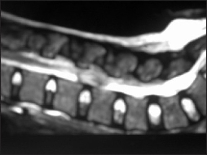
MRISagittal spinal MRI: arachnoiditis, a rare but genuine cause of cauda equina syndrome, described here after spinal anaesthesia.
Source : Jain et al., Indian journal of anaesthesia, 2010, figure 1 · CC BY
Below the cord, the lumbar and sacral roots run down as a free bundle in the canal. They carry the control of the bladder, of the anal sphincter, of genital sensation and of lower limb motor function. A conflict that compresses them all together therefore does not produce one more pain: it produces a loss of functions, some of which do not come back.
The spinal cord ends at about L1 or L2. Below that, the spinal canal contains a bundle of lumbar and sacral nerve roots that run freely in the dural sac before exiting at their own level: that is the cauda equina. These roots carry the innervation of the lower limbs, but also, through the sacral roots S2 to S4, the innervation of the bladder, the anorectum and the genitalia. Compression wide enough to involve them together produces cauda equina syndrome.
The commonest cause is a large central or paracentral disc herniation, most often at L4-L5 or L5-S1, which abruptly fills the canal. This is the point at which this article meets the one devoted to sciatica from lumbar disc herniation : the same lesion that usually produces radicular pain in a single territory can, if it is central and massive, compress the whole bundle. The other causes are also met in practice: advanced degenerative lumbar stenosis, spondylolisthesis, intracanal tumour or metastasis, epidural abscess, spontaneous or anticoagulant-related epidural haematoma, trauma.56
Four quantified landmarks to keep in mind
Age and sex, Scottish cohort: incidence of 7.2 per 100,000 in women aged 30 to 39.2 Proportion among low back pain, combined estimate from 4 studies.3 Negative MRI scans, development and validation cohort of the SuCESS score.7 Persistent bladder disturbance, meta-analysis of 16 studies and 987 patients.8
The syndrome is rare. A population study conducted across the whole of Scotland, which recorded for one year every patient operated on as an emergency for degenerative compression of the cauda equina, found 149 cases in 5.4 million inhabitants, that is a crude incidence of 2.7 per 100,000 adults per year.2 That work brings two less expected lessons. First, the incidence is higher than had been thought: earlier European estimates hovered around 0.3 to 0.6 per 100,000, that is at least four times lower. Second, the disease preferentially affects young adults, more often women, with a peak of 7.2 per 100,000 in women aged 30 to 39. The typical patient is therefore not the elderly person with osteoarthritis: it is someone in the middle of their working life, exactly the profile that comes to a community practice with low back pain or sciatica.
A coding trap worth knowing
In that same study, the authors compared their clinical case-finding with hospital diagnostic codes. Of 211 patients carrying a new ICD-10 code for cauda equina syndrome, 55 % did not have the syndrome once the records were reviewed.2 Any frequency statistic drawn from coding databases therefore greatly overestimates the phenomenon, and the divergent figures found in the literature often come from that rather than from a genuine difference in population.
The literature long suffered from the absence of a shared definition. A review of 212 articles published over twenty-one years counted seventeen different definitions of the syndrome.9 It is a practical problem and not merely an academic one: two studies announcing opposite results on surgical timing may simply not be talking about the same patients.
The classification that has taken hold, adopted by the standards of the British association of spine surgeons, is a simple dichotomy.910
| Stage | What the patient has | What it changes |
|---|---|---|
| Suspected (no compression) |
Suggestive symptoms, normal MRI or no compression of the cauda equina. This is the commonest situation. | No surgery. But the sorting could not have been done without imaging. |
| Incomplete syndrome (CESI) |
Saddle sensory disturbance, functional urinary disturbance: altered stream, need to strain, loss of the sensation of a full bladder. The patient still passes urine voluntarily. | This is the stage at which early decompression shows its clearest benefit. It is also the one most often missed. |
| Syndrome with retention (CESR) |
Established urinary retention, a bladder that no longer empties, overflow leakage, extensive saddle anaesthesia. | Markedly poorer prognosis for bladder, bowel and sexual function, whatever the operative delay thereafter. |
This distinction is not a refinement of classification: it is the main clinical message of this article. A study that followed 46 patients for a mean of 43 months after surgery, using validated questionnaires, shows that those who had reached the stage of retention had significantly worse results for bladder, bowel and sexual function than those operated on at the incomplete stage.11 In other words, most of the prognosis is decided before hospital, in the interval between the first functional disturbances and the first person who takes them seriously.
Key points
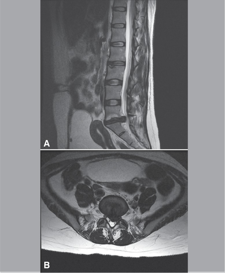
MRISagittal and axial lumbar MRI, T1 sequence: protruding disc herniation at L5-S1 responsible for cauda equina syndrome.
Source : Fuso et al., Acta Ortopedica Brasileira, 2013, figure 1 · CC BY-NC
The classic descriptions list the signs as a list, which suggests they arrive together. In practice they arrive as a cascade, and the first of the cascade are the ones nobody mentions spontaneously.
A Dutch study recorded the prevalence of each symptom at the time of presentation in 75 patients operated on for cauda equina syndrome from disc herniation. Sciatica was present in 97 % of them, a change in saddle sensation in 93 %, voiding dysfunction in 92 %, defecation dysfunction in 74 %.12 The most telling figure in that study is elsewhere: only 26 patients out of 75 had been asked about their sexual function, and 25 of those had a disturbance. The symptom was not rare, it was not sought.
The mean duration of complaints before presentation was 84 hours, with a median of 48 hours.12 That means half of those patients had already had symptoms for two days when they arrived at hospital, before the operative-delay clock even started. It is that earlier delay, the one before hospital, over which a physiotherapist has some hold.
The typical cascade, from earliest to latest
Sequence built from the staged classification of Lavy et al. 20229 and the prevalences recorded at presentation by Korse et al. 2017.12 This is a typical order, not a chronology measured patient by patient: a presentation may start straight away at the stage of retention, particularly in abrupt compressions.
| Rank | Sign | How the patient says it | What it requires |
|---|---|---|---|
| 1 | Saddle sensory disturbance | “It feels odd when I wipe”, “it's like cotton wool”, “like after the dentist” | Same-day emergency referral |
| 2 | Disturbance of genital sensation | “I feel nothing during sex any more”, “I cannot feel the urine passing” | Same-day emergency referral |
| 3 | Functional voiding disturbance | “It takes a while to come”, “I have to strain”, “it feels as though some is left” | Same-day emergency referral |
| 4 | Recent bilateral sciatica, bilateral motor deficit | “Since yesterday it goes down both legs” | Same-day emergency referral |
| 5 | Loss of the sensation of a full bladder | “I never feel like going to the toilet any more”, “I have lost the sensation” | Same-day emergency referral |
| 6 | Anorectal disturbance | “I cannot feel it coming any more”, “I have no control over flatus” | Same-day emergency referral |
| 7 | Urinary retention | “I have not been able to pass urine at all since this morning” | Absolute emergency, but the prognosis is already engaged |
| 8 | Overflow incontinence | “It leaks on its own without my noticing” | Absolute emergency, the most ominous stage |
The costliest misreading
Overflow incontinence reads as incontinence, and therefore as a leakage problem. It is the opposite: the bladder is too full and spills over passively because it can no longer empty. A patient who “leaks” while no longer passing urine normally is a patient in retention. Confusing the two wastes the hours that remain.
Two misleading situations recur in the series and deserve to be known.
The first is paradoxical improvement in pain. A root compressed to the point of losing conduction stops hurting. A patient whose severe sciatica settles markedly within a few hours while weakness or numbness appears is not getting better.
The second is atypical presentation. A published case describes a patient with bilateral weakness, perineal numbness and sphincter disturbance, but also clonus, normally a sign of a central lesion and not of root compression. The presence of that sign cast doubt on the diagnosis and raised the possibility of an inflammatory or demyelinating condition. The lumbosacral MRI showed a large L5-S1 herniation, decompression was carried out, and the clonus resolved completely after surgery.13 The authors' lesson is explicit: the presence of an unexpected sign must not delay surgery when the rest of the picture is consistent.
Key points
This is the most useful chapter of this article, because it is the only point in the pathway where the physiotherapist truly decides what happens next. Screening is decided by words.
Thirty physiotherapists from a British community musculoskeletal service were interviewed about their practice in screening for cauda equina syndrome. All of them routinely asked the questions about bladder, bowel and saddle anaesthesia. Only nine routinely asked the questions about sexual function. Half reported discomfort in raising that area. Fewer than half framed the question before asking it, and five participants out of thirty brought together the four dimensions of a proper enquiry.14
This work is valuable because it does not say that physiotherapists do not know what to ask: it says that they do know and that they skip it anyway, on the single most intimate dimension. The authors also note barriers of language and culture, and the fact that the way of asking these questions had never been studied.
Framing is the dimension most often left out and the least costly to put right. It consists of saying, before asking anything at all, why you are asking. A wording that works:
Framing sentence
“Before we go on, I need to ask you a few rather personal questions, about the bladder, the bowels and intimate sensation. I ask them of every patient who has back pain. It is rare, but there is a condition of the nerves in the lower back that starts like this, and that is treated far better when it is caught early. If anything has changed recently, even a detail, I want to know.”
That sentence does three things at once: it announces the register, it depersonalises the question, and it explains what is at stake without alarming. It also gives the patient permission to report a “detail” they would have kept quiet.
The common principle is to avoid both jargon and euphemism. “Do you have any sphincter disturbance?” means nothing to a patient. “Everything all right in that department?” invites a yes.
| Area | Ask this, in these words | Avoid this, and why |
|---|---|---|
| Bladder, starting |
“When you go to the toilet, does the urine come straight away, or do you have to wait or strain for it to start?” | “Do you have any urinary difficulties?”: too vague, the patient thinks of burning or frequency. |
| Bladder, sensation |
“Can you still feel when your bladder is full? Can you feel the urine passing when you go?” | Asking only about incontinence: loss of sensation comes before leakage, often by several days. |
| Bladder, emptying |
“When you have finished, does it feel as though some is left? Do you have to go back soon afterwards?” | “Do you empty your bladder properly?”: invites a reflex yes. |
| Anorectal | “Can you feel when you need to open your bowels? Can you feel the stool passing? Can you hold on, and hold in flatus?” | “Are you constipated?”: constipation is commonplace and diverts from the real question, which is sensation and control. |
| Saddle sensation |
“When you wipe, or when you sit on the toilet, does it feel the same as before, on both sides?” | “Do you have saddle anaesthesia?”: the term does not belong to the patient. |
| Genital | “Have you noticed any change in sensation in the private area? During sex, for example, or when washing?” | Skipping the question, or asking it last in a low voice: that is what most people do, and it is the least often sought sign. |
| Genital, by patient |
In men: “Have you noticed any change in erections, or in sensation at ejaculation?” In women: “Have you noticed a loss of sensation, or new discomfort during sex?” | One neutral question for everyone: it stays so general that it yields nothing. |
Three methodological precautions
Screening is not a snapshot. A disc herniation can complete itself between two sessions. The approach recommended in the literature written for physiotherapists is to back up the enquiry with an explicit instruction handed to the patient: tell them which signs should take them to the emergency department without waiting for the next appointment, and tell them in plain words rather than in medical terms.16 A service evaluation covering 231 suspected patients seen in an outpatient setting shows that this combination, an explicit safety net and an established chain of communication, is what makes it possible to refer urgently only the minority who need it without missing the others: 21 % were referred to the emergency department, and seven compressions were confirmed on MRI.17
Key points
A clinician who knows the real value of their signs stops expecting from them what they do not give. No sign of this syndrome allows it to be ruled out, and that is precisely what must govern practice.
The largest published prospective evaluation covered 260 patients referred with suspected cauda equina syndrome to a tertiary spinal unit, all examined by spine surgeons then measured by bladder scan before MRI.18 Its results are harsh on clinical examination in isolation. Bilateral sciatica, the classic red flag, has a sensitivity of 32.4 % and a positive predictive value of 17.2 %. It therefore misses two thirds of cases, and when it is present it is wrong five times out of six.
Other work points the same way. A review of 57 patients referred with suspicion, of whom 13 had confirmed compression, concludes that digital rectal examination provides no discrimination : accuracy of 51 %, diagnostic odds ratio of 1.42, with no significant relationship between the number of positive clinical signs and the MRI result.19 Another series identified no clinical sign predictive of compression on MRI.20 A review of 250 referrals finds 13 % of cases confirmed, with no difference in clinical presentation between true and false positives, and concludes that imaging, not clinical triage, is the limiting step.21
Measured diagnostic value, prospective cohort of 260 patients
Katzouraki et al. 2020, prospective study of 260 patients referred with suspicion, bladder scan before lumbosacral MRI.18 The bars are proportional to the percentage, on a scale of 0 to 100 %.
Measuring the residual by bladder ultrasound is the only item on this list that clearly improves performance. With a threshold of 200 ml, the prospective cohort finds a sensitivity of 94.1 % and a negative predictive value of 98.7 %.18 A meta-analysis of seven studies and 734 patients compared thresholds of 100, 200, 300 and 500 ml, and confirms that 200 ml offers the best accuracy, with a sensitivity of 82 % and a specificity of 65 %.22 The authors of that meta-analysis attach an explicit caveat to their conclusion: the residual must not be used in isolation, nor as a rule-out tool.
That caveat has been put to the test. A series of 50 medicolegal files of confirmed syndromes looked for those who had had a bladder scan: 26 had, and in 13 of them, that is half, the residual was 200 ml or less while the diagnosis was clinical and radiological.23 All thirteen were incomplete forms, and all were operated on as emergencies. The authors' conclusion deserves quoting as it stands in a patient's record: a residual of 200 ml or less reduces the probability of the syndrome but does not rule it out, and clinical suspicion must always lead to MRI.
What this changes for a physiotherapist
A patient coming back from the emergency department with “the bladder scan was normal, it is not cauda equina” is not reassured by that, if their symptoms persist or progress. In the medicolegal series cited above, half of the confirmed syndromes that had had a bladder scan were below the 200 ml threshold, and all were incomplete forms.23 Worsening after a normal test justifies re-referral, not waiting.
If no sign is sufficient on its own, combining them raises the probability. A Dutch series of 58 suspected cases, of which 8 compressions were confirmed, shows that retention above 500 ml, or the combination of at least two of bilateral sciatica, subjective urinary retention and rectal incontinence, carries an odds ratio of 48.0 for compression on MRI, with a very wide confidence interval of 3.30 to 697.21 that reflects the small sample size.24
That logic was formalised in 2026 in the SuCESS score, built on 259 patients then validated on two independent cohorts totalling 444 patients. Six clinical variables go into it: saddle anaesthesia, reduced perianal sensation, motor deficit, bilateral sciatica, post-void residual and urinary catheterisation. At a threshold of 3 points out of 8, the score reaches a sensitivity and a negative predictive value of 100 %, identifying all 64 confirmed cases, and would have allowed 38.7 % of urgent MRI scans to be deferred.7
To be read with the right caution
A sensitivity of 100 % obtained on samples of this size remains to be confirmed in other health systems, and the score is a tool for hospital triage, designed to decide on an MRI in patients who have already been referred. It is not a community-practice tool, and must not be used to decide not to refer. A 2025 systematic review of international guidelines in fact finds a consistent position across nine documents: in the presence of red flags, MRI must be performed as an emergency.25
The majority of MRI scans requested urgently for suspicion show no compression: more than 80 % in the SuCESS score cohort7, 81 % in the series behind the 19 % confirmed of the systematic review of incidence.3 These patients are not patients seen “for nothing”. One study followed 62 of them and finds high levels of pain, chronicity and disability, with more functional comorbidity than in patients with a positive MRI, and above all no coherent follow-up pathway.26 A more recent series of 922 patients confirms that those who re-attend for the same symptoms after a normal MRI do not go on to develop compression: of 43 patients who came back within the year, none had a genuine syndrome on the second examination.27
These two results complement each other and set out what to do: the initial MRI remains indispensable to decide, and a recent, normal MRI, in the absence of a new symptom, does not justify repeating the examination but rather organising management of the pain and disability, which is what these patients really need.
The phrase “refer to the family doctor” makes no sense here. What has to be obtained is an MRI and, if it confirms, an operating theatre. That points to a type of facility, not to a correspondent.
Decision tree for low back or radicular pain with neurological signs
Built from the IFOMPT international framework for red flags28, the masterclass for physiotherapists by Greenhalgh et al. 201816, the 2025 systematic review of international guidelines25 and the data on falsely reassuring residuals.23
The international red flag framework published by IFOMPT insists on a point that has changed the way we reason: red flags are not switches, they feed clinical reasoning, and most of them have no established diagnostic value taken in isolation.28 But this syndrome is an exception to watchful waiting, because the useful window is measured in hours and the only test that decides is an MRI. Referring to a correspondent who will then have to request the test adds a link where every link costs hours.
Concretely, what you should aim for is an emergency department with access to MRI and a pathway to a spinal surgery team. The British literature documents the weak point in the circuit: in a series of 250 referrals, 73 patients presented without imaging outside working hours, and the test was put off to the next day for 60 of them, that is 82 %.21 The authors argue for 24-hour MRI access in every hospital that has one. That does not depend on the physiotherapist, but it tells them where their referral has the best chance of getting somewhere fast.
What must appear in the letter or message
Key points
It is the most quoted figure of this syndrome, and the least often read at source. It does not come from a randomised trial, it comes from a meta-analysis of observational studies published in 2000, whose conclusions are more nuanced than the use made of them.
The source is a meta-analysis published in Spine in 2000, which reviewed 104 references, retained 42 meeting its inclusion criteria, and analysed the results of 322 patients operated on for cauda equina syndrome from disc herniation.29 The authors divided the delay into five bands: under 24 hours, 24 to 48 hours, 2 to 10 days, 11 days to one month, more than one month. Two results come out of it, and the second is almost always forgotten.
The same work identifies prognostic factors independent of the delay that weigh heavily in practice: pre-existing chronic low back pain is associated with poorer urinary and rectal results, preoperative rectal dysfunction with poorer urinary continence, and increasing age with poorer postoperative sexual function.29
Two years later, a review published in the British Journal of Neurosurgery challenges head-on the idea of emergency surgery for everyone. Its arguments are not statistical but pathophysiological and operative: by the time of admission, the chance of recovery may already be lost; removing a large central herniation is considerably harder than a routine discectomy and often requires an extended approach; carried out in degraded conditions, as is frequently the case on call, surgery may add to morbidity instead of reducing it. The authors conclude explicitly that when retention with overflow incontinence is already present at presentation, urgent decompression brings no benefit.30
That position is not marginal, and it sheds light on a finding met everywhere: it is not the stopwatch that governs the prognosis, it is the neurological state at the moment the stopwatch starts.
A meta-analysis published in 2026 in The Spine Journal took the question up again across 15 comparative studies and 26,627 adults operated on for a degenerative syndrome, with subgroup analyses planned in advance.1 Its results structure clinical practice far better than the raw 48-hour figure.
| Question | What the data answer |
|---|---|
| Does operating before 48 hours do better than after? | Yes for urinary recovery (pooled odds ratio of about 2.3), motor recovery and overall neurological recovery. |
| Does operating before 24 hours do better than between 24 and 48 hours? | No, no consistent advantage, neurologically, urinary, functional or in hospital terms. |
| Is the benefit the same at every stage? | No. A marked effect in the incomplete forms, no significant difference related to delay in the forms with retention. |
| Does the benefit hold over time? | It concerns mainly early recovery. At twelve months and beyond, the gaps narrow and preoperative bladder state becomes the dominant prognostic factor. |
The authors' conclusion is a “clinically significant” threshold at around 48 hours from symptom onset, with maximum urgency reserved for patients who retain voluntary voiding.1 That is the exact reversal of the intuition that consists of waiting for retention to be sure of the diagnosis.
Another meta-analysis, covering 22 studies and 852 patients, illustrates the fragility of the evidence base better than any commentary. It does find far less persistent bladder dysfunction after decompression carried out within 48 hours, but look at the confidence intervals.31
Persistent long-term bladder dysfunction by operative delay, with confidence intervals
Kumar et al. 2022, meta-analysis of 22 studies and 852 cases. Persistent bladder dysfunction: 24.6 % [95 % CI 1.6 to 50.9] before 48 hours against 50.3 % [95 % CI 10.3 to 90.4] beyond. The authors conclude that a randomised trial is still needed to settle the question.31
The same phenomenon is found in small series. A Spanish study of 18 patients observes better motor and sphincter recovery in those operated on early, but finds no statistically significant difference, and says so.32 A review published in 2025 in JBJS Reviews sums up the state of the question bluntly: the timing of decompression remains debated, some studies showing no difference between 24 and 48 hours while others insist on immediate intervention.33
The three underlying reasons for that disagreement
The institutional position is more clear-cut than the evidence. The standards of the British association of spine surgeons insist on MRI without delay and emergency surgery in the cases that warrant it.10 A 2026 narrative review reports the update of the British national pathway published in 2025, which aims for decompression within 24 hours of confirmation, keeps the bladder scan as an argument in favour of the diagnosis and not as a means of ruling it out, and calls for 24-hour MRI capacity in general hospitals.34
That development does not contradict the nuance of the meta-analyses: it draws the organisational consequence from it. Since the prognosis depends above all on the stage reached at the time of diagnosis, the only variable genuinely modifiable at the scale of a health system is the time it takes to make that diagnosis. It is also the only variable a physiotherapist acts on.
Key points
This is the part that red flag boxes never mention, and yet it is the part that gives the urgency its meaning. Surgery stops the compression; it does not always restore the functions.
A 2026 meta-analysis covering 16 studies and 987 patients operated on for a degenerative syndrome finds persistent bladder dysfunction in 32.6 % of patients beyond twelve months, with a confidence interval of 23.4 to 41.9 % and marked heterogeneity between studies.8 In those who already had a bladder disturbance before the operation, the raw paired data show persistence in 130 patients out of 278, that is 46.8 %. The authors note that the results are consistently worse in patients admitted in retention than in the incomplete forms, and that the evidence on the effect of surgical delay on long-term bladder outcome remains heterogeneous and inconclusive.
A systematic review published the same year on sexual function gives an even higher figure: the reported prevalences run from 48 to 67 %, with a pooled estimate of 58.4 % sexual dysfunction at a distance.4 The disturbance affects both sexes and is not confined to erectile dysfunction: it includes impairment of arousal, orgasm and satisfaction. The authors note associations with age, with the duration of preoperative bladder dysfunction and with markers of sacral root involvement, perianal numbness and anorectal disturbance, but not with operative delay. Baseline sexual function was almost never documented.
Sequelae at a distance, cohort of 61 patients reviewed at a mean of 58 months
Barker et al. 2021, 61 patients out of 82 operated on who answered validated questionnaires, mean follow-up of 58 months. Urinary and faecal incontinence, catheterisation, sexual dysfunction and genital numbness were significantly commoner in patients admitted in retention. Half reported moderate depression or worse.35
These results agree with those of other cohorts. A meta-analysis of 22 studies and 852 cases finds, at a mean follow-up of 39 months, 43.3 % persistent bladder dysfunction, 31.1 % anorectal dysfunction, 53.3 % sensory deficit, 38.4 % motor weakness and 40.1 % sexual dysfunction.31 An insurance database study comparing 2,362 operated patients with 9,448 matched controls operated on the spine without cauda equina syndrome shows that at five years the risk of bladder dysfunction remains independently increased, with an odds ratio of 1.72 (95 % CI 1.56 to 1.89).36
Two lessons from the Scottish cohort followed for a mean of 43 months deserve to be known by any physiotherapist who will see these patients.
The first: asked which symptom they would most want treated, 57 % named pain, ahead of all the functional disturbances. Yet only 7 % had been referred to a pain management clinic.11 The second: the psychological impact is major and underestimated. In the same population, 37 % had scores compatible with a risk of depression over the last thirty days and 45 % over the last twelve months, and bladder and anorectal function were the significant predictors of mental quality of life, more so than physical function.37
A direct practical consequence
A patient referred for rehabilitation after cauda equina syndrome often comes with a request centred on walking and strength, because that is what the system has offered them. Opening up the question of the bladder, the bowels, genital sensation and chronic pain explicitly, as you open the question at the time of screening, allows you to treat what really weighs on their quality of life.
This is the best-documented hole in this file: the needs are massive, the provision is thin, and the level of evidence for the interventions is low. Saying so honestly is better than promising protocols that have not been evaluated.
A British national cohort covering patients operated on between June 2018 and May 2019 measured access to rehabilitation. Inpatient physiotherapy is almost universal: 572 patients out of 610, that is 94 %. Referral to specialist spinal rehabilitation on discharge concerns only 49 patients out of 608, that is 8 %. At one year, residual symptoms remain common: 66 % motor disturbance, 20 % bladder, 17 % anorectal, 13 % sexual. The authors note a marked difference by sex: 27 % of women retain bladder dysfunction against 11 % of men, whereas the rates at presentation were comparable, 84 % and 82 %.38 Their conclusion is that prospective studies of rehabilitation strategies are lacking to guide decisions.
A Japanese prospective cohort followed 93 patients operated on by posterior decompression, with constipation and urinary symptom questionnaires before the operation then at 1, 3, 6 and 12 months. On admission, 38.1 % had defecation disturbance and 33.3 % urinary disturbance. Surgery improved these symptoms in 30 to 50 % of the patients affected, with a first visible effect as early as one month and improvement continuing up to one year.39
That time course is useful to know for two reasons. It lets you explain to the patient that sphincter recovery is slow and that the absence of progress at three months is not a definitive failure. And it indicates that the useful rehabilitation window extends over at least a year, which is well beyond the durations usually prescribed.
Level of evidence of rehabilitation modalities after cauda equina syndrome
Transanal irrigation: clinical trial of Ethans et al. 2024.40 Neurogenic bladder: review of Hao et al. 2025.41 Pelvic floor rehabilitation: case reported by Bodalia et al. 2021.42 Electrical stimulation: case reported by Lim et al. 2023.43 Absence of prospective studies: national cohort of Roy et al. 2025.38 The levels shown are an appraisal of the overall quality of the evidence available in this population, not a published formal GRADE rating.
This is the only area in which a clinical trial has been conducted specifically in patients with cauda equina syndrome. Twelve participants, mean age 46, used a transanal irrigation system for ten weeks; ten completed the study. The neurogenic anorectal dysfunction score improved significantly, as did the faecal incontinence score, the constipation score, quality of life and self-rated bowel function, with colonic transit time reduced by 22 %, and no notable adverse effect.40 The authors recommend considering this option in patients in whom the usual methods of bowel management fail.
The sample is small and there is no control group: it is a solid lead, not definitive evidence. It has the merit of existing, which is true of no other modality in this population.
Management of the neurogenic bladder after lumbosacral injury follows a hierarchy of objectives that it is important to understand before proposing anything at all: protect upper urinary tract function first, restore at least partly lower tract function next, improve control, reduce the residual, prevent urinary infection and improve quality of life. The means run from conservative treatment to drug treatment, catheterisation, neuromodulation and surgery, given sequentially according to the situation.41
That framework indicates the physiotherapist's place: it is real but subordinate to urological assessment, and in particular to urodynamic testing where indicated. A study coupling clinical and urodynamic assessment in 43 patients showed that perianal sensation and overall motor weakness were the determinants of bladder function, and that bladder recovery was directly related to the number of days of delay.44 Proposing pelvic floor work without knowing whether the bladder is hypocontractile or overactive risks treating it the wrong way round.
What you should refrain from claiming
No controlled trial has evaluated pelvic floor rehabilitation specifically after cauda equina syndrome. The most detailed published case describes a trial of pelvic floor treatment combined with injections, with relief described as minimal.42 That does not mean pelvic floor rehabilitation is useless in this indication: it means it has not been evaluated, and that this should be said to the patient rather than promising them a result.
For the day-to-day practice of pelvic floor work itself, the base of competence is that of pelvic and perineal physiotherapy and anorectal rehabilitation, transposed to a neurological population. The site offers courses dedicated to these fields, including anorectal rehabilitation and pelvic floor rehabilitation, as well as a course devoted to triaging neurological disorders in musculoskeletal practice, which is exactly the competence brought to bear at the moment of the screening described above.
Key points
This subpopulation deserves a chapter for a precise reason: every warning sign is explained away in advance there. Low back pain, urinary disturbance and perineal disturbance belong to the normal picture of pregnancy and of the puerperium, which gives the syndrome the best of covers.
Cauda equina syndrome is rare during pregnancy, and symptomatic disc herniation itself is an uncommon cause of low back pain there.45 But the context is precisely the one in which the peak of incidence was measured: women aged 30 to 39, with an incidence of 7.2 per 100,000 per year, are the population most affected in the Scottish cohort.2
Management does not change, and that is the point to remember: cauda equina syndrome and progressive neurological deficits are absolute surgical indications at any stage of pregnancy. MRI is the investigation of choice, and surgery has been shown to be safe in this population, provided the obstetric environment can manage a fetal complication.45 A published case describes a patient successfully operated on prone at 30 weeks' gestation, who then delivered by planned caesarean section at term.46
The four confusions to avoid postpartum
In postpartum pelvic floor rehabilitation, vigilance rests on a simple point: a patient referred for postpartum incontinence who has saddle hypoaesthesia, loss of the sensation of needing to go, or a new anorectal disturbance does not belong to the usual protocol until those signs have been explained.
Three published cases, chosen because they illustrate three different errors in the pathway. None is a reconstruction: these are observations reported in the indexed literature, with their identifiers.
A 30-year-old woman presents at a chiropractic practice with saddle-distribution sensory loss and urinary retention. She had already been assessed in the emergency department and in radiology 48 hours earlier, and her symptoms had worsened since. After a brief history and examination, the practitioner refers her straight back to the emergency department. Given the rapid progression of the picture and the lumbar MRI already done, decompression is carried out the same day. Low back pain and neurogenic symptoms persisted after the operation, prompting a trial of epidural injections and pelvic floor rehabilitation, with relief described as minimal.42
What this case teaches
A woman presents with bilateral lower limb weakness, perineal numbness and sphincter disturbance, but also clonus, a sign of upper motor neurone lesion that does not belong to the classic picture. The diagnostic doubt led to consideration of an inflammatory or demyelinating cause, and to urgent brain and cervicothoracic MRI, which showed nothing. The lumbosacral MRI showed a large L5-S1 herniation. Decompression was carried out, the compression was significant intraoperatively, the patient recovered completely and the clonus disappeared.13 The authors conclude that the presence of clonus does not rule out the diagnosis, and that surgery must not be deferred while a second condition is sought if the clinical and radiological picture is consistent.
A 38-year-old man with a known lumbar disc herniation develops cauda equina syndrome after a vigorous back massage including spinal manipulation: severe numbness of both lower limbs, inability to walk from bilateral weakness, urinary and faecal incontinence. Imaging shows an L4-L5 herniation migrated into the canal, widely compressing the dural sac. He is operated on as an emergency, with laminectomy, decompression, discectomy and fusion, and recovers walking within a week, along with continence.47
An isolated case does not demonstrate a causal relationship, and the authors themselves call the event extremely rare. What it does indicate is usable: in a patient with a known large disc herniation, the appearance of sacral territory signs after a session, whatever the technique, must be taken seriously immediately and not put down to a commonplace post-treatment reaction.
This chapter condenses the article into an approach that holds up in a busy clinic day, without turning every episode of low back pain into a suspected neurosurgical emergency.
This screening is repeated at every session in an at-risk patient, because a herniation can complete itself between two appointments. It does not take the place of the examination, it is added to it.
A safety net, worded to be understood
“If you find it hard to pass urine or cannot manage at all, if you no longer feel your bladder filling, if you lose sensation when you wipe or during sex, if you cannot feel when you need to open your bowels, or if both legs become weak: go to the emergency department the same day, do not wait for our next appointment and do not book with your doctor. Tell the emergency department that your physiotherapist raised cauda equina syndrome.”
This instruction works better written than spoken, and the written form also has documentary value. The literature written for physiotherapists insists on this triad: communication, documentation, safety net.16
It is on everyone's mind and rarely documented. A scoping review counted, between 2009 and 2021 in the United Kingdom, fifteen claims involving physiotherapists, that is 0.7 % of all claims linked to this syndrome.48 The risk is therefore low in absolute terms. A qualitative study of forty physiotherapists and other people involved shows, on the other hand, that going through proceedings has a lasting effect on the wellbeing and practice of those who experience it, in a context perceived as a culture of blame that discourages sharing and collective learning.15
The practical conclusion is not to protect yourself by referring everyone, which would be unmanageable and pointless since more than 80 % of urgent MRI scans are already negative. It is to do the two things that genuinely protect both patient and practitioner: ask the complete set of questions, and write down what was asked and answered.
Is bilateral sciatica enough to raise the diagnosis?
It should raise it, but it is enough neither to retain it nor to rule it out. In the largest prospective cohort, its sensitivity is 32.4 % and its positive predictive value 17.2 %: it misses two thirds of cases.18 It is the combination of signs, and above all the presence of sacral territory signs, that creates the suspicion.
Does a normal bladder scan rule the diagnosis out?
No. The 200 ml threshold has a good negative predictive value in a suspected population18, but in a series of 50 medicolegal files of confirmed syndromes, half of those who had had a bladder scan had a residual of 200 ml or less, and all were incomplete forms operated on as emergencies.23 The residual supports the diagnosis, it does not rule it out.
Should a digital rectal examination be done before referring?
That procedure provides no diagnostic discrimination: accuracy of 51 % and an odds ratio of 1.42 in the study that evaluated it.19 It is not part of the competence required to decide to refer, and its absence must never delay referral. Some guidelines keep it in the hospital assessment.25
What if the patient refuses to go to the emergency department?
Explain concretely what is at stake, namely bladder, anorectal and sexual function, rather than using medical terms. Offer to telephone the department or a relative from the clinic. Then, if they maintain their refusal, record in writing the information given, the time and the patient's decision, and hand them the written instruction on when to seek help. Informed refusal belongs to them; documentation of the information belongs to you.
Can spinal manipulation trigger cauda equina syndrome?
Cases have been reported, notably in a patient with a pre-existing disc herniation after a vigorous massage with manipulation.47 An isolated case does not demonstrate causality and the authors call the event extremely rare. What should be taken from it is operational: in a patient with a known large herniation, the appearance of sacral signs after a session must never be put down to a post-treatment reaction.
How long can recovery continue?
Longer than is generally thought. In a prospective cohort of 93 patients, improvements in urinary and defecation function appear from the first month and continue to be recorded up to twelve months after surgery.39 There is therefore no reason to conclude to a definitive state at three or six months.
Why is the patient who still passes urine more urgent than the one in retention?
Because it is in that patient that early surgery demonstrates a benefit. The 2026 meta-analysis finds a marked effect of delay in the incomplete forms and no significant difference related to delay in the forms with retention.1 That does not mean a patient in retention is not an emergency, but that the one who is not yet there is the one whose future depends most on how fast you decide.
Should a patient with a normal MRI be re-examined later?
Not routinely. Of 510 patients with a normal initial MRI, 43 re-attended within the year for similar symptoms, and none had compression on the second examination.27 These patients do, on the other hand, have high levels of pain and disability, often with no follow-up pathway26 : what they need is pain management, not another MRI. A new symptom, however, is reassessed.
What is the difference from lumbar stenosis, which also compresses the cauda equina?
In degenerative lumbar stenosis , compression is chronic and progressive, its typical manifestation is neurogenic claudication and progression to a severe deficit remains a rare event. Cauda equina syndrome is an acute compression, and it is that abruptness that makes it an emergency. A stenosis can nevertheless decompensate: the warning signs in this article apply to it as well.
Where do ordinary low back pain and sciatica stand in relation to this syndrome?
They are its background. Non-specific low back pain and sciatica from disc herniation account for the vast majority of the patients you will see, and serious spinal disease represents only a few per cent of them. That is precisely why screening should be routine and brief, rather than exhaustive in a few. Femoral neuralgia raises the same question for the upper roots.
Each reference carries its PubMed identifier and its DOI. The metadata were checked against two independent databases, PubMed and CrossRef.
Behind this article
An author who explains, a reviewer who checks.
How we write and check our content

Physiotherapist · co-founder of Physio Learning
Marked for life by his first four-hour lecture without a single image, he took a master’s in instructional design so that it would never happen to anyone again. He hunts down publication bias and unreadable slides with the same intransigence.

Head of scientific content
Physiotherapist specialising in neuro-musculoskeletal practice and holder of a master’s in public health. He checks the methodological rigour of every article: primary sources, levels of evidence, no exceptions.
