

















A clinical synthesis on femoral neuralgia (high lumbar radiculopathy, L2-L4): recognising it, telling it apart from sciatica, knowing that its spontaneous prognosis is favourable, and sorting honestly through what physiotherapy can offer. Every reference has been checked individually on PubMed.
📝 In brief: clinical summary
- Femoral neuralgia is a high lumbar radiculopathy (L2-L3-L4 roots) in which the pain and the paraesthesia predominate on the anterior aspect of the thigh : thigh pain above the knee in every patient with an L2 lesion, and the medial aspect of the knee in 80% of cases with an L3 lesion, which sets it apart from sciatica (posterior, L5-S1) 1.
- The femoral nerve stretch test (Léri's sign / reverse Lasègue) is the most informative test on examination: positive in 91–95 % of operated high lumbar disc herniations, against 13 to 87% for the classic Lasègue; the femoral stretch test, its crossed version and an abolished patellar reflex carry a likelihood ratio ≥5 for an L2-L4 lesion 12.
- Favourable natural history : spontaneous resorption of the disc herniation occurs in about 66.66 % of cases, all the more so when it is large and migrated (96% for a sequestrated fragment, 70% for an extrusion), an argument for first-line conservative management 1415.
- The early surgery option speeds up relief but does not improve the outcome at 1 and 2 years 16 ; conservative treatment is the first-line option, except where a sign calls for urgent surgery (cauda equina syndrome, major motor deficit) 12.
- Imaging is not indicated from the outset in uncomplicated radiculopathy: it becomes justified after ~6 weeks of treatment without improvement, or in the presence of red flags, and it must be read with nuance, since 64% of patients with a spinal tumour had no red flag at all 1310.
- Differential diagnoses to consider: diabetic lumbosacral radiculoplexus neuropathy (diabetic amyotrophy) in the older or diabetic patient with a proximal deficit 4 and meralgia paraesthetica, a purely sensory mononeuropathy with no motor deficit and no loss of the patellar reflex 7.
- The level of evidence for physiotherapy remains low : no difference demonstrated against a comparator in sciatica 21 ; neural mobilisation might nonetheless reduce pain and disability in the short term, on data with marked heterogeneity 23.
📍 What exactly is femoral neuralgia?
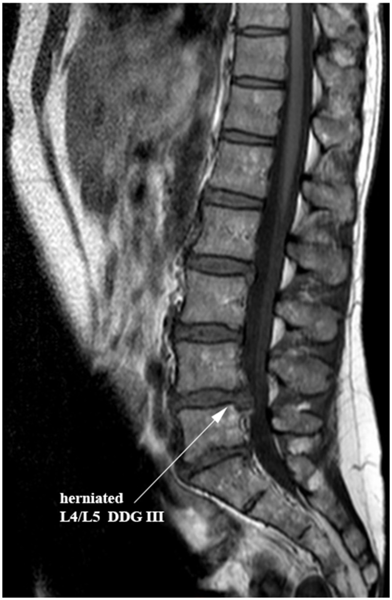
MRISagittal lumbar MRI: L4-L5 disc herniation, arrow on the herniation compressing the adjacent nerve root.
Source : Mern et al., PLoS ONE, 2017, figure 1 · CC BY
Femoral neuralgia, also called crural neuralgia or, more precisely, high lumbar radiculopathy, is the radicular pain that arises from irritation or compression of one of the upper lumbar nerve roots: L2, L3 or L4. Where sciatica involves the posterior territory of the lower limb (L5-S1 roots), femoral neuralgia projects onto the anterior aspect of the thigh. This topographical contrast is not a teaching convenience: it is the physiotherapist's first reasoning tool when faced with thigh pain 1.
A definition anchored in topography
Femoral neuralgia is above all a diagnosis built on topography. The subjective distribution of the pain and the paraesthesia says more about the level involved than the objective neurological examination alone 1. The clearest data come from a surgical series of high lumbar disc herniations: in L2/L3 herniations, pain and/or numbness was located in the thigh above the knee in all patients; in L3/L4 herniations, 80 % described involvement of the medial aspect of the knee 1.
A simple, robust clinical rule can therefore be laid down: pain or numbness on the anterior aspect of the thigh, above the knee, is a specific sign of an L2 lesion; involvement of the medial aspect of the knee points towards L3 1. It is this anterior, proximal signature that separates femoral neuralgia from sciatica, which is posterior and runs below the knee.
Anatomy: the anterior course of the femoral nerve
The L2, L3 and L4 roots contribute to the femoral (crural) nerve, which runs in the anterior compartment of the thigh. That explains the signs looked for on examination. On the motor side, a high lumbar radiculopathy typically affects the quadriceps femoris (a deficit found in 27 to 70% of patients in a surgical series), the iliopsoas (60 to 95%) and, less consistently, the tibialis anterior (0–43 %) 1. The patellar reflex, which depends on L3-L4, may be abolished.
When it comes to neural tension testing, the test that matters is the femoral nerve stretch test (FNST, Léri's sign, sometimes called the “reverse Lasègue”), not the classic Lasègue. In the same series the Lasègue (straight leg raise) was positive in 13 to 87% of patients depending on the group, against 91–95 % for the femoral test 1. This hierarchy is the reverse of the one seen in sciatica, where the Lasègue is central.
An analysis of likelihood ratios confirms this trio: in patients with confirmed root involvement, the femoral test, its crossed version and an abolished patellar reflex are associated with a likelihood ratio ≥5 (up to infinity) for an L2, L3 or L4 lesion 2. These are precisely the manoeuvres the clinician calls on when faced with anterior thigh pain.
Key points
- Femoral neuralgia = L2-L4 roots, pain on the anterior aspect of the thigh (vs L5-S1 sciatica, which is posterior).
- Pain above the knee → think L2 ; medial aspect of the knee → think L3 1.
- The femoral test (Léri's sign), its crossed version and an abolished patellar reflex are the most discriminating manoeuvres 2.
- Possible motor deficit of the quadriceps, the iliopsoas, and more rarely the tibialis anterior.
Femoral neuralgia or sciatica: a distinction that structures everything
| Criterion | Femoral neuralgia (crural neuralgia) | Sciatica |
|---|---|---|
| Roots | L2 – L3 – L4 | L5 – S1 |
| Painful territory | The anterior aspect of the thigh, proximal to the knee (L2), medial aspect of the knee (L3) | The posterior aspect, running below the knee |
| Key neural tension test | Femoral nerve stretch (Léri's sign): 91-95% positive | Lasègue (straight leg raise) |
| Reflex assessed | Patellar (L3-L4) | Achilles (S1) |
| Muscles to test | Quadriceps, iliopsoas, tibialis anterior | Dorsiflexors, triceps surae |
Topographical distribution and examination data after Kido 2016 and Suri 2011 (operated high lumbar disc herniations).
Causes: the high disc herniation and its traps
The most frequently described cause is the high lumbar disc herniation (L1-L2, L2-L3 levels). These herniations are rarer than low ones and present differently: patients are significantly older and the incidence of autonomic dysfunction is higher, hence a clinical picture that is often atypical and less easy to attach to a precise level 3. That misleading quality makes it essential to keep the differential diagnoses in mind.
Two of them deserve particular vigilance:
- Diabetic lumbosacral radiculoplexus neuropathy (diabetic amyotrophy). In an older and/or diabetic patient with pain, weakness and wasting affecting mainly the proximal muscles of the thigh, asymmetrically, this should be considered rather than a simple discogenic femoral neuralgia. It typically begins unilaterally and focally in the leg, thigh or buttock, then spreads, involving motor, sensory and autonomic fibres, with associated weight loss and a monophasic course 4.
- Meralgia paraesthetica. This is a purely sensory mononeuropathy of the lateral femoral cutaneous nerve, causing paraesthesia and pain over the anterolateral aspect of the thigh, without motor deficit or loss of the patellar reflex, which clearly separates it from an L2-L4 lesion. Its diagnosis is essentially clinical 7.
Finally, even though serious spinal conditions (cancer, infection, fracture, cauda equina compression) remain rare in patients with spinal pain (2.5 to 5.1% in prospective studies, of which 0.0-2.1% cancers 8 ), they are reason enough never to reduce every thigh pain straight away to a run-of-the-mill discogenic femoral neuralgia.
Epidemiology: a real entity, but little studied in its own right
This has to be said plainly to the reader: femoral neuralgia suffers from a shortage of specific data. Most of what is known about the value of the clinical examination and about the natural history is extrapolated from lumbosacral radiculopathies in general, which are dominated by sciatica, and where diagnostic accuracy studies are themselves rare and heterogeneous 9. In that systematic review, the femoral nerve stretch test showed a sensitivity of 1.00 (CI 0.40-1.00) and a specificity of 0.83 (0.52-0.98), but drawn from a very small number of studies: a positive test strengthens the suspicion, yet confidence intervals that wide call for caution 9.
What can be stated without over-interpreting: femoral neuralgia corresponds to involvement of the high roots, less common than sciatica, arising on rarer high disc herniations and in older patients 3. For the rest, exact prevalence and risk factors of its own, the literature does not allow a verdict, and it would be dishonest to pretend otherwise.
This epistemic humility is not an admission of helplessness: on the contrary, it points towards a rigorous approach based on the anterior topography, a targeted examination (femoral test, patellar reflex, strength of the quadriceps and iliopsoas) and the elimination of differential diagnoses, before treatment is even discussed.
🔎 How do you recognise it on examination?
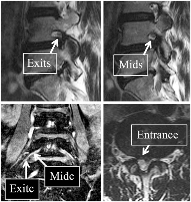
MRIProset sequence MRI, four slices: mild compression of the L4 nerve root within the nerve root canal, arrows on the narrowed areas.
Source : Ahn et al., Asian Spine Journal, 2016, figure 1 · CC BY-NC
💪 Where to look for the motor deficit
In high lumbar radiculopathy the deficit is not confined to the quadriceps: the iliopsoas is the most often affected.
Frequency of motor deficit by muscle (bars = midpoint of the reported range, exact values in the legend). Source: Kido et al., 2016 (PMID 27053156).
🔎 In femoral neuralgia it is Léri's sign that speaks, not the Lasègue
The femoral nerve stretch test (Léri / reverse Lasègue) is far more informative than the classic Lasègue in high lumbar radiculopathy.
Test positivity in a series of 58 operated high lumbar disc herniations. The Lasègue, so useful for sciatica (L5-S1), loses its value in femoral neuralgia: it is the femoral nerve that must be stretched. Source: Kido et al., 2016 (PMID 27053156).
Femoral neuralgia is a high lumbar radiculopathy involving the L2, L3 or L4 roots. The whole point of the clinical examination is to recognise this anterior and proximal picture and to tell it apart from sciatica (L5-S1), which is posterior and runs below the knee 1. The physiotherapist does not make a structural diagnosis, which is the role of imaging when it is indicated, but gathers a bundle of topographical, motor and reflex arguments. That bundle points to the level involved, supports the radicular hypothesis and, above all, helps rule out the differential diagnoses.
Anterior thigh pain, above the knee, signals femoral neuralgia; pain running down the back of the leg suggests sciatica.
Topography of the pain: the first clue
The subjective distribution of the pain and the paraesthesia is often more useful than the objective neurological examination alone for identifying the level involved in high lumbar herniations 1. The basic rule is simple: in femoral neuralgia the pain predominates on the anterior aspect of the thigh, and its exact height varies with the root involved.
- L2 lesion : pain and/or paraesthesia in the proximal thigh, above the knee, is present in every patient; it is a specific sign of an L2 lesion 1.
- L3 lesion : the pain reaches the medial aspect of the knee in 80% of patients, which points strongly to L3 1.
- L4 lesion : the territory extends towards the medial aspect of the leg.
This topographical reasoning stays indicative and does not amount to a diagnosis of level: the evidence comes from surgical series of lateral herniations, and overlapping territories are common. It guides the targeted examination that follows, without replacing it.
Léri's sign (reverse Lasègue): the key test
Faced with anterior thigh pain, the reference test is not the Lasègue of sciatica but the femoral nerve stretch test (FNST), historically called Léri's sign or the reverse Lasègue. With the patient prone (or side-lying, unaffected side down), the physiotherapist flexes the knee then extends the hip: tensioning the femoral nerve reproduces the anterior thigh pain when L2-L4 is involved.
Its superiority over the classic Lasègue in high radiculopathy is clear. In a series of 58 operated high lumbar disc herniations, the FNST was positive in 91–95 % of cases, against only 13 to 87% for the straight leg raising test depending on the group 1. In other words, in femoral neuralgia the Lasègue can be misleadingly negative: it is Léri's sign that has to be sought.
In patients whose root involvement has been confirmed, the femoral stretch test, its crossed version (reproduction of pain on the affected side during the contralateral manoeuvre) and an abolished patellar reflex are among the most discriminating manoeuvres for a mid-lumbar L2, L3 or L4 lesion, each associated with a likelihood ratio ≥ 5 that may tend towards infinity 2. These are precisely the manoeuvres the physiotherapist calls on when faced with anterior thigh pain.
The quadriceps deficit (and that of its neighbours)
The motor examination looks for weakness in the L2-L4 myotomes. The quadriceps femoris (knee extension, locking the knee going up and down stairs, standing up from a chair) is the emblematic target, but it is neither the most consistent nor the only one affected. In Kido's series, weakness involved:
| Muscle tested | Function | Frequency of weakness 1 |
|---|---|---|
| Iliopsoas | Hip flexion | 60-95 % |
| Quadriceps femoris | Knee extension | 27-70 % |
| Tibialis anterior | Ankle dorsiflexion | 0-43 % |
In practice, an isolated quadriceps deficit is less common than is believed: the iliopsoas is in fact the most often affected 1. The physiotherapist therefore tests the whole L2-L4 chain, quantifies strength (manual muscle testing, comparison with the other side) and monitors progress, progressive motor worsening being one of the few reasons for prompt medical review.
The patellar reflex
The patellar reflex (L2-L4 reflex arc, mainly L3-L4) completes this triad. Its abolition or reduction on the affected side is one of the most informative signs: patellar reflex testing has shown a likelihood ratio ≥ 5 for involvement of the mid-lumbar roots 2. A brisk or normal patellar reflex does not rule out femoral neuralgia, but its asymmetrical abolition, combined with Léri's sign and the anterior topography, strongly reinforces the hypothesis of a high root lesion.
What the physiotherapist tests and interprets: the synthesis
No single sign makes the diagnosis on its own. It is the convergence (anterior topography + positive FNST + quadriceps or psoas weakness + abolished patellar reflex) that builds the presumption. The table below summarises the value of the manoeuvres, with their level of evidence.
| Examination finding | What it points to | Level of evidence |
|---|---|---|
| Anterior thigh topography | Localises L2 (proximal) vs L3 (medial knee) | Moderate: surgical series 1 |
| Léri's sign / FNST | L2-L4 root involvement | Moderate: LR ≥ 5 2 ; sensitivity and specificity fragile 9 |
| Crossed FNST | Reinforces root involvement | Moderate 2 |
| Quadriceps / psoas deficit | Confirms the myotome affected | Low: frequency highly variable 1 |
| Abolished patellar reflex | L2-L4 involvement | Moderate: LR ≥ 5 2 |
Caution is needed on the performance figures. Evidence specific to femoral neuralgia is thin: most data are extrapolated from lumbosacral radiculopathies in general, where diagnostic accuracy studies are rare and heterogeneous 9. In that review the FNST showed a sensitivity of 1.00 (CI 0.40-1.00) and a specificity of 0.83 (0.52-0.98), but drawn from a very small number of studies, and confidence intervals that wide mean a positive test has to be taken as an argument, not as proof. It has to be said plainly, to the patient as in the notes: these manoeuvres strengthen the suspicion, they do not demonstrate it.
Do not confuse: the differential diagnoses to rule out
Recognising femoral neuralgia also means knowing when the picture is not one. Three traps deserve to be second nature for the physiotherapist.
- Meralgia paraesthetica : a mononeuropathy that is purely sensory , affecting the lateral femoral cutaneous nerve: paraesthesia and pain over the anterolateral aspect of the thigh, without motor deficit or loss of the patellar reflex 7. The absence of a motor sign and of a reflex sign is what separates it here from an L2-L4 lesion.
- Diabetic amyotrophy (lumbosacral radiculoplexus neuropathy): in an older or diabetic patient, pain with weakness and wasting of the proximal muscles of the thigh, often asymmetrical and preceded by weight loss, should suggest not a herniation but this plexopathy; it starts focally then spreads, involving motor, sensory and autonomic fibres 456.
- Atypical high lumbar herniation : L1-L2 and L2-L3 herniations are rarer than low ones: patients are significantly older and more often show autonomic dysfunction, hence a picture that is sometimes hard to attach to a precise level 3.
Key points
- Anterior territory of the thigh: proximal = L2, medial aspect of the knee = L3 1.
- Léri's sign (FNST), not the Lasègue: positive in 91-95% of high herniations; its crossed version and an abolished patellar reflex carry an LR ≥ 5 12.
- L2-L4 motor deficit : psoas most often affected (60-95%), quadriceps 27-70% 1.
- Caution : specific evidence is thin and confidence intervals are wide, so the tests point, they do not prove 9.
- Differentials : meralgia paraesthetica (purely sensory), diabetic amyotrophy (older or diabetic patient), atypical high herniation 753.
🚩 Differential diagnosis and red flags
Pain over the anterior aspect of the thigh is not synonymous with discogenic femoral neuralgia. Before attaching the picture to an L2-L4 high lumbar radiculopathy, the physiotherapist has to rule out the great mimics methodically (hip disease, meralgia paraesthetica, diabetic amyotrophy, psoas mass) and spot the signals that call for a medical opinion or imaging. The compass remains the neurological examination: in a true root lesion, the femoral nerve stretch test (Léri's sign / reverse Lasègue), its crossed version and an abolished patellar reflex show likelihood ratios ≥5, up to infinity, for an L2, L3 or L4 lesion 2. Conversely, anterior thigh pain without quadriceps deficit, with a preserved patellar reflex and a negative femoral stretch, should make the diagnosis be reconsidered.
Key points
- Femoral neuralgia = anterior thigh pain (L2-L4), with a positive femoral nerve stretch test and often a deficit of the quadriceps or the iliopsoas 1.
- A picture that is purely sensory, with no motor deficit and no change in the patellar reflex, points to meralgia paraesthetica, not to an L2-L4 root 7.
- In an older or diabetic patient with weight loss and proximal weakness and wasting, think of diabetic amyotrophy rather than a herniation 4.
- Serious conditions are rare (2.5-5.1% of patients with low back pain), but the absence of a red flag is not fully reassuring: 64% of spinal tumours showed none at all 8.
The hip, the first mechanical mimic
Hip disease (osteoarthritis, impingement, peri-articular tendon disease) readily projects groin and anterior thigh pain that overlaps the L2-L3 territory. Clinically, the direction is set at the bedside: pain reproduced by passive hip movement (flexion with internal rotation), restricted joint range, no neurological disturbance. That is precisely the opposite of the radicular profile described above. Here, what the confirmed data contribute is indirect but decisive: the neurodynamic manoeuvres and the patellar reflex act as discriminators. A negative femoral nerve stretch test, normal quadriceps strength and a brisk, symmetrical patellar reflex push a mid-lumbar root lesion away 2 and bring the attention back to the joint. In practice, coherence between topography, mechanical provocation of the hip and the neurological examination settles matters better than any isolated test, all the more so because the diagnostic accuracy evidence for the neurological examination itself remains thin and heterogeneous 9.
Meralgia paraesthetica: a purely sensory trap
This is the differential most classically confused with an L2 femoral neuralgia. Meralgia paraesthetica is a mononeuropathy that is purely sensory , affecting the lateral femoral cutaneous nerve: it produces paraesthesia and pain over the anterolateral aspect of the thigh, without motor deficit or loss of the patellar reflex 7. That is the whole difference from an L2-L4 lesion, where a deficit of the quadriceps (27-70%) and of the iliopsoas (60-95%) and an altered patellar reflex are expected 1. The diagnosis of meralgia is essentially clinical, and its management follows a graded ladder beginning with conservative treatment, then corticosteroid injections, with surgery reserved for failures 7.
The topographical landmark helps: L2 root pain sits on the anterior aspect of the thigh above the knee, and L3 pain on the medial aspect of the knee 1, whereas meralgia stays confined to a more superficial anterolateral territory. But topography alone is never enough: it is the absence of a motor and reflex signature that marks out meralgia.
Diabetic amyotrophy: do not confuse plexopathy with herniation
In an older and/or diabetic patient with thigh pain, quadriceps weakness and wasting, the reflex is to think of an L3-L4 herniation, and that is sometimes a mistake. Diabetic lumbosacral radiculoplexus neuropathy (Bruns-Garland diabetic amyotrophy) is a lumbosacral plexopathy that typically begins unilaterally and focally in the leg, thigh or buttock, then spreads, involving motor, sensory and autonomic fibres 4. It is associated with weight loss and affects mainly the proximal muscles of the thigh asymmetrically, usually in a monophasic way and often preceded by significant weight loss 6. The course is monophasic, with at least partial recovery, but with prolonged morbidity from pain and paralysis 5.
Two clues should alert the physiotherapist and prompt referral for a neurological opinion: unexplained weight loss and dysautonomia. This last point ties in with a useful fact: lumbar herniations located high (L1-L2, L2-L3) are common causes of femoral neuralgia; they occur in significantly older patients and come with a higher incidence of autonomic dysfunction, hence atypical presentations 3. In other words, in the age range where discogenic femoral neuralgia becomes rarer and more misleading, diabetic amyotrophy climbs the list of hypotheses.
Psoas mass and retroperitoneal disease
A mass developing in the psoas region (abscess, tumour, haematoma) can compress the lumbar plexus and the femoral nerve and mimic femoral neuralgia, with groin pain, an antalgic posture in hip flexion and a progressive deficit. The literature we can cite here does not deal specifically with these masses, and that has to be said plainly: the data available concern serious spinal conditions in general. They remain rare, with a prevalence of 2.5 to 5.1% among patients with low back pain in prospective studies, of which cancer 0.0-2.1% and infection 0.0-1.9% 8. But their rarity does not excuse a lack of vigilance: a motor deficit that progresses, night or rest pain, fever, a context of immunosuppression or a history of cancer should take the case out of the physiotherapy frame and trigger a work-up. A history of cancer is, moreover, the only red flag that genuinely raises the probability of a malignant cause 11.
Red flags: to be read with nuance, not as a green light
The key, counter-intuitive message is that the absence of a red flag is not fully reassuring. In a prospective evaluation, 64% of patients with a spinal tumour showed no red flag at all, and a negative answer to one or two screening questions does not significantly reduce the probability of serious disease 10. Conversely, taken in isolation, most red flags barely change the pre-test probability, hence the value of combining the signs before concluding 11. Two items do stand out all the same: recent trauma and age over 50 are associated with vertebral fracture 10, the prevalence of fracture being about 1% in primary care and 5% in secondary care 11.
| Clinical situation | Course of action | Level of evidence |
|---|---|---|
| Cauda equina syndrome (urinary retention, sphincter hypotonia) or major motor deficit | Refer urgently: surgery to be discussed without delay | Strong recommendation 12 |
| History of cancer, weight loss, fever, night or rest pain, trauma, age > 50 | Medical opinion + targeted imaging (MRI preferred) | Moderate 10 |
| Progressive neurological deficit or radicular pain persisting > 6 weeks despite treatment | Imaging and specialist opinion | Moderate 13 |
| Uncomplicated femoral neuralgia, stable examination, no red flag | Conservative management, no imaging from the outset | Low to moderate 13 |
When to image, when to refer
The rule is clear and reassuring for the patient: uncomplicated low back pain or radiculopathy is regarded as a benign, self-limiting condition that warrants no imaging from the outset 13. Imaging is discussed after about 6 weeks of medical treatment and physiotherapy without improvement, or in the presence of red flags raising the suspicion of cauda equina syndrome, cancer, fracture or infection 13. This waiting is legitimised by the favourable natural history of disc herniation: spontaneous resorption occurs in about two thirds of cases 14, all the more so when the herniation is large, at 96% for sequestrated fragments against 13% for bulges 15. When it is indicated, most guidelines prefer MRI to CT, bearing in mind that herniations are common in asymptomatic people, which exposes the clinician to false positives 12.
Two situations are exceptions to this waiting and call for referral without waiting the 6 weeks: cauda equina syndrome and a major motor deficit, which may require urgent surgery 12. Outside these emergencies, referral for epidural injection or surgery is considered only for persistent symptoms or a progressive neurological deficit, all the more so because a randomised trial in sciatica shows that early surgery speeds up relief but does not improve the outcome from one year onwards 16, an argument transposable, with caution, to femoral neuralgia, which is less studied in its own right.
The practical synthesis fits in one sentence: triage on the neurological examination and the context, stay conservative and without imaging in the uncomplicated form, but never read the absence of a red flag as a guarantee. Combining the signs, monitoring motor progress and reassessing at 6 weeks are the real safety net.
🌱 What is the prognosis? What you can tell the patient
🌱 The bigger the herniation, the better its chances of disappearing on its own
Counter-intuitive but solid: spontaneous regression on imaging is all the more common when the herniation is large and extruded.
Frequency of spontaneous regression by morphological type. Across all herniations, spontaneous resorption occurs in about two thirds of cases 14. A strong argument for conservative management. Source: Chiu et al., 2015 (PMID 25009200).
The question every patient with femoral neuralgia asks, sometimes without daring to, is simple: “Will it go away?” The honest answer is largely reassuring, provided it is not turned into a promise. Femoral neuralgia shares the natural history of lumbar radiculopathies caused by disc herniation: an underlying tendency to spontaneous improvement, over several weeks to several months. But the evidence specific to femoral neuralgia (L2-L4 roots) is thin: most of the solid data come from sciatica and from lumbar radiculopathies in general, and transpose only with caution. Telling the patient “it usually turns out well” is justified; guaranteeing a precise timescale or a certain cure is not.
A spontaneously favourable natural history
The central message is solid: most lumbar radiculopathies are self-limiting, symptoms subsiding over a few weeks to a few months, and clinical improvement goes hand in hand with morphological resorption of the herniation 17. That finding is the foundation that justifies management that is conservative first, over the first weeks to months, in most patients.
Quantitatively, a meta-analysis of 11 cohort studies puts the overall incidence of spontaneous resorption of a lumbar disc herniation, documented on CT or MRI, at about 66.66% (95% CI 51-69%); on that basis the authors suggest that conservative treatment could become the first-line choice 14. In other words, in nearly two out of three patients the herniation disappears by itself, with no surgical procedure.
These figures cover lumbar herniation as a whole and not femoral neuralgia alone: they are to be extrapolated with caution, since high L2-L4 radiculopathy is far less studied. But the biological logic, namely that the body resorbs the herniated fragment, has no reason to stop at the upper levels of the spine.
The bigger the herniation, the more it regresses: the reassuring paradox
The most counter-intuitive point when informing the patient deserves to be explained: the probability of spontaneous regression depends heavily on the morphological type of the herniation, and it is the largest and most migrated herniations that regress most often 15.
| Type of herniation | Spontaneous regression | Complete resolution |
|---|---|---|
| Sequestrated (free fragment) | 96 % | 43 % |
| Extruded | 70 % | 15 % |
| Protrusion | 41 % | – |
| Disc bulge | 13 % | – |
After Chiu 2015 (Clinical Rehabilitation). The larger and more migrated the herniation, the greater its chance of regressing.
This fact transforms the way a worrying MRI is discussed. A patient told they have a “large free-fragment herniation” often hears a sentence being passed; statistically it is on the contrary the situation with the greatest chance of resorbing on its own, at 96% regression and 43% complete disappearance for a sequestrated fragment. The landmark MRI follow-up study already confirmed it: migrated herniations shrink markedly or even disappear, whereas simple protrusions change little 18.
It is not the size of the herniation on the image that makes the prognosis, but the body's ability to resorb it, and it does so all the better when the herniation is large.
Timescales: clinical first, imaging second
A valuable detail for framing expectations: felt improvement often precedes visible improvement. In half of the improved cases in Komori's follow-up 18, the clinical signs settled before the MRI showed the herniation shrinking. In practical terms, the patient can feel better while “the image has not moved yet”, which argues against routine follow-up MRI and in favour of trusting the lived experience and the examination.
On timescales, the literature speaks of “weeks to months” 17 without a precise limit. That is also what structures the use of imaging: in uncomplicated radiculopathy, regarded as benign and self-limiting, no imaging is justified from the outset; it is discussed after about 6 weeks of medical treatment and physiotherapy without improvement, or when red flags appear 13. That 6-week threshold gives the patient an honest landmark: the natural history is given time to do its work before reassessment.
Key points: what you can tell the patient
- Most of the time it passes: about two thirds of lumbar herniations resorb spontaneously 14, over a few weeks to months 17.
- A large herniation is not bad news: sequestrated herniations regress in 96% of cases 15.
- Feeling better comes before the image: no routine follow-up MRI; the clinical picture is what counts 18.
- Operating early does not change the outcome at one year: the initial conservative option is legitimate 16.
- It cannot be predicted precisely: individual prognostic factors are uncertain 19.
Early surgery: relieved sooner, not better healed
Many patients ask whether it would not be better to “sort the problem out” surgically straight away. The reference data come from sciatica and extrapolate to femoral neuralgia: a randomised trial shows that early surgery speeds up relief, but that outcomes are comparable to those of prolonged conservative care from one year onwards, with no divergence in the second year 16. In other words, surgery buys time on the pain, not a better final result. That supports the choice of initial conservative management, keeping surgical referral for persistent symptoms, a progressive neurological deficit and, of course, emergencies (cauda equina syndrome, major motor deficit) 12.
Prognostic factors: acknowledging what we do not know
This is where the temptation to give a verdict has to be resisted. The patient wants to know “which way it will go”; the literature does not allow us to tell them reliably. A systematic review of non-surgically treated sciatica reports contradictory and above all negative results on the influence of initial pain intensity, neurological deficit, signs of root tension, symptom duration and imaging; age, sex, smoking, a history of sciatica and physically demanding work do not appear to influence the course, and the heterogeneity of the studies prevents any firm conclusion 19.
The honest position is therefore this: “The indicators we believe to be prognostic are not reliably so.” Neither the size of the herniation nor the initial intensity of the pain solidly predicts the future. This uncertainty is not an embarrassing admission of ignorance: it protects the patient from a falsely pessimistic prognosis (“your deficit is severe, this will take a long time”) that the data do not support.
What physiotherapy can, and cannot, promise
The favourable trajectory comes first from the natural history, not from the demonstrated superiority of any technique. A systematic review concludes that no type of conservative treatment (injections, traction, physiotherapy, rest) is clearly superior to the others in lumbosacral radicular syndrome 20. More severely still, a meta-analysis of 18 trials in sciatica finds no difference between physiotherapy and comparator for pain or disability, and concludes that the evidence is insufficient to formulate clinical recommendations 21. Since femoral neuralgia is studied even less, humility is called for.
That does not condemn rehabilitation: neural mobilisation (neurodynamics) added to management might improve pain, function and disability in the short term, with benefit found in 6 of the 8 trials analysed and no harmful effect 22, a meta-analysis quantifying a significant reduction in pain and disability 23 , but with high heterogeneity and data extrapolated from lumbar radiculopathies in general. The message to the patient is therefore twofold: physiotherapy actively accompanies a course that is usually favourable, but its own effect on the prognosis is not solidly demonstrated. Management that is active and humble.
Reassure without trivialising
Reassure, yes, but not at the price of “it is nothing”. Two safeguards. First, femoral neuralgia is not always a herniation: in an older or diabetic patient with pain, weakness and quadriceps wasting, diabetic amyotrophy (lumbosacral radiculoplexus neuropathy) has to be considered, monophasic in course but with prolonged morbidity 5. Second, although serious spinal conditions remain rare 8, the absence of red flags is not fully reassuring, since 64% of spinal tumours came with no red flag at all 10. The favourable prognosis is therefore stated within a frame: after ruling out what must be ruled out, and keeping the door open to reassessment if the expected course does not arrive.
🎯 What rehabilitation?
Let us say it straight away: femoral neuralgia suffers from a lack of evidence that is its own. Almost all the rehabilitation data we have come from sciatica (L5-S1) or from lumbar radiculopathies in general, and are then extrapolated to high lumbar radiculopathy (L2-L4). That extrapolation is reasonable, since the radicular mechanism and the natural history of the disc are the same, but it remains an extrapolation, and the honest physiotherapist says so to the patient. On that fragile base one solid certainty nonetheless stands out: the natural history is largely favourable, which makes active conservative management the legitimate first-line option in the absence of red flags.
Key points
- Evidence specific to femoral neuralgia is thin : we extrapolate mainly from sciatica and from lumbar radiculopathies.
- The solid foundation is not a technique, it is the favourable natural history : ~66% spontaneous resorption of disc herniations 14.
- Priority to staying active and to education; passive techniques are only adjuncts serving the return to activity.
- No conservative treatment has been shown to be clearly superior to the others 20.
- The neurodynamic approach is the best supported adjunct in the short term, but on heterogeneous data 22.
The foundation: natural history and conservative management
Before any technique, the therapeutic message rests on one fact: most lumbar radiculopathies get better on their own. Symptoms subside over a few weeks to a few months, and clinical improvement goes hand in hand with morphological resorption of the herniation 17. A meta-analysis of 11 cohorts puts spontaneous resorption at about 66.66% (95% CI 51-69%), which leads its authors to propose conservative treatment as the first-line choice 14.
A counter-intuitive fact the patient needs to hear: the more the herniation is large and migrated, the greater its chance of disappearing: 96% regression for sequestrated herniations, 70% for extruded ones, 41% for protrusions and only 13% for simple bulges 15. A worrying scan is therefore not an unfavourable prognosis, quite the opposite. And while early surgery speeds up relief in disc-related sciatica, it does not improve the outcome at one and two years 16 : data drawn from sciatica, but which supports the option of waiting and rehabilitating.
In femoral neuralgia the physiotherapist's best ally is no technique at all: it is the time that repairs the disc.
A cautious corollary: on the trajectory itself, reliable prognostic factors are hard to identify. A systematic review of non-operated sciatica finds contradictory and above all negative results for initial pain intensity, neurological deficit, signs of root tension, symptom duration and imaging; age, sex, smoking, a history of sciatica and physically demanding work do not appear to weigh 19. In other words, it is hard to predict who will do well quickly, all the more reason to support everyone actively.
Staying active and education
The first “treatment” is a principle of conduct: keep activity within the limits of pain and avoid deconditioning. Initial management of radiculopathy or sciatica, and by extension of femoral neuralgia, is conservative, except for signs calling for urgent surgery: cauda equina syndrome (urinary retention, reduced anal sphincter tone) or a major motor deficit 12. Epidural injection or surgery is discussed only for persistent symptoms or a progressive neurological deficit.
Education logically builds on the natural history (reassurance about disc resorption) and on the right use of imaging. Imaging is not indicated from the outset in uncomplicated low back pain or radiculopathy, regarded as benign and self-limiting; it becomes justified after ~6 weeks without improvement, or when red flags appear 13. Herniations are, moreover, common in asymptomatic people, hence a real risk of anxiety-provoking false positives 12. Education therefore carries, by implication, a note of vigilance: the absence of red flags is never fully reassuring, since 64% of spinal tumours showed none at all 10, and faced with an older or diabetic patient with quadriceps weakness and wasting, think of diabetic amyotrophy rather than a herniation 4.
Therapeutic exercise
Exercise is at the heart of active management, but its level of evidence in radicular pain is moderate to low and above all not specific to femoral neuralgia. Two readings coexist, and they must be presented without merging them.
On one side, a recent meta-analysis on lumbar disc herniation reports large effects of conservative physiotherapy on pain and function: an overall effect across all modalities of SMC = 2.28 (95% CI 1.51-3.05), exercise at SMC = 1.97 (95% CI 0.46-3.48) and traction leading at SMC = 2.52 (95% CI 1.57-3.37) 24. On the other, these spectacular effect sizes rest on marked heterogeneity and must be read with caution: they say that something favourable is happening, not that one modality is magic.
Manual therapy and neurodynamics
These two passive families are useful as adjuncts to the active return to movement, not as a treatment in their own right. It is neurodynamics (neural mobilisation) that has the most coherent file, while that file remains a limited one.
A systematic review concludes that neural mobilisation added to conservative management might improve pain, function and disability in the short term in low back and radicular pain: 6 of the 8 RCTs show a benefit on every outcome, and none observes a harmful effect 22. A meta-analysis points the same way, with marked effects on pain (Hedges' g = −1.097; 95% CI −1.482 to −0.712) and on disability (g = −0.964; 95% CI −1.475 to −0.453), but with high heterogeneity (I² ~85-89%) and data from general lumbar radiculopathy, not from femoral neuralgia alone 23. The technique is coherent with the clinical picture of femoral neuralgia, where the femoral nerve is involved: the femoral nerve stretch test (Léri's sign) is precisely the most informative manoeuvre in L2-L4 lesions 1.
| Modality | Level of evidence (radiculopathy / sciatica) | Specific to femoral neuralgia? |
|---|---|---|
| Neurodynamics (neural mobilisation) | Moderate in the short term, marked heterogeneity 2223 | No: extrapolated, but coherent with femoral nerve involvement |
| Therapeutic exercise | Large effects reported but heterogeneous 24 | No: lumbar herniation overall |
| Manual therapy / manipulation | Greatest reduction in leg pain in the short term, but very low confidence 25 | No |
| Traction | Highest effect reported (SMC 2.52) but very heterogeneous 24 | No |
| Staying active / education | Based on the favourable natural history 1417 | Extrapolated |
On manual therapy proper, the most recent network meta-analysis (50 RCTs, 4,920 participants) places spinal manipulation and exercise combined with neural mobilisation among the interventions producing the largest short-term reductions in leg pain, but with a level of confidence that is very low, the efficacy of non-surgical interventions remaining very uncertain 25.
What the evidence does not allow us to assert
It has to be said plainly to the reader rather than settled artificially. Two major reviews call for humility. First, no type of conservative treatment for lumbosacral radicular syndrome (injections, traction, physiotherapy, rest) is clearly superior to the others, and in the short term physiotherapy has not proved superior to an inactive treatment 20. Second, a systematic review with meta-analysis of 18 trials (2,699 patients) in sciatica finds no difference between physiotherapy and comparator for pain or disability, in the short, medium or long term, on data mostly at high risk of bias: the authors conclude that the evidence is insufficient to formulate clinical recommendations 21.
Active management, yes, but humble: the favourable trajectory owes itself first to the natural history, not to the demonstrated superiority of a technique.
The practical reading is not nihilistic. It is this: since femoral neuralgia usually resolves favourably on its own, and since no modality has shown clear superiority, rehabilitation is best when it is active, educational, low in iatrogenic risk and focused on the return to activity, with neurodynamics and manual therapy serving as adjuncts to relieve and to make movement possible. We offer, we relieve, we get people moving again, without overselling the certainty of an effect that, in femoral neuralgia precisely, has never been demonstrated on its own.
📋 What do the guidelines say?
Femoral neuralgia, a high lumbar radiculopathy of the L2-L3-L4 roots in which the pain and the paraesthesia predominate on the anterior aspect of the thigh 1 , suffers from a paradox: it is a common reason for consultation, but the evidence that is its own is thin. Most guidance is extrapolated from low back pain and from sciatica, which are better studied. The dominant message remains coherent: in the uncomplicated form, management is first conservative and active, with imaging and invasive procedures reserved for specific situations.
A spontaneously favourable natural history
The foundation of the whole first-line strategy is the prognosis. Most lumbar radiculopathies are self-limiting, symptoms subsiding over a few weeks to a few months, and clinical improvement goes hand in hand with morphological resorption of the herniation 17. A meta-analysis of 11 cohorts puts spontaneous resorption of a lumbar disc herniation at about 66.66 % of cases under conservative treatment 14, which leads its authors to suggest conservative care as the first choice.
A counter-intuitive fact worth explaining to the patient: the larger and more migrated the herniation, the more it regresses. The spontaneous regression rate reaches 96% for sequestrated (free) herniations, 70% for extrusions, 41% for protrusions and only 13% for disc bulges; complete disappearance occurs in 43% of sequestrations and 15% of extrusions 15. The landmark MRI follow-up study confirms it: migrated fragments shrink markedly or even disappear, whereas simple protrusions barely move, and in half of the improved cases, the clinical improvement precedes the radiological one 18. These data concern lumbar radiculopathy in general and are to be extrapolated with caution to L2-L4 femoral neuralgia, which is less specifically studied.
Early surgery: sooner, not better
Should surgery be done early to go faster? The reference randomised trial in disc-related sciatica answers clearly: early surgery speeds up relief, but the results are comparable from one year onwards between surgery and prolonged conservative care, with no divergence in the second year 16. In other words, the operation buys time, not a better medium-term result. That supports the initial conservative option, on data drawn from sciatica and extrapolated to femoral neuralgia. Surgery or epidural injection is considered only for persistent symptoms or a progressive neurological deficit, and urgent surgery is discussed only in the face of cauda equina syndrome (urinary retention, anal sphincter hypotonia) or a major motor deficit 12.
Physiotherapy: act, but with humility about the evidence
This is where the rule of honesty applies. The level of evidence for physiotherapy interventions in radicular pain is low. A systematic review of 18 trials (2,699 patients) in sciatica finds no difference between physiotherapy and comparator for pain or disability, in the short, medium and long term, on data that are mostly at high risk of bias and very heterogeneous; its authors conclude that there is not enough evidence to formulate clinical recommendations 21. Earlier still, no conservative modality (injections, traction, physiotherapy, rest) proved clearly superior to the others in lumbosacral radicular syndrome 20. So it has to be owned: the favourable trajectory comes first from the natural history, not from any demonstration that one technique is superior.
Some avenues nonetheless remain encouraging, subject to heterogeneity. Neural mobilisation (neurodynamics) added to conservative treatment might improve pain, function and disability in the short term: 6 of the 8 RCTs in one review show a benefit on every outcome, and none observes a harmful effect 22. A meta-analysis finds a significant reduction in pain (Hedges' g = −1.10) and in disability (g = −0.96) in lumbar radiculopathy, but with high heterogeneity (I² ≈ 85-89%) and data that are not specific to femoral neuralgia 23. In disc herniation, a meta-analysis reports large effects of traction (SMC = 2.52), of exercise (SMC = 1.97) and of combined modalities (SMC = 2.28), again to be interpreted with caution given the marked heterogeneity 24. Finally, a network meta-analysis (50 RCTs, 4,920 participants) places spinal manipulation and exercise + neural mobilisation at the top for short-term leg pain, but with a level of confidence that is very low : the efficacy of non-surgical interventions remains very uncertain 25.
| Element of management | What the evidence says | Level |
|---|---|---|
| Stay conservative as first-line (outside red flags) | Favourable natural history, frequent spontaneous resorption; early surgery = faster but no better at 1 year | Solid (extrapolated from sciatica/herniation) |
| Neural mobilisation / neurodynamics | Possible short-term benefit on pain and disability, but marked heterogeneity | Moderate to low |
| Physiotherapy “in general” vs comparator | No difference demonstrated; evidence insufficient to recommend | Low |
| Prognostic factors (initial pain, imaging, deficit, etc.) | Contradictory and mostly negative results; firm conclusions impossible | Low |
On the individual prognosis it is better, moreover, to stay modest: a systematic review of non-operated sciatica identifies no reliable factor, since initial pain intensity, neurological deficit, signs of root tension, symptom duration and imaging give contradictory results, while age, sex, smoking, a history of sciatica and physically demanding work do not appear to influence the course 19.
Imaging: rarely from the outset
The rule is constant: low back pain or radiculopathy that is uncomplicated is benign and self-limiting, and warrants no imaging at the outset 13. Imaging, MRI rather than CT, is discussed after about 6 weeks of medical treatment and physiotherapy without improvement, in the case of a progressive neurological deficit, or when red flags appear 1312. Restraint is all the more justified because disc herniations are common in asymptomatic people: imaging too early risks finding a “culprit” that is not one 12.
Red flags: necessary but imperfect
Serious spinal conditions remain rare : a review of 41,320 patients finds a prevalence of 2.5 to 5.1% in prospective studies (cancer 0.0-2.1%, infection 0.0-1.9%, cord or cauda equina compression 0.1-1.9%) 8. Even so, false reassurance has to be avoided: 64% of patients with a spinal tumour showed no red flag, and a negative answer to one or two screening questions does not significantly reduce the probability of serious disease 10. Recent trauma and age > 50 were associated with fracture. The lesson: red flags are to be combined, they are not used in isolation. Taken alone, most barely change the probability; a history of cancer is the most informative for a malignant cause (post-test probability 33%). The prevalence of fracture is about 1% in primary care and 5% in secondary care, and that of cancer about 0.5% in primary care 11.
Do not confuse: the differentials to keep in mind
Not every anterior thigh pain is a discogenic femoral neuralgia. Two clinical traps dominate:
- Diabetic amyotrophy (lumbosacral radiculoplexus neuropathy): in an older and/or diabetic patient, pain with weakness and wasting that is proximal in the quadriceps, beginning focally or asymmetrically in the thigh then spreading, often preceded by weight loss, points to this diagnosis rather than to a herniation 456. The course is monophasic, with at least partial recovery but prolonged morbidity.
- Meralgia paraesthetica : a mononeuropathy that is purely sensory , of the lateral femoral cutaneous nerve, producing anterolateral paraesthesia and pain without motor deficit or loss of the patellar reflex, which distinguishes it from an L2-L4 lesion. Clinical diagnosis, graded management: conservative first, then corticosteroid injection, surgery as a last resort 7.
Remember too that lumbar herniations located high (L1-L2, L2-L3), common causes of femoral neuralgia, affect significantly older patients and more often come with autonomic dysfunction, hence a picture that is sometimes atypical and harder to attach to a precise level 3. The targeted examination remains the best compass: the femoral nerve stretch test (Léri's sign), its crossed version and an abolished patellar reflex are the most discriminating manoeuvres for an L2-L4 lesion 2 , even if their accuracy rests on very few studies with wide confidence intervals 9.
Education and staying active: the real lever
In the absence of a demonstrably superior passive technique, the most robust axis is educational. Explaining the favourable natural history, namely that most herniations regress, all the more so when they are large, makes it possible to reassure without trivialising and to defuse the fear of movement. The aim is to offer management that is active but humble : staying active within the limits of pain, reassurance grounded in the prognosis, and referral for imaging or a surgical opinion only if symptoms persist beyond a few weeks, if the deficit progresses, or when combined red flags are present.
Key points
- First-line = conservative. Favourable natural history; spontaneous resorption ≈ 66% 14, all the more frequent when the herniation is large or migrated 15.
- Early surgery = faster, not better at 1 year 16 ; emergency reserved for cauda equina or a major motor deficit 12.
- Imaging is not routine : after ~6 weeks of failure or when red flags appear 13.
- Red flags to be combined : 64% of spinal tumours have none 10 ; serious conditions rare, 2.5-5.1% 8.
- Physiotherapy evidence is low : tell the patient about the uncertainty 21 ; neurodynamics = a short-term avenue, heterogeneous 22.
- Think of the differentials : diabetic amyotrophy and meralgia paraesthetica.
🗂️ What do concrete case reports teach us?
Nothing replaces reasoning at the patient's bedside. To anchor the data presented above, here is a published case, with its PubMed identifier. It was chosen because it documents precisely the scenario this chapter wants to make recognisable: a femoral neuralgia that was not one, and that resisted for a year. Every decision taken in it rests strictly on the evidence already cited: triage of red flags, the hierarchy of the clinical examination, then therapeutic progression. The aim is not to provide a rigid protocol, but to show how a physiotherapist puts together topography, discriminating tests and natural history in order to act without over-medicalising.
Published case: a “femoral neuralgia” treated for a year that was an osteoid osteoma
The case. Saçaklıdır and colleagues report a man aged 36 years who presented with low back pain radiating to the right lower limb for a year26.
What had already been tried. Physiotherapy, platelet-rich plasma injections, pregabalin, duloxetine. Only indometacin relieved him26.
The diagnosis. A radiograph of the femur was requested, and it made the diagnosis of an osteoid osteoma of the proximal femoral diaphysis26. The authors recall that this benign bone tumour readily arises from the metaphyseal and diaphyseal regions of long bones, that pain is often its first symptom, and that it can mimic many diseases26.
What that changes in a session, concretely. Two signals were present and transpose as they stand to the clinic. The first is the duration : a year of radicular pain that yields to nothing is not a radiculopathy doing badly, it is a hypothesis to revisit. The second is the selective response to non-steroidal anti-inflammatory drugs, which is a classic feature of osteoid osteoma and which the patient reports spontaneously if asked what relieves them.
The physiotherapist does not have to make this diagnosis. What they do have to do is notice that a well-conducted treatment has been failing for too long, and say so to the referrer rather than renewing the sessions.
Key points: what this published case teaches
- Always screen for red flags before mechanical reasoning, and never take their absence alone as reassurance 1011.
- In an older or diabetic patient with weight loss and a proximal deficit, think of diabetic amyotrophy, not only of a disc herniation 46.
- The discriminating examination in femoral neuralgia = Léri's sign, patellar reflex, anterior distribution, not the Lasègue 12.
- With no red flag, the route is conservative, and the natural history favourable ; there is no rush to imaging or surgery 141613.
- We act with humility : the evidence specific to femoral neuralgia is thin and often extrapolated from sciatica 921.
These two trajectories, imaginary once again, do not claim to cover the real diversity of patients. They are a reminder of what matters: the same complaint, “I have pain at the front of my thigh”, may cover a benign radiculopathy destined to settle on its own or a condition that demands an entirely different path. Clinical discernment, more than any protocol, is what makes the difference.
🧭 How is this applied in practice?
Once the diagnostic reasoning is done, what matters most to the physiotherapist remains: what to do, in what order, and with what words. This section sets out a field algorithm, the key messages to pass on to the patient, the common pitfalls and the criteria for referring on. Two ideas run through it. First, femoral neuralgia calls, in the vast majority of cases, for first-line conservative management, carried by a favourable natural history. Second, the evidence specific to femoral neuralgia is thin: most of the data are extrapolated from lumbosacral radiculopathies or from sciatica, and we shall say so to the patient as much as to the reader.
A three-step algorithm
The practical approach is organised in three steps: screen, treat, reassess.
Step 1: screen (red flags and differential diagnoses). Before any rehabilitation, rule out the emergency. Cauda equina syndrome (urinary retention, reduced anal sphincter tone, saddle anaesthesia) or a major motor deficit call for urgent medical referral, as they may justify surgery without delay 12. Also look for signs suggesting a serious underlying condition (cancer, fracture, infection), which remain rare: a systematic review covering 41,320 patients with low back pain finds a prevalence of serious spinal disease of 2.5 to 5.1% in prospective studies, including 0.0 to 2.1% of cancer and 0.1 to 1.9% of spinal cord or cauda equina compression 8. One caution, though: the absence of a red flag is not fully reassuring. In a prospective evaluation, 64% of patients with a spinal tumour had no red flag at all, and a negative answer to one or two screening questions does not significantly lower the probability of serious disease 10. Reasoning should therefore rest on combinations of signs rather than question by question: a history of cancer is the most informative red flag (post-test probability of malignancy 33%, CI 22-46%), whereas most isolated signs barely change the probability 11. Recent trauma and age over 50 years point towards fracture 10.
Two differential diagnoses deserve a particular reflex when faced with anterior thigh pain. In an older and/or diabetic patient with pain, weakness and proximal wasting of the quadriceps, consider diabetic amyotrophy (lumbosacral radiculoplexus neuropathy) rather than a herniation: it begins focally and asymmetrically in the thigh, the leg or the buttock and then spreads, is associated with weight loss and affects motor, sensory and autonomic fibres 456. When faced with anterolateral thigh paraesthesia without motor deficit or loss of the patellar reflex, think of meralgia paraesthetica, a purely sensory mononeuropathy of the lateral femoral cutaneous nerve 7.
Step 2: confirm clinically and treat. The picture of femoral neuralgia is confirmed on examination. The distribution points to the level: pain or numbness on the anterior aspect of the thigh above the knee = a specific sign of L2; the medial aspect of the knee = suggestive of L3 1. The most discriminating manoeuvres are the femoral nerve stretch test (Léri's sign / reverse Lasègue), its crossed version and an abolished patellar reflex: in patients with confirmed root involvement, each is associated with a likelihood ratio ≥ 5 for an L2-L4 lesion 2. In a series of operated high herniations, the femoral nerve stretch test was positive in 91 to 95% of cases against 13 to 87% for the classic Lasègue 1 : it is therefore this test, and not the Lasègue of sciatica, that should be favoured here. Some measure is still called for: a systematic review reports for this test a sensitivity of 1.00 (CI 0.40-1.00) and a specificity of 0.83 (0.52-0.98), but drawn from a very small number of studies with very wide confidence intervals 9. A positive test strengthens the suspicion; it does not prove it.
In the absence of a red flag and of a major deficit, management is active and conservative. Reasonable options include exercise, neural mobilisation and manual techniques, alongside a return to activity. Neural mobilisation (neurodynamics) added to usual care might improve pain, function and disability in the short term in low back and radicular pain 22, with a meta-analysis finding a significant reduction in pain (Hedges' g = −1.097; CI −1.482 to −0.712) and in disability 23. These figures call for caution: high heterogeneity (I² ~85-89%) and data extrapolated from lumbar radiculopathies in general.
Step 3: reassess. Set a reassessment horizon at around 6 weeks. Imaging is not indicated from the outset in uncomplicated radiculopathy, which is regarded as benign and self-limiting; it becomes justified after about 6 weeks of medical treatment and physiotherapy without improvement, or in the face of red flags 1312. If the symptoms recede, carry on. If they persist beyond that, or if a neurological deficit progresses, refer on (see below).
Key messages to pass on to the patient
Education is a cornerstone, all the more so as the prognosis works in your favour.
- “This is a nerve problem, not a broken leg.” Femoral neuralgia is a high radiculopathy (L2-L4) in which the anterior thigh pain is explained by irritation of a root, to be distinguished from posterior sciatica 1.
- “The body often repairs itself.” Most radiculopathies are self-limiting within a few weeks to months, with clinical improvement accompanying resorption of the herniation 17. A counter-intuitive fact worth explaining: the larger and the more migrated the herniation, the more it regresses, at 96% for sequestrated fragments, 70% for extrusions, 41% for protrusions and 13% for bulges 15.
- “An MRI right away would not change much.” Early imaging is not recommended in uncomplicated radiculopathy 1312, all the more so as herniations are common in people with no symptoms.
- “Moving is safe and desirable.” Early surgery speeds up relief but does not improve the outcome at one and two years compared with conservative care in sciatica 16 : there is no rush to operate on an uncomplicated presentation.
- Honesty about uncertainty. Telling the patient that physiotherapy often relieves symptoms but that the evidence remains limited is more accurate, and sturdier in the long run, than promising a result.
Common mistakes to avoid
- Using the Lasègue as the reference test. In femoral neuralgia it is the femoral nerve stretch test that is informative (91-95% positivity against 13-87% for the Lasègue): relying on the Lasègue alone leads to under-diagnosis 1.
- Confusing femoral neuralgia with sciatica. The distribution settles it: anterior/medial (L2-L4) against posterior running below the knee (L5-S1) 1.
- Requesting or pressing for imaging too early. Outside red flags and a progressive deficit, it is not indicated before ~6 weeks and exposes the patient to false positives 1312.
- Feeling reassured by the absence of red flags. 64% of spinal tumours presented none at all 10 : stay alert to how things evolve.
- Forgetting the neurological differentials. Do not put every thigh pain down to a herniation: diabetic amyotrophy in the older or diabetic patient 5, meralgia paraesthetica when the picture is purely sensory 7. High herniations, moreover, affect older patients, with more autonomic dysfunction and an often atypical picture 3.
- Overselling a technique. No conservative treatment has shown clear superiority over the others in lumbosacral radicular syndrome 20, and the overall evidence for physiotherapy in sciatica is judged insufficient for firm recommendations 21. Active management, but humble.
When to refer on?
Referring on is decided on simple criteria, to be sorted according to urgency.
| Situation | What to do | Support |
|---|---|---|
| Cauda equina syndrome, major motor deficit | Medical referral, urgent (surgery possible) | Jensen 2019 |
| Combined red flags (history of cancer, trauma, age > 50 years, deterioration in general condition) | Medical opinion / targeted imaging | Premkumar 2018; Downie 2013 |
| No improvement after ~6 weeks of well-conducted treatment | Medical reassessment, imaging can be considered | Hutchins 2021 |
| Progressive neurological deficit, refractory pain | Discussion of epidural injection / surgery | Jensen 2019 |
| Suspected diabetic amyotrophy or meralgia paraesthetica | Referral for diagnostic confirmation | Dyck 2001; Ahmed 2025 |
An honest prognostic nuance is called for: the factors usually invoked (initial pain intensity, deficit, tension signs, duration, imaging) give contradictory and above all negative results for predicting the course, and age, sex, smoking and the physical demands of work do not appear to influence it 19. We cannot yet predict finely who will recover quickly, which is one more reason to reassess regularly rather than give a prognosis from the outset.
Key points
- Screen, treat, reassess. Rule out cauda equina and a major deficit, then first-line conservative care, with reassessment at ~6 weeks.
- The right test for anterior thigh pain is the femoral nerve stretch test (Léri's sign), not the Lasègue 12.
- Reassure with facts : ~66.7% spontaneous resorption of herniations 14, identical outcomes at one year between early surgery and conservative care 16.
- No imaging from the outset outside red flags or failure at 6 weeks 13, but stay alert: their absence is not enough to reassure 10.
- Think of the differentials : diabetic amyotrophy, meralgia paraesthetica.
- Stay humble : thin specific evidence, extrapolated from sciatica 21 (active management, but without overselling it).
Bibliography
Each reference checked individually on PubMed (clickable PMID). 25 sources. Click a superscript note marker in the text: the bibliography opens and highlights the source.
- Kido T, et al. (2016). Journal of Orthopaedic Science. PMID 27053156. doi:10.1016/j.jos.2016.03.003.
- Suri P, Rainville J, Katz JN, et al. (2011). Spine (Phila Pa 1976). PMID 20543768. doi:10.1097/BRS.0b013e3181c953cc.
- Lee DS, et al. (last author Park MS) (2013). Journal of Korean Neurosurgical Society. PMID 24379943. doi:10.3340/jkns.2013.54.5.379.
- Dyck PJ, et al. (1999). Neurology. PMID 10599791. doi:10.1212/wnl.53.9.2113.
- Dyck PJB, Norell JE, Dyck PJ (2001). Brain. PMID 11353735. doi:10.1093/brain/124.6.1197.
- Bhanushali MJ, et al. (2008). Neurology India. PMID 19127036. doi:10.4103/0028-3886.44814.
- Ahmed MS, Varrassi G, Hadjiconstanti D, Zis P (2025). Pain and Therapy. PMID 39673032. doi:10.1007/s40122-024-00693-4.
- Galliker G, et al. (2020). The American Journal of Medicine. PMID 31278933. doi:10.1016/j.amjmed.2019.06.005.
- Tawa N, Rhoda A, Diener I (2017). BMC Musculoskeletal Disorders. PMID 28231784. doi:10.1186/s12891-016-1383-2.
- Premkumar A, et al. (2018). The Journal of Bone and Joint Surgery. American Volume. PMID 29509613. doi:10.2106/JBJS.17.00134.
- Downie A, Williams CM, Henschke N, Hancock MJ, Ostelo RWJG, de Vet HCW, et al. (2013). BMJ. PMID 24335669. doi:10.1136/bmj.f7095.
- Jensen RK, et al. (2019). BMJ. PMID 31744805. doi:10.1136/bmj.l6273.
- Hutchins TA, et al. (2021). Journal of the American College of Radiology. PMID 34794594. doi:10.1016/j.jacr.2021.08.002.
- Zhong M, et al. (2017). Pain Physician. PMID 28072796.
- Chiu CC, Chuang TY, Chang KH, et al. (2015). Clinical Rehabilitation. PMID 25009200. doi:10.1177/0269215514540919.
- Peul WC, et al. (2008). BMJ. PMID 18502911. doi:10.1136/bmj.a143.
- Casey E (2011). Physical Medicine and Rehabilitation Clinics of North America. PMID 21292142. doi:10.1016/j.pmr.2010.10.001.
- Komori H, Shinomiya K, Nakai O, Yamaura I, Takeda S, Furuya K (1996). Spine (Phila Pa 1976). PMID 8720408. doi:10.1097/00007632-199601150-00013.
- Ashworth J, Konstantinou K, Dunn KM (2011). BMC Musculoskeletal Disorders. PMID 21943339. doi:10.1186/1471-2474-12-208.
- Luijsterburg PAJ, Verhagen AP, Ostelo RWJG, van Os TAG, Peul WC, Koes BW (2007). European Spine Journal. PMID 17415595. doi:10.1007/s00586-007-0367-1.
- Dove L, Jones G, Kelsey LA, Cairns MC, Schmid AB (2023). European Spine Journal. PMID 36580149. doi:10.1007/s00586-022-07356-y.
- Peacock M, et al. (2023). The Journal of Manual & Manipulative Therapy. PMID 35583521. doi:10.1080/10669817.2022.2065599.
- Lin LH, et al. (2023). Life (Basel, Switzerland). PMID 38137856. doi:10.3390/life13122255.
- Thavarajasingam SG, et al. (2025). Brain & Spine. PMID 41209688. doi:10.1016/j.bas.2025.105632.
- Zhu Z, et al. (2025). The Journal of Pain. PMID 40373933. doi:10.1016/j.jpain.2025.105431.
- Saçaklıdır R, Huseynli L, Şencan S, Gündüz OH (2022). Proximal femur diaphysis osteoid osteoma mimicking lumbar radiculopathy. Turkish Journal of Physical Medicine and Rehabilitation. PMID 37674793. doi:10.5606/tftrd.2023.10653.
❓ Frequently asked questions
How do you tell femoral neuralgia from sciatica?
Femoral neuralgia is a high lumbar radiculopathy (L2-L3-L4 roots) in which the pain and the paraesthesia sit on the anterior aspect of the thigh, above the knee, whereas sciatica (L5-S1) is posterior and runs below the knee. Pain or numbness in the thigh above the knee is a specific sign of an L2 lesion, and the medial aspect of the knee points to an L3 lesion 1.
Which clinical test should be favoured when faced with anterior thigh pain?
The femoral nerve stretch test (Léri's sign, or reverse Lasègue) is far more informative than the classic Lasègue in L2-L4 lesions: in a series of operated high lumbar disc herniations it was positive in 91 to 95% of cases, against 13 to 87% for the straight leg raise 1. The femoral stretch test, its crossed version and an abolished patellar reflex carry a likelihood ratio ≥5 for a mid-lumbar lesion 2.
Does femoral neuralgia resolve on its own?
The natural history of lumbar disc herniation is mostly favourable: spontaneous resorption occurs in about 66.66% of cases 14, all the more so when the herniation is large: 96% for a sequestrated fragment, 70% for an extrusion, 41% for a protrusion and 13% for a bulge 15. This justifies first-line conservative management.
Should femoral neuralgia be operated on quickly?
No, except in an emergency. A randomised trial in sciatica shows that early surgery speeds up relief but leads to outcomes comparable to conservative care from one year onwards, with no divergence in the second year 16. Conservative treatment remains the first-line option, except where signs call for urgent surgery, such as cauda equina syndrome or a major motor deficit 12.
When should imaging (MRI) be requested?
Imaging is not indicated from the outset in uncomplicated radiculopathy, which is regarded as benign and self-limiting; it becomes justified after about 6 weeks of medical treatment and physiotherapy without improvement, or in the presence of red flags 13. One nuance deserves care: 64% of patients with a spinal tumour had no red flag at all 10, and serious conditions remain rare 8.
Which differential diagnoses must not be missed?
In an older or diabetic patient with pain and a proximal deficit of the lower limb, diabetic lumbosacral radiculoplexus neuropathy (diabetic amyotrophy) must be considered; it begins focally and asymmetrically in the thigh and then spreads 4. Meralgia paraesthetica is another key differential: a purely sensory mononeuropathy of the lateral femoral cutaneous nerve, with no motor deficit and no loss of the patellar reflex 7.

