

















In brief
Facet joint osteoarthritis, or lumbar facet joint arthropathy, is a degenerative and inflammatory condition of the facet (zygapophyseal) joints of the lumbar spine and a major cause of chronic low back pain. The pain sits low in the back, is made worse by extension, by standing and by walking, and is relieved by flexion and by sitting; imaging is not very specific and a definite diagnosis rests on medial branch anaesthetic blocks (the gold standard). Management is stratified: education, exercise and manual therapy first line, exercise being the cornerstone with no one type superior; radiofrequency neurotomy is reserved for refractory cases. It accounts for 15 to 45 % of chronic low back pain.
A clinical synthesis based on the ASIPP 2020 guidelines (Manchikanti), the Cohen 2020 multispecialty consensus, the MINT trial (Juch JAMA 2017) and the Hayden Cochrane 2021 exercise NMA.
Clinical synthesis
- Lumbar facet joint arthropathy is a major source of chronic low back pain, identified as the main aetiology in 15–45 % of patients across series (Cohen & Raja 2007, Manchikanti 2020).
- Its radiological prevalence rises sharply with age (very high after 60), but the correlation between imaging findings and the pain the patient feels is weak (Brinjikji 2015).
- Major risk factors: age, a high BMI, female sex, and above all prior disc degeneration (Kalichman 2007, Gellhorn 2013).
- The pathophysiology combines post-discal mechanical overload, cartilage breakdown, synovitis (IL-1β, IL-6, TNF-α mediators) and osteophyte formation that can lead to foraminal stenosis.
- The typical pain sits low in the back and is made worse by extension, prolonged standing and walking, and relieved by flexion and by sitting.
- A definite diagnosis rests only on controlled medial branch anaesthetic blocks, regarded as the gold standard (Cohen 2020).
- Imaging (MRI, CT, radiographs) is non-specific for facet pain because degenerative abnormalities are so prevalent in asymptomatic people (Brinjikji 2015).
- No clinical test on its own has sufficient diagnostic accuracy (Hancock 2007, Maas 2017). The reasoning must rest on a convergence of clues.
- Management is stratified: first education + exercise + manual therapy, then targeted interventions and radiofrequency neurotomy in refractory pain (Foster 2018, Cohen 2020).
- Exercise is the cornerstone, but no one type stands out consistently : the two reference network meta-analyses do not rank the same modalities first, on low-quality evidence (Owen 2020 BJSM, Hayden NMA J Physiother 2021). Patient adherence is what counts.
- Manual therapy is a useful adjunct to exercise for short-term relief (Coulter 2018 Spine J).
- Radiofrequency neurotomy of the medial branches gives relief lasting 6 to 24 months in good responders (Cohen 2020), but the MINT trial (Juch JAMA 2017) called its efficacy into question in an unstratified population, hence the importance of rigorous selection.
- Pain neuroscience education (PNE) is essential for addressing kinesiophobia and catastrophising (Louw 2011).
- Self-management is the key to preventing recurrence; physical exercise reduces the risk of recurrence by ~33 % (Shiri 2018).
- The physiotherapist must identify spinal red flags (Finucane 2020 IFOMPT) and refer urgently where necessary (cauda equina syndrome, fracture, malignancy, infection).
- In older people, the course can be complicated by degenerative lumbar spinal stenosis with neurogenic claudication, and that is the main prognostic turning point of the condition.
- Measuring outcomes with validated PROMs (ODI, RMDQ, NPRS) is crucial for showing progress and for calculating the minimal clinically important difference (MCID).
Contents
- What are the fundamentals to know about lumbar facet joint arthropathy?
- How do you assess and diagnose lumbar facet joint arthropathy with confidence?
- Which treatment strategies are the most effective for lumbar facet joint arthropathy?
- How do you secure a lasting recovery and prevent recurrence?
- Older people and degenerative lumbar spinal stenosis: when facet arthropathy changes its face
- What do real clinical cases teach us about facet arthropathy?
- How do you apply these recommendations concretely in your practice?
What are the fundamentals to know about lumbar facet joint arthropathy?
How is this condition defined, who is affected and what are the risk factors?
The facet (or zygapophyseal) joints are paired synovial joints formed by the inferior articular processes of one vertebra and the superior articular processes of the vertebra below. Together with the intervertebral disc they form the three-joint complex that provides segmental stability and guides mobility.¹,⁴ Like any synovial joint (knee, hip), they can develop an osteoarthritic process combining cartilage breakdown, synovial inflammation and subchondral bone remodelling.⁴ Facet arthropathy is a major cause of chronic low back pain : the estimates from the interventional literature put its prevalence at 15 to 45 % of patients with chronic low back pain, depending on how rigorous the diagnostic criteria are (controlled anaesthetic blocks in particular).²,³,⁵ This wide range reflects diagnostic heterogeneity: without comparative blocks, the risk of overdiagnosis is high (false positives can reach 30-40 %).³ The radiological prevalence of facet arthropathy rises sharply with age. The reference review by Kalichman & Hunter (Sem Arthritis Rheum 2007) brought together the available data: present in fewer than 60 % of people under 45, but almost universal in those over 60.⁴ The Brinjikji et al. meta-analysis (AJNR 2015) confirms that the prevalence of degenerative spinal abnormalities (including facet arthropathy) in asymptomatic people is very high and rises linearly with age, a decisive point for clinical interpretation.⁶ There is therefore a weak correlation between radiological severity and the intensity of the pain the patient reports.¹,⁴ 📈📊 Radiological prevalence of lumbar facet arthropathy by age band
A synthesis of the Kalichman 2007 and Brinjikji 2015 data on asymptomatic and all-comer cohorts
Indicative data brought together from the asymptomatic imaging cohorts. Source: Kalichman L, Hunter DJ. Semin Arthritis Rheum. 2007;37(2):69-80 (PMID 17379279); Brinjikji W et al. AJNR. 2015;36(4):811-816 (PMID 25430861). Clinical caveat: the radiological presence does not prove the source of pain; the correlation between imaging and symptoms stays weak.
- Advanced age is the strongest predictor ⏳: cumulative mechanical wear, proteoglycan changes in the cartilage, paravertebral sarcopenia and a loss of subchondral bone quality.⁴,⁷
- A high body mass index (BMI) 🍔: overweight and obesity increase the compressive loads on the lumbar spine and accelerate degeneration (with an additional metabolic effect through leptin and pro-inflammatory cytokines).⁴,⁷
- Female sex ♀: women show a higher prevalence and severity overall, possibly linked to hormonal factors (oestrogens after the menopause) and to biomechanical differences (facet orientation, lumbar lordosis).⁴
- Intervertebral disc degeneration 🎯: this is the major initiating factor. The loss of disc height transfers load onto the posterior facets and can overload them by 3 to 4 times; this biomechanical mechanism is central to the secondary development of facet arthropathy (Gellhorn, Katz & Suri, Nat Rev Rheumatol 2013).⁷
- Genetic factors and a general predisposition to osteoarthritis : the concordance with knee and hip osteoarthritis suggests a common background; patients with multi-site osteoarthritis more often have symptomatic facet arthropathy.⁴,⁷
- Repeated microtrauma and certain occupations that expose the lumbar spine to extension and rotation stresses (carrying loads, vibration, sports with hyperextension) can speed up the breakdown.¹,⁴
What happens in the body and how does facet arthropathy evolve naturally?
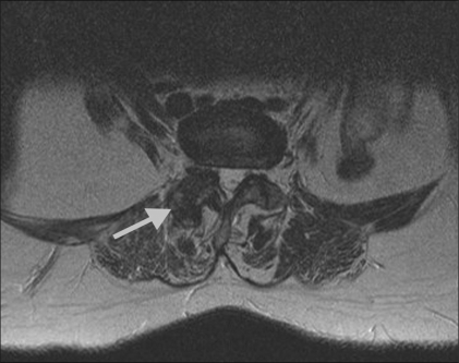
MRIAxial lumbar MRI: advanced right-sided facet joint osteoarthritis, arrow on the affected joint.
Source : Chaturvedi et al., The Indian Journal of Radiology & Imaging, 2009, figure 1 · CC BY
- Breakdown of the articular cartilage : thinning, fissuring, erosion of the surfaces. This breakdown is mediated by matrix metalloproteinases (MMP-3, MMP-13) and by ADAMTS-5.⁴,⁷
- Synovitis (synovial inflammation) 🔥: the synovial membrane thickens, becomes hypertrophic and releases pro-inflammatory mediators, interleukin-1β (IL-1β), interleukin-6 (IL-6) and tumour necrosis factor-α (TNF-α), which accelerate cartilage destruction and sensitise the local nerve endings (peripheral sensitisation).¹,⁷,⁸
- Subchondral bone remodelling and osteophyte formation : sclerosis, subchondral cysts and osteophytes around the joint, contributing to the facet hypertrophy seen on imaging.⁴,⁷
- Innervation of the facet 🧠: each facet joint receives a double innervation from the medial branches of the dorsal ramus of the spinal nerves at its own level and at the level above (key anatomy for the radiofrequency neurotomy strategy: a single facet level requires denervation of two medial branches).²,³
- Foraminal stenosis (narrowing of the intervertebral foramen and compression of the emerging root);
- Central or lateral canal stenosis (narrowing of the spinal canal);
- Degenerative spondylolisthesis (most often L4-L5) linked to facet incompetence.
🧬 The pathophysiological cascade of facet arthropathy
From post-discal biomechanical overload to degenerative lumbar spinal stenosis
A summary diagram based on Gellhorn AC et al. Nat Rev Rheumatol. 2013;9(4):216-224 and Perolat R et al. Insights Imaging. 2018;9(5):773-789 (PMC 6206372).
Key points
- Lumbar facet joint arthropathy accounts for 15–45 % of chronic low back pain (Cohen & Raja 2007, Manchikanti 2020).
- The radiological prevalence rises sharply with age (Kalichman 2007, Brinjikji 2015) but its correlation with pain is weak.
- The major initiating factor is disc degeneration, which overloads the posterior facets through the loss of intervertebral height (Gellhorn 2013).
- The pathophysiology combines mechanical overload, synovitis (IL-1β, IL-6, TNF-α) and bone remodelling with osteophyte formation.
- The course can lead to degenerative lumbar spinal stenosis and to neurological compression in older people, and that is the main prognostic turning point.
Bibliography
- Perolat R, Kastler A, Nicot B, Pellat JM, Tahon F, Attye A, et al. Facet joint syndrome: from diagnosis to interventional management. Insights Imaging. 2018;9(5):773-789. PMID 30090998.
- Manchikanti L, Kaye AD, Soin A, Albers SL, Beall D, Latchaw R, et al. Comprehensive Evidence-Based Guidelines for Facet Joint Interventions in the Management of Chronic Spinal Pain: ASIPP Guidelines. Pain Physician. 2020;23(3S):S1-S127. PMID 32503359.
- Cohen SP, Bhaskar A, Bhatia A, et al. Consensus practice guidelines on interventions for lumbar facet joint pain from a multispecialty, international working group. Reg Anesth Pain Med. 2020;45(6):424-467. doi:10.1136/rapm-2019-101243.
- Kalichman L, Hunter DJ. Lumbar facet joint osteoarthritis: a review. Semin Arthritis Rheum. 2007;37(2):69-80. PMID 17379279.
- Cohen SP, Raja SN. Pathogenesis, diagnosis, and treatment of lumbar zygapophysial (facet) joint pain. Anesthesiology. 2007;106(3):591-614. PMID 17325518.
- Brinjikji W, Luetmer PH, Comstock B, et al. Systematic literature review of imaging features of spinal degeneration in asymptomatic populations. AJNR Am J Neuroradiol. 2015;36(4):811-816. PMID 25430861.
- Gellhorn AC, Katz JN, Suri P. Osteoarthritis of the spine: the facet joints. Nat Rev Rheumatol. 2013;9(4):216-224. PMID 23147891.
- Schwarzer AC, Aprill CN, Derby R, Fortin J, Kine G, Bogduk N. The relative contributions of the disc and zygapophyseal joint in chronic low back pain. Spine. 1994;19(7):801-806. PMID 8202798.
- Foster NE, Anema JR, Cherkin D, Chou R, Cohen SP, Gross DP, et al. Prevention and treatment of low back pain: evidence, challenges, and promising directions. Lancet. 2018;391(10137):2368-2383. PMID 29573872.
How do you assess and diagnose lumbar facet joint arthropathy with confidence?
Which questions should you ask to understand the patient and their history?
The history is the first step in the clinical diagnosis, and the most valuable. Although no symptom is pathognomonic of facet arthropathy, certain features raise the diagnostic probability significantly when they converge. The suggestive clinical features:- Location of the pain 📍: low back pain, paravertebral, most often unilateral (sometimes bilateral). It can radiate towards the buttock, the hip or the back of the thigh but classically stops above the knee , an important point of difference from radiculopathy.²,⁴
- Character of the pain : a dull, poorly localised ache, sometimes described as a « tight depth ». Rarely sharp or stabbing.²,⁴
- Aggravating factors ⚠️: the pain worsens with the positions that load the facets, lumbar extension, hyperextension, prolonged standing, prolonged walking, and rotation or side bending towards the painful side. High heels, standing work and reaching up to open windows are classic provocative situations.²,⁴
- Relieving factors ✅: the pain eases in flexion (bending forward), on prolonged sitting, and with the hips flexed in bed (knees bent). These positions « open » the facet space and unload the joint.²,⁴
- Circadian rhythm : brief morning stiffness (generally < 30 minutes), start-up pain after a period of inactivity, improvement with moderate movement but worsening with overuse.²,⁴
- Course : chronic and fluctuating, with painful episodes alternating with quiet periods; no systemic neurological signs (no radiation below the knee, no sclerotomal paraesthesia).²,⁴
📋 Suggestive clues from the history (to be weighed, not pathognomonic)
A synthesis of Cohen 2020, Perolat 2018 and Maas 2017: modest diagnostic value in isolation, growing value in combination
Source: a synthesis of Maas ET et al. Eur J Pain. 2017;21(3):403-414 (PMID 27723170); Cohen SP et al. Reg Anesth Pain Med. 2020;45(6):424-467. Methodological note: the individual likelihood ratios stay modest; the diagnostic value comes from a coherent accumulation.
Which clinical tests should you perform and which other conditions should you rule out?
The physical examination aims to reproduce the familiar pain of the patient by loading the facet joints. The systematic reviews (Hancock 2007; Maas 2017) are unanimous, however: no single test has sufficient sensitivity and specificity to confirm or rule out a facet origin with certainty.¹,⁵ The usual tests (to be read in the overall context):- Palpation of the articular pillars 🖐️: deep palpation of the facets can reproduce the pain, but specificity is very low (many neighbouring structures are tender).
- Kemp test / quadrant test : extension + rotation + side bending towards the painful side to compress the facet. Sensitivity and specificity judged modest; reproducibility average.⁵
- Revel test (a pre-MBB cluster) : a set of accumulated history variables (age > 65, pain relieved by walking, and so on), of limited use in practice according to recent reviews.²,⁵
- Straight leg raise (Lasègue) : negative classically (no root compression), useful in the differential.
- Neurological examination : normal in pure facet arthropathy (strength, sensation and tendon reflexes preserved). Any neurological sign calls for the diagnosis to be reconsidered towards root compression or canal stenosis.
| Clinical test | Sensitivity | Specificity | Level of evidence |
|---|---|---|---|
| Paravertebral palpation | Moderate | Low | Low |
| Kemp test (extension-rotation) | Moderate | Low-moderate | Low |
| History cluster (Revel 1998) | Low | Low | Low |
| Imaging (MRI/CT) | High (abnormalities) | Very low (pain) | Low |
| Medial branch block (MBB) | ≥80 % | High | High (gold standard) |
- Rule out serious conditions (red flags): fracture, infection, malignancy, cauda equina syndrome;
- Characterise an associated foraminal or canal stenosis;
- Plan a guided intervention (injection, radiofrequency neurotomy).
🚩 Spinal red flags to rule out systematically (Finucane 2020, Verhagen 2016)
- Suspected vertebral fracture : recent trauma, known osteoporosis, long-term corticosteroid therapy, new pain in an older person.
- Suspected malignancy : a history of cancer, unexplained weight loss > 5 % in 6 months, non-mechanical night pain unrelieved by rest, progressive pain with no improvement.
- Suspected infection : fever, immunosuppression, IV drug use, recent infection, haemodialysis, intense and rapidly worsening pain.
- Cauda equina syndrome ⚡: saddle anaesthesia, sphincter disturbance (urinary retention, faecal incontinence), a bilateral lower limb motor deficit, reduced perineal sensation, a neurosurgical emergency.
- Progressive neurological deficit : worsening paraesthesia, motor weakness, hyporeflexia, urgent medical referral.
- Systemic inflammation : prolonged morning stiffness (> 1 h), pain improved by exercise (but not by rest), other joints involved, uveitis, psoriasis, think of spondyloarthritis.
⚠️ Any red flag calls for medical referral before or during physiotherapy management. Red flags taken in isolation have a low predictive value; it is their clustering and the context that signal urgency (Verhagen 2016, Finucane 2020 IFOMPT).
- Discogenic pain : worse in flexion and on prolonged sitting, relieved by partial extension, with possible radiation along the matching dermatome.
- Sacroiliac joint syndrome : pain located lower down (around the PSIS), specific provocation tests (the Laslett cluster: compression, distraction, sacral thrust, femoral thrust, Gaenslen).
- Myofascial syndrome : trigger points in quadratus lumborum, gluteus medius and piriformis.
- Lumbar radiculopathy : pain radiating below the knee, a neurological deficit (sensory, motor, reflexes).
- Degenerative lumbar spinal stenosis : neurogenic claudication (leg pain on walking, relieved by trunk flexion), a reduced walking distance, often a late complication of facet arthropathy.
- Hip pathology (hip osteoarthritis): groin pain, restricted flexion and internal rotation (FABER test).
- Inflammatory spondyloarthritis : an inflammatory picture with ASAS criteria, HLA-B27, syndesmophytes, sacroiliitis on MRI.
Should these patients be classified, and for what benefit?
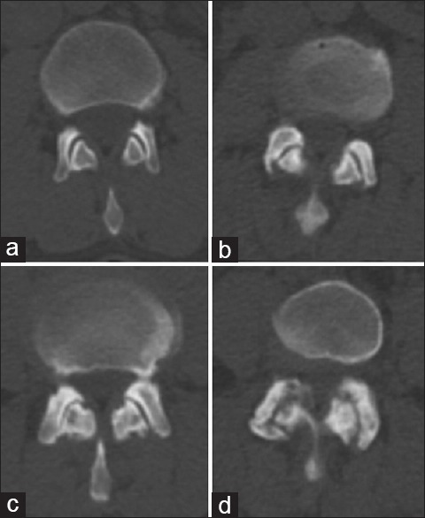
CT scanCT, four axial sections: severity scale of facet joint osteoarthritis, from normal (a) to severe (d).
Source : Shur et al., Indian Journal of Nuclear Medicine, 2015, figure 5 · CC BY-NC-SA
- Worsens in lumbar extension, on prolonged standing and on prolonged walking;
- Improves in flexion and on sitting (to be told apart from discogenic pain, which responds the other way round);
- Stays localised to the lower back or radiates little (rarely below the knee).
- Targeted treatment direction : exercises favouring lumbar flexion and motor control, avoiding prolonged hyperextension positions, working on hip mobility in flexion to unload the lower back.⁸,⁹
- Managing expectations : explaining to the patient that their pain follows mechanical patterns improves adherence and self-management (Foster 2018).⁸
- Stratified care : reserving the more invasive investigations (diagnostic blocks) for patients refractory to well-conducted conservative management for at least 6 to 12 weeks.²,³
Criticism and controversies: the diagnostic paradox
The diagnosis of facet arthropathy remains fertile ground for debate. Three major controversies deserve attention 🎯: 1. The gold standard paradox. Medial branch anaesthetic blocks, regarded as the gold standard, are themselves criticised: a high false positive rate (up to 38-40 % across meta-analyses), the need for comparative blocks to bring that rate down, limited access (an interventional radiologist or a trained PRM physician), cost and risks (infection, vascular or nerve injury, dural puncture).²,³ This creates a situation in which most diagnoses made in ambulatory practice remain « suspected diagnoses » based on clinical clusters with modest performance. 2. The gap between imaging and pain. Telling a patient about an MRI report that mentions « facet osteoarthritis » or « posterior joint hypertrophy » can generate a powerful nocebo effect: a belief in irreversible fragility, kinesiophobia, avoidance behaviour, withdrawal from work. Educating them about the real (often small) meaning of these findings becomes an essential therapeutic act.⁶,⁸ 3. The biopsychosocial dimension. As with any chronic low back pain, psychosocial factors (catastrophising, kinesiophobia, depression, expectations, occupational and medico-legal context) influence the prognosis at least as much as the anatomical ones. A strictly « anatomical » diagnosis of facet arthropathy is not enough to steer successful management (Foster 2018, Maher 2017).⁸,⁹Key points
- The diagnostic gold standard for facet arthropathy is the controlled medial branch anaesthetic block (MBB), with a reduction threshold of ≥ 80 % and comparative blocks (Cohen 2020, Manchikanti 2020).
- The suggestive clinical signs are: low paravertebral back pain, worse with extension and prolonged standing, relieved by flexion and by sitting, with no radiation below the knee (Perolat 2018, Maas 2017).
- Imaging is very poorly specific : most asymptomatic older people show signs of facet arthropathy (Brinjikji 2015).
- No single clinical test has sufficient accuracy (Hancock 2007, Maas 2017); the diagnosis rests on a convergence of clues.
- Spinal red flags to rule out systematically (Finucane 2020 IFOMPT); differential diagnosis: discogenic, sacroiliac, radiculopathy, canal stenosis, spondyloarthritis.
Bibliography
- Maas ET, Juch JNS, Ostelo RWJG, et al. Systematic review of patient history and physical examination to diagnose chronic low back pain originating from the facet joints. Eur J Pain. 2017;21(3):403-414. PMID 27723170.
- Cohen SP, Bhaskar A, Bhatia A, et al. Consensus practice guidelines on interventions for lumbar facet joint pain from a multispecialty, international working group. Reg Anesth Pain Med. 2020;45(6):424-467. doi:10.1136/rapm-2019-101243.
- Manchikanti L, Kaye AD, Soin A, et al. Comprehensive Evidence-Based Guidelines for Facet Joint Interventions in the Management of Chronic Spinal Pain: ASIPP Guidelines. Pain Physician. 2020;23(3S):S1-S127. PMID 32503359.
- Perolat R, Kastler A, Nicot B, et al. Facet joint syndrome: from diagnosis to interventional management. Insights Imaging. 2018;9(5):773-789. PMID 30090998.
- Hancock MJ, Maher CG, Latimer J, et al. Systematic review of tests to identify the disc, SIJ or facet joint as the source of low back pain. Eur Spine J. 2007;16(10):1539-1550. PMID 17566796.
- Brinjikji W, Luetmer PH, Comstock B, et al. Systematic literature review of imaging features of spinal degeneration in asymptomatic populations. AJNR Am J Neuroradiol. 2015;36(4):811-816. PMID 25430861.
- Schwarzer AC, Aprill CN, Derby R, et al. The relative contributions of the disc and zygapophyseal joint in chronic low back pain. Spine. 1994;19(7):801-806. PMID 8202798.
- Foster NE, Anema JR, Cherkin D, et al. Prevention and treatment of low back pain: evidence, challenges, and promising directions. Lancet. 2018;391(10137):2368-2383. PMID 29573872.
- Maher C, Underwood M, Buchbinder R. Non-specific low back pain. Lancet. 2017;389(10070):736-747. PMID 27745712.
- Finucane LM, Downie A, Mercer C, et al. International Framework for Red Flags for Potential Serious Spinal Pathologies. J Orthop Sports Phys Ther. 2020;50(7):350-372. PMID 32438853.
- Verhagen AP, Downie A, Popal N, Maher C, Koes BW. Red flags presented in current low back pain guidelines: a review. Eur Spine J. 2016;25(9):2788-2802. PMID 27376890.
Which treatment strategies are the most effective for lumbar facet joint arthropathy?
Where do you start? What is the recommended hierarchy of interventions?
The consensus hierarchy 🚶♂️ (Foster Lancet 2018, Cohen 2020, Manchikanti ASIPP 2020):🪜 Stratified hierarchy of interventions for lumbar facet pain
From the least invasive (education + exercise) to the most invasive (fusion surgery), with indicative levels of evidence
A hierarchy brought together from Cohen SP et al. Reg Anesth Pain Med. 2020;45(6):424-467 and Manchikanti L et al. Pain Physician. 2020;23(3S):S1-S127. Caveat: the line 2 and later interventions must stay tied to a continuing active rehabilitation programme.
What place does exercise hold and is there a superior approach?
Therapeutic exercise is the cornerstone 🏋️♂️ of the treatment of any non-specific chronic low back pain, and therefore a fortiori of facet arthropathy. The reference Cochrane review (Hayden et al., Cochrane Database Syst Rev 2021; CD009790, the most recent version to date) concludes that exercise is superior to no treatment, to placebo and to usual care on pain and on function.⁵ The most important conclusion of the literature of the past 10 years, however, is that no type of exercise has shown consistent superiority from one meta-analysis to another. The network meta-analysis by Owen et al. (BJSM 2020) compared 89 trials (5,578 patients): Pilates came top for pain, resistance training and motor control or stabilisation for function, resistance and aerobic work for mental health; stretching and the McKenzie method, by contrast, did not stand out from the control group, (note that an expression of concern was issued about this publication; the general conclusions remain consistent with other NMAs but must be read with nuance).⁶ ⚠️ The companion network meta-analysis to the Cochrane review (Hayden et al., J Physiother 2021, 217 trials, 20,969 patients) places Pilates, the McKenzie method and functional restoration ahead of the other types. The two rankings overlap only on Pilates, rest on low-quality evidence and on indirect comparisons: no type stands out consistently, and both teams conclude that the exercise to encourage is the one the patient enjoys and will keep up. The guiding principles therefore matter more than the specific technique:- Motor control and segmental stabilisation : the Cochrane review by Saragiotto 2016 (CD012004) confirms the efficacy of motor control exercise (MCE) targeting transversus abdominis and multifidus, with a modest but significant effect on pain and function.⁷
- Progressive strengthening : strengthening the trunk, the posterior chain (glutes, hamstrings) and the deep stabilisers. No single « winning » protocol.
- Graded exposure ✅: a progressive return to the movements and postures avoided through fear of pain, the basis of graded exposure and of graded activity.
- Moderate-intensity aerobic work : walking, cycling, swimming, which reduces central sensitisation and improves overall function.⁵
- Patient adherence = factor number one : choose an activity that means something to the patient and that they will enjoy keeping up in the long term. The best results come from compliance, not from the « perfect » technique.⁵,⁶
| Exercise modality | Effect on pain | Effect on function | Level of evidence |
|---|---|---|---|
| Motor control (MCE) | Moderate | Moderate | High (Cochrane) |
| Pilates | Moderate | Moderate | Moderate |
| McKenzie (MDT) | Moderate | Moderate | Moderate |
| General strengthening | Moderate | Moderate | High (Hayden 2021) |
| Yoga / Tai Chi | Moderate | Moderate | Moderate |
| Walking / aerobic work | Moderate | Moderate | Moderate |
| Aquatic therapy | Moderate | Moderate | Moderate (often in older people) |
Manual therapies and technologies: how effective are they really?
The manual therapy (joint mobilisation, high-velocity manipulation) is a useful adjunct to exercise. The meta-analysis by Coulter et al. (Spine J 2018) on chronic low back pain finds moderate quality evidence for a small reduction in pain (SMD -0.28; 95 % CI -0.47 to -0.09) and in disability (SMD -0.33; 95 % CI -0.63 to -0.03) at the end of treatment, against other active treatments; manipulation produces a larger effect there than mobilisation, and the authors judge multimodal programmes promising without having been able to pool them in a meta-analysis.⁸ It can help engage the patient in active rehabilitation by producing initial relief. The passive modalities (ultrasound, TENS, heat, electrotherapy) have weak to very weak evidence in the recent reviews.¹,² Their use, where it happens, must stay occasional, as an aid in managing pain peaks, and never replace the active component of treatment. ⚠️ There is a teaching risk: reaching routinely for these modalities can reinforce the patient's passive stance (« the physio has to cure me ») and hold back self-management.Beyond the physical: how do you educate the patient and address psychological factors?
🧠 Every modern biopsychosocial model (Foster 2018, NICE 2016, Kamper 2015) stresses that psychosocial factors are major predictors of chronicity and of functional impact, often more powerful than the anatomical factors.¹,⁹ Pain neuroscience education (PNE) is a validated approach that helps the patient understand that:- Pain is a complex production of the nervous system, not a simple indicator of tissue damage;
- The presence of abnormalities on imaging does not mean fragility;
- Movement and exercise modulate the perception of pain positively (the analgesic effect of exercise);
- Prolonged rest and avoidance are harmful in the medium and long term (deconditioning, kinesiophobia).
- Kinesiophobia (fear of movement), the TSK-11;
- Catastrophising , the Pain Catastrophizing Scale (PCS);
- Depressive or anxious symptoms , PHQ-9, GAD-7, HADS;
- Mistaken beliefs about pain, the Fear-Avoidance Beliefs Questionnaire (FABQ);
- Occupational context and compensation (blue and black flags).
Key points
- Management is stratified : line 1 = education + exercise + manual therapy, then increasingly invasive interventions according to the response (Foster 2018, Cohen 2020).
- No type of exercise stands out consistently : the two reference network meta-analyses do not rank the same modalities first; patient adherence and the principles of progressive loading are what count (Owen 2020 BJSM, Hayden NMA J Physiother 2021).
- The manual therapy is a useful adjunct to exercise for short-term relief (Coulter 2018). The passive modalities have limited efficacy.
- The radiofrequency neurotomy is effective in carefully selected patients (Cohen 2020), but the MINT trial (Juch JAMA 2017) calls for nuance: rigorous selection by comparative blocks is essential.
- Pain neuroscience education and addressing the psychosocial factors (kinesiophobia, catastrophising) are essential (Louw 2011, Kamper 2015).
Bibliography
- Foster NE, Anema JR, Cherkin D, et al. Prevention and treatment of low back pain: evidence, challenges, and promising directions. Lancet. 2018;391(10137):2368-2383. PMID 29573872.
- Cohen SP, Bhaskar A, Bhatia A, et al. Consensus practice guidelines on interventions for lumbar facet joint pain. Reg Anesth Pain Med. 2020;45(6):424-467. doi:10.1136/rapm-2019-101243.
- Manchikanti L, Kaye AD, Soin A, et al. ASIPP Guidelines: Facet Joint Interventions. Pain Physician. 2020;23(3S):S1-S127. PMID 32503359.
- Juch JNS, Maas ET, Ostelo RWJG, et al. Effect of Radiofrequency Denervation on Pain Intensity Among Patients With Chronic Low Back Pain: The MINT Randomized Clinical Trials. JAMA. 2017;318(1):68-81. PMID 28672319.
- Hayden JA, Ellis J, Ogilvie R, et al. Exercise therapy for chronic low back pain. Cochrane Database Syst Rev. 2021;9:CD009790. doi:10.1002/14651858.CD009790.pub2.
- Owen PJ, Miller CT, Mundell NL, et al. Which specific modes of exercise training are most effective for treating low back pain? Network meta-analysis. BJSM. 2020;54(21):1279-1287. PMID 31666220. (Note: an expression of concern was published; to be read alongside other NMAs.)
- Saragiotto BT, Maher CG, Yamato TP, et al. Motor control exercise for chronic non-specific low-back pain. Cochrane Database Syst Rev. 2016;1:CD012004. PMID 26742533.
- Coulter ID, Crawford C, Hurwitz EL, et al. Manipulation and mobilization for treating chronic low back pain: a systematic review and meta-analysis. Spine J. 2018;18(5):866-879. PMID 29371112.
- Kamper SJ, Apeldoorn AT, Chiarotto A, et al. Multidisciplinary biopsychosocial rehabilitation for chronic low back pain. BMJ. 2015;350:h444. PMID 25180773.
- Louw A, Diener I, Butler DS, Puentedura EJ. The effect of neuroscience education on pain, disability, anxiety, and stress in chronic musculoskeletal pain. Arch Phys Med Rehabil. 2011;92(12):2041-2056. PMID 22133255.
- Maher C, Underwood M, Buchbinder R. Non-specific low back pain. Lancet. 2017;389(10070):736-747. PMID 27745712.
- McCormick ZL, Vorobeychik Y, Gill JS, et al. Randomized prospective trial of cooled versus traditional radiofrequency ablation of the medial branch nerves for the treatment of lumbar facet joint pain. Reg Anesth Pain Med. 2019;44(3):389-397. PMID 30777903.
How do you secure a lasting recovery and prevent recurrence in lumbar facet joint arthropathy?
How do you make the patient an active player in their own recovery through self-management?
Self-management 💡 is the cornerstone of any lasting management. A patient-centred approach, aligned with the biopsychosocial model, turns the patient from a passive recipient into an active manager of their own health.³ The pillars of self-management:- In-depth therapeutic education : taking apart the mistaken beliefs (« my back is fragile », « I have osteoarthritis, so I must not move »), a realistic explanation of the limited meaning of imaging abnormalities, demystifying chronic pain.⁴,⁵
- A personalised independent exercise programme : a set of simple, repeatable home exercises the patient can keep up on their own. Favour enjoyable and adaptable approaches (Pilates, yoga, Tai Chi, Nordic walking, cycling, swimming).⁶
- Behavioural strategies : setting progressive SMART goals (Specific, Measurable, Achievable, Realistic, Time-bound), activity planning, graded exposure, stress management.⁵
- Managing flare-ups : preparing the patient to recognise and handle pain peaks without panicking or cutting back on activity, staying as active as possible.
- A support network : encouraging physical activity in groups (associations, clubs), favouring social interaction and avoiding isolation.
When and how do you plan a safe return to activity?
🏋️ The return to sport and to activities of daily living (ADLs) must follow a progression by functional criteria, not a rigid calendar. The general principles that apply:- Phase 1, pain control and restoration of functional range : moderate daily walking, gentle mobility, diaphragmatic breathing exercises, activation of the deep muscles (transversus abdominis, multifidus).⁶,⁷
- Phase 2, progressive strengthening and muscular endurance : dynamic stabilisation exercises, posterior chain strengthening (squat, hip thrust, deadlifts adapted to the tolerance threshold), progressive planks.
- Phase 3, reintroduction of specific loads : sporting or occupational movements reproduced progressively, with a reduced initial volume and controlled progression. The criterion for progressing: no significant increase in pain in the 24 h after the effort.
- Phase 4, return to full activity : volume and intensity increased gradually, with training load monitored to avoid overtraining.
🛡️ Reduction in the risk of low back pain recurrence through physical exercise
Shiri 2018 meta-analysis, Am J Epidemiol, 16 RCTs/NRCTs
Source: Shiri R, Coggon D, Falah-Hassani K. Am J Epidemiol. 2018;187(5):1093-1101 (PMID 29053873). A meta-analysis of 13 RCTs + 3 NRCTs.
Criticism and controversies: « core stability », adherence and the limits of the model
Two important nuances deserve attention ⚖️: 1. The concept of « core stability » has been both popularised and called into question. Presented at first as a central goal of lumbar rehabilitation (Hodges, Richardson, Jull in the 1990s and 2000s), it has recently been criticised for over-simplification. The aim is not to create a rigid muscular « corset », which even increases the compressive loads, but rather to promote adaptive muscular coordination and dynamic control of movement.⁶ Excessive focus on transversus abdominis alone is out of date. 2. Long-term adherence remains a major challenge. Exercise programmes have a preventive effect only if they are kept up. Adherence rates typically fall from 60-70 % at 3 months to under 30 % at 12 months. Strategies for improving adherence include building it into the daily routine, short and enjoyable exercises, regular follow-up (in person or by telehealth), positive feedback, and taking the patient's preferences into account.⁸Key points
- Self-management is the central target of lasting treatment: patient education + independent exercise + behavioural strategies (Foster 2018, Maher 2017).
- Regular physical exercise reduces the risk of low back pain recurrence by ~33 % (Shiri 2018).
- The return to activity is planned by functional criteria (mobility, control, pain after effort) and not by a rigid calendar.
- The aim is not a rigid trunk but adaptive dynamic coordination ; the concept of « core stability » has evolved towards a broader view.
- Long-term adherence remains challenge number one: enjoyable, short exercises, built into the routine, with regular follow-up.
Bibliography
- Hayden JA, Ellis J, Ogilvie R, et al. Exercise therapy for chronic low back pain. Cochrane Database Syst Rev. 2021;9:CD009790. doi:10.1002/14651858.CD009790.pub2.
- Foster NE, Anema JR, Cherkin D, et al. Prevention and treatment of low back pain. Lancet. 2018;391(10137):2368-2383. PMID 29573872.
- Kamper SJ, Apeldoorn AT, Chiarotto A, et al. Multidisciplinary biopsychosocial rehabilitation for chronic low back pain. BMJ. 2015;350:h444. PMID 25180773.
- Maher C, Underwood M, Buchbinder R. Non-specific low back pain. Lancet. 2017;389(10070):736-747. PMID 27745712.
- Louw A, Diener I, Butler DS, Puentedura EJ. The effect of neuroscience education on pain, disability, anxiety, and stress in chronic musculoskeletal pain. Arch Phys Med Rehabil. 2011;92(12):2041-2056. PMID 22133255.
- Saragiotto BT, Maher CG, Yamato TP, et al. Motor control exercise for chronic non-specific low-back pain. Cochrane Database Syst Rev. 2016;1:CD012004. PMID 26742533.
- Owen PJ, Miller CT, Mundell NL, et al. Which specific modes of exercise training are most effective for treating low back pain? Network meta-analysis. BJSM. 2020;54(21):1279-1287. PMID 31666220.
- Shiri R, Coggon D, Falah-Hassani K. Exercise for the prevention of low back pain: systematic review and meta-analysis of controlled trials. Am J Epidemiol. 2018;187(5):1093-1101. PMID 29053873.
Older people and degenerative lumbar spinal stenosis: when facet arthropathy changes its face
Why does this subgroup deserve a dedicated section?
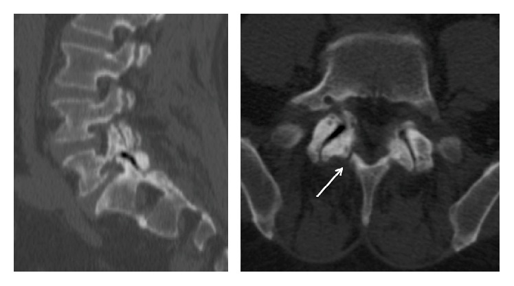
CT scanSagittal and axial lumbar CT: severe facet joint osteoarthritis with an intra-articular vacuum sign, the arrow marking an associated L5 isthmic lysis.
Source : Ruiz Picazo et al., Case Reports in Orthopedics, 2016, figure 3 · CC BY
- Pain, paraesthesia or weakness in one or both legs appearing on walking or on prolonged standing;
- Characteristic relief on trunk flexion (leaning forward on a trolley, uphill more easily than downhill, on a bicycle);
- A reduced walking distance, progressively disabling;
- A neurological examination often unremarkable at rest, but richer after exertion.
⚖️ Neurogenic versus vascular claudication, the differential diagnosis in older people
A crucial distinction in geriatrics, and the two can coexist
A clinical synthesis after Katz JN, Harris MB. Lumbar spinal stenosis. N Engl J Med. 2008;358(8):818-825.
- Sarcopenia : the age-related loss of muscle mass and strength, which worsens segmental instability (weak multifidus) and reduces exercise tolerance. EWGSOP2 criteria (grip strength, gait speed, lean mass).
- Frailty (the frailty syndrome): Fried criteria (weight loss, weakness, slowness, low activity, exhaustion), which raise the risk of falls and of dependency.
- Osteoporosis : a raised risk of vertebral fracture (new acute pain in an older person → a red flag).
- Multimorbidity and polypharmacy : drug interactions, the risk of falls (benzodiazepines, antipsychotics, antihypertensives).
- Physical deconditioning and a progressive loss of independence.
- Social isolation and depressive symptoms, which amplify the perception of pain.
How do you adapt the assessment and the management?
🎯 The physiotherapy management of an older person with facet arthropathy and/or lumbar stenosis must be adapted and global : A specific initial assessment:- Screen for sarcopenia (grip strength, gait speed < 1 m/s, chair stand test);
- Assess the risk of falls (Tinetti, Berg, Timed Up and Go > 13.5 s);
- Walking distance (a 6-minute walk test or an inclined treadmill test to unmask the claudication);
- A brief cognitive assessment (MMSE, MoCA) if cognitive decline is suspected as affecting adherence;
- Depressive symptoms (the Geriatric Depression Scale);
- A careful neurological examination (reflexes, strength, sensation, sphincters);
- Identification of the specific red flags (see the box).
🚩 Specific red flags in older people with facet arthropathy or suspected stenosis
- Cauda equina syndrome ⚡: saddle anaesthesia, sphincter disturbance, a bilateral motor deficit, a neurosurgical emergency.
- A progressive motor deficit (weak foot extension, plantarflexion, quadriceps), prompt neurosurgical referral.
- New pain in an older person, non-mechanical, at night, resistant to analgesics, rule out vertebral fracture, malignancy and infection.
- Unexplained weight loss, a deterioration in general condition, night sweats , a cancer work-up.
- A history of cancer (breast, prostate, lung, kidney, thyroid, myeloma), spinal metastases to rule out (MRI, bone scan).
- Suspected osteoporotic fracture : corticosteroid therapy, early menopause, a family history, a low-energy peripheral fracture.
- Suspected infection : fever, intense pain worse at night, immunosuppression, dialysis, recent spinal surgery.
- Systemic inflammatory symptoms (late-onset spondyloarthropathy, polymyalgia rheumatica), blood tests.
⚠️ The prevalence of serious conditions is higher in this age band, so heightened vigilance is essential (Finucane 2020, Verhagen 2016).
- Favour exercises in flexion (a « flexion-biased programme »): positions and movements that open the canal space (supine knees to chest, four-point kneeling in flexion, cycling, progressive lumbar flexion exercises). This is the Bodack & Monteiro approach, shown to be effective in lumbar stenosis.²
- Aerobic exercise on a cycle ergometer 🚴 (the flexed position is well tolerated): improves functional capacity without provoking the claudication.
- Multi-modal strengthening targeting the deep postural muscles (multifidus, transversus), the hip extensors (glutes), the trunk flexors and the stability of the cross chains, without excessive hyperextension.
- An anti-sarcopenia programme : progressive strengthening of the large muscle groups (adapted squats, leg press, resistance band exercises) with controlled progression.
- Fall prevention : balance exercises (tandem, single-leg, unstable surfaces), strengthening the dorsiflexors of the foot, Nordic walking with poles.
- Environmental adaptation and ergonomic advice : using a shopping trolley (an analgesic effect in flexion), grab rails at home, suitable footwear.
- A multidisciplinary approach : working with a geriatrician, a PRM physician, an occupational therapist, a dietitian (protein > 1.0-1.2 g/kg/day in older people to limit sarcopenia), and a psychologist where needed.
Key points
- The degenerative lumbar spinal stenosis is the major late complication of facet arthropathy in older people, and the leading indication for spinal surgery after 65.
- The typical clinical picture is intermittent neurogenic claudication, relieved by flexion (differential: vascular claudication).
- A global geriatric assessment is essential: sarcopenia, frailty, osteoporosis, polypharmacy, fall risk, cognition.
- Red flags are more frequent in this age band: heightened vigilance (Finucane 2020).
- Therapeutic strategy: flexion exercises favoured, prevention of sarcopenia and of falls, a multidisciplinary approach, preserving independence.
Bibliography
- Gellhorn AC, Katz JN, Suri P. Osteoarthritis of the spine: the facet joints. Nat Rev Rheumatol. 2013;9(4):216-224. PMID 23147891.
- Katz JN, Harris MB. Clinical practice. Lumbar spinal stenosis. N Engl J Med. 2008;358(8):818-825. PMID 18287604.
- Finucane LM, Downie A, Mercer C, et al. International Framework for Red Flags for Potential Serious Spinal Pathologies. J Orthop Sports Phys Ther. 2020;50(7):350-372. PMID 32438853.
- Verhagen AP, Downie A, Popal N, Maher C, Koes BW. Red flags presented in current low back pain guidelines: a review. Eur Spine J. 2016;25(9):2788-2802. PMID 27376890.
- Hayden JA, Ellis J, Ogilvie R, et al. Exercise therapy for chronic low back pain. Cochrane Database Syst Rev. 2021;9:CD009790. doi:10.1002/14651858.CD009790.pub2.
- Brinjikji W, Luetmer PH, Comstock B, et al. Systematic literature review of imaging features of spinal degeneration in asymptomatic populations. AJNR Am J Neuroradiol. 2015;36(4):811-816. PMID 25430861.
- Cruz-Jentoft AJ, Bahat G, Bauer J, et al. Sarcopenia: revised European consensus on definition and diagnosis (EWGSOP2). Age Ageing. 2019;48(1):16-31. PMID 30312372.
- Fried LP, Tangen CM, Walston J, et al. Frailty in older adults: evidence for a phenotype. J Gerontol A Biol Sci Med Sci. 2001;56(3):M146-M156. PMID 11253156.
What do real clinical cases teach us about lumbar facet joint arthropathy?
Analysis of a "classic" case: from assessment to conservative resolution
The typical management of a patient with facet arthropathy follows a course documented in several clinical series.¹,² A frequent clinical profile combines:- A patient aged 50-65, with a history of sedentary work or of prolonged standing;
- Chronic low back pain with flare-ups, paravertebral and unilateral;
- Worse on extension, on prolonged standing and on stairs;
- Relieved in flexion, on sitting, and lying with knees to chest;
- MRI showing facet arthropathy at L4-L5 / L5-S1, with no disc herniation and no significant root compression;
- A normal neurological examination.
- Patient education about the condition, taking apart the nocebo (« osteoarthritis visible on MRI »), explaining the dissociation between imaging and symptoms.³,⁴
- Joint mobilisation (Maitland grade III-IV techniques on the hypomobile segments, gentle lumbar flexion mobilisation, hip mobilisation).⁵
- A targeted exercise programme : motor control (transversus, multifidus), strengthening the hip extensors, stretching the hip flexors, lumbar flexion exercises (knees to chest, child pose, flexion in four-point kneeling).⁶
- Ergonomic advice : avoiding prolonged hyperextension, alternating positions, adjusting the workstation, progressive walking.
The diagnostic challenge: when facet arthropathy mimics a radiculopathy
🎯 Facet pain can radiate into the territory of a dermatome and generate a pseudo-radicular presentation, one of the great traps of the differential diagnosis. Several clinical series illustrate it: a patient with « sciatica » radiating into the buttock and the back of the thigh, with no objective neurological deficit, may in fact have pain of facet origin confirmed by a positive anaesthetic block.⁸ A particularly instructive case is that of facet synovial cysts (developed in section 5.3 below), which can cause a true radiculopathy through compression, illustrating the shifting boundary between « pure » facet pain and a radicular complication.⁸,⁹ The clinical clues that should suggest a facet origin in a « sciatica-like » presentation:- Radiation stopping above the knee;
- No neurological deficit (strength, sensation and reflexes preserved);
- Worse in extension and relieved in flexion (the opposite of discogenic radiculopathy);
- A negative Lasègue;
- MRI showing advanced facet arthropathy with no compressive disc herniation.
A complex case: the facet synovial cyst
🧩 The facet synovial cyst is a direct complication of advanced facet arthropathy: the hypertrophied synovial membrane can herniate as a cyst backwards, into the spinal canal, where it can compress the underlying nerve root and create a true radiculopathy. Several published cases illustrate this scenario.⁸,⁹ Arthur et al. (2012) report two women aged 52 and 54 with sciatica and a progressive neurological deficit, foot drop in one and calf weakness in the other, imaging revealing a large facet synovial cyst compressing a nerve root.⁸ Activity modification, gabapentinoids and NSAIDs having failed, both women eventually had to be operated on for excision of the cyst and root decompression. One of them was still receiving spinal manipulation while her deficit was setting in: the authors draw from it the rule to refer without delay for axial imaging and a surgical opinion any patient whose deficit is progressing under treatment. More encouraging: Scrofani et al. (Medicina 2024) published a comprehensive review with two illustrative cases of spontaneous resolution of lumbar synovial cysts under conservative treatment, without surgery.⁹ The authors count only nine cases of documented spontaneous resolution in the literature, to which their two observations are added. Rare though it is, this possibility justifies an initial conservative trial (possibly including a periradicular corticosteroid injection) in patients with no progressive deficit. In their two cases, the cyst had disappeared on the follow-up MRI within a few months.⁹Critique and controversy: the value and the limits of clinical cases
💡 Analysing clinical cases calls for methodological vigilance:- The weakest level of evidence (level 5 CEBM, very low GRADE);
- A major publication bias
- : the spectacular cases (complete resolution, atypical presentation) are over-represented;
- No generalisation is possible: a case illustrates, it never demonstrates efficacy; Where a spectacular case and a meta-analysis diverge,.
| Level of evidence (Oxford CEBM) | Type of study | Strength of the evidence |
|---|---|---|
| 1a | Meta-analysis of RCTs | Very high |
| 1b | A single good-quality RCT | High |
| 2a-2b | Cohort studies | Moderate |
| 3 | Case-control and cross-sectional studies | Low-moderate |
| 4 | Case series | Low |
| 5 | Case reports + expert opinion | Very low |
Key points
- The « classic » clinical picture combines paravertebral low back pain, worse in extension, relieved in flexion, with no neurological signs. Multimodal conservative management (education + exercise + manual therapy) resolves most uncomplicated cases in 8-12 weeks.
- Facet arthropathy can mimic a radiculopathy ; the key clues are radiation stopping above the knee, the absence of a neurological deficit, and the « extension-biased » pattern.
- The facet synovial cysts are a possible complication (Arthur 2012); a spontaneous resolution under conservative treatment is possible in some cases (Scrofani 2024, a review of nine published cases).
- A case report = level 5 evidence (the weakest). It illustrates, it never demonstrates efficacy; where it diverges from a meta-analysis, follow the meta-analysis.
Bibliography
- Cohen SP, Bhaskar A, Bhatia A, et al. Consensus practice guidelines on interventions for lumbar facet joint pain. Reg Anesth Pain Med. 2020;45(6):424-467. doi:10.1136/rapm-2019-101243.
- Perolat R, Kastler A, Nicot B, et al. Facet joint syndrome: from diagnosis to interventional management. Insights Imaging. 2018;9(5):773-789. PMID 30090998.
- Foster NE, Anema JR, Cherkin D, et al. Prevention and treatment of low back pain. Lancet. 2018;391(10137):2368-2383. PMID 29573872.
- Maher C, Underwood M, Buchbinder R. Non-specific low back pain. Lancet. 2017;389(10070):736-747. PMID 27745712.
- Coulter ID, Crawford C, Hurwitz EL, et al. Manipulation and mobilization for treating chronic low back pain: a systematic review and meta-analysis. Spine J. 2018;18(5):866-879. PMID 29371112.
- Saragiotto BT, Maher CG, Yamato TP, et al. Motor control exercise for chronic non-specific low-back pain. Cochrane Database Syst Rev. 2016;1:CD012004. PMID 26742533.
- Hayden JA, Ellis J, Ogilvie R, et al. Exercise therapy for chronic low back pain. Cochrane Database Syst Rev. 2021;9:CD009790. doi:10.1002/14651858.CD009790.pub2.
- Arthur B, Lewkonia P, Quon JA, Street J, Bishop PB. Acute sciatica and progressive neurological deficit secondary to facet synovial cysts: a report of two cases. J Can Chiropr Assoc. 2012;56(3):173-178. PMC 3430450.
- Scrofani R, De Simone M, Migliorini F, et al. Spontaneous Resolution of Symptomatic Synovial Cysts of the Lumbar Spine: A Comprehensive Review with Two Illustrative Cases. Medicina (Kaunas). 2024;60(7):1115. PMC 11278635.
- Manchikanti L, Kaye AD, Soin A, et al. ASIPP Guidelines: Facet Joint Interventions. Pain Physician. 2020;23(3S):S1-S127. PMID 32503359.
How do you apply these recommendations concretely in your practice?
When, and to which other health professionals, should you refer?
The physiotherapist, in direct access in particular, acts as a first-line clinician 🚩 whose first responsibility is to identify the patients who need a medical or further assessment.¹,² Triage rests on recognising « flags » of different colours. Red flags : signs and symptoms suggestive of a serious underlying condition, calling for prompt medical referral.¹,² The main spinal red flags were brought together in the international IFOMPT 2020 framework (Finucane et al.):- A history of cancer or unexplained weight loss;
- Fever, immunosuppression, IV drug use;
- Significant recent trauma, osteoporosis;
- A progressive or severe neurological deficit;
- Cauda equina syndrome (saddle anaesthesia, sphincter disturbance), an emergency;
- Non-mechanical night pain, unrelieved by rest;
- Systemic symptoms (sweats, chills).
- PRM physician / rheumatologist : refractory chronic pain, suspected spondyloarthritis, discussion of interventional options;
- Neurosurgeon / orthopaedic surgeon : degenerative lumbar spinal stenosis with a major functional impact, a progressive neurological deficit, segmental instability;
- Pain specialist : severe chronic pain, a candidate for radiofrequency neurotomy;
- Psychologist specialising in pain : severe kinesiophobia, catastrophising, comorbid depression, post-traumatic stress;
- Geriatrician : a frail older person with multimorbidity;
- Dietitian : a high BMI, sarcopenia (protein intake);
- Occupational therapist : adapting the workstation or the home environment.
How do you measure outcomes and overcome the barriers to implementation?
📈 Measuring progress objectively is an essential clinical act. The PROMs (patient-reported outcome measures) recommended for chronic low back pain:| PROM | Domain assessed | Range | MCID (minimal clinically important difference) |
|---|---|---|---|
| NPRS (Numerical Pain Rating Scale) | Pain intensity | 0-10 | ≈ 2 points (or a reduction of ≥ 30 %) |
| ODI (Oswestry Disability Index) | Functional disability | 0-100 | ≈ 10 points |
| RMDQ (Roland-Morris Disability Questionnaire) | Functional disability | 0-24 | ≈ 3-5 points |
| STarT Back | Risk of chronicity | 9 items | Risk stratification (low/medium/high) |
| TSK-11 | Kinesiophobia | 11-44 | ≈ 4 points |
| PCS (Pain Catastrophizing Scale) | Catastrophising | 0-52 | ≈ a 38 % reduction |
| EQ-5D-5L | Quality of life | Utility index | ≈ 0.08-0.10 |
- The 6-minute walk test (aerobic capacity, claudication in older people);
- Timed Up and Go (fall risk);
- The 30-second chair stand test (lower limb strength);
- Sit-and-reach (posterior chain flexibility).
- Audit and feedback 📊: the Ivers 2012 Cochrane review, collecting practice data and giving constructive comparative feedback.⁶
- Local opinion leaders and « champions »: respected peers who promote good practice.
- Interactive training (workshops, mentoring, simulation), more effective than simply circulating guidelines.
- Building PROMs into the workflow (digitised, automated).
- Shared decision-making with the patient: better adherence, satisfaction and outcomes.
Criticism and controversies: between the evidence ideal and pragmatic reality
⚖️ Several grey areas persist in applying the recommendations: 1. The « implementation gap » : we know what works, but research on how to build it into varied settings is still in its infancy. The strategies that work are often costly and hard to generalise. 2. The red flag paradox : useful but poorly specific in isolation, they can generate excessive medicalisation if applied mechanically. Probabilistic clinical reasoning remains more valuable than a checklist.² 3. Standardisation versus personalisation : PROMs and standardised care pathways are needed for research and for quality, but they do not always capture the patient's individual goals. Expert skill lies in navigating between these two poles.Key points
- Referral is crucial: red flags → prompt medical assessment; yellow flags → psychologist, biopsychosocial management; blue and black flags → occupational physician.
- PROMs recommended for chronic low back pain: NPRS (pain), ODI or RMDQ (function), TSK-11 (kinesiophobia), STarT Back (risk stratification).
- Know the MCID of each PROM so as to show a clinically significant improvement (ODI ≈ 10 points, NPRS ≈ 2 points, RMDQ ≈ 3-5 points).
- Overcome the barriers to implementation through audit and feedback (Cochrane Ivers 2012), local opinion leaders, interactive training and shared decision-making.
Bibliography
- Finucane LM, Downie A, Mercer C, et al. International Framework for Red Flags for Potential Serious Spinal Pathologies. J Orthop Sports Phys Ther. 2020;50(7):350-372. PMID 32438853.
- Verhagen AP, Downie A, Popal N, Maher C, Koes BW. Red flags presented in current low back pain guidelines: a review. Eur Spine J. 2016;25(9):2788-2802. PMID 27376890.
- Kamper SJ, Apeldoorn AT, Chiarotto A, et al. Multidisciplinary biopsychosocial rehabilitation for chronic low back pain. BMJ. 2015;350:h444. PMID 25180773.
- Foster NE, Anema JR, Cherkin D, et al. Prevention and treatment of low back pain. Lancet. 2018;391(10137):2368-2383. PMID 29573872.
- Maher C, Underwood M, Buchbinder R. Non-specific low back pain. Lancet. 2017;389(10070):736-747. PMID 27745712.
- Ivers N, Jamtvedt G, Flottorp S, et al. Audit and feedback: effects on professional practice and healthcare outcomes. Cochrane Database Syst Rev. 2012;6:CD000259. PMID 22696318.
- Louw A, Diener I, Butler DS, Puentedura EJ. The effect of neuroscience education on pain, disability, anxiety, and stress in chronic musculoskeletal pain. Arch Phys Med Rehabil. 2011;92(12):2041-2056. PMID 22133255.
- Cohen SP, Bhaskar A, Bhatia A, et al. Consensus practice guidelines on interventions for lumbar facet joint pain. Reg Anesth Pain Med. 2020;45(6):424-467. doi:10.1136/rapm-2019-101243.
- Hayden JA, Ellis J, Ogilvie R, et al. Exercise therapy for chronic low back pain. Cochrane Database Syst Rev. 2021;9:CD009790. doi:10.1002/14651858.CD009790.pub2.
What next after reading this?
This article is part of a collection of evidence-based clinical syntheses. A question, a comment, a correction to suggest? Contact us directly through the WhatsApp button at the bottom right of the screen.

