

















Clinical review of scoliosis (adolescent idiopathic and adult): how to screen for it and measure it, how to assess the risk of progression, and what bracing (the BrAIST trial) and specific exercises (Schroth) are really worth, without promising to “correct” a curve. Every reference has been checked individually on PubMed.
📝 In brief: clinical summary
- Scoliosis is a three-dimensional deformity of the spine confirmed by a Cobb angle ≥ 10° with axial vertebral rotation; the idiopathic form accounts for about 80% of cases 1.
- Adolescent idiopathic scoliosis (AIS) affects 1 to 3% of at-risk children aged 10 to 16 and predominates in girls, a predominance that sharpens with severity: the girl-to-boy sex ratio goes from 1.4:1 for curves of 10–20° to 7.2:1 beyond 40° 45.
- Screening rests on the Adams test coupled with the scoliometer: at a threshold of 7° of trunk rotation angle, sensitivity and specificity for a Cobb > 10° are about 83% and 87%; lowering the threshold to 5° takes sensitivity to 100% but makes specificity fall to 47% 2.
- The risk of progression depends on the size of the curve and on skeletal maturity (Risser sign): in the skeletally immature child (Risser 0–1), a curve of 20–29° progresses in 68% of cases, against 23% in the more mature child 8.
- For high-risk adolescent curves, the randomised BrAIST trial showed that a brace reduces progression to the surgical threshold: 72% success against 48% under observation alone, with a benefit that grows with the hours worn (up to 90–93% beyond 12.9 h/day) 14.
- Specific exercises (Schroth method, PSSE) added to standard care modestly improve the Cobb angle (−3.5° at 6 months; meta-analysis −3.18°): a recommended adjunct that slows worsening without “correcting” the curve 1516.
- Over the very long term, the natural history of untreated AIS is rather reassuring: survival comparable to the general population at 50 years, patients who stay functional, but chronic low back pain more frequent (61% vs 35%), which justifies simple monitoring of small curves 10.
🦴 What is scoliosis?
👧 The bigger the curve, the more female it is
The girl-to-boy ratio is almost balanced for small curves but becomes heavily lopsided for large ones, a useful marker for follow-up.
Girl-to-boy ratio by size of the curve. Adolescent idiopathic scoliosis affects 1 to 3% of 10-16 year-olds 4. Source: Konieczny et al., 2013 (PMID 24432052).
Scoliosis is one of the commonest spinal deformities met in practice, and one of the most poorly understood. Before discussing management, the physiotherapist needs a rigorous definition: scoliosis is neither “poor posture” nor a simple asymmetry noticed in the shower. It is a structural deformity of the spine, one that answers precise diagnostic criteria and whose prognosis depends closely on age and skeletal maturity.
A three-dimensional definition, with figures attached
Scoliosis is defined as a three-dimensional deformity of the spine. It combines a lateral curve in the frontal (coronal) plane, axial rotation of the vertebrae and a change in the sagittal profile. According to the Scoliosis Research Society, the diagnosis is confirmed when the Cobb angle is greater than or equal to 10° and axial rotation of the vertebrae is identifiable 1. This twin condition is essential: an isolated lateral curve, with no rotational component, is not a structural scoliosis but a postural scoliosis, reducible and benign.
The 10° threshold is not arbitrary: below it, the deviation falls within normal variation and is not considered pathological 2. That figure, measured on a radiograph of the whole spine, separates the merely asymmetrical from the genuinely scoliotic patient.
In aetiological terms, idiopathic scoliosis, of unknown cause, is set apart from secondary scoliosis. Idiopathic scoliosis accounts for about 80% of cases, the remaining 20% being secondary to another condition: congenital (vertebral malformation) or neuromuscular 1. This distinction is decisive for the physiotherapist: a secondary scoliosis follows a different logic, in which the deformity is only a symptom of an underlying disease.
Key points
- Scoliosis is a three-dimensional deformity: frontal curve + vertebral rotation.
- Diagnosis confirmed if Cobb angle ≥ 10° with identifiable rotation 1.
- Without rotation, this is a postural scoliosis that is reducible, not a structural scoliosis.
- About 80% of scoliosis cases are idiopathic and 20% are secondary (congenital, neuromuscular).
Adolescent idiopathic scoliosis (AIS)
The adolescent idiopathic scoliosis is the commonest form of structural spinal deformity 3. It is defined by a lateral deviation of the spine with a Cobb angle ≥ 10°, in the absence of congenital or neuromuscular abnormality, appearing around puberty. Its aetiopathogenesis remains unknown, and its natural history was long poorly understood 4.
AIS affects 1 to 3% of children in the at-risk population aged 10 to 16 4 and some reviews widen the range to 1–4% of adolescents in early puberty 3. The overall prevalence reported in the literature spans 0.47–5.2 % depending on the populations studied and the screening methods used 5. This wide variability calls for caution: there is no single figure, only a range that depends on the measurement threshold and on the counting method.
Epidemiology: a female predominance that sharpens with severity
The most useful figure in practice concerns sex. Small curves affect girls almost as often as boys, but the female predominance rises sharply with the size of the curve and the girl-to-boy ratio goes from 1.4:1 for curves of 10–20° to 7.2:1 for curves above 40° 5. In other words, it is above all the progressive and severe curves that concern girls.
This asymmetry has a direct clinical consequence: a girl in full growth, with a curve already established, deserves closer monitoring than a boy with a comparable curve, because her risk of progressing to a severe form is higher.
Screening without over-referring: the Adams test and the scoliometer
The physiotherapist is often first in line to spot a scoliosis. Clinical screening rests on the Adams test (forward bending of the trunk), which brings out the rib hump linked to vertebral rotation, completed by the scoliometer, which measures the angle of trunk rotation (ATR) 2. Thresholds of 5° to 7° of ATR have been proposed as criteria for referral to radiography.
The choice of threshold illustrates a fundamental trade-off between sensitivity and over-referral. With a threshold of 7° of rotation, sensitivity and specificity for a Cobb angle > 10° are about 83% and 87% ; lowering the threshold to 5°, sensitivity reaches 100% but specificity falls to 47%, at the price of many false positives referred needlessly for radiography 2. In a large screening study covering 33,596 girls, a 5° threshold referred 5.2% of children, against 1.4% at 7° and only 0.3% at 10°; the positive predictive value of a 5° threshold was no more than 28.3% for a scoliosis ≥ 10° 6.
| ATR threshold (scoliometer) | Referral rate to radiography | Trade-off |
|---|---|---|
| 5° | ≈ 5.2 % | Maximum sensitivity, many false positives (specificity ≈ 47%) |
| 7° | ≈ 1.4 % | Good balance (sensitivity ≈ 83%, specificity ≈ 87%) |
| 10° | ≈ 0.3 % | Few referrals but a risk of missing curves |
It has to be said plainly: the optimal threshold remains hard to set 6. The scoliometer points towards radiography; only measurement of the Cobb angle on film confirms the diagnosis. The physiotherapist should therefore present screening as a triage, not as a verdict.
What the physiotherapist needs to understand about the risk of progression
Not every scoliosis progresses. The risk of progression of an untreated scoliosis depends jointly on skeletal maturity (Risser sign), on the size and type of the curve, and on age 7. In the historical cohort of 727 patients of Lonstein and Carlson, 23.2% progressed, the incidence of progression being linked to the type and magnitude of the curve, to age at presentation, to the Risser sign and to menarcheal status 7.
The interaction between remaining growth and curve size is striking. In the skeletally immature child (Risser 0–1), a curve of 5–19° progresses in 22% of cases, against 68% for a curve of 20–29°; in the more mature child (Risser 2–4), those rates fall to 1.6% and 23% respectively 8. The same curve therefore carries a quite different prognosis depending on whether much or little growth is left.
A systematic review with meta-analysis identifies as factors of progression an initial severity > 25° of Cobb, an age at diagnosis < 13 years, a pre-menarcheal status and skeletal immaturity, but stresses that the predictive value of these markers remains limited and the level of evidence low so no method can be recommended as a reliable diagnostic criterion of progression 9. This is an uncertainty to own in front of families: children “at risk” are identified, but individual progression is not predicted with certainty.
A natural history more reassuring than was once believed
For a long time scoliosis was presented as a major threat to life and function. The 50-year follow-up of untreated idiopathic scolioses strongly qualifies that account: survival was comparable to that of the general population (estimated probability ≈ 0.55 against 0.57 expected) and patients remained productive and functional at a high level 10. However, chronic low back pain was more frequent than in controls: 61% against 35% 10. These data justify simple monitoring for small curves, with no dramatisation.
Scoliosis does not stop at adolescence: adult scoliosis
The physiotherapist also meets scoliosis in adults. Adult scoliosis (degenerative de novo, or an aged adolescent idiopathic scoliosis) is defined by a deformity in the coronal plane with a Cobb angle > 10° in a skeletally mature subject 11. It is a potentially disabling condition, whose frequency grows with ageing.
A meta-analysis (5 studies, 4,069 participants) puts the overall prevalence of de novo adult scoliosis at 37.6 %, higher in women (41.2%) than in men (27.5%), and strongly linked to age: 13% before the age of 60 against 36% after 12. Vertebral deformities affect 32 to 68% of people over the age of 65 11.
Contrary to a received idea, scoliosis can carry on progressing in adulthood, but slowly and in a linear fashion at about 0.82°/year for aged idiopathic scolioses and 1.64°/year for de novo degenerative scolioses, over a mean follow-up of 27 years 13. This slow pace justifies spaced radiographic monitoring rather than systematic treatment. Here the dominant picture is no longer the growth prognosis but pain with low back pain present in 60 to 80% of patients, mostly on the convex side of the curve 11.
What the physiotherapist should remember
- Tell apart postural scoliosis (reducible) and structural scoliosis (Cobb ≥ 10° + rotation).
- The adolescent's prognosis depends on the size AND on skeletal maturity (Risser): same curve, different prognosis depending on the growth that is left 8.
- Screening (Adams + scoliometer) is a triage that is imperfect; the referral threshold arbitrates between false positives and missed curves 6.
- In adults, the issue tips towards pain and function, with slow and linear progression 13.
🔎 How is it screened for and measured?
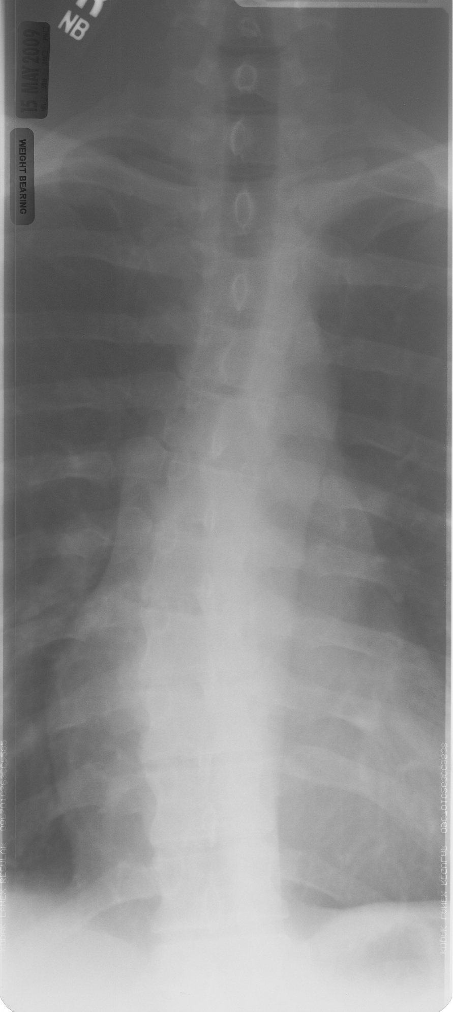
X-rayWeight-bearing anteroposterior spine radiograph: right convex thoracic scoliosis of approximately 20 degrees, apex at T9, with a compensatory left convex mid-lumbar scoliosis.
Source : Vicarious, Wikimedia Commons · CC BY-SA
Screening for a scoliosis is not just a matter of “seeing a crooked back”. The approach is graded: a simple clinical examination points the way, a scoliometer puts a figure on it, and only radiography confirms and measures the deformity. The physiotherapist's role is central upstream (spotting, quantifying rotation, and knowing when to refer for imaging) and then downstream, in follow-up. The thresholds and their limits still have to be known, because no clinical test is perfect.
What we are really looking for: a deformity in three dimensions
A scoliosis is not a simple lateral curve: it is a three-dimensional deformity of the spine. According to the Scoliosis Research Society, the diagnosis is confirmed only when two conditions are met: a Cobb angle ≥ 10° in the frontal plane and an axial rotation of the vertebrae that is identifiable 1. A curve below 10°, or a simple postural scoliosis with no rotation, does not meet this diagnosis. That rotational component is crucial: it is what produces the palpable rib “hump” on the back, and it is what clinical screening seeks to unmask.
About 80 % of scolioses are idiopathic (with no identified cause), the remaining 20 % being secondary to a congenital or neuromuscular condition 1. The commonest form is adolescent idiopathic scoliosis (AIS), which affects 1 to 3 % of at-risk children aged 10 to 16 4. In adults, the definition changes in nature: scoliosis is spoken of when the Cobb angle exceeds 10° in the coronal plane on a skeletally mature spine 11.
Key points
- Diagnosis = Cobb angle ≥ 10° + vertebral rotation 1. Without rotation, this is not a structural scoliosis.
- The Adams test spots it, the scoliometer quantifies rotation, the radiograph alone confirms and measures.
- Referral threshold for imaging: 5°–7° of trunk rotation on the scoliometer 2.
- The Risser sign (skeletal maturity) governs the risk of progression and therefore the monitoring.
The Adams test: the reference screening manoeuvre
The forward bend test of the trunk (Adams test) is the commonest screening method 2. The principle is simple: the subject, feet together and knees straight, bends slowly forward, arms relaxed, hands together. The examiner looks at the back tangentially, from behind and from the side: vertebral rotation, invisible in standing, then makes one hemithorax bulge (a rib hump at thoracic level) or a lumbar prominence. A difference in height between the two sides of the back is the sign of the rotational component.
This test is free, quick and involves no radiation, but it is purely qualitative: it says “there is an asymmetry” without saying “how much”. An untrained eye underestimates small rotations and over-reads ordinary postural asymmetries. It is precisely to objectify what the eye perceives that a scoliometer is almost always added to it 2.
The scoliometer: putting a figure on trunk rotation
The scoliometer (Bunnell inclinometer) is laid flat on the back, at the apex of the rib hump, during the Adams test. It measures the angle of trunk rotation (ATR) in degrees. It is that figure which decides whether or not the child is referred for a radiograph.
In other words, lowering the threshold to 5° guarantees that no true scoliosis is missed, at the price of a flood of useless radiographs (fewer than one referral in two will be confirmed). Raising it to 7° spares radiation but lets some early curves slip through the net. There is no universal “good” threshold: it is a trade-off to be owned.
Large-scale screening data illustrate this bluntly. In a study covering more than 33,000 girls, a 5° ATR threshold led to referring 5.2 % of children, against 1.4 % at 7° and only 0.3 % at 10° 6. Above all, the positive predictive value of a 5° threshold was no more than 28.3 % for a scoliosis ≥ 10°, meaning that nearly three referred children out of four had a normal or sub-threshold radiograph.
The conclusion of that work is honest and deserves to be passed on to the reader: the optimal referral threshold remains hard to determine 6. The scoliometer is an excellent tool for triage, not a diagnostic tool. It points the way; it never concludes on its own.
The Cobb angle: the reference measure, on a radiograph
The Cobb angle is the gold-standard measure of severity, taken on a standing full-spine radiograph in the frontal view. A line is drawn along the upper endplate of the most tilted vertebra at the top of the curve, another along the lower endplate of the most tilted vertebra at the bottom: the angle formed by their perpendiculars is the Cobb angle. It is what defines the diagnosis (≥ 10°), grades management and tracks progression over time.
An essential point for the physiotherapist: this measurement is not made clinically. Neither the Adams test, nor the scoliometer, nor any surface measurement gives the Cobb angle. They only decide whether imaging is warranted. Once the radiograph is available, the Cobb angle becomes the follow-up figure shared with the doctor and the surgeon.
The scoliometer points towards radiography; only the Cobb angle confirms the diagnosis and measures severity.
The Risser sign: measuring maturity, not the curve
In the adolescent, the question is not only “how big is the curve?” but “will it still grow?”. A scoliosis progresses essentially during spinal growth, so assessing the skeletal maturity that is left is decisive. That is the role of the Risser sign, graded from 0 to 5 on the pelvic radiograph according to ossification of the iliac crest (0 = a great deal of growth left, 5 = maturity reached).
The same 25° curve therefore carries a quite different prognosis at Risser 0 and at Risser 4: in the skeletally immature child it progresses in two-thirds of cases; in the adolescent close to maturity, in fewer than a quarter. In the founding cohort of 727 patients, 23.2 % progressed, progression being linked to the type and size of the curve, to age at presentation, to the Risser sign and to menarcheal status 7.
The female predominance, another marker for follow-up, sharpens strongly with severity: the girl-to-boy ratio goes from 1.4:1 for curves of 10–20° to 7.2:1 beyond 40° 5. Progressive and severe curves therefore concern girls in the great majority, which justifies heightened vigilance.
Caution: no marker predicts progression reliably
We have to be frank with the reader here, because the temptation to over-read these figures is strong. A systematic review with meta-analysis identified as factors of progression an initial Cobb angle > 25°, an age at diagnosis < 13 years, a pre-menarcheal status and skeletal immaturity, but the authors conclude that the predictive value of all these markers remains limited and the level of evidence low. No method can today be recommended as a reliable criterion for predicting progression 9. These markers guide how often to monitor; they do not allow the future of a given curve to be announced with certainty.
What the physiotherapist assesses, and when to refer for imaging
Beyond the screening tests alone, the physiotherapy assessment documents: overall posture and the balance of the shoulders and pelvis, the site and side of the rib hump on the Adams test, the ATR measured with the scoliometer, the flexibility and reducibility of the curve, muscular and respiratory status, and, in adults, pain, often present on the convex side 11. In adults, scoliosis is common and readily painful: low back pain affects 60 to 80 % of patients 11, and the reason for consulting is the discomfort, not the deformity itself.
When should a radiograph be requested? The practical rule is clear: a positive Adams test with an ATR ≥ 5–7° on the scoliometer warrants medical referral for imaging 2. To these are added, whatever the ATR: a marked trunk asymmetry, a curve worsening rapidly in a skeletally immature child, atypical or night pain, or any neurological sign, all of which should prompt referral without delay. Conversely, a sense of measure is needed: the 50-year follow-up of untreated idiopathic scolioses is rather reassuring (survival comparable to the general population, functional patients) even if chronic low back pain is more frequent there (61 % vs 35 % of controls) 10. That finding justifies simple monitoring of small curves, rather than systematic treatment: to screen is not to medicalise.
In short, the physiotherapist is a key link in detection: they know how to do the Adams test, put a figure on rotation, read a threshold while knowing its margin of error, and refer at the right moment. Diagnostic confirmation, measurement of the Cobb angle and the reading of the Risser sign belong to imaging and to the doctor, in a chain where every tool has its place and none replaces the others.
📈 Will it get worse? The risk of progression
This is the question that dominates the first consultation, for the adolescent as much as for the parents: will this curve get worse? The honest answer is that it depends on a few measurable parameters, but that none of them allows a certain prediction for a given individual. The risk of progression of an untreated scoliosis is essentially governed by three elements: the size of the curve (Cobb angle), skeletal maturity (Risser sign) and the growth that is left, to which are added age at diagnosis, menarcheal status and the type of curve 78.
The three drivers of progression
A scoliosis progresses all the more when growth is still to come. That is why skeletal maturity, estimated by the Risser sign (ossification of the iliac crest), weighs as much as the size of the curve. An immature spine (Risser 0–1) carries a clearly higher risk than a spine close to the end of growth (Risser 2–4). The menarcheal status of the girl is a practical marker: the period of greatest risk lies around the pubertal growth spurt, before and shortly after the first period 7.
The second driver is the initial size of the curve: the higher the Cobb angle at diagnosis, the greater the probability of worsening. In the long run, it is even the absolute magnitude of the curve that emerges as the most predictive factor 8. A systematic review with meta-analysis takes as an alert threshold an initial severity above 25° of Cobb, together with an age at diagnosis below 13 years and a pre-menarcheal status 9.
The third element is the background. The female predominance, moderate for small curves, sharpens strongly with severity: the girl-to-boy ratio goes from 1.4:1 for curves of 10° to 20° up to 7.2:1 for curves above 40° 5. In other words, it is above all girls who develop progressive and severe curves, a concrete argument for closer monitoring in that population.
Key points
- Three factors dominate the risk of progression: the Cobb angle, the Risser sign and the growth that is left.
- The less mature the child (low Risser) and the larger the curve, the higher the risk of worsening.
- Progressive and severe curves concern girls in the great majority (ratio up to 7.2:1 beyond 40°).
- No marker taken on its own allows the course of an individual case to be predicted with certainty.
Probabilities in figures
The historical data give useful orders of magnitude. In the reference cohort of Lonstein & Carlson (727 patients), 23.2% of scolioses progressed, the incidence of progression being linked to the type and magnitude of the curve, to age at presentation, to the Risser sign and to menarcheal status 7.
The combined effect of maturity and size reads clearly when the two parameters are crossed. In the skeletally immature child (Risser 0–1), a curve of 5–19° progresses in 22% of cases, but a curve of 20–29° progresses in 68% of cases. In the more mature child (Risser 2–4), those same rates fall to 1.6% and 23% respectively 8.
| Situation | Curve 5–19° | Curve 20–29° |
|---|---|---|
| Skeletally immature child (Risser 0–1) | 22% progression | 68% progression |
| Child close to maturity (Risser 2–4) | 1.6% progression | 23% progression |
After Wong & Tan 2010, from the data of Lonstein & Carlson.
These figures illustrate a simple rule: a moderate curve in a child in full growth deserves particular vigilance, whereas the same curve in an adolescent close to skeletal maturity is far more stable.
What science cannot yet predict
We have to be frank about the limits of these predictions. The systematic review by Noshchenko 9 does identify risk factors (Cobb > 25°, age < 13 years, pre-menarcheal status, skeletal immaturity) but stresses that the predictive value of all these markers remains limited and the level of evidence low.
No method can today be recommended as a reliable diagnostic criterion for the progression of an adolescent scoliosis.
In practice this means reasoning in group probabilities, not in individual certainties. Two adolescents with identical curves and Risser grades may progress differently. It is that uncertainty, and not an excess of caution, that justifies repeated monitoring over time because it is the trajectory seen across several radiographs, far more than a single measurement, that really informs on the risk of progression.
What determines the monitoring
The follow-up strategy flows directly from these risk factors. It starts upstream, at screening: the Adams test (forward bending of the trunk) coupled with the scoliometer points towards radiography beyond a trunk rotation angle of 5 to 7° 2. The choice of threshold is a trade-off owned as such: at 7°, sensitivity and specificity for a Cobb > 10° are about 83% and 87%; at 5°, sensitivity reaches 100% but specificity falls to 47%, at the price of many false positives 2. Only measurement of the Cobb angle on a radiograph confirms the diagnosis.
Once the scoliosis is confirmed, three approaches emerge according to severity and maturity 1 :
| Profile | Approach | Level of evidence |
|---|---|---|
| Small curve, low risk | Simple spaced radiographic monitoring | Reassuring natural history |
| Curve 20–40°, skeletally immature child | Brace (± specific exercises) | High (randomised BrAIST trial) |
| Adjunct at any stage during growth | Specific exercises (Schroth / PSSE) | Moderate, adjunct |
For small curves, observation alone is justified by a reassuring natural history: in the 50-year follow-up of untreated idiopathic scolioses, survival was comparable to that of the general population and patients remained functional at a high level, even if chronic low back pain was more frequent there (61% against 35% in controls) 10.
For high-risk curves in the adolescent (Cobb 20–40°, immaturity), the brace has proved its effectiveness in the randomised BrAIST trial: the success rate (avoiding progression to the surgical threshold of 50°) was 72% with a brace against 48% under observation alone, with a clear dose-response relationship, since wearing it for at least 12.9 hours a day was associated with 90–93% success 14. It is the most solid level of evidence in the field. Specific exercises (Schroth / PSSE) bring a real but modest benefit on the Cobb angle 15 and remain an adjunct, not a way of “correcting” the deformity 1.
And in adulthood?
Contrary to a received idea, a scoliosis can carry on progressing after the end of growth, but slowly and in a linear fashion. In a study with a mean follow-up of 27 years, the annual progression of single lumbar and thoracolumbar curves was about 0.82°/year for aged idiopathic scolioses (which began in adolescence) and 1.64°/year for de novo degenerative scolioses 13.
This slow pace changes the logic of monitoring: in adults, spaced radiographic follow-up is preferred to systematic treatment, management being guided above all by symptoms (pain, functional impact) and not by the angle alone. Degenerative adult scoliosis is in fact common and grows with age, with low back pain present in 60 to 80% of patients 11 and it is that pain, more than the curve itself, which most often prompts the consultation.
In short, “will it get worse?” calls not for a single answer but for an individualised assessment: the younger and less mature the child, and the larger the curve, the more real the risk, with no marker allowing a certain verdict. Hence the importance of structured follow-up over time, which remains the best tool for telling stable curves from progressing ones.
🎽 Observation, brace, surgery: what to choose according to severity?
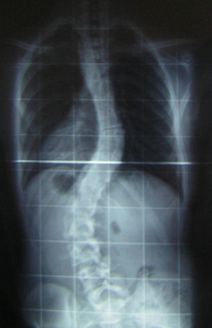
X-rayPosteroanterior spine radiograph, film viewed on a lightbox: adolescent idiopathic scoliosis with a double curve, thoracic of 30 degrees and lumbar of 53 degrees.
Source : Silverjonny, Wikimedia Commons · Domaine public
🎽 The brace works, and the trial that proved it
In adolescents with a progressive, at-risk curve, the brace clearly reduces progression to the surgical threshold. And the more it is worn, the better it works.
“Success” = no progression to the surgical threshold (≥ 50°). Randomised BrAIST trial. The relationship is dose-dependent: the success rate reaches 90-93% in those who wear the brace at least 12.9 h/day. Source: Weinstein et al., NEJM 2013 (PMID 24047455).
There is no single treatment for scoliosis, but a graded strategy: the choice between simple observation, brace and surgery depends above all on the size of the curve (Cobb angle), on the risk of progression and on the skeletal maturity that is left. This section sets out the decision thresholds, what the best available trial (BrAIST) really showed about the brace, the place of surgery and, above all, what is firmly established as against what remains uncertain.
The decision reasoning: size, maturity, progression
Scoliosis is defined by a Cobb angle ≥ 10° together with an identifiable axial vertebral rotation 12. That 10° threshold confirms the diagnosis, but does not on its own dictate what to do: two curves of the same size do not carry the same prognosis, depending on the growth that is left. The risk of progression depends jointly on the initial size and on skeletal maturity assessed by the Risser sign 78. In practice, in a skeletally immature child (Risser 0–1), a curve of 20–29° progresses in about 68% of cases, against only 23% in a more mature child (Risser 2–4); for a curve of 5–19°, those rates fall to 22% and 1.6% respectively 8. It is that combination, a large curve plus a lot of growth left, that defines a “high-risk” scoliosis and tips the balance from observation towards active treatment.
A systematic review with meta-analysis identifies as factors of progression an initial severity above 25°, an age at diagnosis below 13 years, a pre-menarcheal status and skeletal immaturity, but it stresses that the predictive value of these markers remains limited and the level of evidence low, with no method that can be recommended as a reliable criterion of progression 9. In other words, a risk is estimated, an individual trajectory is not predicted: hence the importance of repeated radiographic monitoring rather than a decision frozen at diagnosis.
Small curves and the end of growth: observation is often enough
For curves of small size, monitored therapeutic abstention is legitimate, and it rests on reassuring natural-history data. The 50-year follow-up of untreated idiopathic scolioses showed survival comparable to that of the general population (estimated probability ~0.55 against 0.57 expected), with patients remaining productive and functional at a high level 10. The main impact reported was chronic low back pain, more frequent than in controls (61% against 35%), a point not to be played down, but one that does not make a small scoliosis a disabling disease in itself. These data justify not over-treating and reserving intervention for genuinely progressive curves.
Monitoring rests on a clinical examination (Adams forward bending test, scoliometer measuring the angle of trunk rotation) and, if needed, a radiograph to measure the Cobb angle, the only measurement that confirms and quantifies the deformity 62. The referral threshold for radiography is debated: at 5° of rotation, sensitivity reaches 100% but specificity falls to 47%, whereas at 7° sensitivity and specificity for a Cobb > 10° are close to 83% and 87% 2. Lowering the threshold screens more but multiplies the false positives sent for radiography: at 5° the referral rate was 5.2% with a positive predictive value of only 28.3%, against a 1.4% referral rate at 7° 6. A trade-off therefore has to be owned, with no “perfect” threshold.
Progressive curves: the brace, the best level of evidence (BrAIST)
In the skeletally immature adolescent with an at-risk curve (typically Cobb 20–40°), the thoracolumbosacral orthosis (TLSO) is the reference treatment, and it is the randomised BrAIST trial that gives it its most solid foundation 14. The success criterion there was clear and clinically relevant: reaching skeletal maturity without the curve exceeding 50°, that is, without crossing the surgical threshold.
The result is clear-cut: the success rate was 72% in the brace group against 48% under observation alone 14. Above all, the trial showed a significant dose-response relationship between the number of hours worn each day and the success rate (p < 0.001): wearing it on average for at least 12.9 hours a day was associated with success rates of 90 to 93%. The trial was in fact stopped early because of the benefit observed. This message is crucial for physiotherapy practice and for patient education: the effectiveness of the brace depends directly on adherence, which shifts part of the challenge towards supporting the wearing of it.
| Strategy | Typical indication | What the data show | Level of evidence |
|---|---|---|---|
| Observation | Small curves, low risk, end of growth | Reassuring natural history at 50 years (normal survival, good functional level); more frequent low back pain 10 | Observational, but consistent |
| Brace (TLSO) | Curve ~20–40°, skeletally immature at-risk patient | 72% success vs 48%; dose-response, 90–93% if ≥ 12.9 h/day 14 | High (RCT) |
| Specific exercises (PSSE / Schroth) | Adjunct, often combined with observation or with the brace | Modest effect on the Cobb angle: −3.5° at 6 months 15 and −3.18° in meta-analysis 16 | Moderate, adjunct |
| Surgery | Curve ≥ ~50°, progression despite treatment, disabling adult forms | Threshold defined as failure of conservative treatment; indicated in symptomatic forms 1411 | Clinical consensus |
An important nuance to pass on to the patient and the family: the brace does not “straighten” the spine. The SOSORT recommendations remind us that the brace effectively prevents progression to the surgical threshold, even if on average the curves do not reduce 1. The aim is to stabilise, not to correct. That distinction avoids false expectations and refocuses the conversation on adherence and on avoiding surgery.
The surgical threshold: about 50° and beyond
In the adolescent, surgery enters the discussion when the curve reaches or exceeds about 50°: it is precisely that 50° threshold which defines “failure” of conservative treatment in BrAIST, the logic of the whole pathway being to avoid getting there 14. The whole graded strategy (observation, brace, adjunctive exercises) aims to prevent that threshold being crossed during growth. The sources kept here do not detail the techniques or the functional results of spinal fusion in the adolescent; we therefore limit ourselves to what is established: the decision threshold and the aim of avoiding it.
In adults: conservative first, surgery for disabling forms
Adult scoliosis (degenerative de novo or aged idiopathic) is defined by a Cobb angle > 10° in a skeletally mature subject 11. It is common and becomes more so with age: a meta-analysis puts the prevalence of the de novo form at 37.6%, higher in women, rising from 13% before the age of 60 to 36% after 12. The cardinal symptom is not the deformity but pain: low back pain is present in 60 to 80% of patients, mostly on the convex side of the curve 11.
The reasoning differs from that in the adolescent: in adults we no longer reason in “hours worn” but in slow trajectory. Progression exists but stays moderate and linear, about 0.82°/year for aged idiopathic scolioses and 1.64°/year for de novo degenerative forms over a mean follow-up of 27 years 13. This pace justifies spaced radiographic monitoring rather than systematic treatment.
First-line management is conservative and centred on pain and function: physiotherapy, exercise, analgesia. A prospective cohort of 30 women treated with 15 physiotherapy sessions over three months showed a significant improvement in pain, disability and quality of life (p < 0.001), with, however, a modest level of evidence given the small numbers and the absence of randomisation 17. We must also be honest about the uncertainty: a systematic review concludes that the quantity and quality of data on injections, bracing and yoga in adults are insufficient to recommend for or against these methods 18. Surgery is considered only in case of failure of medical treatment, disabling pain or neurological deficit, and it is frequently indicated in symptomatic forms (axial pain, neurogenic claudication), medical treatment often proving of little use in those cases 11.
Key points
- The decision is graded, based on the size of the curve (Cobb) and on skeletal maturity (Risser), not on a single threshold 89.
- Small curves: observation is legitimate, since the natural history at 50 years is reassuring, apart from more frequent low back pain 10.
- Brace (BrAIST): the best level of evidence, with 72% success against 48% and a benefit strongly dependent on hours worn (90–93% if ≥ 12.9 h/day). It stabilises, it does not correct 141.
- Surgical threshold ≈ 50°: the whole conservative pathway aims not to reach it 14.
- Adults: slow progression, management conservative first and centred on pain; surgery reserved for disabling forms or those with a deficit. The level of evidence for conservative treatments in adults remains low 131118.
🤸 Specific exercises (Schroth, PSSE): what are they worth?
🤸 Specific exercises (Schroth): a real but modest effect
Added to standard management, specific exercises limit the curve a little. “A little”: they do not correct it, and saying so honestly is part of the care.
Randomised trial with blinded assessor: largest curve 3.5° smaller in the Schroth group (95% CI 1.1° to 5.9°; p = 0.006). A network meta-analysis (17 trials, 857 patients) confirms a modest benefit 19. The level of evidence for exercises remains lower than that for the brace 1. Source: Schreiber et al., 2016 (PMID 28033399).
Because scoliosis is a three-dimensional deformity of the spine, the diagnosis being made only in the presence of a Cobb angle ≥ 10° together with axial rotation of the vertebrae, 1 so the idea of a rehabilitation acting on all three planes is appealing. That is the promise of scoliosis-specific exercises, grouped under the acronym PSSE (physiotherapeutic scoliosis-specific exercises), of which the Schroth method is the most studied. But between what families expect and what the literature really shows, a clear boundary has to be drawn: these exercises can slow a worsening and improve how it is lived with, they do not correct an established curve durably. This section takes stock without over-promising.
What are we talking about?
PSSE are physiotherapy programmes designed specifically for scoliosis, as opposed to general rehabilitation or untargeted strengthening. They are offered as a complement to standard management (observation, brace as the case may be), and not in its place. Their place is framed by the international SOSORT 2018 recommendations, which number 68 recommendations shared between bracing (25), PSSE to prevent progression during growth (12), PSSE while the brace is worn or around surgery (6) and assessment (14) 1. In other words, specific exercises are not a stand-alone treatment but one link in a graded strategy, according to the severity of the curve and skeletal maturity.
What the trials show on the Cobb angle
The reference datum comes from a randomised controlled trial with a blinded assessor and a blinded statistician 15. Fifty adolescents with an idiopathic scoliosis (curves of 10° to 45°) received either standard care alone or that same care plus six months of Schroth exercises. At six months, the largest curve was on average 3.5° smaller in the Schroth group than in the control group, a statistically significant result.
The benefit tended to be more marked the more severe the starting curve 15. Two reservations are called for, however: the sample remains modest (50 patients) and the size of the effect, a few degrees, sits within the order of magnitude of the measurement variability of the Cobb angle itself. This is therefore a real signal, but one of small size.
What the meta-analyses confirm
Two recent syntheses converge and reinforce that signal without inflating it. A meta-analysis devoted to the Schroth method (6 studies, 144 patients) finds a mean reduction in the Cobb angle of 3.18° (95% CI −4.30 to −2.07), with zero heterogeneity between studies (I² = 0%), together with an improvement in quality of life and in the angle of trunk rotation 16. A broader systematic review with network meta-analysis (17 randomised trials, 857 patients) concludes that PSSE improve the Cobb angle compared with conventional rehabilitation (standardised mean difference = −0.7; 95% CI −0.95 to −0.44; p = 0.001), the Schroth approach emerging as the most effective on posture and trunk deformity 19.
| Outcome measured | Effect of PSSE / Schroth | Source |
|---|---|---|
| Cobb angle (RCT) | −3.5° vs controls at 6 months | Schreiber 2016 |
| Cobb angle (Schroth meta-analysis) | −3.18° on average | Ceballos-Laita 2023 |
| Cobb angle vs conventional rehabilitation | SMD −0.7 (in favour of PSSE) | Dong 2024 |
| Quality of life / trunk rotation | Modest improvement | Ceballos-Laita 2023 |
The message is consistent from one source to another: a modest but reproducible gain of a few degrees, together with an improvement in comfort and quality of life. None of these data allows anyone to announce that a curve will be “straightened” by exercises alone.
A level of evidence to state honestly
This is where rigour is needed. The level of evidence for PSSE remains lower than that of the brace. In the SOSORT 2018 recommendations, out of 68 recommendations in total, only three reached grade A backed by a level I of evidence, two for the brace and one for assessment, none for PSSE 1. The recommendations concerning exercises sit mostly at grades B (13 PSSE recommendations) and C, with a single PSSE recommendation reaching a recommendation level of “I” with a level of evidence of “II” 1. In plain terms, specific exercises are a recommended adjunct, not a treatment whose corrective power has been shown at the highest level.
The contrast with the brace lights up that hierarchy. The randomised BrAIST trial showed, for high-risk adolescent curves, a success rate (no progression to the surgical threshold of 50°) of 72% with a brace against 48% under observation alone, with a clear dose-response relationship, wearing it at least 12.9 hours a day being associated with 90–93% success 14. No trial on PSSE reaches that level of proof. The two approaches are in any case not in the same category: the brace has a targeted indication (progressive curve, skeletal immaturity), whereas exercises address a wider group, including alongside the brace.
Specific exercises can slow worsening and improve how the condition is lived with; they do not replace the brace when the brace is indicated.
And in adults?
In adults, the question is no longer really to change a curve, often old and stiffened, but to treat pain and its functional impact. Yet the level of evidence there is even more limited. A systematic review of symptomatic degenerative scoliosis concludes that the quantity and quality of the data on injections, bracing and yoga are insufficient to recommend for or against these methods 18. Active physiotherapy remains the first-line conservative treatment and does improve pain and functional capacity in practice, but in small, non-randomised cohorts; caution is therefore needed about how far these results reach. Conservative management keeps all its sense as a first recourse, before surgery is considered, without being presentable as a “validated” treatment of the adult deformity.
Uncertainty owned
On one point, the literature explicitly invites modesty: nobody can say today that specific exercises, on their own, change the natural history of a scoliosis in the way the brace does. The effects measured are real but small, the numbers limited, and long-term follow-up is lacking. Rather than deciding beyond what the data show, it is better to say so to the patient and the family: what we expect from exercises is that they help to stabilise, mobilise and live better with the curve, not that they make it disappear.
Key points
- PSSE (Schroth among them) bring a modest but reproducible benefit on the Cobb angle: about −3.5° in the reference trial 15, −3.18° in meta-analysis 16.
- They are added to standard management, they do not replace it, and they are framed by the 68 SOSORT 2018 recommendations 1.
- The level of evidence remains lower than that of the brace with no grade A recommendation for PSSE, against an effectiveness shown for the brace in the BrAIST trial (72% vs 48%) 114.
- In adults, the aim is analgesic and functional; the evidence for conservative treatments there is insufficient to conclude 18.
- Never promise to “correct” the curve with exercises alone: slowing the worsening and improving quality of life are honest and attainable aims.
👵 And adult scoliosis?
Long regarded as a disease of children and adolescents, scoliosis does not vanish at skeletal maturity: on the contrary, it is a frequent reason for consultation and for physiotherapy in middle-aged and older people. In adults, the definition stays geometric: a deformity of the spine in the coronal plane with a Cobb angle above 10° in a skeletally mature subject 11. But the stakes change radically in nature. In the adolescent, we monitor and slow a curve in full growth; in the adult, we manage a deformity already established, whose main problem is no longer appearance or rapid progression, but pain and its functional impact.
Two adult scolioses not to be confused
Under the single term “adult scoliosis” lie situations of quite different origin. The deformity may be iatrogenic, may result from a degenerative process appearing on a spine that was straight until then, the de novo scoliosis or it may correspond to an adolescent idiopathic scoliosis that has “aged” and worsened over time 11. This distinction is not merely academic: it shapes the prognosis and the speed of progression.
De novo degenerative scoliosis arises from asymmetric wear of the discs and the facet joints. It often starts around the age of fifty and becomes clinically loud around 70 11. Its frequency is far higher than one imagines: a systematic review with meta-analysis (5 studies, 4,069 participants aged 41 to 94) puts its overall prevalence at 37.6%, higher in women (41.2%) than in men (27.5%), and strongly linked to age, 13% before the age of 60 against 36% after 12. More broadly, vertebral deformities affect 32 to 68% of people over the age of 65 11.
In other words, this is not a clinical curiosity but a massive epidemiological reality in an ageing population, and therefore a major field of action for the physiotherapist.
Pain, the central symptom
Where the adolescent is most often symptom-free, the adult comes first of all because it hurts. Low back pain is present in 60 to 80% of patients with adult scoliosis, mainly on the convex side of the curve, and is explained by the degenerative changes and the muscular fatigue linked to spinal imbalance 11. To this axial pain may be added, in advanced forms, signs of nerve compression such as neurogenic claudication 11.
This scoliosis–pain association nevertheless deserves qualifying, because it feeds an anxiety that is sometimes out of proportion in patients. The 50-year follow-up of untreated idiopathic scolioses is enlightening here: patient survival was comparable to that of the general population and they remained productive and functional at a high level. Chronic low back pain was certainly more frequent than in controls (61% against 35%), but it stayed compatible with an active life 10. That reassuring result invites us not to dramatise a curve discovered in adulthood: having a scoliosis is not a sentence to disability.
In adults we do not treat an angle on a radiograph: we treat a person who is in pain and whose function we want to preserve.
A real but slow progression
Contrary to a received idea, the curve of a scoliosis can carry on worsening after the end of growth, but it does so slowly and steadily. In a study with a mean follow-up of 27 years, the annual progression of single lumbar and thoracolumbar curves was about 0.82°/year for aged idiopathic scolioses (which began in adolescence) and 1.64°/year for de novo degenerative scolioses, with a kinetic that is linear 13. De novo degenerative scoliosis therefore progresses about twice as fast as an aged idiopathic scoliosis.
This slowness has an important practical consequence: it justifies spaced radiographic monitoring rather than a hasty therapeutic escalation. A few tenths of a degree to a degree and a half a year do not call for an urgent decision; they invite us to follow the course over time and to act on the symptoms.
Conservative management in the front line
In the symptomatic adult, the aim of treatment is not to “straighten” the spine, which no non-surgical approach achieves, but to reduce pain and disability and to preserve independence. Physiotherapy, exercise and analgesia are logically the first-line treatment.
The available data point towards a benefit, while remaining of low evidential level. In a prospective cohort of 30 women treated with 15 sessions of physiotherapy and exercise over three months, pain (VAS), disability (RMDQ) and quality of life (SRS-22, SF-36) all improved significantly (p < 0.001) 17. These encouraging results must nevertheless be read with caution: small numbers and the absence of a control group and of randomisation limit how far the conclusions reach.
We have to be honest with the reader here: the level of evidence for non-surgical treatments of degenerative adult scoliosis is low. A systematic review concludes explicitly that the quantity and quality of the data on injections, bracing and yoga are insufficient to recommend for or against the use of these methods 18. In adults we are therefore not in the situation of the adolescent, where the brace has a high-level randomised trial (BrAIST). This does not mean that rehabilitation is useless, since clinical experience and the cohorts suggest the opposite, but that we still lack solid trials to rank the modalities precisely. This uncertainty should be shared with the patient rather than hidden.
When should surgery be considered?
Surgery is not the first-line treatment of adult scoliosis. It is considered only in case of failure of conservative treatment, disabling pain or neurological deficit 17. In advanced symptomatic forms (stubborn axial pain, neurogenic claudication from associated stenosis), surgery is on the other hand frequently indicated, all the more so as purely medical treatment often proves of little use there 11.
The clinical message therefore lies in a balance. On one side, adult scoliosis is a potentially disabling condition, which affects a heterogeneous group of patients and some forms of which do indeed belong in the operating theatre 11. On the other, its progression is generally slow 13 and the very long-term natural history of idiopathic scolioses is more favourable than was feared 10. Between these two realities, physiotherapy holds a central place: supporting the pain, maintaining mobility and strength, preserving function, and referring to the surgeon only when the impact really justifies it.
Key points on adult scoliosis
- Definition unchanged but a different issue: Cobb > 10° in the adult, where the dominant problem is pain, not growth 11.
- Common: de novo degenerative scoliosis is thought to affect ~37.6% of adults, up to 36% after the age of 60 12.
- Painful: low back pain in 60 to 80% of patients, mostly on the convex side 11, but compatible with a functional life in the long term 10.
- Slow and linear progression: ~0.82°/year (aged idiopathic) to 1.64°/year (de novo), hence spaced monitoring 13.
- Conservative first: physiotherapy and exercise improve pain and quality of life 17, but the level of evidence remains low 18 and that should be said to the patient.
- Surgery is reserved for failure of conservative treatment, for disabling pain or for a neurological deficit 1711.
🗂️ What do concrete case reports teach us?
Trial and cohort data describe averages; in the consulting room, the physiotherapist meets people. To connect the two, here is a published study, with its PubMed identifier. It illustrates no real patient: these are supports for reasoning, meant to show how thresholds, probabilities of progression and levels of evidence come together in a clinical decision. No individual prognostic figure can be deduced from these accounts: they express population trends, not a prediction for a given person.
Published case: what conservative treatment shifts, and what the Cobb angle does not measure
The study. Zagalaz-Anula and colleagues analysed the perception of the visual vertical and postural balance in adolescent girls with an idiopathic scoliosis, according to whether management rested on simple observation or on a conservative treatment20.
The setting. The authors recall that adolescent idiopathic scoliosis is defined by a lateral curvature of the spine of at least 10 degrees of Cobb angle, of unknown aetiology, and that some studies have found in these patients a perception of the visual vertical comparable to that of healthy subjects20.
Why this study rather than two invented adolescent girls. Because it deals with a dimension that teaching vignettes almost never address: what treatment changes in the way the person perceives their own vertical. That is precisely the physiotherapist's ground, and it is not the Cobb angle.
The reservation. The authors themselves present their work as a preliminary study20. It therefore does not change the brace indications or the thresholds set out earlier in this article, which rest on data of a quite different evidential level. It opens a question, it does not close it.
Key points
- One and the same logic: the decision is read at the intersection of the size of the curve, skeletal maturity and the aim (slowing progression in the adolescent, relieving pain in the adult).
- Screening points, the radiograph decides: the scoliometer imposes a sensitivity/false-positive trade-off (5° = 100% sensitivity but 47% specificity), and only a Cobb angle ≥ 10° confirms 2.
- In the at-risk adolescent, the brace has the best evidence: 72% success vs 48% under observation, with a dose effect 14 and the Schroth exercises are a modest adjunct, not a correction 151.
- In adults, slow progression and weak evidence: ~0.8–1.6°/year 13 and conservative care relieves, but the data remain insufficient for a firm verdict 18.
- No certain individual prognosis: the markers of progression have a limited predictive value 9 and these accounts illustrate trends, not destinies.
🧭 How is this applied in practice?
Translating the evidence into clinical decisions means constantly crossing two variables: the severity of the curve (Cobb angle) and skeletal maturity (Risser sign, menarcheal status, growth left). It is that pair which determines the risk of progression and therefore the intensity of management 78. The physiotherapist is neither the prescriber of the brace nor the one who makes the radiographic diagnosis, but holds a pivotal place: screening, referral, specific rehabilitation and support for adherence.
An algorithm graded by severity and maturity
The grading logic drawn from the SOSORT 2018 recommendations sets out three levels of response (observation, specific exercises, brace), to which surgery is added 1. The table below sums up that grading; it is a framework for reasoning, not a rigid protocol, because each situation also depends on the growth that is left and on the dynamics of progression documented on successive radiographs.
| Clinical situation | Usual approach | Level of evidence |
|---|---|---|
| Curve 10–20°, growing child | Close clinical and radiographic monitoring; specific exercises (PSSE) possible | Moderate 1 |
| Curve 20–40°, immaturity (low Risser) | Brace (medical indication) + specific exercises as an adjunct | High for the brace 14 |
| Curve ≥ 45–50°, surgical threshold | Specialist surgical opinion | Consensus 1 |
| Small curve with skeletal maturity reached | Simple observation, reassurance | Reassuring natural history 10 |
Two numerical markers frame the referral. First, the risk of progression is not uniform since in the skeletally immature child (Risser 0–1), a curve of 5–19° progresses in about 22% of cases, against 68% for a curve of 20–29°; in the more mature child (Risser 2–4), those rates fall to 1.6% and 23% respectively 8. The initial size and the growth that is left are therefore the two decision levers. Second, the brace has proved its effectiveness for at-risk curves.
This effectiveness is dose-dependent since an average wear of at least 12.9 hours a day is associated with success rates of 90 to 93% 14. This is a central argument to pass on to the family when a brace is prescribed: the benefit is decided by the hours actually worn.
The place of specific exercises: realistic, not magical
Scoliosis-specific exercises (PSSE, including the Schroth method) are the heart of the physiotherapy intervention, but their power must be presented honestly. Added to standard treatment, they modestly reduce the Cobb angle: in a randomised blinded trial of 50 adolescents (curves 10–45°), at six months the Schroth group had a largest curve 3.5° smaller than the controls (95% CI −1.1° to −5.9°; p = 0.006) 15. A meta-analysis finds a consistent but small effect (mean reduction of 3.18°; 95% CI −4.30 to −2.07) 16, and a network meta-analysis confirms their superiority over conventional rehabilitation 19.
Specific exercises slow the worsening and improve posture; they do not “straighten” a spine that is already deformed.
This nuance is not cosmetic: in the SOSORT 2018 recommendations, out of 68 recommendations in total, only three reached grade A backed by a level I of evidence, two for the brace and one for assessment, none for PSSE 1. In other words, the level of evidence for exercises remains lower than that for the brace. Promising a correction from exercises alone would expose the family to disappointment and might delay an indication for a brace. The right wording: exercises are a recommended adjunct, useful for slowing progression and for maintaining mobility and self-elongation, never a substitute for the brace when the brace is indicated.
Key messages to pass on to parents
The announcement of a scoliosis often generates disproportionate anxiety. The practitioner's role is to inform without dramatising, drawing on natural-history data.
- “Most small curves do not progress to anything serious.” In the historical cohort of 727 patients, only 23.2% progressed, progression being linked to the type and size of the curve, to age, to the Risser sign and to menarcheal status 7.
- “Over the very long term, the prognosis is rather reassuring.” The 50-year follow-up of untreated idiopathic scolioses shows survival comparable to that of the general population and patients who are productive and functional; chronic low back pain is, however, more frequent there (61% vs 35% of controls) 10. That message justifies simple monitoring of small curves without trivialising the follow-up.
- “If a brace is prescribed, the number of hours worn matters more than anything.” It is the modifiable factor most strongly associated with success 14.
- “Exercises help, but they do not replace the brace.” Setting expectations from the start avoids misunderstandings 1.
- “Scoliosis affects girls above all in its progressive forms.” The girl-to-boy sex ratio goes from 1.4:1 for curves of 10–20° to 7.2:1 beyond 40° 5 which is useful for explaining closer monitoring in the adolescent girl.
When and to whom to refer
Screening rests on the Adams test (forward bending of the trunk), completed by the scoliometer which measures the angle of trunk rotation (ATR) 2. The threshold for referral to radiography classically lies between 5° and 7° of ATR, but that choice is an owned trade-off between sensitivity and over-referral, not a settled truth.
Lowering the threshold to 5° takes sensitivity to 100% but makes specificity fall to 47%, multiplying the false positives sent for radiography 2. In mass school screening, a 5° threshold led to referring 5.2% of children, against 1.4% at 7° and 0.3% at 10°, with a positive predictive value of only 28.3% at 5° for a scoliosis ≥ 10° 6. It has to be said plainly: the optimal threshold remains undetermined in the literature 6 and the practitioner decides according to the context (age, dynamics, growth left, legitimate parental worry), not according to an absolute figure.
In practice, a specialist opinion is sought (doctor, orthopaedic surgeon) for: an ATR above the chosen threshold together with a clinical asymmetry; a curve documented as being in active growth, all the more so in a pre-menarcheal girl; any progression on successive films; and of course a curve approaching the surgical threshold (≈ 45–50°). It must be kept in mind that no marker reliably predicts progression: the review by Noshchenko et al. 9 identifies as risk factors an initial Cobb > 25°, an age < 13 years, a pre-menarcheal status and skeletal immaturity, while stressing that their predictive value remains limited and that no method can be recommended as a diagnostic criterion of progression.
The particular case of the adult
In adults, scoliosis (degenerative de novo or aged idiopathic) is defined by a Cobb > 10° on a mature spine 11. It is common, with the prevalence of vertebral deformities reaching 32 to 68% after the age of 65, and it shows itself above all through pain, low back pain being present in 60 to 80% of patients, mainly on the convex side 11. Progression there is slow and linear: about 0.82°/year for an aged idiopathic scoliosis and 1.64°/year for a degenerative form 13, which justifies spaced monitoring rather than systematic treatment.
First-line conservative management targets pain and function. A prospective cohort (15 sessions of physiotherapy and exercise over three months) shows a significant improvement in pain, disability and quality of life (p < 0.001) 17. Beware, however, of how fragile the evidence is since a systematic review concludes that the data on injections, bracing and yoga are insufficient to recommend for or against these methods in the symptomatic adult 18. The patient should therefore be told of this uncertainty rather than sold a technique.
Key points
- Two variables govern the decision: severity (Cobb angle) and skeletal maturity (Risser). The risk of progression climbs sharply in the skeletally immature child with a curve > 20° 8.
- Grading: observation for small curves, specific exercises as an adjunct, a brace for at-risk curves (20–40°, skeletally immature), surgical opinion around 45–50°.
- Brace = the most solid evidence (72% success vs 48%), with a benefit that grows with the hours worn, at least ~12.9 h/day 14.
- Schroth exercises (PSSE): a real but modest effect (~−3.5° of Cobb), a level of evidence below that of the brace, and they slow, they do not correct 151.
- Refer on an ATR above the chosen threshold (5–7°, an owned trade-off, optimal threshold unsettled), a curve in active growth, or documented progression 26.
- Message for parents: most small curves do not progress; the very long-term prognosis is rather reassuring 10.
Bibliography
Every reference checked individually on PubMed (clickable PMID). 19 sources. Click a superscript note call in the text: the bibliography opens and highlights the source.
- Negrini S, Donzelli S, Aulisa AG, et al. (SOSORT) (2018). Scoliosis and Spinal Disorders. PMID 29435499. doi:10.1186/s13013-017-0145-8.
- Kuznia AL, Hernandez AK, Lee LU (2020). American Family Physician. PMID 31894928.
- Cheng JC, Castelein RM, Chu WC, Danielsson AJ, Dobbs MB, Grivas TB, Weinstein SL, et al. (2015). Nature Reviews Disease Primers. PMID 27188385. doi:10.1038/nrdp.2015.30.
- Weinstein SL, Dolan LA, Cheng JCY, Danielsson A, Morcuende JA (2008). The Lancet. PMID 18456103. doi:10.1016/S0140-6736(08)60658-3.
- Konieczny MR, Senyurt H, Krauspe R (2013). Journal of Children's Orthopaedics. PMID 24432052. doi:10.1007/s11832-012-0457-4.
- Huang SC (1997). Spine (Phila Pa 1976). PMID 9306527. doi:10.1097/00007632-199709010-00007.
- Lonstein JE, Carlson JM (1984). The Journal of Bone and Joint Surgery. American Volume. PMID 6480635.
- Wong HK, Tan KJ (2010). Indian Journal of Orthopaedics. PMID 20165671. doi:10.4103/0019-5413.58601.
- Noshchenko A, Hoffecker L, Lindley EM, et al. (2015). World Journal of Orthopedics (World J Orthop). PMID 26301183. doi:10.5312/wjo.v6.i7.537.
- Weinstein SL, Dolan LA, Spratt KF, Peterson KK, Spoonamore MJ, Ponseti IV (2003). JAMA. PMID 12578488. doi:10.1001/jama.289.5.559.
- Cristante AF, Teixeira e Silva R, Costa GHR, Marcon RM (2021). Revista Brasileira de Ortopedia. PMID 33627892. doi:10.1055/s-0040-1709736.
- McAviney J, Roberts C, Sullivan B, et al. (2020). European Spine Journal (Eur Spine J). PMID 32440771. doi:10.1007/s00586-020-06453-0.
- Marty-Poumarat C, Scattin L, Marpeau M, Garreau de Loubresse C, Aegerter P (2007). Spine (Phila Pa 1976). PMID 17495780. doi:10.1097/01.brs.0000263328.89135.a6.
- Weinstein SL, Dolan LA, Wright JG, Dobbs MB (2013). The New England Journal of Medicine. PMID 24047455. doi:10.1056/NEJMoa1307337.
- Schreiber S, Parent EC, Khodayari Moez E, et al. (2016). PLoS One. PMID 28033399. doi:10.1371/journal.pone.0168746.
- Ceballos-Laita L, Carrasco-Uribarren A, Cabanillas-Barea S, Pérez-Guillén S, Pardos-Aguilella P, Jiménez-Del-Barrio S (2023). European Journal of Physical and Rehabilitation Medicine. PMID 36692412. doi:10.23736/S1973-9087.23.07654-2.
- Bayram F, Aydin AL, Ozturk O, et al. (2024). Maedica (Maedica - a Journal of Clinical Medicine, Bucur). PMID 38736938. doi:10.26574/maedica.2024.19.1.23.
- Schoutens C, Cushman DM, McCormick ZL, Conger A, van Royen BJ, Spiker WR (2020). Pain Medicine. PMID 31617915. doi:10.1093/pm/pnz253.
- Dong H, Zhang J, Yang X, et al. (2024). American Journal of Physical Medicine & Rehabilitation. PMID 38726971. doi:10.1097/PHM.0000000000002524.
- Zagalaz-Anula N, León-Morillas F, Andradre-Ortega JA, Ibáñez-Vera AJ, de Oliveira-Sousa SL, Lomas-Vega R (2021). Case Report: Conservative Treatment of Adolescent Idiopathic Scoliosis Can Alter the Perception of Verticality. Frontiers in Pediatrics. PMID 33569361. doi:10.3389/fped.2020.609555.
❓ Frequently asked questions
What is scoliosis and how is it diagnosed?
Scoliosis is a three-dimensional deformity of the spine, defined as a lateral curvature of at least 10° of Cobb angle in the absence of any underlying congenital or neuromuscular abnormality. According to the Scoliosis Research Society, the diagnosis is confirmed when the Cobb angle is ≥ 10° and axial rotation of the vertebrae is identifiable; idiopathic scoliosis accounts for about 80% of cases 12.
Who is affected by adolescent idiopathic scoliosis?
Adolescent idiopathic scoliosis (AIS) is the commonest form: it affects 1 to 3% of children aged 10 to 16 in the at-risk population, with a reported overall prevalence of 0.47 to 5.2%. It predominates in girls, and that predominance rises sharply with severity: the girl-to-boy ratio goes from 1.4:1 for curves of 10 to 20° to 7.2:1 for curves above 40° 45.
How is scoliosis screened for, and when is a radiograph needed?
Screening rests on the Adams test (forward bending of the trunk), most often completed by a scoliometer that measures the angle of trunk rotation. Thresholds of 5° and 7° have been proposed as criteria for referral to radiography. At 7°, sensitivity and specificity for a Cobb > 10° are about 83% and 87%; at 5°, sensitivity reaches 100% but specificity falls to 47%, at the price of more false positives. Only measurement of the Cobb angle on a radiograph confirms the diagnosis 26.
What is the risk that a scoliosis will get worse?
The risk of progression depends jointly on the size of the curve, skeletal maturity (Risser sign), age at presentation and menarcheal status. In the skeletally immature child (Risser 0–1), a curve of 20–29° progresses in 68% of cases against 22% for a curve of 5–19°; in the more mature child (Risser 2–4), those rates fall to 23% and 1.6%. In the historical cohort of 727 patients, 23.2% progressed 87.
Is bracing effective in avoiding surgery?
Yes. For high-risk adolescent curves (Cobb 20–40°, skeletally immature patient), the randomised BrAIST trial showed that a brace prevents progression to the surgical threshold (≥ 50°): the success rate was 72% with a brace against 48% under observation alone. There is a clear dose-response relationship: wearing the brace for an average of at least 12.9 hours a day was associated with success rates of 90 to 93%. The trial was stopped early because of the benefit observed 14.
Can exercises (the Schroth method) correct a scoliosis?
Scoliosis-specific exercises (PSSE), the Schroth method among them, bring a real but modest benefit. In a randomised blinded trial of 50 adolescents, at 6 months the Schroth group had a largest curve 3.5° smaller than the controls (95% CI −1.1° to −5.9°; p = 0.006); a meta-analysis finds a mean reduction of 3.18° in the Cobb angle. These exercises aim to slow progression and improve posture, not to “correct” an established curve durably. Their level of evidence remains below that of bracing 1516.
Further reading in the journal
- Scheuermann's disease is the sagittal-plane growth deformity, rigid and wedge-shaped, that often coexists with a mild scoliosis
- Non-specific low back pain (acute/chronic)
- Thoracic spine pain (chest pain of spinal origin)
- Osteoporosis and the prevention of fragility fractures

