

















In brief
Osteoporosis is a silent systemic skeletal disease characterised by low bone mass and deterioration of the microarchitecture, diagnosed by a DXA T-score of -2.5 or below or by a fragility fracture (hip, vertebra, wrist). Its worldwide prevalence is 18.3 %, with a postmenopausal female predominance (23.1 % against 11.7 % in men). Management is tiered: universal basic measures (calcium, vitamin D, multicomponent exercise, stopping smoking and alcohol), then bisphosphonates as first-line drug treatment in patients at high risk. Falls prevention is a priority, multifactorial programmes reducing them by 23 to 34 %.
A clinical synthesis based on the most recent international guidelines: NOGG UK 2022/2024, BHOF 2022, ESCEO 2019, FRAX, the exercise consensus Strong, Steady & Straight and prospective data 2024-2026.
Clinical synthesis
- Osteoporosis is a silent systemic skeletal disease characterised by low bone mass and deterioration of the microarchitecture. The diagnosis rests on a DXA T-score ≤ -2.5 or on the occurrence of a fragility fracture.1
- Worldwide prevalence 18.3 % (Salari 2021), with a clear postmenopausal female predominance (23.1 % against 11.7 % in men). Fragility fractures mainly affect the hip, the vertebrae and the wrist.2
- The imbalance in bone remodelling (resorption > formation) is accelerated by the fall in oestrogen after the menopause. Trabecular bone loses its connections, cortical bone thins, and mechanical strength collapses.3
- Major risk factors: age, female sex, a previous fragility fracture (RR ×2 to ×5), family history, low BMI, smoking, alcohol, inactivity, calcium and vitamin D deficiency, long-term glucocorticoids.4
- Assessment combines a targeted history, DXA (T-score), TBS for the microarchitecture and blood tests to rule out secondary causes (up to 30 % of cases: myeloma, hyperparathyroidism, coeliac disease, hyperthyroidism).5
- The FRAX® tool calculates the 10-year probability of a major osteoporotic fracture and of hip fracture, and stratifies the risk (low / moderate / high / very high).6
- Management is tiered : universal basic measures (calcium 1000-1200 mg a day, vitamin D 800-1000 IU a day, multicomponent exercise, stopping smoking and alcohol) for everyone, then drug treatment for patients at high risk.7
- The bisphosphonates (alendronate, risedronate, zoledronate) are the first-line drug treatment; denosumab is effective but exposes the patient to rebound fractures if it is stopped without substitution; the bone-forming agents (teriparatide, romosozumab) are reserved for very high risk.8
- Exercise is non-negotiable : the combination of progressive strengthening, moderate to high impact and balance work is the most effective (Strong Steady Straight 2022 consensus, LIFTMOR-Watson 2018, Too Fit To Fracture 2014).9
- The prevention of falls is an absolute priority: 90 % of hip fractures follow a fall. Multifactorial programmes (exercise, environment, medication review) reduce falls by 23 to 34 % (Sherrington 2019 Cochrane).10
- Every fragility fracture must trigger a systematic investigation of the underlying osteoporosis. Fracture Liaison Services (FLS) significantly increase the rate of post-fracture screening and treatment.11
- Adherence to treatment remains a major challenge (below 50 % at 1 year), and is correlated with an increased risk of fracture. Therapeutic education and shared decision-making are essential levers.12
- The passive manual therapies and whole-body vibration have no solid evidence of effectiveness for directly increasing BMD; they do not replace the proven active approaches.13
- Long-term bisphosphonates (≥ 5 years) can rarely cause atypical femoral fractures (AFF); the anti-fracture benefit far outweighs it in patients at high risk, but it justifies periodic reassessment and a discussion of drug holidays.14
- Always look for red flags (focal bone pain plus weight loss, hypercalcaemia, unexplained anaemia → myeloma or metastasis) before attributing a vertebral fracture to primary osteoporosis.15
Contents
- What are the fundamentals to know about osteoporosis and the prevention of fragility fractures?
- How do you assess and diagnose osteoporosis with certainty?
- Which treatment strategies are the most effective for osteoporosis?
- How do you secure lasting recovery and prevent further fragility fractures?
- What do real clinical cases teach us about osteoporosis?
- How do you apply these recommendations concretely in your practice?
What are the fundamentals to know about osteoporosis and the prevention of fragility fractures?
How is this condition defined, who is affected and what are the risk factors?
The fragility fracture is defined as a fracture occurring after a low-energy trauma (typically a fall from the patient's own height or less), which excludes high-energy traumatic fractures.⁴ The most frequent sites are the hip, the vertebrae, the wrist (distal radius) and the proximal humerus.⁵ The Johnell-Kanis 2006 estimate counted 9 million osteoporotic fractures a year worldwide (1.6 million of them at the hip), that is 5.8 million DALYs lost and 0.83 % of the global burden of non-communicable diseases.⁵ The Salari 2021 meta-analysis (pooling 86 studies representing more than 100,000 subjects) puts the worldwide prevalence of osteoporosis at 18.3 %, with a marked asymmetry: 23.1 % in women against 11.7 % in men.⁶ The Hernlund 2013 report (IOF / EFPIA collaboration for the EU27) put at 22 million women and 5.5 million men the Europeans with osteoporosis, with 3.5 million fragility fractures a year (610,000 hips, 520,000 vertebral, 560,000 forearm).⁷ Prevalence rises exponentially with age; after 80, more than one woman in two is affected.¹📊 Prevalence of osteoporosis by sex (Salari 2021 meta-analysis)
Percentage of the population with a BMD T-score ≤ -2.5 (DXA)
Source: Salari N, Ghasemi H, Mohammadi L, et al. J Orthop Surg Res. 2021;16(1):609. doi:10.1186/s13018-021-02772-0
- Non-modifiable factors: advancing age (the risk doubles every 7 to 8 years after 50), female sex, early menopause (< 45 years), a personal history of fragility fracture (the most powerful predictor, multiplying the risk by 1.9 to 2.1 independently of BMD according to the Kanis 2004 meta-analysis),⁹ a family history of hip fracture (a 1st degree relative), ethnic origin (white and Asian at higher risk).⁸
- Modifiable factors: A low BMI (< 19 kg/m²), current smoking, excessive alcohol intake (≥ 3 units a day), inactivity, low calcium intake and vitamin D deficiency, long-term oral glucocorticoids (≥ 5 mg a day of prednisone equivalent for > 3 months, the main cause of secondary osteoporosis).¹⁰
- Associated conditions and medicines (secondary osteoporosis): rheumatoid arthritis, type 1 diabetes, untreated coeliac disease, hyperthyroidism, hyperparathyroidism, multiple myeloma, male hypogonadism, aromatase inhibitors, anti-androgens, long-standing antiepileptics.¹
⚖️ Relative weighting of the clinical risk factors (FRAX®)
Indicative order of impact on the 10-year probability of a major osteoporotic fracture
Approximate relative risks, independent of BMD. Source: Kanis JA. Osteoporos Int. 2008;19:385-397 (the founding FRAX paper) and Kanis 2018, a brief history of FRAX.8,9
What happens in the body, and how does osteoporosis progress naturally?
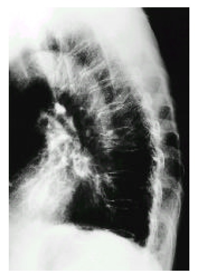
X-rayLateral chest radiograph showing vertebral compression fractures of T5 and T8 with marked kyphosis, typical of severe osteoporosis.
Source : Min et al., The Korean Journal of Internal Medicine, 1999, figure 2 · CC BY-NC
- Trabecular bone (vertebral bodies, metaphyses): thinning and loss of the horizontal connections, which turns a 3D network into isolated vertical pillars with little resistance to compression, hence vertebral collapse.¹¹
- Cortical bone (diaphyses, femoral neck): thinning through endosteal resorption and increased cortical porosity, which reduces resistance to bending and shear, hence femoral neck fractures.¹⁴
📈 The fracture cascade: every fracture multiplies the next risk
RR of a further fracture after an initial fracture (Kanis 2004 meta-analysis, 11 cohorts)
Sources: Kanis 2004 meta-analysis Bone (PMID 15268886); Johnell & Kanis 2005 hip fracture mortality.9,15
Key points
- Osteoporosis is a silent skeletal disease defined by a DXA T-score ≤ -2.5 or by the occurrence of a fragility fracture (wrist, vertebra, hip, humerus, pelvis).
- Worldwide prevalence 18.3 % with a clear asymmetry: 23.1 % in women against 11.7 % in men (Salari 2021). 9 million fragility fractures a year worldwide (Johnell-Kanis 2006).
- Major risk factors built into FRAX®: age, female sex, a personal history of fracture (the most powerful: RR ≈ 2), a family history of hip fracture, long-term glucocorticoids, low BMI, smoking, alcohol, rheumatoid arthritis.
- Pathophysiology: an imbalance in bone remodelling with osteoclastic resorption > osteoblastic formation, accelerated by the fall in oestrogen after the menopause and by the oxidative stress of ageing (the complementary paradigms of Khosla 2012 and Manolagas 2010).
- The natural course follows a fracture cascade : a first fracture multiplies the next risk by 2 to 5, with a mortality of 20-30 % one year after a femoral neck fracture.
Bibliography
- Kanis JA, Cooper C, Rizzoli R, Reginster JY ; Scientific Advisory Board of the European Society for Clinical and Economic Aspects of Osteoporosis (ESCEO). European guidance for the diagnosis and management of osteoporosis in postmenopausal women. Osteoporos Int. 2019;30(1):3-44. PMID 30324412.
- World Health Organization. Assessment of fracture risk and its application to screening for postmenopausal osteoporosis. WHO Technical Report Series 843. Geneva : WHO ; 1994. (The founding document defining the T-score ≤ -2.5 threshold.)
- Siris ES, Adler R, Bilezikian J, et al. The clinical diagnosis of osteoporosis : a position statement from the National Bone Health Alliance Working Group. Osteoporos Int. 2014;25(5):1439-1443. PMID 24577348.
- Compston JE, McClung MR, Leslie WD. Osteoporosis. Lancet. 2019;393(10169):364-376. PMID 30696576.
- Johnell O, Kanis JA. An estimate of the worldwide prevalence and disability associated with osteoporotic fractures. Osteoporos Int. 2006;17(12):1726-1733. PMID 16983459.
- Salari N, Ghasemi H, Mohammadi L, Behzadi MH, Rabieenia E, Shohaimi S, Mohammadi M. The global prevalence of osteoporosis in the world : a comprehensive systematic review and meta-analysis. J Orthop Surg Res. 2021;16(1):609. doi:10.1186/s13018-021-02772-0 (PMC8522202).
- Hernlund E, Svedbom A, Ivergård M, Compston J, Cooper C, Stenmark J, McCloskey EV, Jönsson B, Kanis JA. Osteoporosis in the European Union : medical management, epidemiology and economic burden. Arch Osteoporos. 2013;8:136. PMID 24113837.
- Kanis JA, Johansson H, Harvey NC, McCloskey EV. A brief history of FRAX. Arch Osteoporos. 2018;13(1):118. PMID 30382424.
- Kanis JA, Johnell O, De Laet C, Johansson H, et al. A meta-analysis of previous fracture and subsequent fracture risk. Bone. 2004;35(2):375-382. PMID 15268886.
- Buckley L, Guyatt G, Fink HA, et al. 2017 American College of Rheumatology Guideline for the Prevention and Treatment of Glucocorticoid-Induced Osteoporosis. Arthritis Care Res (Hoboken). 2017;69(8):1095-1110. PMID 28585410.
- Florencio-Silva R, Sasso GR, Sasso-Cerri E, Simões MJ, Cerri PS. Biology of bone tissue : structure, function, and factors that influence bone cells. Biomed Res Int. 2015;2015:421746. PMID 26247020.
- Khosla S, Oursler MJ, Monroe DG. Estrogen and the skeleton. Trends Endocrinol Metab. 2012;23(11):576-581. PMID 22595550.
- Manolagas SC. From estrogen-centric to aging and oxidative stress : a revised perspective of the pathogenesis of osteoporosis. Endocr Rev. 2010;31(3):266-300. PMID 20051526.
- Cummings SR, Melton LJ. Epidemiology and outcomes of osteoporotic fractures. Lancet. 2002;359(9319):1761-1767. PMID 12049882.
- Johnell O, Kanis JA. An estimate of the worldwide prevalence, mortality and disability associated with hip fracture. Osteoporos Int. 2004;15(11):897-902. PMID 15490120.
How do you assess and diagnose osteoporosis with certainty?
Which questions should you ask to understand the patient and their history?
📝 The history systematically looks for the clinical risk factors built into FRAX® and identified by the ESCEO 2019 and BHOF 2022 guidelines.¹,³ Essential questions:- A personal history of fragility fracture (wrist, vertebra, hip, humerus, pelvis after a low-energy trauma), the most powerful independent predictor.⁴
- Family history : femoral neck fracture in a 1st degree relative.¹
- Age and hormonal status : age at menarche, age at menopause (early menopause < 45 years is a major factor), prolonged amenorrhoea in young women (the triad or REDs).⁵
- Low BMI (< 19 kg/m²) or recent unintentional weight loss (≥ 5 % over 6 months).⁶
- Lifestyle : smoking, alcohol (≥ 3 units a day), dietary calcium intake (1000-1200 mg a day recommended), sun exposure and vitamin D status, level of physical activity.¹,⁷
- Medicines that carry risk : oral glucocorticoids (prednisone equivalent ≥ 5 mg a day for > 3 months, a major cause of secondary osteoporosis), aromatase inhibitors, anti-androgens, thyroid hormones at suppressive doses, enzyme-inducing antiepileptics (phenytoin, carbamazepine, phenobarbital), long-term PPIs, SSRIs, antiretrovirals.⁸
- Comorbidities : rheumatoid arthritis, type 1 diabetes, coeliac disease (look for IgA anti-tTG in a young man or a premenopausal woman with unexplained osteoporosis), inflammatory bowel disease, hypogonadism, primary hyperparathyroidism, hyperthyroidism.¹
- History of falls (number in the past year, circumstances), fear of falling (kinesiophobia), balance impairment, sarcopenia, all essential components of the risk of fracture, and not captured by FRAX®.⁹
- Height loss measured (≥ 4 cm) or reported (≥ 6 cm): strongly suggests undiagnosed vertebral collapse → an indication for spinal imaging.¹⁰
Which clinical tests should be done, and which other conditions must be ruled out?
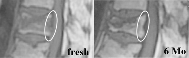
MRIMRI of the lumbar spine, sagittal T1-weighted view: change in posterior wall signal of a collapsed vertebra, circled, between the recent (fresh) stage and 6 months.
Source : Qasem et al., Journal of Orthopaedic Surgery and Research, 2014, figure 2 · CC BY
- DXA (dual-energy X-ray absorptiometry) : gold standard for measuring BMD at the femoral neck, the total hip and the lumbar spine L1-L4. Osteoporosis is diagnosed at a T-score ≤ -2.5 at the lowest site; osteopenia for -2.5 < T-score < -1.0.² The Z-score is used in premenopausal women and in men < 50 (osteoporosis if Z < -2.0).
- TBS (Trabecular Bone Score) : a software texture analysis of the lumbar DXA image that estimates the quality of the trabecular microarchitecture independently of BMD. A low TBS (< 1.230) significantly increases fracture risk and improves FRAX® prediction, particularly in the osteopenic range and in people with type 2 diabetes (Silva 2014, McCloskey 2016).¹¹,¹²
- First-line blood tests : full blood count, ESR, CRP, corrected calcium, phosphate, creatinine plus eGFR, alkaline phosphatase, 25-OH vitamin D, TSH, serum protein electrophoresis (myeloma), urine dipstick, 24-hour urinary calcium. These tests rule out the secondary causes of osteoporosis, present in up to 30 % of cases.¹³
- Spinal imaging : Vertebral Fracture Assessment (VFA) built into the DXA, or lateral T4-L4 radiographs if there is clinical suspicion (height loss ≥ 4 cm, kyphosis, unexplained acute thoracic pain). An undiagnosed prevalent vertebral fracture justifies treatment even if BMD is in the osteopenic range.¹⁰
🚩 Red flags when osteoporosis is suspected
- Focal bone pain, weight loss and night sweats → suspected multiple myeloma or bone metastasis (work-up: serum protein electrophoresis, immunofixation, light chains, bone scan or spinal MRI)
- Hypercalcaemia → primary hyperparathyroidism, myeloma, osteolytic metastasis, sarcoidosis
- Unexplained anaemia, renal failure and monoclonal proteinuria → multiple myeloma
- Very high alkaline phosphatase plus bone pain → Paget's disease, metastasis, osteomalacia
- Persistent hypophosphataemia → osteomalacia (severe vitamin D deficiency, malabsorption, tubulopathy)
- Severe osteoporosis in a young man or a premenopausal woman → a broadened aetiological work-up is essential (hypogonadism, hyperthyroidism, coeliac disease, hypercortisolism, mastocytosis)
- Vertebral fracture above T4 or fracture after minimal trauma in a young person with no risk factors → always consider a secondary or malignant cause
⚠️ Any red flag → prompt medical referral for an aetiological work-up before starting anti-osteoporotic treatment.
Should patients be classified and risk stratified with FRAX?
📊 Yes, risk stratification is the key step that goes beyond reading the T-score alone. It identifies who will gain most from drug treatment. The FRAX® tool (WHO Collaborating Centre, Sheffield, launched in 2008, with country-specific models) calculates the 10-year probability of a major osteoporotic fracture (hip, clinical vertebral, humerus, wrist) and of hip fracture alone, combining: age, sex, BMI, previous fracture, parental hip fracture, smoking, alcohol ≥ 3 units a day, glucocorticoids, rheumatoid arthritis, secondary osteoporosis, and femoral neck BMD (optional).⁸ Further adjustments are possible: corticosteroid dose dependence, type 2 diabetes (underestimated by FRAX), TBS, falls score.¹| Risk level (NOGG / IOF) | Criterion | Treatment strategy | Evidence |
|---|---|---|---|
| Low | Below the FRAX intervention threshold | Lifestyle and dietary advice, exercise, calcium and vitamin D | GRADE high |
| Intermediate | Close to the FRAX intervention threshold | Measure BMD if not already done, recalculate FRAX, shared decision | GRADE moderate |
| High | Above the FRAX intervention threshold OR T-score ≤ -2.5 OR previous fragility fracture | Oral or IV bisphosphonate first line | GRADE high |
| Very high | Recent vertebral or hip fracture (< 2 years) OR multiple fractures OR T-score ≤ -3.0 OR fracture while on treatment OR FRAX hip > 4.5 % | Bone-forming agent first line (teriparatide, romosozumab) then switch to an antiresorptive | GRADE high (Gregson 2022 NOGG) |
🧭 Simplified FRAX algorithm: from screening to the treatment decision
Adapted from NOGG UK 2022/2024, ESCEO 2019 and BHOF 2022
Summary algorithm. Sources: Kanis ESCEO 20191, LeBoff BHOF 20223, Gregson NOGG 202215, Camacho AACE 202016.
Key points
- Assessment combines a targeted history (clinical risk factors), DXA (T-score), TBS for microarchitecture, blood tests to rule out secondary causes (30 % of cases), and spinal imaging if collapse is suspected.
- The FRAX® tool calculates the 10-year probability of a major fracture and of hip fracture and stratifies risk as low, high or very high. It guides the treatment decision beyond the T-score alone.
- The « very high risk » subgroup (recent fracture, T-score ≤ -3, multiple fractures) benefits from a bone-forming agent first line (teriparatide, romosozumab) then a switch to an antiresorptive, the key innovation of the 2020-2024 guidelines.
- Red flags never to overlook: focal bone pain plus weight loss → myeloma; hypercalcaemia → hyperparathyroidism; severe osteoporosis in a young man → a broadened aetiological work-up is mandatory (coeliac disease, hypogonadism, mastocytosis).
- The real challenge is not the tool but the treatment gap : < 20 % of patients are treated after a fracture. Fracture Liaison Services are the most effective organisational solution.
Bibliography
- Kanis JA, Cooper C, Rizzoli R, Reginster JY. European guidance for the diagnosis and management of osteoporosis in postmenopausal women. Osteoporos Int. 2019;30(1):3-44. PMID 30324412.
- Compston JE, McClung MR, Leslie WD. Osteoporosis. Lancet. 2019;393(10169):364-376. PMID 30696576.
- LeBoff MS, Greenspan SL, Insogna KL, et al. The clinician's guide to prevention and treatment of osteoporosis. Osteoporos Int. 2022;33(10):2049-2102. PMID 35478046.
- Kanis JA, Johnell O, De Laet C, Johansson H, et al. A meta-analysis of previous fracture and subsequent fracture risk. Bone. 2004;35(2):375-382. PMID 15268886.
- Cauley JA. Public health impact of osteoporosis. J Gerontol A Biol Sci Med Sci. 2013;68(10):1243-1251. PMID 23902935.
- De Laet C, Kanis JA, Odén A, Johanson H, Johnell O, et al. Body mass index as a predictor of fracture risk : a meta-analysis. Osteoporos Int. 2005;16(11):1330-1338. PMID 15928804.
- Cummings SR, Melton LJ. Epidemiology and outcomes of osteoporotic fractures. Lancet. 2002;359(9319):1761-1767. PMID 12049882.
- Buckley L, Guyatt G, Fink HA, et al. 2017 American College of Rheumatology Guideline for the Prevention and Treatment of Glucocorticoid-Induced Osteoporosis. Arthritis Care Res (Hoboken). 2017;69(8):1095-1110. PMID 28585410.
- Sherrington C, Fairhall NJ, Wallbank GK, et al. Exercise for preventing falls in older people living in the community. Cochrane Database Syst Rev. 2019;1(1):CD012424. PMID 30703272.
- Siminoski K, Warshawski RS, Jen H, et al. The accuracy of historical height loss for the detection of vertebral fractures in postmenopausal women. Osteoporos Int. 2006;17(2):290-296. PMID 16143833.
- Silva BC, Leslie WD, Resch H, et al. Trabecular bone score : a noninvasive analytical method based upon the DXA image. J Bone Miner Res. 2014;29(3):518-530. PMID 24443324.
- McCloskey EV, Odén A, Harvey NC, et al. A meta-analysis of trabecular bone score in fracture risk prediction and its relationship to FRAX. J Bone Miner Res. 2016;31(5):940-948. PMID 26498132.
- Hennings J, Lalla M, Wallin G. Causes of secondary osteoporosis. Best Pract Res Clin Endocrinol Metab. 2008;22(5):737-754. (The classic source cited by every guideline for the concept ; the detailed blood work-up is in Kanis 2019 and LeBoff 2022.)
- Shoback D, Rosen CJ, Black DM, Cheung AM, Murad MH, Eastell R. Pharmacological management of osteoporosis in postmenopausal women : an Endocrine Society guideline update. J Clin Endocrinol Metab. 2020;105(3):587-594. PMID 32068863.
- Gregson CL, Armstrong DJ, Bowden J, et al. UK clinical guideline for the prevention and treatment of osteoporosis. Arch Osteoporos. 2022;17(1):58. PMID 35378630.
- Camacho PM, Petak SM, Binkley N, et al. American Association of Clinical Endocrinologists / American College of Endocrinology Clinical Practice Guidelines for the Diagnosis and Treatment of Postmenopausal Osteoporosis — 2020 Update. Endocr Pract. 2020;26(Suppl 1):1-46. PMID 32427503.
- Eastell R, Rosen CJ, Black DM, Cheung AM, Murad MH, Shoback D. Pharmacological management of osteoporosis in postmenopausal women : an Endocrine Society clinical practice guideline. J Clin Endocrinol Metab. 2019;104(5):1595-1622. PMID 30907953.
Which treatment strategies are the most effective for osteoporosis?
Where do you start? What is the recommended hierarchy of interventions?
The hierarchy is clear:- Assessment and risk stratification (FRAX + DXA + TBS + secondary work-up) → see chapter 2.
- Universal baseline measures for every patient at risk:
- Calcium intake : 1000-1200 mg a day, from food first (dairy products, calcium-rich mineral waters, green vegetables). Supplement only when a deficiency is documented.⁵
- Vitamin D : 800-1000 IU a day, adjusted to reach a 25-OH-D ≥ 75 nmol/L (30 ng/mL). Deficiency affects ≥ 30 % of people in France and should be corrected systematically.⁸
- Stopping smoking and moderating alcohol (< 2 units a day).⁵
- Multicomponent exercise (see below), a non-negotiable part of management.¹,²
- Falls prevention : medication review (psychotropics, antihypertensives that carry risk), correcting vision, suitable footwear, home adaptation.⁹
- Drug treatment for patients at high risk or who have had a fragility fracture.
| Class | Drug | Fracture risk reduction | Main indication | Level of evidence |
|---|---|---|---|---|
| Oral bisphosphonate | Alendronate 70 mg weekly Risedronate 35 mg weekly | Vertebral -45 %; hip -30 %; non-vertebral -16 to -23 % | 1st line, high risk | GRADE high |
| IV bisphosphonate | Zoledronate 5 mg yearly | Vertebral -70 %; hip -41 %; non-vertebral -25 % | High risk, oral intolerance, poor adherence | GRADE high |
| Anti-RANKL antibody | Denosumab 60 mg every 6 months SC | Vertebral -68 %; hip -40 %; non-vertebral -20 % | Renal failure (eGFR < 35), bisphosphonate intolerance | GRADE high |
| PTH bone-forming agent | Teriparatide 20 µg a day SC (24-month course) | Vertebral -65 %; non-vertebral -35 % | Very high risk, recent fracture, antiresorptive failure | GRADE high |
| Anti-sclerostin | Romosozumab 210 mg monthly SC (12 months) | Vertebral -73 %; non-vertebral -19 % (vs alendronate, ARCH) | Very high risk; switching to an antiresorptive is mandatory | GRADE moderate (a cardiovascular signal to watch) |
| SERM | Raloxifene 60 mg a day | Vertebral -30 %; no effect on the hip | Women at high breast cancer risk | GRADE moderate |
| MHT | Oestradiol ± progestogen | Any fracture -25 to -40 % | Women < 60 years or menopause < 10 years, vasomotor symptoms | GRADE moderate (NOGG 2024) |
- The denosumab antibody is very effective but carries a risk of rebound fracture if it is stopped without a follow-on treatment: multiple vertebral fractures from the rebound effect can occur 7-18 months after the last injection. Switching to a bisphosphonate within 7 months of the last dose is mandatory under current guidelines.¹⁰
- The romosozumab antibody showed superiority over alendronate in the ARCH trial (12 months of romosozumab then alendronate, against alendronate alone) for preventing vertebral (-48 %) and non-vertebral (-19 %) fractures. A cardiovascular signal (ischaemic heart disease in particular) calls for caution in patients at high cardiovascular risk; the 2024 meta-analyses are reassuring but heterogeneous.¹¹
- Menopausal hormone therapy (MHT) is repositioned as a first-line option in the NOGG 2024 guidelines for young postmenopausal women (< 60 years, < 10 years since the menopause) at high fracture risk and low thromboembolic and breast cancer risk, a notable change from previous decades.⁴
What is the place of exercise, and is there a superior approach?
🏋️♀️ Exercise is non-negotiable at every stage. It has two aims: (1) to stimulate osteogenesis in order to preserve or improve BMD, and (2) to reduce the risk of falling through muscle strengthening, balance and coordination. The most recent reviews converge on a multicomponent combination.¹,²,¹²,¹³ Essential components of an optimal programme (synthesis of Brooke-Wavell 2022, Giangregorio 2014, Beck ESSA 2017, Pinheiro 2020 WHO):- Progressive resistance training (heavy loads) : the most powerful stimulus for bone. LIFTMOR-type programmes (Watson 2018, RCT n=101): 5 sets of 5 repetitions at ≥ 85 % of 1RM (squat, deadlift, press, pull-up with impact), twice a week, 8 months → femoral neck +0.3 %, lumbar spine +2.9 %, with better strength and function.¹³ Safe in postmenopausal women with osteopenia or osteoporosis when it is supervised.
- Impact activities : brisk walking, light jogging, stair climbing, targeted jumps. Even brief impacts (10-20 jumps a day at twice body mass) stimulate bone formation at weight-bearing sites.¹²
- Balance and proprioception training : Tai Chi, exercises on unstable surfaces, Otago-type programmes. Sherrington 2019 (Cochrane, 108 RCTs, 23,407 participants): combining balance with resistance reduces the rate of falls by 34 %, and Tai Chi alone by 19 %.¹
- Posture and thoracic extension : strengthening the trunk extensors (without forced spinal flexion), particularly for patients with vertebral collapse. It improves posture and reduces the risk of falling forwards.¹⁴
🏋️ Comparative effectiveness of exercise modes (fall reduction, Sherrington 2019)
Cochrane meta-analysis, 108 RCTs, 23,407 participants
Relative reductions in the rate of falls against control. Source: Sherrington C, et al. Cochrane Database Syst Rev. 2019;1(1):CD012424. PMID 30703272
Manual therapies, whole-body vibration: how effective are they really?
In the light of the available evidence:- Manual therapies (mobilisations, manipulations) : no high-quality evidence that they increase BMD or prevent osteoporotic fractures. They may be useful as an adjunct for treating associated musculoskeletal pain or for improving mobility, but they are not a treatment for osteoporosis in themselves. Use them with caution in patients with severe osteoporosis (high-velocity manipulation carries an iatrogenic fracture risk).¹⁵
- Whole-body vibration (WBV) : recent meta-analyses (Marín-Cascales 2018, n = 28 RCTs; Luo 2019; and the 2024 systematic overview) conclude that the effect is modest on lumbar and femoral BMD, mainly in women < 65 years with a normal BMI. Effects clearly inferior to a conventional resistance plus impact programme. Possible use as an adjunct in very frail patients who cannot take on intensive active exercise, but never a replacement for the proven active approaches.¹⁶
Beyond the physical: how do you educate the patient and manage the fear of falling?
The effectiveness of any treatment strategy depends on the patient's adherence. Yet adherence to oral anti-osteoporotic treatments is disastrous : below 50 % at 1 year and < 30 % at 2 years in real-world studies, which largely cancels out the benefits demonstrated in RCTs.¹⁷ The effective levers are:- Structured therapeutic education about the disease, its silent nature, the benefit of treatment, the expected side effects and how to handle them.
- Shared decision-making (Shared Decision-Making): present the options with their benefits and risks in numbers (Number Needed to Treat).
- Choosing the dosing schedule that suits the patient: annual IV zoledronate for patients with poor compliance, subcutaneous denosumab for patients with renal failure, and so on.
- Proactive follow-up : telephone reminders, a coordinating nurse, mobile apps.
Key points
- Management is tiered : baseline measures (calcium 1000-1200 mg a day, vitamin D 800-1000 IU a day, multicomponent exercise, stopping smoking) for everyone, then drugs for patients at high risk.
- Bisphosphonates first line (alendronate, risedronate, zoledronate); denosumab effective but the switch is mandatory; bone-forming agents (teriparatide, romosozumab) first line for the « very high risk » group.
- The most effective exercise is multicomponent : progressive resistance (LIFTMOR 2018) + moderate impact + balance (Tai Chi). Falls reduced by up to 34 % (Sherrington 2019 Cochrane).
- The manual therapies and whole-body vibration have no robust evidence of effect on BMD; a possible adjunct role, but not a central one.
- Adherence to oral treatments is below 50 % at 1 year. Therapeutic education, shared decision-making and the choice of dosing schedule are the key levers.
- The fear of falling is a vicious circle to break actively: graded exposure, self-efficacy, CBT.
Bibliography
- Sherrington C, Fairhall NJ, Wallbank GK, Tiedemann A, Michaleff ZA, Howard K, Clemson L, Hopewell S, Lamb SE. Exercise for preventing falls in older people living in the community. Cochrane Database Syst Rev. 2019;1(1):CD012424. PMID 30703272.
- Brooke-Wavell K, Skelton DA, Barker KL, et al. Strong, steady and straight : UK consensus statement on physical activity and exercise for osteoporosis. Br J Sports Med. 2022;56(15):837-846. PMID 35577538.
- Eastell R, Rosen CJ, Black DM, Cheung AM, Murad MH, Shoback D. Pharmacological management of osteoporosis in postmenopausal women : an Endocrine Society clinical practice guideline. J Clin Endocrinol Metab. 2019;104(5):1595-1622. PMID 30907953.
- Gregson CL, Armstrong DJ, Bowden J, et al. UK clinical guideline for the prevention and treatment of osteoporosis. Arch Osteoporos. 2022;17(1):58. PMID 35378630.
- LeBoff MS, Greenspan SL, Insogna KL, et al. The clinician's guide to prevention and treatment of osteoporosis. Osteoporos Int. 2022;33(10):2049-2102. PMID 35478046.
- Camacho PM, Petak SM, Binkley N, et al. AACE / ACE Clinical Practice Guidelines for the Diagnosis and Treatment of Postmenopausal Osteoporosis — 2020 Update. Endocr Pract. 2020;26(Suppl 1):1-46. PMID 32427503.
- Shoback D, Rosen CJ, Black DM, Cheung AM, Murad MH, Eastell R. Pharmacological management of osteoporosis in postmenopausal women : an Endocrine Society guideline update. J Clin Endocrinol Metab. 2020;105(3):587-594. PMID 32068863.
- Holick MF, Binkley NC, Bischoff-Ferrari HA, et al. Evaluation, treatment, and prevention of vitamin D deficiency : an Endocrine Society clinical practice guideline. J Clin Endocrinol Metab. 2011;96(7):1911-1930. PMID 21646368.
- Hopewell S, Adedire O, Copsey BJ, et al. Multifactorial and multiple component interventions for preventing falls in older people living in the community. Cochrane Database Syst Rev. 2018;7(7):CD012221. PMID 30035305.
- Tsourdi E, Zillikens MC, Meier C, et al. Fracture risk and management of discontinuation of denosumab therapy: a systematic review and position statement by ECTS. J Clin Endocrinol Metab. 2020. PMID 33103722.
- Saag KG, Petersen J, Brandi ML, et al. Romosozumab or alendronate for fracture prevention in women with osteoporosis (ARCH). N Engl J Med. 2017;377(15):1417-1427. PMID 28892457.
- Giangregorio LM, Papaioannou A, MacIntyre NJ, et al. Too Fit To Fracture : exercise recommendations for individuals with osteoporosis or osteoporotic vertebral fracture. Osteoporos Int. 2014;25(3):821-835. PMID 24281053.
- Watson SL, Weeks BK, Weis LJ, Harding AT, Horan SA, Beck BR. High-intensity resistance and impact training improves bone mineral density and physical function in postmenopausal women with osteopenia and osteoporosis : the LIFTMOR randomized controlled trial. J Bone Miner Res. 2018;33(2):211-220. PMID 28975661.
- Beck BR, Daly RM, Singh MA, Taaffe DR. Exercise and Sports Science Australia (ESSA) position statement on exercise prescription for the prevention and management of osteoporosis. J Sci Med Sport. 2017;20(5):438-445. doi:10.1016/j.jsams.2016.10.001.
- Pinheiro MB, Oliveira J, Bauman A, Fairhall N, Kwok W, Sherrington C. Evidence on physical activity and osteoporosis prevention for people aged 65+ years : a systematic review to inform the WHO guidelines on physical activity and sedentary behaviour. Int J Behav Nutr Phys Act. 2020;17(1):150. PMID 33239014.
- Marín-Cascales E, Alcaraz PE, Ramos-Campo DJ, Martinez-Rodriguez A, Chung LH, Rubio-Arias JÁ. Whole-body vibration training and bone health in postmenopausal women : a systematic review and meta-analysis. Medicine (Baltimore). 2018;97(34):e11918. PMID 30142802.
- Hiligsmann M, Cornelissen D, Vrijens B, et al. Determinants, consequences and potential solutions to poor adherence to anti-osteoporosis treatment : results of an expert group meeting organized by the European Society for Clinical and Economic Aspects of Osteoporosis, Osteoarthritis and Musculoskeletal Diseases (ESCEO) and the International Osteoporosis Foundation (IOF). Osteoporos Int. 2019;30(11):2155-2165. PMID 31388696.
- Whipple MO, Hamel AV, Talley KMC. Fear of falling among community-dwelling older adults: A scoping review to identify effective evidence-based interventions. Geriatr Nurs. 2018;39(2):170-177. PMID 28941942.
How do you secure lasting recovery and prevent further fragility fractures?
How do you make the patient an active participant through self-management?
Self-management equips the patient with the knowledge, skills and confidence needed to manage their condition actively day to day. Three pillars: 1. Therapeutic education 📚. Structured programmes improve knowledge and adherence, the sine qua non of effectiveness: without adherence there is no reduction in fracture risk, even with the best treatment.¹,² 2. Regular physical activity 🏋️♀️ (see chapter 3 for the modes). On duration: long-term programmes, preferably supervised at least in part, with a structured progression and periodic review. Beyond the direct bone benefits, exercise improves mood, quality of life and self-efficacy, all of them levers for long-term adherence.³ 3. Environmental falls prevention 🏠. More than 90 % of hip fractures follow a fall.⁴ Assess the home (Home Safety Assessment) and adapt it:- Remove unsecured rugs, secure electrical cables
- Improve lighting (hallway, stairs, bathroom, with a night light)
- Fit grab rails (bathroom, toilet)
- Non-slip mats in the shower or bath, bath mat
- Adapt the stairs (handrails on both sides, markings)
- Suitable footwear (flat non-slip sole, ankle support)
- Medication review (psychotropics, hypnotics, antihypertensives that risk orthostatic hypotension)
- Correcting vision (cataract above all, suitable glasses)
When and how do you plan a safe return to sport and to activities?
Returning to an active life, sport included, is a legitimate and encouraged goal. ✅ It must be gradual, individualised and stratified. Initial assessment. Before considering any intensification, assess:- A history of fragility fractures (vertebral ones in particular)
- A recent BMD (DXA) and TBS
- The 10-year FRAX
- Functional assessment: strength (handgrip, sit-to-stand), balance (timed up-and-go, Berg), endurance (6-minute walk test)
- Cardiovascular and pulmonary comorbidities (exercise test if indicated)
- Phase 1, foundation (4-8 weeks) : moderate whole-body strengthening (60-70 % 1RM, 8-12 repetitions, 2-3 times a week), daily balance exercises, low-impact activities (swimming, aqua aerobics, stationary cycling, moderate walking). Establishing good posture, strengthening the trunk extensors (without forced flexion).⁶
- Phase 2, introducing moderate impact (8-16 weeks) : brisk walking with changes of direction, stair climbing, hiking on varied ground, dancing, doubles tennis. Gradual increase in resistance load (up to 80 % 1RM).⁷
- Phase 3, higher-impact activities (only for patients at low fracture risk and with no history of vertebral fracture): moderate jogging, targeted jumps, singles racket sports, cross-country skiing. LIFTMOR-type programmes for those who tolerate high intensity.⁸
🚩 Activities and movements to avoid or approach with caution
- Forced forward flexion of the trunk (touching the toes with straight legs, the classic sit-up, rowing in kyphosis) → a major anterior compressive load on the vertebral bodies, with a risk of collapse
- Loaded spinal rotation (aggressive golf, throwing, some yoga movements)
- Sports with a high risk of falling or collision : difficult downhill skiing, ice skating, horse riding, combat sports, singles and team sports → an unfavourable benefit-risk balance in patients at high risk
- Uncontrolled jumping or trampolining in patients with multiple vertebral fractures or a severe T-score
- Head-down inversion (inverted yoga) when there are vertebral collapses
- Running down steep slopes (repeated braking forces on the spine)
⚠️ The golden rule: every patient who has had a vertebral fracture must be given specific instructions on spinal flexion and twisting before returning to unsupervised sport.
Key points
- Self-management rests on three pillars: therapeutic education, regular physical activity (multicomponent), and environmental falls prevention (home adaptation, medication review, vision correction, footwear).
- Returning to sport is encouraged but stratified : phase 1 (low-impact foundation) → phase 2 (moderate impact) → phase 3 (high impact, reserved for low risk). References Too Fit To Fracture (Giangregorio 2014) and Strong Steady Straight (Brooke-Wavell 2022).
- To be avoided or approached with caution: loaded forward flexion of the trunk (sit-ups, touching the toes with straight legs), loaded rotation, sports with a high risk of falling, uncontrolled jumping in patients with vertebral fractures.
- The golden rule is individualisation according to the risk profile (BMD + FRAX + history + functional capacity + preferences) with a structured progression.
Bibliography
- Hiligsmann M, Cornelissen D, Vrijens B, et al. Determinants, consequences and potential solutions to poor adherence to anti-osteoporosis treatment : ESCEO / IOF expert group meeting. Osteoporos Int. 2019;30(11):2155-2165. PMID 31388696.
- LeBoff MS, Greenspan SL, Insogna KL, et al. The clinician's guide to prevention and treatment of osteoporosis. Osteoporos Int. 2022;33(10):2049-2102. PMID 35478046.
- Pinheiro MB, Oliveira J, Bauman A, Fairhall N, Kwok W, Sherrington C. Evidence on physical activity and osteoporosis prevention for people aged 65+ years : a systematic review to inform the WHO guidelines. Int J Behav Nutr Phys Act. 2020;17(1):150. PMID 33239014.
- Johnell O, Kanis JA. An estimate of the worldwide prevalence, mortality and disability associated with hip fracture. Osteoporos Int. 2004;15(11):897-902. PMID 15490120.
- Hopewell S, Adedire O, Copsey BJ, et al. Multifactorial and multiple component interventions for preventing falls in older people living in the community. Cochrane Database Syst Rev. 2018;7(7):CD012221. PMID 30035305.
- Giangregorio LM, Papaioannou A, MacIntyre NJ, et al. Too Fit To Fracture : exercise recommendations for individuals with osteoporosis or osteoporotic vertebral fracture. Osteoporos Int. 2014;25(3):821-835. PMID 24281053.
- Brooke-Wavell K, Skelton DA, Barker KL, et al. Strong, steady and straight : UK consensus statement on physical activity and exercise for osteoporosis. Br J Sports Med. 2022;56(15):837-846. PMID 35577538.
- Watson SL, Weeks BK, Weis LJ, Harding AT, Horan SA, Beck BR. High-intensity resistance and impact training improves bone mineral density and physical function in postmenopausal women with osteopenia and osteoporosis : the LIFTMOR RCT. J Bone Miner Res. 2018;33(2):211-220. PMID 28975661.
What do real clinical cases teach us about osteoporosis?
Analysis of a classic case: wrist fracture and the fracture cascade
The commonest case is the postmenopausal woman who sustains a first distal radius fracture (Colles fracture), often after a fall from standing height. It is the typical osteoporotic sentinel fracture , and it occurs on average 10 to 15 years before the hip fracture.¹ Without intervention the sequence continues: vertebral collapse (60-70 years), then femoral neck fracture (≥ 75 years). The modern protocol, now standardised in Fracture Liaison Services (FLS), systematically triggers:- Orthopaedic management of the fracture
- Identifying the fracture as a fragility fracture (mechanism: a fall from standing height)
- Risk assessment: DXA + TBS + FRAX, blood tests
- Starting anti-osteoporotic treatment matched to the risk profile
- Setting up the exercise and falls prevention programme
- Structured follow-up at 3, 6 and 12 months
The diagnostic challenge: when osteoporosis mimics (or hides) another condition
Osteoporosis is silent, but its manifestations (chronic thoracic pain, vertebral collapse, height loss) can be confused with other conditions, or conversely a serious condition can hide behind a picture of osteoporosis. 😥 Multiple myeloma is the most feared mimic: it causes diffuse osteolytic lesions that can resemble severe osteoporosis with multiple vertebral collapses (D'Souza 2023, a JAMA review).⁴ The warning signs that point to myeloma, always to be looked for when a case of severe osteoporosis is unusual, are:- Unexplained anaemia (often normochromic normocytic)
- Recent-onset renal failure
- Hypercalcaemia
- A very high erythrocyte sedimentation rate, raised total protein
- A monoclonal peak on serum protein electrophoresis (SPE), the key test in case of doubt
- Focal bone pain out of proportion to the BMD
- Vertebral involvement above T4 (rare in primary osteoporosis)
- No response to analgesics or to anti-osteoporotic treatment
- Bone metastases (breast, lung, prostate, kidney, thyroid cancer, melanoma)
- Primary hyperparathyroidism (hypercalcaemia + raised PTH)
- Osteomalacia (defective mineralisation, severe vitamin D deficiency, malabsorption)
- Paget's disease (very high alkaline phosphatase)
- Systemic mastocytosis (rare but a cause of severe osteoporosis in young people)
- Fibrous dysplasia
Complex case studies: a young man, glucocorticoid-induced disease, atypical femoral fracture
Case 1, osteoporosis in a young man (a teaching case: undiagnosed coeliac disease). A 28-year-old man with chronic back pain, height loss and iron-deficiency anaemia. DXA: T-score -3.2 at the spine. Blood tests: vitamin D deficiency, low calcium, positive IgA anti-tTG. Diagnosis: coeliac disease with malabsorption of calcium and vitamin D, causing severe secondary osteoporosis. Treatment: a strict gluten-free diet, calcium and vitamin D supplementation and a bisphosphonate. BMD recovery can be substantial after several years of strict diet. Key lesson: any osteoporosis in a young man or a premenopausal woman calls for a broadened aetiological work-up.⁵ Case 2, glucocorticoid-induced osteoporosis. A 55-year-old woman with rheumatoid arthritis treated with prednisone 10 mg a day for 3 years. Recent atraumatic vertebral fracture (T8). DXA: T-score -2.8. Glucocorticoid-induced osteoporosis (GIOP) is the commonest cause of secondary osteoporosis; the pathophysiology is twofold: reduced bone formation (apoptosis of osteoblasts and osteocytes) and increased resorption (RANKL induction). Preventive management is crucial from the moment prolonged corticosteroid treatment begins (≥ 3 months, dose ≥ 5 mg a day of prednisone equivalent) under the ACR 2017 recommendations (Buckley): calcium plus vitamin D for everyone, and a bisphosphonate or denosumab if the risk is moderate to high.⁶ Case 3, atypical femoral fracture (AFF) on long-term bisphosphonate. A 76-year-old woman on alendronate for 9 years for postmenopausal osteoporosis. Progressive right subtrochanteric pain for 3 months, then a spontaneous fracture. Characteristic radiographs: a transverse or short oblique fracture of the femoral shaft, with lateral cortical thickening, in the subtrochanteric region or the upper shaft. Work-up: confirmation by bone scan or MRI (contralateral lesions are often associated). The Black 2020 NEJM study (a cohort of 196,129 women) confirms that AFFs are rare but that their incidence rises with the duration of bisphosphonate exposure (RR ×3 after 3 years, ×8 after 5 years, ×43 after 8 years), while remaining overall < 1 per 1,000 patient-years.⁷ The net benefit remains very largely in favour of treatment for patients at high risk, but these data justify:- An assessment of the benefit-risk balance every 3 to 5 years
- Discussion of a treatment break (drug holiday) in patients at low residual risk after 3-5 years of oral bisphosphonate or 3 years of IV zoledronate
- Continuing treatment in patients still at high risk (reasonable to continue up to 10 years for alendronate, 6 years for zoledronate)
- Faced with any thigh pain on long-term bisphosphonate: bilateral radiographs of the whole femur
📊 Pyramid of the levels of evidence, the GRADE / Oxford CEBM hierarchy
To read the clinical cases in the light of the literature
Simplified GRADE / Oxford CEBM hierarchy. Practical implication: if a seductive clinical case diverges from a meta-analysis, follow the meta-analysis. Case reports generate hypotheses, flag rare presentations or illustrate clinical reasoning; they never demonstrate effectiveness.
Key points
- A fragility fracture, above all of the wrist, vertebra or hip, is a sentinel fracture that must trigger a systematic investigation of the underlying osteoporosis through a Fracture Liaison Service.
- Always consider the secondary causes of osteoporosis, particularly in a young man, in a premenopausal woman, or in the case of an atypical presentation (coeliac disease, hyperparathyroidism, hypogonadism, hyperthyroidism, mastocytosis, inflammatory bowel disease).
- Faced with an unusual case of « severe osteoporosis » (anaemia, hypercalcaemia, focal bone pain, fracture above T4) → always consider multiple myeloma (SPE, immunofixation, light chains) before starting anti-osteoporotic treatment.
- The atypical femoral fractures on long-term bisphosphonates are rare but real. The anti-fracture benefit of the class far outweighs that risk in patients at high risk, but it justifies periodic review and discussion of treatment breaks in patients at low residual risk.
- Clinical cases illustrate; they do not demonstrate . If there is a divergence with a meta-analysis, follow the meta-analysis (level 1a) rather than the isolated case (level 5).
Bibliography
- Cummings SR, Melton LJ. Epidemiology and outcomes of osteoporotic fractures. Lancet. 2002;359(9319):1761-1767. PMID 12049882.
- Wu CH, Tu ST, Chang YF, et al. Fracture liaison services improve outcomes of patients with osteoporosis-related fractures : a systematic literature review and meta-analysis. Bone. 2018;111:92-100. PMID 29555309.
- Li N, Hiligsmann M, Boonen A, et al. The impact of fracture liaison services on subsequent fractures and mortality : a systematic literature review and meta-analysis. Osteoporos Int. 2021;32(8):1517-1530. PMID 33829285.
- Rajkumar SV, Kumar S. Multiple myeloma current treatment algorithms. Blood Cancer J. 2020;10(9):94. PMID 32989217.
- Larussa T, Suraci E, Nazionale I, et al. Bone mineralization in celiac disease. Gastroenterol Res Pract. 2012;2012:198025. PMID 22737164.
- Buckley L, Guyatt G, Fink HA, et al. 2017 American College of Rheumatology Guideline for the Prevention and Treatment of Glucocorticoid-Induced Osteoporosis. Arthritis Care Res (Hoboken). 2017;69(8):1095-1110. PMID 28585410.
- Black DM, Geiger EJ, Eastell R, et al. Atypical femur fracture risk versus fragility fracture prevention with bisphosphonates. N Engl J Med. 2020;383(8):743-753. PMID 32813950.
- Adler RA, El-Hajj Fuleihan G, Bauer DC, et al. Managing osteoporosis in patients on long-term bisphosphonate treatment : report of a task force of the American Society for Bone and Mineral Research. J Bone Miner Res. 2016;31(1):16-35. PMID 26350171.
How do you apply these recommendations concretely in your practice?
When and to which other professionals should you refer?
A modern physiotherapist's competence lies in recognising the limits of their scope and in identifying the situations that need additional expertise. Referral is not an admission of failure but a mark of competence and clinical safety. Identifying red flags is the first non-negotiable step. The Finucane 2020 international synthesis (JOSPT, IFOMPT) offers a workable clinical framework: what counts is less the presence of an isolated red flag (low positive predictive value) than the combination of signs and the clinical course.¹🚩 Red flags specific to osteoporosis and fragility fractures
- Acute thoracic pain in an older person after an ordinary movement → suspected acute vertebral collapse, immediate imaging
- Focal bone pain at night, weight loss and sweats → myeloma / metastasis / occult cancer
- Rapidly progressive kyphosis plus height loss ≥ 4 cm → multiple vertebral collapses, imaging essential
- Progressive thigh or hip pain on long-term bisphosphonate → suspected atypical femoral fracture (bilateral femoral radiographs)
- Vertebral fracture above T4 or fracture in a young person with no risk factor → secondary or malignant cause
- Severe osteoporosis in a man < 60 years or a premenopausal woman → a broadened aetiological work-up is mandatory
- Neurological signs with spinal pain (radicular pain, motor deficit, sphincter disturbance, cauda equina syndrome) → urgent imaging plus a surgical opinion
- Multiple fractures despite well-conducted anti-osteoporotic treatment → reconsider the diagnosis (a secondary cause? adherence? an indication for a bone-forming agent?)
⚠️ Any red flag → prompt medical referral (general practitioner, rheumatologist, emergency department if there are neurological signs) before physiotherapy follow-up continues.
- Rheumatologist / endocrinologist : indication for or change of drug treatment, complex secondary osteoporosis, bone-forming agents
- Nutrition physician / dietitian : optimising calcium and vitamin D intake, managing eating disorders
- Occupational therapist : home adaptation, assistive devices, adapting activities of daily living
- Psychologist : kinesiophobia, depression, anxiety, chronic pain management
- Sports physician : return to sport after a fracture, osteoporosis in athletes
- Gynaecologist / endocrinologist : MHT, early menopause, hypogonadism
- Orthopaedic surgeon : severe fracture, vertebroplasty or kyphoplasty after an unstable collapse
How do you measure outcomes and overcome the barriers to implementation?
📊 Measuring objectively and subjectively is essential. The PROMs (patient-reported outcome measures) most used in osteoporosis practice are:- VAS / numerical rating scale for pain (0-10)
- Patient-Specific Functional Scale (PSFS) : activities that matter to the patient (0-10 per activity)
- Oswestry Disability Index (ODI) or Roland-Morris for chronic thoracic pain
- QUALEFFO-41 : Quality of Life Questionnaire of the European Foundation for Osteoporosis, specific to quality of life in osteoporosis
- FES-I (Falls Efficacy Scale International) : fear of falling
- SF-36 / EQ-5D : general quality of life
- FRAX : recalculated periodically to follow how the overall risk changes
- A lack of clinical time
- A lack of skills in literature searching and critical appraisal
- Insufficient organisational support (no time set aside for continuing education)
- Limited access to scientific databases
- Reimbursement models that reward quantity rather than quality
- Interactive continuing education (workshops, mentoring) rather than passive reading
- Clinical leadership : EBP « champions » within organisations
- Audit and feedback : analysing your own practice and comparing it with the recommendations
- Technological tools : electronic records with automatic reminders (FRAX calculation, post-fracture screening)
- Engaging the patient as an active partner (Shared Decision-Making)
Key points
- Medical referral is required in the face of any red flag (acute thoracic pain, a fracture in a young person, thigh pain on long-term bisphosphonate, severe osteoporosis in a young man, neurological signs).
- The interprofessional collaboration (rheumatologist, endocrinologist, gynaecologist, occupational therapist, psychologist, sports physician) is essential for complex cases and for coordinating care.
- Measure outcomes with validated PROMs (VAS, PSFS, QUALEFFO-41 specific to osteoporosis, FES-I for fear of falling) plus functional tools (timed up-and-go, Berg, handgrip).
- The Fracture Liaison Services are the most effective organisational answer to the treatment gap after a fracture.
- The barriers to EBP (time, training, support) are overcome by a combination of strategies: interactive continuing education, clinical leadership, audit and feedback, technological tools, and active patient engagement.
Bibliography
- Finucane LM, Downie A, Mercer C, et al. International framework for red flags for potential serious spinal pathologies. J Orthop Sports Phys Ther. 2020;50(7):350-372. PMID 32438853.
- Wu CH, Tu ST, Chang YF, et al. Fracture liaison services improve outcomes of patients with osteoporosis-related fractures : a systematic literature review and meta-analysis. Bone. 2018;111:92-100. PMID 29555309.
- Li N, Hiligsmann M, Boonen A, et al. The impact of fracture liaison services on subsequent fractures and mortality : a systematic literature review and meta-analysis. Osteoporos Int. 2021;32(8):1517-1530. PMID 33829285.
- Ivers N, Jamtvedt G, Flottorp S, et al. Audit and feedback : effects on professional practice and healthcare outcomes. Cochrane Database Syst Rev. 2012;(6):CD000259. PMID 22696318.
- Greenhalgh T, Howick J, Maskrey N. Evidence based medicine : a movement in crisis? BMJ. 2014;348:g3725. PMID 24927763.
- Chiarotto A, Boers M, Deyo RA, et al. Core outcome measurement instruments for clinical trials in nonspecific low back pain. Pain. 2018;159(3):481-495. PMID 29194127.
- LeBoff MS, Greenspan SL, Insogna KL, et al. The clinician's guide to prevention and treatment of osteoporosis. Osteoporos Int. 2022;33(10):2049-2102. PMID 35478046.
What next after reading this?
This article is part of a collection of evidence-based clinical syntheses. A question, a comment, a correction to suggest? Contact us directly through the WhatsApp button at the bottom right of the screen.

