














OnlineFIFPL


Neurological rehabilitation
Neurology and geriatrics
Bryan Littré · Lucas Meilhou
Go further
Vertebral compression fracture: a quarter to a third are recognised, and 19.2 % recur within a year. Red flags, imaging, vertebroplasty and exercise.
An osteoporotic vertebral fracture is not rare, it is rarely named: only a quarter to a third are recognised at the time they occur.
19.2%vertebral refracture within the year following an incident fracture
What follows covers each of these points in detail, with sources. It is there if you need it.
This topic is taught in a course: Neurology and geriatrics, online with Bryan Littré.
1 / 14
13 article chapters · 57 min in total
Under-diagnosis◔ 4 min
The vertebral body gives way without impact, under an everyday load: carrying a shopping bag, coughing, bending.
Suspecting it◔ 6 min
No single red flag is enough; it is their combination that is informative.
Imaging◔ 5 min
The lateral radiograph shows the deformity, not whether it is recent: that is for MRI.
Differentials◔ 3 min
The real trap is not confusing the compression fracture: it is not thinking of it at all.
Refracture risk◔ 5 min
A first vertebral fracture multiplies by 5.1 the risk of another within the year.
Preventing◔ 5 min
Three levers, and none consists of treating the bone directly: spotting, reducing falls, strengthening.
Vertebroplasty◔ 6 min
Twenty years of trials on vertebroplasty give results that contradict one another.
Bracing◔ 2 min
The spinal brace is prescribed very widely, but the literature that supports it is thin.
Rehabilitation◔ 5 min
Physiotherapy after vertebral fracture has not demonstrated much, but is still worth doing well.
Spinal flexion◔ 5 min
Loaded flexion is to be avoided in the early phase, but lifelong bans are counterproductive.
Case reports◔ 4 min
Four cases show the possible trajectories, from a good outcome to a missed diagnosis.
In practice◔ 3 min
Four decisions sum it all up: thinking of the fracture, knowing when to stop, knowing what to do, treating the bone.
Questions◔ 4 min
The useful marker is not a fixed duration but a direction: the pain should decrease.
Train on this
Neurological rehabilitation
Bryan Littré · Lucas Meilhou
Next comes practice: the course that teaches this topic, with Bryan Littré.
Our courses on this topic may be covered by DPC or FIFPL, the French continuing-education funding schemes, depending on your profession and situation.
A 76-year-old woman comes in with back pain that started « just like that », with no fall and no awkward movement. It hurts when she gets up from a chair, and eases when she lies down. The picture looks like ordinary mechanical thoracic spine pain, and in one case in two to one in three it is a vertebral fracture that has just happened and that nobody will name.1
This article deals with vertebral fracture as a reason for consulting : how to spot it when nothing points to it, what should interrupt the session, and what can be done next. For the bone disease itself (densitometry, T-score thresholds, anti-osteoporotic drugs, primary prevention), see Osteoporosis and the prevention of fragility fractures.
Three figures that govern what to do
What the literature establishes about detection, recurrence and mortality.
Sources: clinical recognition, Schousboe 2016, review of the epidemiology of vertebral fractures1 ; recurrence at one year, Lindsay 2001, analysis of 2,725 women from the placebo arms of four trials4 ; mortality, Bliuc 2009, Dubbo prospective cohort6.
What to take away in one minute
Because they have neither the noise nor the staging of a fracture. There is no spectacular fall, no sudden loss of function, no deformity visible under the skin. There is a back that hurts in someone whose back is expected to hurt.
The word « fracture » suggests an event. This one is not. The osteoporotic vertebral body does not break under an impact: it gives way under a load that healthy bone would have taken without flinching. Carrying a shopping bag, pulling a sheet, coughing, bending to lace a shoe, sitting down a little heavily, and the history often reports nothing more. In some patients there is not even that: the fracture is found months later on a radiograph taken for something else.
The reference figure comes from Schousboe's review published in the Journal of Clinical Densitometry : only a quarter to a third of vertebral fractures are recognised clinically at the time they occur, the others requiring lateral imaging of the spine to be detected.1 This is a wording that must be read exactly: it does not say that two thirds of fractures are asymptomatic, it says that they are not recognised. The nuance moves where the responsibility lies. Many of these patients were in pain. They did seek help. They were told it was their age.
The osteoporotic vertebral fracture is not silent: it is inaudible. It speaks the same language as ordinary thoracic spine pain, in a population where ordinary thoracic spine pain is the norm.
Under-diagnosis does not stop at the practice door. It runs through the imaging chain as well. An Irish study reviewed the chest CT angiograms performed in patients admitted for hip fracture between 2010 and 2017: of the 225 patients whose images were available, 40 % had at least one vertebral fracture and 20 % had several. Only one radiology report in five mentioned it. And only 24 % of patients left hospital on anti-osteoporotic treatment, with no difference between those who had a vertebral fracture and those who did not.2
In other words, in patients already admitted for a fragility fracture, whose vertebrae had already been photographed, whose fracture was already on the image, the chain did not conclude. That is the chain the physiotherapist can interrupt, because they are often the professional who sees these patients for the longest and the most often.
Four measures of the gap between what happens and what gets treated
Every step of the chain loses patients: the fracture occurs, the image exists, the report says nothing, the treatment never starts.
Source: Kelly et al. 2021, Journal of Clinical Densitometry, retrospective cohort of 2,122 hip fractures, 225 CT angiograms re-read blind and graded according to Genant.2
The European prospective EPOS study remains the reference measure on this continent: 14,011 men and women aged 50 and over, recruited in 29 centres, radiographed and then re-radiographed on average 3.8 years later. The age-standardised incidence of morphometric fractures was 10.7 per 1,000 person-years in women and 5.7 in men, with a marked rise with age in both sexes.3
Two practical lessons hide in there. The first is that men are not spared: the ratio is about two to one, not ten to one. A 78-year-old man complaining of back pain deserves exactly the same vigilance as a woman of the same age. The second is that incidence climbs steeply with age, which shifts the pre-test probability with every decade: the same clinical picture does not mean the same thing at 60 and at 82.
Incidence of vertebral fractures in Europe
Two ways of reading the same films, two sexes, and the order of magnitude stays the same.
Source: European Prospective Osteoporosis Study Group 2002, Journal of Bone and Mineral Research ; 14,011 subjects aged 50 and over, 29 European centres, paired films at 3.8 years on average, fracture defined by a height loss of 20 % and 4 mm.3
Key points
The honest answer is: not with a single sign. Twenty years of research have demolished the idea that one red flag on its own is enough. What remains, and what can be used in the clinic, is a way of counting.
The 2023 Cochrane review by Han and Hancock brought together fourteen diagnostic studies comparing history-taking and physical examination with reference imaging. Its conclusion is blunt: most red flags are not useful for screening for vertebral fracture in a patient with low back pain. Those that do inform, in primary care, are trauma (positive likelihood ratio 6.42; 95 % CI 2.94 to 14.02 for osteoporotic fracture specifically) and corticosteroid therapy (2.46; 1.13 to 5.34). Older age informs mainly for unspecified vertebral fracture. And above all: combinations of red flags do better than items taken on their own.8
Downie's review published in the BMJ in 2013 had already given the measure in post-test probabilities, which speak more plainly than likelihood ratios: in a patient with low back pain, prolonged corticosteroid therapy raises the probability of fracture to 33 % (CI 10 to 67), older age to 9 % (3 to 25), severe trauma to 11 % (8 to 16). And when several red flags are present at the same time, the probability of spinal fracture rises to 90 % (34–99).9 The intervals are wide, since these are small studies, but the direction is constant.
One tool deserves to be known, because it was built for and by manual therapists, and because the 2023 Cochrane review kept it as the most informative combination in its review. Roman and Cook analysed more than 1,400 patients from a spine surgery centre and isolated a cluster of five features:10
Two positive items or fewer give a sensitivity of 0.95 (CI 0.83 to 0.99) and a negative likelihood ratio of 0.16 (0.04 to 0.51): fracture becomes unlikely, which is the most useful function day to day. Four items out of five give a positive likelihood ratio of 9.6 (3.7 to 14.9), recalculated to 9.62 (5.88 to 15.73) by the Cochrane review from the raw data.810
It must be said what this tool is not. It was derived in the population of a specialised surgical centre, where the prevalence of fracture is far higher than in a community practice, and the authors themselves write that prospective validation in a separate sample is still needed. It has not taken place. The cluster is therefore a structured aide-memoire, not an approved test: it organises doubt, it does not replace it.
What the signs available in the clinic are really worth
Positive likelihood ratios. Above 5, a sign usefully shifts the probability; between 1 and 2, it changes almost nothing.
Sources: Roman cluster, Roman et al. 201010 ; weight, wall-occiput and rib-pelvis, Green et al. 2004, JAMA, clinical examination review11 ; trauma and corticosteroid therapy for osteoporotic fracture, 2023 Cochrane review8 ; height loss, Mikula et al. 2017, 66,021 patients.12 The confidence intervals appear in the body of the text; they are wide for several of these signs.
The clinical examination review published in JAMA by Green and colleagues assessed the physical manoeuvres for diagnosing osteoporosis and occult spinal fracture. Three of them need no equipment:11
Documented height loss deserves a mention of its own, because it is often quoted without its figures. Mikula's study of 66,021 patients shows a specificity that climbs fast but a sensitivity that collapses : at 1, 2, 3 and 4 cm of height loss, the sensitivity for detecting a vertebral fracture is 42, 32, 19 and 14 % and the specificity 70, 82, 92 and 95 %.12 Clinical translation: a documented height loss of 3 or 4 cm justifies investigation, but the absence of height loss rules out nothing at all and even at the most sensitive threshold, 1 cm, close to six fractures in ten go undetected.
Red flags: what should stop or change the session
None of these items requires « doing nothing at all ». Each one requires not doing what was planned and getting a medical opinion before resuming.
⚠️ The safety rule fits in one sentence: until a recent fracture has been ruled out, do not manipulate, do not mobilise forcefully, do not apply posteroanterior pressure on the spinous processes, and do not load the spine in flexion.
Key points
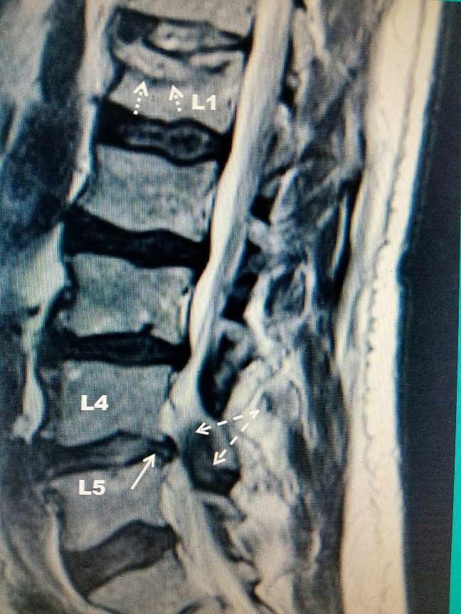
MRISagittal T2-weighted MRI of the lumbar spine: L1 compression fracture indicated by an arrow, with associated stenosis at L4-L5.
Source : Hatgis et al., Cureus, 2017, figure 1 · CC BY
The physiotherapist does not prescribe imaging, but is often the one who triggers the request. Knowing which examination answers which question avoids sending a patient away with a radiograph that will not say what you wanted to know.
« Is there a vertebral deformity? » The radiograph of the thoracic and lumbar spine lateral view answers that one. The lateral view is not a detail: it is the projection on which loss of vertebral body height is read, and it is precisely the imaging the literature names as necessary to recognise the fractures that escape clinical detection.1 Grading follows Genant's semiquantitative method, still the reference thirty years after its publication, and whose founding paper establishes above all its inter- and intra-observer reproducibility.13 In its working form it grades the loss of vertebral body height: grade 1 for about 20 to 25 %, grade 2 for 25 to 40 %, grade 3 beyond 40 %.
« Is this fracture recent? » The radiograph does not say. A grade 2 compression on a film carries no date: it may be six weeks or six years old. It is MRI that answers, through the presence of bone marrow oedema (high signal on STIR or fat-saturated T2, low signal on T1). It is the examination that separates the vertebra that has just given way from the one that gave way long ago, and it is that distinction that governs everything else: the first demands caution, the second allows progressive loading.
CT can settle the question in some cases when MRI is unavailable or contraindicated. A German study of 192 fractures measured the ability of multislice CT alone to separate recent or subacute fractures from old ones: the area under the curve reaches 0.854 and 0.861 depending on the reader, with a sensitivity of 97.2 and 94.5 % but a specificity of only 58.1 and 65.1 % when doubtful cases are classified as recent.14 In other words, CT misses few recent fractures but over-diagnoses many: it reassures badly. One sign is the exception, double trabecular compaction, present in about half of recent fractures but highly specific (93.2 and 88.6 %).14
« Does this patient have untreated osteoporosis? » That one is bone densitometry, and it is the general practitioner or the rheumatologist who requests it. The point the physiotherapist needs to know is that in France the updated recommendations of the Société française de rhumatologie and the GRIO recommend drug treatment in women with a severe fracture, vertebral fracture belonging to that category, without requiring a prior densitometric threshold.35 A patient coming out of a vertebral fracture with no bone treatment at all is not a borderline case: it is a departure from the recommendation, and flagging it is part of the job.
| Examination | Answers | Does not answer | What it changes in the session |
|---|---|---|---|
| Lateral radiograph, thoracic and lumbar spine | Is there a deformity? Of what Genant grade13? | Since when. An old compression fracture and yesterday's have the same image. | Confirms the underlying bone and the number of levels involved; on its own it does not allow loading to be authorised. |
| MRI with STIR or fat-saturated T2 sequence | Is the fracture recent? Is there bone marrow oedema? Is there retropulsion of the posterior wall, neurological compression? | Bone mineral density. | This is the examination that does or does not authorise progressive loading and the return to resisted exercise. |
| CT | The cortex, the geometry, the posterior wall; approximate dating. | Reliable dating: specificity of 58 and 65 % depending on the reader for « recent ».14 | Useful as a second line; does not remove the need for MRI when the question is how fresh the fracture is. |
| Bone densitometry | The T-score, hence the state of the bone and the drug indication. | The existence of a fracture, which it does not see outside a dedicated vertebral assessment module. | Nothing directly, but its absence in a fractured patient is a signal to pass on.35 |
Decision tree for spinal pain in the older patient
The question is not « should everyone be imaged », it is « does this particular patient cross a threshold ».
Editorial construction from the published thresholds: red flags and combinations, 2023 Cochrane review8 and Downie 20139 ; five-item cluster, Roman 201010 ; Genant grading13. This algorithm has not been validated prospectively as such.
Key points
The differential diagnosis works in both directions. You have to be able to recognise the compression fracture beneath thoracic pain you would have called mechanical, and to know that a compression fracture visible on imaging does not necessarily explain today's pain.
The commonest trap is not confusing the compression fracture with another disease: it is not thinking of it at all, because the picture fits something ordinary perfectly. Three spinal conditions, each covered in detail elsewhere on this site, occur in the same patient and sometimes on the same day.
| Entity | What points towards it | What points against it | What to do |
|---|---|---|---|
| Vertebral compression fracture | Clear, dated onset even without trauma; segmental pain on percussion; pain on going from sitting to standing; relief lying down; height loss; corticosteroid therapy. | Long-standing pain fluctuating for years; purely posterolateral pain reproduced by combined movements. | Lateral imaging; forceful techniques suspended until the result is in. |
| Thoracic spine pain | Interscapular or paravertebral pain, often postural, made worse by sustained positions and eased by movement. | An abrupt, dated onset; pain that worsens week after week. | Active management; imaging not routine. |
| Non-specific low back pain | Long history, repeated episodes, examination with no fixed painful point. | A first-ever episode after 70 should give pause before being labelled non-specific. | Standard biopsychosocial model; reassess if the trajectory departs from expectation. |
| Lumbar facet joint arthropathy | Pain on extension and rotation, relieved by flexion; non-radicular buttock referral. | Pain relieved by lying strictly flat and reproduced by percussion of the spinous process. | Conservative treatment; lateral imaging remains useful if the bone is osteoporotic. |
| Pathological fracture: metastasis, myeloma | Non-positional night pain, deterioration in general condition, history of cancer, involvement of the posterior wall or the pedicle on imaging. | Strictly mechanical pain in a patient who is otherwise well. | Medical referral without delay. This is not a physiotherapist's diagnosis, it is a reason to refer. |
The reverse reasoning deserves just as much attention. In someone of 80, finding a vertebral deformity on a radiograph is nothing exceptional: it is even expected. Automatically attributing today's pain to that image is to repeat on the thoracic spine the mistake physiotherapy took twenty years to unlearn on the lumbar spine. An old deformity, with no marrow oedema on MRI, is not an active fracture: it is a scar.
A compression fracture on a radiograph dates from nothing. It is bone marrow oedema on MRI that separates the vertebra that is hurting from the one that has hurt.
This distinction has a direct consequence for what you allow yourself. Faced with a recent fracture, caution is required over loading and over flexion. Faced with an old, painless deformity in a patient whose pain comes from elsewhere, the same caution becomes an unjustified brake, and the international recommendations insist on exactly this point: do not turn a history of fracture into a list of lifelong bans.28
On the same spine, elsewhere on this site
Key points
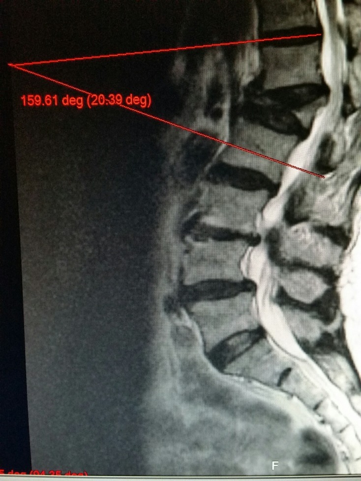
MRISagittal MRI of the lumbar spine: severely collapsed vertebra with marked kyphotic angulation, a sequela of an untreated fracture.
Source : Jacobson et al., Cureus, 2017, figure 1 · CC BY
Because the fracture is not an accident, it is a symptom. It reveals bone that gives way under ordinary stresses, and that bone does not become normal again because the pain has settled.
This is the figure to know by heart. Lindsay and colleagues analysed the 2,725 postmenopausal women randomised to the placebo arms of four large osteoporosis trials. Among the 381 participants who developed an incident vertebral fracture during the study, the incidence of a new vertebral fracture in the following year was 19.2 % (95 % CI 13.6 to 24.8). And the presence of at least one vertebral fracture at baseline multiplied by 5.1 the risk of fracturing during the first year of the study.4
One woman in five. Within the year. This figure circulates widely in distorted forms, « 20 % », « one in five », sometimes « 25 % », without the primary source ever being cited. Here it is, with its population: postmenopausal women of mean age 74, on placebo, in trials where fractures were detected radiographically, including those that had never shown themselves clinically.4
Klotzbuecher's statistical synthesis, published in the Journal of Bone and Mineral Research, gives the overall picture across all fracture sites. Its most striking result: the strongest association in the whole literature is the one between a previous vertebral fracture and a later vertebral fracture, with a risk about four times higher, which rises further with the number of fractures already present. For the other site combinations (hip, wrist, spine), the relative risk is around 2.5
The fracture cascade, in measured risks
The spine predicts the spine better than any other site predicts anything at all.
Sources: refracture at one year, Lindsay et al. 2001, JAMA, 381 patients who fractured out of 2,725 on placebo4 ; relative risks, Klotzbuecher et al. 2000, JBMR, statistical synthesis of the literature: 2.0 (CI 1.8 to 2.1) in peri- and postmenopausal women, 2.2 (1.9 to 2.6) in the other studies.5
The vertebral fracture is not only a predictor of fracture. It is a marker of overall frailty. The Dubbo prospective cohort, published in JAMA, followed women and men aged 60 and over for nearly twenty years: the standardised mortality ratio after a clinical vertebral fracture is 1.82 in women (CI 1.52 to 2.17) and 2.12 in men (1.66 to 2.72), with excess mortality persisting five years after the event.6
This figure has to be read with the caution an association deserves: the vertebral fracture does not kill directly in most cases. It occurs in people who are frailer, more undernourished, more comorbid, and it then worsens what made it likely in the first place, through pain, immobilisation, loss of function, kyphosis, and the reduction in abdominal and thoracic volume. The chain is plausible and documented link by link, but overall causality is not demonstrated by this type of study, and this article will not pretend that it is.
What this figure does not say
A standardised mortality ratio of 1.82 means that people who have had a clinical vertebral fracture die 1.82 times as often as the general population of the same age and sex.6 It does not mean that the fracture causes this excess of deaths, nor that preventing the fracture would avoid them. It is a signal of severity, to be treated as such: a patient who has just had a vertebral fracture deserves to have their overall condition looked at, not only their back.
Red flags: recognising the next fracture
A refracture does not present like the first one: the patient is already known, already followed up, and their back pain no longer surprises anyone. These are the signals that should reopen the file, in the middle of a course of care that was going well.
⚠️ Faced with one of these signals, forceful techniques and loading are suspended, and the patient is referred for imaging before the programme picks up where it left off.
Key points
Three things, and none of them consists of treating the bone. Spotting that the osteoporosis is untreated, reducing falls, and giving the patient back a spine that holds. The first two are the least spectacular and the best demonstrated.
It is the simplest and most cost-effective contribution. It calls for no new skill: it calls for a question to be asked. Since your fracture, have you had a bone density scan? Are you taking any treatment for your bones?
The probability that the answer is no is high. In the Irish cohort already cited, only 24 % of patients admitted for hip fracture left on anti-osteoporotic treatment, with no difference between those who had a vertebral fracture on CT and those who did not.2 And the French recommendations of the Société française de rhumatologie and the GRIO are explicit: in women with a severe fracture (a category that includes vertebral fracture), drug treatment is recommended, all the available agents being usable.35
What this spotting produces is not hypothetical. Post-fracture care pathways, or fracture liaison services, have been the subject of a systematic review with meta-analysis covering 37 studies, 34 of them at low risk of bias: the risk of a secondary fragility fracture is reduced, relative risk 0.68 (CI 0.55 to 0.83) beyond two years, with moderate certainty of evidence according to GRADE.7 A one-third reduction in the risk of refracture, obtained through organisation and not through a technical procedure.
The physiotherapist does not prescribe bisphosphonates. But they are often the only clinician to see this patient twenty times in three months, and the only one in a position to notice that nobody has prescribed anything.
This is the area where the evidence is the strongest in this whole article, and it does not bear specifically on vertebral fracture. Sherrington's Cochrane review, in the abridged version published in the British Journal of Sports Medicine, establishes on 59 studies and 12,981 participants that exercise reduces the rate of falls by 23 % (rate ratio 0.77; CI 0.71 to 0.83), with high certainty and that label is rare and deserves emphasis.30
The detail of the modalities matters as much as the overall result:
And one finding of direct use to the profession: the subgroup analysis shows a larger effect in the trials where the intervention was delivered by a health professional, most often a physiotherapist.30
The logic of this argument has to be named, because it is indirect. The evidence bears on falls, not on vertebral fractures, and a large share of vertebral fractures occur without a fall. Reducing falls by 23 % therefore does not reduce vertebral fractures by 23 %. But in a patient who has already fractured, fall prevention protects the hip, the wrist and the pelvis, whose risk is also doubled.5 It is a real benefit, on neighbouring targets, and it is better presented that way than over-interpreted.
This is the most specific line, and the most fragile in evidence. It rests on a prospective study by Sinaki carried out at the Mayo Clinic: 50 postmenopausal women followed for ten years after a two-year randomised trial, 27 of whom had performed progressive resistive strengthening of the spinal extensors. At ten years, the incidence of vertebral fracture was 1.6 % of the vertebral bodies examined in the exercise group against 4.3 % in the controls, a relative risk of 2.7 against the controls (p = 0.029). Extensor strength was still significantly higher eight years after the programme stopped.27
It is an encouraging result and it must be given its exact weight: fifty women, a single team, an outcome measured per vertebral body and not per patient, and observational follow-up after the randomised phase. This is not the demonstration that strengthening prevents fractures; it is the best signal available in favour of that hypothesis, and it has never been replicated on a large scale.
What each lever reduces, and with what certainty
Three levers available to the physiotherapist, three different targets. None of them directly reduces vertebral fractures.
Sources: falls, Sherrington et al. 2020, abridged Cochrane review30 ; post-fracture pathway, Danazumi et al. 2024, 37 studies, the authors' GRADE assessment7 ; extensors, Sinaki et al. 2002, 10-year follow-up of 50 women.27 The certainty labels repeat those of the authors for the first two rows; the third is an editorial judgement, no GRADE assessment having been published on that study.
What the physiotherapist does not do
They do not set the indication for an anti-osteoporotic treatment, they do not stop it, they do not comment on it in terms of the choice of agent. The role described here is a role of spotting and passing on : noting the absence of assessment or treatment, writing it in a letter, and getting on with your own work. A patient sent back to their doctor with a precise, dated observation gets more than a patient who has had the pharmacology of bone explained to them.
Key points
This is the subject on which it is most tempting to take a side, and the one where taking a side would be dishonest. Twenty years of trials have produced results that contradict one another, and the way they contradict one another is instructive.
The principle is simple: inject acrylic cement into the fractured vertebral body to stabilise it and relieve the pain. Kyphoplasty adds a balloon inflated before the injection, meant to restore part of the lost height. Both techniques are widely practised. Their evidence base is one of the most disputed in musculoskeletal medicine.
2010, VERTOS II. Open-label Dutch and Belgian trial, 202 patients randomised, fractures less than six weeks old with oedema on MRI and pain of at least 5 out of 10. Vertebroplasty clearly beats conservative treatment: a difference of 2.6 points on the visual analogue scale at one month and 2.0 at one year, with no serious complication.18
But the trial contains a figure that its conclusions do not put forward and that deserves to be read: of the 431 eligible patients identified, 229, that is 53 %, saw their pain resolve spontaneously during the assessment phase, before randomisation.18 More than one patient in two got better on their own, within a few days. No open-label trial can separate the effect of the cement from that natural course plus the waiting.
2016, VAPOUR. Australian double-blind trial against a placebo procedure, 120 patients, restricted to fractures less than six weeks old with pain of at least 7 out of 10 and an optimised vertebral filling technique. Positive result: 44 % of treated patients against 21 % of controls had pain below 4 out of 10 at fourteen days (difference 23 percentage points, CI 6 to 39; p = 0.011).16
2018, VERTOS IV. Dutch double-blind trial against a sham procedure, 180 patients, acute fractures. Negative result: no statistically significant difference over twelve months of follow-up, not on pain, not on quality of life, not on disability, not on painkiller use, with both groups improving significantly.17
2018, the Cochrane review. Twenty-one trials, five of them against placebo. Verdict: high to moderate quality evidence that there is no important benefit on pain, disability, quality of life or treatment success. Mean pain was 5 out of 10 on placebo and improved by 0.7 points by vertebroplasty, against a minimal clinically important difference set at 15 %. The authors state that the subgroup analyses show no different effect according to the duration of pain, acute or subacute, and that the open-label trials comparing with usual care have probably overestimated the benefit.15
2023, VERTOS V. A change of ground: this time the fractures are chronic, pain for more than three months with persistent oedema on MRI, and the comparator is not a placebo but an anaesthetic injection. 80 patients. Positive but modest result: a difference of 1.3 points on the visual analogue scale at twelve months (CI 0.1 to 2.6), a significant difference on quality of life too, but not on disability.19
Twenty years of vertebroplasty trials
The result depends closely on the comparator and on the population included. That is what makes synthesis difficult, and the debate legitimate.
Sources: Klazen 201018, Clark 201616, Firanescu 201817, Buchbinder 201815, Carli 202319. The labels « positive » and « negative » summarise the primary outcome of each trial as published by its authors.
Two readings compete, and neither is absurd.
The first: vertebroplasty does not work, and the positive trials are positive for reasons of method. That is the position of the Cochrane review. VERTOS II is open-label, therefore inflated by expectation; VAPOUR is small, single-centre for its main recruitment, and its outcome is a dichotomised threshold rather than a mean. Against placebo, across five trials, pain does not move in any clinically important way.15
The second: vertebroplasty does work, but for a population that the negative trials have diluted. That is the reading of the VAPOUR authors: fractures less than six weeks old in patients in severe pain, treated with sufficient filling, would be the target, and the negative trials would have included too many old or mildly painful fractures, in whom the natural course erases the difference.16 To which the Cochrane review replies that its subgroup analyses by duration of pain do not find that differential effect.15
A third element has been added recently, and it concerns the survival argument. Several observational studies had reported lower mortality after kyphoplasty. An analysis of 235,317 Medicare beneficiaries revisited the question with increasingly rigorous matching: the apparent advantage (hazard ratio 0.84) disappears and then reverses when matching is done on medical complications and on age and comorbidities, giving a hazard ratio of 1.32 and then 1.81 against kyphoplasty.20 The « survival gain » was an artefact of indication: the least ill patients were the ones being operated on.
What the physiotherapist can say to a patient who asks
That there is an operation, that it consists of injecting cement, that it is sometimes useful in one precise situation (a recent fracture, very painful, resistant to painkillers), and that the literature is divided on its benefit in the other cases. That the decision belongs to the doctor, and that the patient has every right to ask why it is being offered to them in particular. What must not be said: neither « it is useless » nor « you must have it ».
Key points
The direct comparison exists. A meta-analysis of sixteen studies compared balloon kyphoplasty with percutaneous vertebroplasty: kyphoplasty reduces the wedge angle (standardised mean difference 0.98; CI 0.40 to 1.57), restores more vertebral height and reduces the risk of cement leakage (relative risk 0.62; 0.47 to 0.80). But these radiographic gains do not translate clinically : no significant difference on the visual analogue pain scale or on the Oswestry Disability Index.21
It is a textbook case of the gap between a surrogate outcome and the outcome that matters. The vertebra is straighter, cement leakage less frequent, and the patient has neither less pain nor less difficulty. For the physiotherapist the practical consequence is simple: the technique used beforehand does not change the rehabilitation they go on to deliver.
The spinal brace is one of the most intuitive and least demonstrated moves in this whole area of management. It is prescribed very widely; the literature that supports it would fit in a pocket.
The systematic review by Newman, Minns Lowe and Barker brought together twelve studies, eight randomised or randomised pilot trials and four non-randomised studies, totalling 626 participants. Its result deserves to be quoted as it stands: only three studies, covering 153 people, evaluated bracing after an acute vertebral fracture, and none was of high quality. The data are lacking to say that a brace changes vertebral deformity, and it is with rigid braces that complications were the most numerous.22
The review does find more favourable signals in the subacute phase and in prolonged rehabilitation, across nine studies and 473 participants: three of them suggest that a semi-rigid backpack-type thoracolumbar brace can improve strength, pain, posture and quality of life; one study finds a balance benefit with a weighted kyphosis-correcting orthosis. The authors conclude that these leads « should be explored in studies of sufficient size and quality, including men ».22
The useful question is not « is a brace needed », it is « what is this one for, for how long, and what will replace its effect when it comes off ».
That wording is what makes the debate workable. A brace worn for a few weeks to let a patient get upright and walk during the most painful phase does a job that nothing else does. A brace worn for six months « to hold the back up » installs dependence and deprives the extensors of the stimulus they need, at exactly the moment when the literature suggests their strength counts.27
| Phase | What has been studied | What can be concluded from it | Reasonable course of action |
|---|---|---|---|
| Acute fracture | 3 studies, 153 participants, none of high quality. | Inconclusive. No data showing an effect on deformity. Complications at their highest with rigid braces. | If a brace, the lightest one that allows the patient to get upright, for a short duration stated from the outset. |
| Subacute phase and prolonged rehabilitation | 9 studies, 473 participants, variable quality. | Favourable signal for the semi-rigid backpack-type thoracolumbar brace on strength, pain, posture and quality of life (3 studies). | Conceivable alongside an active programme, never in its place. |
| Balance and established kyphosis | 1 study on the weighted kyphosis-correcting orthosis. | Improvement in balance reported, on a single study. | A lead, not a recommendation. To be reassessed when trials of sufficient size appear. |
Key points
You have to start with the bad news, because it is structuring: physiotherapy after vertebral fracture has not demonstrated much. Knowing this changes the way it is delivered, not the decision to deliver it.
The 2019 update of the Cochrane review, by Gibbs and Giangregorio, brought together nine trials and 749 participants, 68 of them men. Its conclusions, in order:23
The authors conclude that a high-quality trial is still needed, and give the required sample size calculation: about 2,500 untreated participants, or 4,400 on anti-osteoporotic treatment.23 No trial of that size has been conducted since.
The PROVE trial, run in Oxford, is the largest ever carried out in the physiotherapy of vertebral fracture: 615 adults with back pain, osteoporosis and at least one vertebral fracture, randomised into three arms, seven individual manual therapy sessions over twelve weeks, seven home exercise sessions, or a single education session.24
At twelve months, on the two primary outcomes, quality of life measured by the QUALEFFO-41 and extensor endurance measured by the timed loaded standing, no statistically significant difference between the three groups. The endurance gain was 9.8 seconds in the exercise arm, 13.6 seconds in the manual therapy arm and 4.2 seconds in the single-session arm, with confidence intervals crossing zero for exercise.24
Two secondary results deserve attention. At four months, significant differences did exist: endurance and balance in the manual therapy arm; endurance in those under 70, balance, mobility and walking in the exercise arm. And the authors write it in black and white: « adherence was problematic », and « the benefits at four months were not sustained », while concluding that « short-term benefits are likely to be valuable ».24
At the other end of the size spectrum, Bennell's pilot trial, run in Melbourne on twenty participants, is clearly positive: compared with a no-treatment group, ten weeks of individual physiotherapy and daily exercise reduce pain on movement by 1.8 points (CI 0.1 to 3.5) and at rest by 2.0 points, improve QUALEFFO function by 4.8 points and endurance measured by the timed loaded standing by 46.7 seconds (16.1 to 77.3). Nine of the eleven participants in the treated group rated their pain « much better » against one of nine in the controls.25
Twenty patients, a comparator with no intervention, no blinding possible on the experience of pain: this pilot does not demonstrate, it suggests. But the juxtaposition with PROVE is illuminating. Bennell compares physiotherapy with nothing ; PROVE compares seven sessions with one education session. That is not the same question, and the fact that PROVE is negative means that seven sessions do no better than one well-conducted session, not that physiotherapy is useless.
PROVE does not say that rehabilitation is useless. It says that one well-conducted education session does almost as well as seven sessions, which is information about dosage, not about principle.
Moderate
Exercise for fall prevention. A 23 % reduction in the rate of falls in the general older population, high certainty in the Cochrane review, downgraded here to moderate because the population studied is not that of people carrying a vertebral fracture and because the target is the fall, not the vertebral fracture.30
Moderate
Improvement in physical performance. The Timed Up and Go improves by 1.13 seconds, the only moderate-quality result in the 2019 Cochrane review, and judged by its own authors to be not clinically important.23
Low
Strengthening of the spinal extensors to reduce fractures. One study, 50 women, 1.6 % against 4.3 % of vertebral bodies fractured at ten years, relative risk 2.7. Never replicated.27
Low
Manual therapy and exercise on pain and function in the short term. Real benefits at four months in PROVE, which do not persist at twelve months; Bennell's pilot positive but on twenty patients and with no active comparator.2425
Low
Avoiding loaded spinal flexion in the early phase. A single comparative study, very small numbers, but a massive result consistent with the biomechanics, and never contradicted since 1984.26
Very low
Semi-rigid thoracolumbar brace in the secondary phase. Three studies out of twelve in Newman's review suggest a benefit on strength, pain, posture and quality of life; variable methodological quality, men almost absent.22
Very low
Exercise to reduce fractures or falls in people carrying a vertebral fracture. A single trial for each outcome, no difference, very low quality evidence according to the Cochrane review itself.23
Not demonstrated
Rigid brace after an acute fracture. Three studies, 153 participants, none of high quality, no demonstrated effect on deformity, and the highest complication rate in the review.22
The levels in this table are an editorial judgement applying the GRADE principles, and not a published GRADE assessment. Two rows repeat certainty judgements made by the authors of the reviews cited (exercise and falls; physical performance); the others are derived from the number of studies, their size, their risk of bias and the consistency of the results. No formal GRADE assessment exists on the rehabilitation of osteoporotic vertebral fracture.
Key points
This is the most asked and the most poorly handled question. The answer comes in two steps that seem to contradict each other and do not: loaded flexion is to be avoided in the early phase, and generic lifelong bans are counterproductive.
From a single study, published in 1984 in the Archives of Physical Medicine and Rehabilitation. Sinaki and Mikkelsen followed 59 women with postmenopausal spinal osteoporosis and back pain, allocated to four exercise programmes. After one to six years of follow-up with radiographs before and after, the new fractures or wedge deformities were distributed as follows:26
New fractures according to the type of exercise prescribed
The founding study of 1984: a massive result on tiny numbers. Both deserve to be read together.
Source: Sinaki and Mikkelsen 1984, Archives of Physical Medicine and Rehabilitation ; 59 postmenopausal osteoporotic women with back pain, follow-up of one to six years, radiographs before and after. Significant difference in favour of extension over flexion (p < 0.001) and over the combined programme (p < 0.01). Group sizes range from 6 to 25 patients : the 89 % in the flexion group represents eight patients out of nine.26
This chart has to be looked at with both eyes. On one side, the gap is spectacular and the statistical significance is there: 16 % against 89 %, p < 0.001.26 On the other, the flexion group had nine patients. A study of nine people normally founds nothing. Yet it founds a rule taught throughout the world for forty years, for three reasons: the effect is very large, it is consistent with the mechanics of the vertebral body, since flexion transfers the load onto the anterior column, precisely where the wedge compression happens, and above all no study has ever contradicted it, for the obvious reason that no ethics committee would today allow osteoporotic patients to be randomised to a flexion programme.
This is a case where the strongest evidence that will ever exist is weak evidence, and where waiting for it would be unreasonable. The practical conclusion is therefore: in the early phase, loaded spinal flexion is not taught and is not used as an exercise. That does not mean immobilising the patient in extension.
The international consensus Too Fit To Fracture, from a two-round Delphi process bringing together researchers and clinicians, is explicit on this point. Its key conclusions:28
The central argument of the consensus is that a list of bans produces a patient who no longer moves, and that a patient who no longer moves loses strength, balance and bone mass, that is to say the three things that were protecting them. The right answer is not to ban flexion: it is to teach picking an object up by bending the hips and knees rather than the spine, and to repeat it until it becomes the default movement.
A ban protects today's vertebra. A technique protects every vertebra of the next ten years, because the patient takes it away with them.
The question has been open since the LIFTMOR trial, run in Australia in 101 postmenopausal women with low bone mass (T-score below −1.0), randomised between eight months of high-intensity resistance and impact training (five sets of five repetitions at more than 85 % of maximum, twice thirty minutes a week, supervised) and a low-intensity home programme. The results are clear in favour of high intensity: lumbar spine bone mineral density +2.9 % against −1.2 %, femoral neck +0.3 % against −1.9 %, cortical thickness, height, and every functional test. A single adverse event was reported.29
This trial has circulated widely as proof that « you can lift heavy even with osteoporosis ». It has to be read for what it is. LIFTMOR included women with low bone mass, selected and screened for conditions and treatments interfering with bone and physical function; this was not a population of women with a recent vertebral fracture. The training was supervised in a gym by professionals, not prescribed to be done alone. Nothing in this trial permits heavy loading of a vertebra that still shows marrow oedema on MRI.
What LIFTMOR does establish, on the other hand, deserves to be kept in mind: in a well-supervised osteopenic and osteoporotic population, high intensity did not produce the wave of fractures that traditional caution had led people to fear. That moves the marker of what is reasonable, not what to do in the acute phase.
The rule of conduct in one line per phase
Key points
Four case reports, chosen because they show the four trajectories this diagnosis can take: the one that goes well, the one discovered too late, the one that turns around, and the one that begins as ordinary low back pain.
Case 1: the ordinary trajectory, and it is a good one
Papa 2012, Journal of the Canadian Chiropractic Association 56(1):29-39: PMID 22457539
A 74-year-old man presents with acute thoracolumbar pain that came on after lifting an object. The radiograph shows generalised demineralisation and a moderate wedge fracture of L1.
Management is entirely conservative: postural education, activity modification, interferential current, taping in extension, instrument-assisted soft tissue technique, and prescribed rehabilitation exercises. The follow-up outcomes are a verbal pain scale, medication use and the return to activities of daily living. Lasting resolution of symptoms, with no recurrence of pain at the twelve-month review.
What this case teaches. The commonest scenario is this one: a moderate-grade fracture in an independent patient, who does well without any invasive procedure. It is the reminder that often goes missing when only the complications come to mind. What this case does not teach: it is a single observation, without a comparator, and improvement at twelve months is also what natural history does, since VERTOS II showed that more than one eligible patient in two improved spontaneously within a few days.1831
Case 2: the one that went unseen for two years
Massoud and Alashkar 2026, Cureus 18(3):e104527: PMID 41783551
A 78-year-old woman presents after a fall. The work-up finds an acute vertebral fracture and, beside it, a chronic vertebral fracture and a chronic diaphragmatic hernia that had gone unnoticed for more than two years.
The authors draw from their case a conclusion that also holds for the physiotherapy clinic: falls are common in older people, the prevalence of osteoporosis among them is high, and the resulting injuries can be subtle and overlooked by patient and doctor alike. Finding them makes it possible not only to treat them early, but to put appropriate preventive measures in place.
What this case teaches. It is the clinical illustration of Schousboe's figure.1 Two years of unnamed fracture in a patient who will certainly have met clinicians during that time. The physiotherapist seeing an older patient for back pain is, statistically, seeing this kind of situation.32
Case 3: the low back pain that becomes an emergency, Kümmell's disease
Kuppan et al. 2022, Journal of Orthopaedic Case Reports 12(10):34-38: PMID 36874888
A 65-year-old woman presents with low back pain that has been going on for four weeks. Progressive weakness and bladder dysfunction then appear. The radiographs show a compression fracture of T12 with an intravertebral vacuum sign. MRI finds intravertebral fluid and significant cord compression. Posterior decompression, stabilisation and transpedicular grafting; histology confirms Kümmell's disease. The patient recovers her strength, her bladder control and independent walking.
The authors recall the mechanism: Kümmell's disease is an osteoporotic vertebral fracture progressing to avascular necrosis of the vertebral body, with an initially asymptomatic period followed by progressive pain, kyphosis and neurological deficit after trivial trauma. They stress that osteoporotic fractures are more prone to non-union because of poor vascular and mechanical support.
What this case teaches. Spinal pain that worsens over the weeks in an osteoporotic patient is not a fracture healing badly: it is a signal. The physiotherapist is the professional best placed to spot this reversal of trajectory, because they are the one who sees the patient again.33
Case 4: late collapse in the very old patient
Picazo et al. 2014, European Spine Journal 23(12):2696-2702: PMID 23778750
The authors report the case of an elderly man with an osteoporotic fracture progressing to severe late collapse with neurological involvement. Treatment consisted of bilateral posterior transpedicular decompression and instrumented fusion with cemented pedicle screws; the postoperative course was uneventful and the patient recovered from his deficit and regained his walking ability.
The paper sets the frame in a sentence that can be taken as it stands: vertebral fracture is common in osteoporotic people and causes no clinical problem in the majority of cases ; only a small number of patients suffer serious neurological complications related to late collapse.
What this case teaches. The proportion matters as much as the event. The point is not to turn every compression fracture into a neurological threat, which would be untrue and frightening, but to know that the complication exists, that it comes late, and that its first signs pass through the physiotherapy appointment before they reach the emergency department.34
Why these four
These are real published case reports, with their PubMed identifier, and not reconstructed vignettes. They have the drawback of their genre: a clinical case demonstrates nothing, it illustrates. Three of them describe situations that went wrong, which is the natural bias of case publication, since what gets published is what surprises. Case 1 is there to restore the proportion: the commonest trajectory is the one that goes well.
Key points
Everything above comes down to four decisions: thinking of the fracture, knowing when to stop, knowing what to do next, and not letting a patient leave when nobody is treating their bone.
Nothing spectacular is being asked. Three minutes added to the history and two measurements are enough to change the probability.
| Phase | Main objective | What you do | What you do not do |
|---|---|---|---|
| Phase 1, recent fracture or suspicion not lifted roughly weeks 0 to 4 |
Avoid making things worse; keep the patient upright and walking; do not settle into bed rest. | Education and explanation of the diagnosis; transfers without spinal flexion (log-roll technique); daily walking in short bouts; breathing work; gentle isometrics of the extensors within the pain-free range; adapting the home. | Manipulation. Forceful mobilisation. Posteroanterior pressure on the spinous processes. Loaded flexion. Added axial load. Stretching into global flexion. |
| Phase 2, consolidation roughly weeks 4 to 12 |
Restore extensor endurance and balance; install the spine-sparing techniques. | Progressive strengthening of the spinal extensors27 ; daily balance training2830 ; systematic teaching of the hip hinge applied to real everyday movements; progressive weaning off the brace if one was prescribed. | Going straight to heavy loading. Extending the brace with no end date. Settling for non-weight-bearing exercise only. |
| Phase 3, maintenance beyond 3 months |
Reduce the risk of falls and of new fracture; make the patient independent. | A programme combining balance, functional exercise and strengthening, the combination that reduces falls the most30 ; moderate intensity preferred after a vertebral fracture28 ; periodic reassessment of height and posture. | Keeping generic bans that no longer serve a purpose. Ending follow-up without having checked that the bone treatment is in place. |
This is the least technical and the most useful part. A letter to the general practitioner containing three precise lines does more than one extra session. What it must contain:
It is this third point that counts most over time. Three quarters of patients admitted for a fragility fracture leave with no treatment for the bone2, whereas the French recommendations provide for treating severe fractures35, and organising post-fracture pathways reduces the risk of refracture by about a third.7 The physiotherapist prescribes nothing. They can point out that something is missing.
Key points
There is no published standard time frame, and the round numbers in circulation deserve caution. What can be said from the data: in VERTOS II, 53 % of eligible patients saw their pain resolve spontaneously during the assessment phase, that is within a few days to a few weeks.18 The useful marker is therefore not a duration but a direction : the pain of a recent fracture should decrease. Pain that increases over the weeks raises the suspicion of non-union or of progressive collapse and calls for an opinion.33
Not until a recent fracture has been ruled out, and that is the cautious position this article supports. We should be honest about the nature of that recommendation: it does not rest on comparative trials, which do not exist and will not exist, but on mechanics and on potential severity. The reasoning is asymmetrical: the expected benefit of a manipulation on this terrain is modest, the cost of a mistake is one more vertebra.
That the decision rests with the doctor, and that the literature is divided. Against placebo, the 2018 Cochrane review finds no clinically important benefit15 ; the VAPOUR trial, for its part, is positive but in very specific patients: a fracture less than six weeks old and pain of at least 7 out of 10.16 The question the patient can legitimately put to their doctor is: « am I in that situation? »
The evidence after an acute fracture is inconclusive, and it is with rigid braces that complications are most numerous.22 When a brace is prescribed, two questions must have an answer from day one: for how long, and what will take over. A brace that supports getting upright for three weeks has nothing to do with a brace worn for six months.
By distinguishing what is risky from what is not, rather than by handing out a list of bans. That is exactly the position of the international consensus Too Fit To Fracture : provide spine-sparing techniques, the hip hinge for picking things up, the log roll for getting up, rather than generic restrictions of the « do not carry anything over five kilos » kind.28 The patient who knows how to do it moves; the patient who has been given a list stops.
Good for confirming, bad for excluding. In 66,021 patients, a height loss of 3 cm has a specificity of 92 % but a sensitivity of 19 %; at 4 cm, specificity rises to 95 % and sensitivity falls to 14 %.12 In other words, a clear height loss justifies looking further, but the absence of height loss lets the great majority of fractures through.
Yes, in a proportion that often comes as a surprise. The European EPOS study measures an incidence of 5.7 per 1,000 person-years in men against 10.7 in women: the ratio is about two to one.3 And the excess mortality after a clinical vertebral fracture is higher in men (standardised ratio 2.12) than in women (1.82).6 An older man with back pain should not receive any less vigilance.
The available data are reassuring without being definitive. The 2019 Cochrane review reports four exercise-related adverse effects across nine trials and 749 participants: one costal cartilage fracture, one rib fracture, one knee pain, one skin irritation, and it rates the certainty of that information as very low.23 The LIFTMOR trial, with training at more than 85 % of maximum, reports only one adverse event in 101 women with low bone mass, but in a supervised setting and outside recent fracture.29
It is indispensable for everything else (independence, morale, bowel function, preventing deconditioning), but it should not be credited with what it does not have. In the Cochrane review on fall prevention, the authors explicitly declare themselves uncertain as to the effect of programmes based mainly on walking, on dance or on strengthening alone; it is the balance and functional programmes, and the combined programmes, that carry the effect.30
The question arises mainly if the pain does not follow the expected trajectory, or if a new episode occurs: the risk of a new vertebral fracture in the following year is 19.2 %4, which makes a new painful episode anything but trivial. It is a medical decision, but the physiotherapist is often the one who notices the change of trajectory and who has to flag it.
Thirty-five references, each resolved through its PubMed identifier and read before citation. The figures in the text point to the source that establishes them, at the place where they are written.
Method and limitations
Sources searched in PubMed through the NCBI E-utilities services, crossing osteoporotic vertebral fracture with epidemiology, under-diagnosis, red flags, imaging, vertebroplasty and kyphoplasty, braces, exercise, fall prevention and case reports. Every identifier was resolved and every abstract read before citation; the four clinical cases are real published case reports, cited with their identifier.
The main limitation of this article : the strongest evidence it contains does not bear on the question asked. Certainty is high for exercise against falls30, high to moderate for the absence of benefit of vertebroplasty against placebo15 and it is low or very low for almost everything that bears directly on rehabilitation after vertebral fracture, the 2019 Cochrane review stopping explicitly at a finding of insufficient evidence.23 Two central claims of practice rest on tiny and never-replicated studies: the protective effect of extensor strengthening (50 women)27 and the danger of loaded flexion (9 patients in the group concerned)26. They are kept here because they are consistent with the mechanics and because no data contradict them, not because they are demonstrated.
A second limitation : the levels in the table of modalities are an editorial judgement applying the GRADE principles, and not a published GRADE assessment; no formal assessment of that kind exists on the rehabilitation of osteoporotic vertebral fracture. Finally, the Roman diagnostic cluster has never undergone the prospective validation its authors called for.10
Article written on 15 August 2026.
Behind this article
An author who explains, a reviewer who checks.
How we write and check our content

Physiotherapist · co-founder of Physio Learning
Marked for life by his first four-hour lecture without a single image, he took a master’s in instructional design so that it would never happen to anyone again. He hunts down publication bias and unreadable slides with the same intransigence.

Head of scientific content
Physiotherapist specialising in neuro-musculoskeletal practice and holder of a master’s in public health. He checks the methodological rigour of every article: primary sources, levels of evidence, no exceptions.
