














OnlineFIFPL

Sports physiotherapy
Become an expert in Pilates-based rehabilitation - e-learning
Marie Reboul
Go further
Scheuermann kyphosis or bad posture? The test that separates them, the radiographic criteria, the brace, and what exercise is honestly worth here.
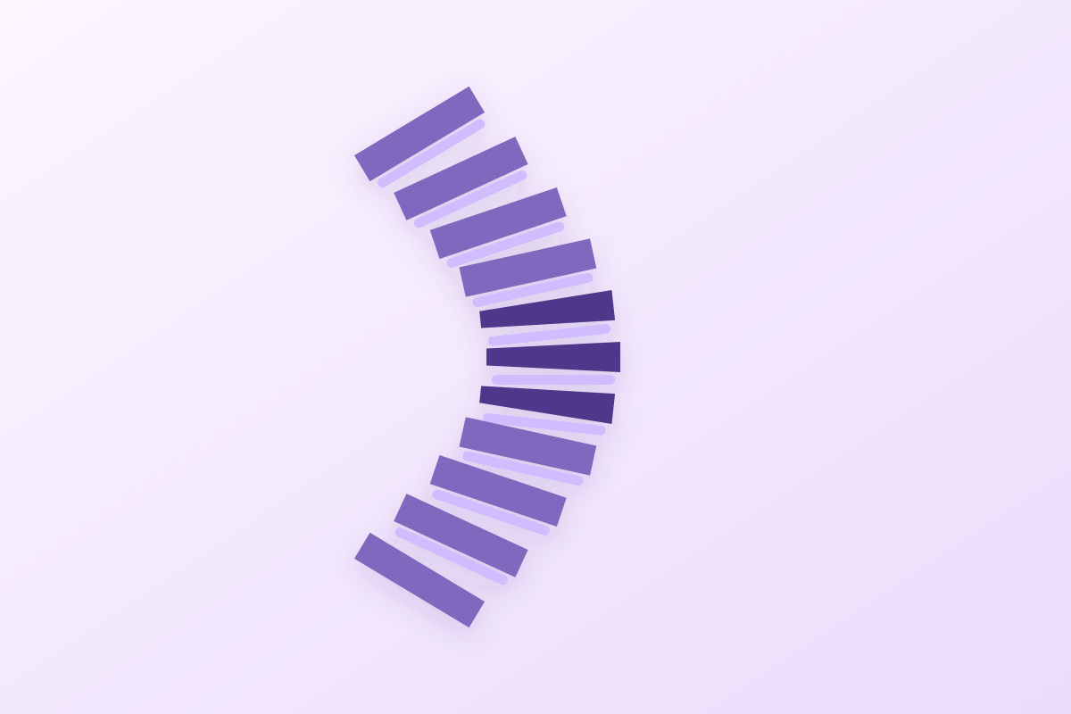
Ask the adolescent to straighten up actively: a postural kyphosis disappears, a Scheuermann kyphosis stays; the distinction takes ten seconds, not the radiographic angle.
74%genetic share of Scheuermann's disease
What follows covers each of these points in detail, with sources. It is there if you need it.
This topic is taught in a course: Become an expert in Pilates-based rehabilitation - e-learning, online with Marie Reboul.
1 / 13
12 article chapters · 48 min in total
Key points◔ 1 min
From diagnosis to surgery, it is never the angle alone that decides.
Distinguishing◔ 5 min
A kyphosis is often, according to Lowe, wrongly put down to simple bad posture.
Radiograph◔ 4 min
The diagnosis rests on vertebral wedging, not on a kyphosis angle.
Forms◔ 4 min
Thoracic form thresholds do not apply to the thoracolumbar form.
Epidemiology◔ 4 min
This disease is 74 % genetic, without that cancelling the role of mechanical loading.
Prognosis◔ 4 min
The parameter to watch in adulthood is not the angle, but sagittal balance.
Brace◔ 5 min
In Sachs, eight patients in ten with irregular brace wear got worse.
Exercise◔ 7 min
Promising to straighten the kyphosis through physiotherapy is not supported by the literature.
Surgery◔ 4 min
Surgery is rare, and it is not the angle alone that decides on operating.
Clinical cases◔ 3 min
Five published cases each illustrate a point that large series do not show.
In practice◔ 3 min
In clinic: what to do, what to refer, and what never to promise.
Questions◔ 4 min
Exercise improves pain and strength, but does not straighten the curve itself.
Train on this
Sports physiotherapy
Marie Reboul
Next comes practice: the course that teaches this topic, with Marie Reboul.
Our courses on this topic may be covered by DPC or FIFPL, the French continuing-education funding schemes, depending on your profession and situation.
In brief
A Scheuermann kyphosis is not bad posture, and that is the error that costs the most: the literature has been repeating for twenty-five years that the deformity is frequently put down to "bad posture", which delays diagnosis. A ten-second manoeuvre separates them: you ask the adolescent to straighten up, then you examine them in hyperextension or prone. A postural kyphosis disappears, a Scheuermann kyphosis stays. The consequence is practical and it is brutal: postural correction exercises and reminders to stand up straight do not straighten a structural curve. What can usefully be worked on is pain, flexibility, the strength of the trunk extensors and monitoring during growth. The brace, for its part, only works while growth remains, and surgery is not decided on the angle.
Clinical summary based on the natural history study by Murray, Weinstein and Spratt (JBJS Am 1993, 32 years of follow-up), Garrido's cohort of untreated patients (Spine Deform 2021, 27 years of follow-up), the assessment of radiographic criteria in the Rotterdam study (Makurthou, Spine 2013) and the only randomised trial of specific exercise published in this condition (Bezalel, Asian Spine J 2019): 58 references verified one by one on PubMed and cross-checked on CrossRef.
Three figures to place the disease
Frequency in the population, weight of the genetic background, rate of progression once growth is complete.
Sources: Makurthou 2013, Spine (PMID 24509552); Damborg 2006, JBJS Am (PMID 17015588) and Damborg 2011, Acta Orthop (PMID 21895506); Garrido 2021, Spine Deform (PMID 34212306). Prevalence depends heavily on the criteria used and on the population: chapter 4 gives the full range, from 2.2 to 9 %.
This article deals with the disease itself
For the presenting complaint in which a Scheuermann hides, and for the other major growth deformity, two separate articles:
Nine points that govern what to do. Each is taken up, detailed and sourced in the corresponding chapter.
Key points
It is the central error in the file, it is documented, and it is corrected in one minute of examination. This chapter sets out the test, then draws the consequences for treatment.
The complaint is nearly always the same. An adolescent, more often a boy, stands stooped. The parents have picked him up on it a hundred times. A PE teacher has spoken of "posture". They arrive at the clinic with a request for postural rehabilitation, and sometimes a prescription that says so in black and white.
How often is that reading wrong? Nobody knows precisely, but the orthopaedic literature has been flagging it as a recurring problem for a quarter of a century. Lowe, in two successive reviews, one of them devoted to the level of evidence, writes that the kyphotic deformity is frequently put down to "bad posture", which leads to a delay in diagnosis and treatment.15 A paediatric review puts it from the families' side: the parents of affected children often mistake it for bad posture.4 Twenty-five years later, the most recent concepts review repeats the same warning.2
The delay is not trivial, for a simple reason: the only treatment capable of altering the shape of the vertebrae works during growth and only during it.1330 A diagnosis made at seventeen in an adolescent at the end of growth closes a door that was open at thirteen.
The distinction is mechanical, not aesthetic. A postural kyphosis is an attitude: the vertebral bodies are normal on the radiograph, the curve is supple and painless, it corrects by itself as soon as the person straightens up.3 By contrast, a Scheuermann kyphosis is a deformity: it is rigid, it rests on visible vertebral lesions, and in the wording of the 2023 review, the kyphotic deformity is rigid and does not correct passively in extension.2
The examination sequence takes four manoeuvres, in this order:
A kyphosis that corrects in hyperextension is not a Scheuermann. A kyphosis that does not correct is not a matter of posture.
The same back, two behaviours
What you see in profile, standing and then in hyperextension. The difference is not in the angle measured standing, it is in what the angle becomes.
Teaching diagram. The behaviour shown follows the description in O'Donnell 2023, Curr Rev Musculoskelet Med (PMID 37615931) for rigidity in extension and the compensations, and Garoflid 2000, Rev Med Suisse Romande (PMID 11109912) for the supple postural round back with a normal radiograph. The outlines are not measurements.
| Feature | Postural kyphosis | Scheuermann kyphosis |
|---|---|---|
| Active correction ("straighten up") | Large, often complete | Slight, the curve persists |
| Hyperextension or prone | The curve disappears3 | The curve stays, it is rigid2 |
| Shape of the curve | A long, smooth arc | A short, angular apex |
| Lateral radiograph | Normal vertebral bodies3 | Wedging ≥ 5° on ≥ 3 contiguous vertebrae7 |
| Other radiographic signs | None | Endplate irregularity, Schmorl's nodes, disc space narrowing6 |
| Pain | Usually absent3 | Common, especially in adolescence and in the lower forms615 |
| Associated tightness | Inconsistent | Iliopsoas, hamstrings, pectorals, anterior aspect of the shoulder2 |
| Course without treatment | Does not become structural3 | Progresses during growth, then 0.45° a year27 |
| What treatment changes | Nothing to correct: monitoring and physical activity3 | The brace remodels during growth1330 |
If the curve is structural, then postural correction exercises and reminders to stand up straight do not straighten it. This is not an editorial opinion: the 2023 concepts review concludes that there is no conclusive research demonstrating a lasting, significant improvement in kyphosis from exercise alone.2 A rehabilitation review devoted to the subject had already written it: physiotherapy is barely mentioned in the literature as an effective treatment for Scheuermann kyphosis, and there is little evidence that it can on its own alter the natural history of the disease.33
That does not mean physiotherapy has nothing to do. It means it changes target. What is measured and worked on is pain, the flexibility of the shortened chains, the strength of the trunk extensors and the monitoring of progression during growth. Chapter 7 sets out what each modality is really worth, with its level of evidence. Deficient trunk extension, in range as in strength, is in fact one of the few objective differences found at thirty-two years of follow-up between patients and matched controls: that is a direct argument for this target.26
Red flags in the adolescent with a round back
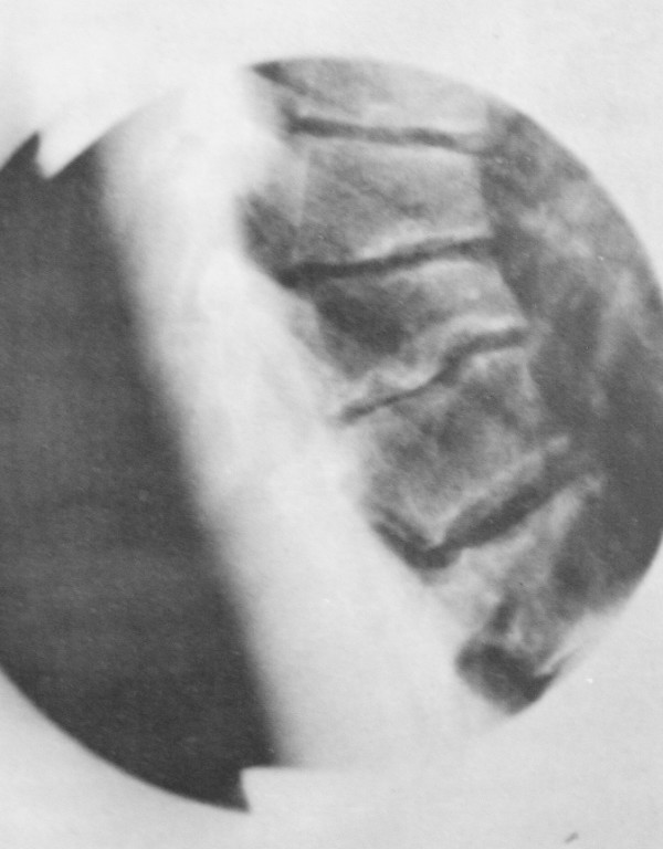
X-rayLateral thoracic spine radiograph showing anterior wedging of several vertebral bodies and endplate irregularity, radiological signs characteristic of late-stage Scheuermann disease.
Source : Weiss et al., Scoliosis, 2009, figure 1 · CC BY
The diagnosis is radiographic. But the most cited criterion, the 45° angle, is also the most fragile: normal adolescent kyphosis is already close to it.
The reference investigation is a radiograph of the whole spine lateral, standing. The historical criterion, set out by Sørensen in 1964, is anterior wedging of at least 5° on at least three contiguous vertebrae.72 It matters to see what that criterion measures: the shape of the vertebrae, not the overall curve. A 70° kyphosis without wedging is not a Scheuermann; three wedged vertebrae with a 48° kyphosis is one.
Around that core, four signs classically accompany the picture and are read on the same film: irregularity of the vertebral endplates, Schmorl's nodes, narrowing of the disc spaces and disc degeneration.6 A Schmorl's node is a herniation of the nucleus pulposus through the cartilaginous and bony endplate into the adjacent vertebral body; it is common on imaging and most often asymptomatic, which rules out making a diagnosis from it alone.12
Many definitions add to the wedging criterion a thoracic kyphosis angle greater than 45°, measured between T5 and T12.13 That threshold circulates everywhere. It deserves a close look.
In 341 normal subjects aged 3 to 18, mean thoracic kyphosis is 44.0 ± 10.9°.9 In other words, the threshold supposed to define the abnormality almost coincides with the mean of normal. In adults, a systematic review of 34 studies covering 7,633 healthy people concludes that kyphosis exceeds 40° in 65 % of cases, and that the widely accepted 40° threshold therefore wrongly classifies a majority of normal subjects.10 Kyphosis also increases with age (Spearman correlation 0.52 between T5 and T12), with no difference between the sexes.10
An angle on its own therefore does not make the diagnosis. What does is the combination of characteristic wedging and clinical rigidity.
One last reason makes the overall angle uninformative on its own: thoracic kyphosis is not a single arc. In 373 asymptomatic young adults, it breaks down into two unequal arcs, a stable upper arc of 25.8° on average and a variable lower arc of 19.8° (p < 0.001), with an apex sitting around T8 and T9 depending on spinal morphology.11 Two patients with the same total angle may therefore have very different spines, and it is where the apex sits, not the sum, that governs mechanical behaviour.
The Rotterdam study did this work in 2,753 participants aged 45 to 89, applying Sørensen's criteria and those of Sachs to lateral radiographs.8 The results are instructive:
This last point is the most useful in the clinic. It is not the angular threshold that decides, it is the joint presence of the vertebral lesions. The authors indeed conclude that there is no absolute reference for the radiographic definition, but that standardised scoring of independent signs gives satisfactory inter-observer agreement.8
| Definition | What it requires | What you need to know about it |
|---|---|---|
| Sørensen 1964, the reference | Anterior wedging ≥ 5° on ≥ 3 contiguous vertebrae72 | The criterion is about the shape of the vertebrae, not the overall angle |
| Broadened definition with an angular threshold | Sørensen's criterion plus T5-T12 kyphosis > 45°13 | The threshold discriminates poorly: normal kyphosis in a child is already 44 ± 11°9 |
| Sachs criteria | Endplate signs and wedging, scored separately8 | Assessed in a population in Rotterdam: inter-observer agreement of 78 to 79 %8 |
| Definition used in the European EVOS study | At least one Schmorl's node or one irregular endplate, plus T4-T12 kyphosis > 40° or a wedged vertebra23 | A broader definition: gives 8 % prevalence in the over-50s23 |
| Thoracolumbar form, called atypical | Wedging of 5° on 3 consecutive vertebrae and total Cobb ≥ 10° between T8 and L216 | Does not meet the classic Sørensen criteria; prevalence of 2.8 % on CT1614 |
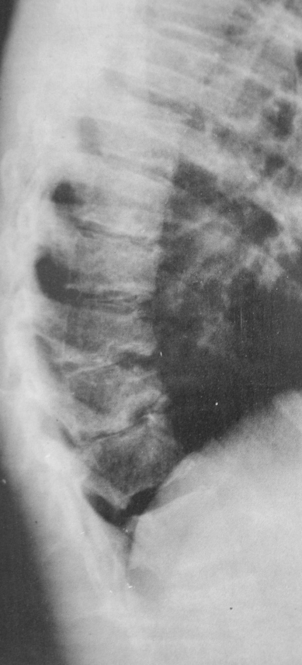
X-rayLateral thoracolumbar spine radiograph showing Schmorl's nodes and marked endplate irregularity, which may be mistaken for infection or tumour if the clinical context is unknown.
Source : Weiss et al., Scoliosis, 2009, figure 2 · CC BY
Key points
The diagnosis rests on wedging, not on the angle. The 45° threshold is convenient but discriminates poorly, and raising or lowering it by ten degrees changes almost nothing in the measured prevalence.8 A standing lateral radiograph of the whole spine is enough to settle the question in the vast majority of cases; MRI is reserved for neurological signs and preoperative work-up.48
Two locations, two presenting complaints, two decision thresholds. Treating a low form with the benchmarks of the high one leads to underestimating its severity.
The classic form sits in the thoracic spine and makes the hump. It is visible, it worries those around the patient, and it brings them to the clinic for cosmetic as much as painful reasons. The thoracolumbar or lumbar form differs on almost every point.
Blumenthal, Roach and Herring proposed the first classification of it in 1987, from cases at the Texas Scottish Rite Hospital.14 They describe two distinct radiographic pictures: a "classic" form and an "atypical" form made up of endplate changes, disc space narrowing and anterior Schmorl's nodes, without meeting Sørensen's criteria. Their observation matters in practice: this form tended to occur in more athletic adolescents, or in those with a history of increased axial loading of the spine.14
Greene, Hensinger and Hunter had described nineteen adolescents with this picture two years earlier. Symptoms sat mainly at the thoracolumbar junction, intense exertion or a traumatic event was clearly associated with the onset of trouble in 16 of the 19 patients, and a grade I or II spondylolysis or spondylolisthesis at L5-S1 was found in 32 % of them.17 None developed a progressive kyphotic deformity during follow-up, and most responded to rest, exercise and temporary avoidance of the activity involved.17
A comparative measurement supports this link with sporting load. In 120 elite skiers under 17 compared with 39 controls of the same age with no high-level practice, alpine skiers and ski jumpers had a significantly higher rate of anterior vertebral endplate lesions in the thoracolumbar spine. The authors put it down to excessive loads and repeated trauma to the immature spine at high speed, particularly in a forward-leaning position.18
The French update by Palazzo, Sailhan and Revel makes the observation that ought to change the clinical eye: lumbar involvement is probably as common as the thoracic form and may be more painful, and the course of thoracolumbar and lumbar forms is unknown.15 A recent CT study of 1,287 examinations in subjects aged 18 to 40 finds a prevalence of 2.8 % for the atypical form defined on the T8-L2 segment.16
This is where the distinction stops being academic. Lowe writes that a kyphosis greater than 80° in the thoracic spine or 65° in the thoracolumbar spine is almost never treated successfully without surgery in a symptomatic patient.1 The JAAOS review puts the surgical indications at 70 to 75° for thoracic curves and to 25 to 30° for thoracolumbar curves.7
A figure of 28° at the junction is therefore anything but reassuring, whereas it would be in the thoracic spine. The reason is mechanical: the thoracolumbar junction is normally straight, and any kyphosis there is a reversal of the curve, not an exaggeration. A finite element model comparing twenty patients with a Scheuermann thoracolumbar kyphosis to twenty healthy subjects finds, in forward flexion, von Mises stresses up to 2.8 times higher at S1 and up to 2.33 times higher in the intervertebral disc.19
| Thoracic form | Thoracolumbar or lumbar form | |
|---|---|---|
| Presenting complaint | Visible deformity, "he stands badly" | Pain at the junction, often on exertion1715 |
| Background | Adolescent in a growth spurt | Sporting adolescent, repeated axial loading1417 |
| Sørensen criteria | Met | Often not met in the atypical form14 |
| Dominant image | Wedging, angular kyphosis | Irregular endplates, anterior Schmorl's nodes, disc space narrowing14 |
| Threshold for discussing surgery | 70–75°7 | 25–30°7 |
| Threshold beyond which conservative treatment fails | 80°1 | 65°1 |
| Associated lesion to look for | Mild scoliosis common26 | L5-S1 spondylolysis or spondylolisthesis, 32 % in one series17 |
| Course | Documented at 27 and 32 years2627 | Unknown, stated as such by the authors15 |
A methodological remark is needed here. The two large natural history series overwhelmingly concern thoracic forms: in Garrido, 57 of the 66 patients assessed had a thoracic kyphosis and only 9 a thoracolumbar deformity.27 What we know about the long-term course therefore describes mainly the high form. That is exactly what Palazzo flags in writing that the course of the low forms is unknown.15
Key points
Faced with thoracolumbar junction pain in a sporting adolescent, think of the low form of Scheuermann even without a visible hump and even if Sørensen's criteria are not met.1417 Look for an associated spondylolysis. And above all, do not transpose the thresholds of the thoracic form: 30° at the junction weighs as much as 75° in the thoracic spine.7
Strong heredity, a frequency that varies widely with the definition used, and a mechanism that remains a mechanical hypothesis on weakened ground.
This is the best established point in the file, and it comes from a twin registry. Damborg and his team surveyed 46,418 Danish twins born between 1931 and 1982, from the Odense registry that covers every twin birth in the country over a hundred and thirty years.20 The results:
The same study finds no variation in prevalence over fifty years of ages examined.21 That is a strong argument against the idea of a disease caused by recent ways of living, and a direct counter-argument to the line that blames adolescent kyphosis on the phone or the school bag.
Published figures range from 0.4 to 10 %.7 The low end corresponds to young populations examined with Sørensen's strict criteria: in 454 patients aged 15 to 40 screened by thoracic radiograph, prevalence is 2.2 % (95 % CI 0.9 to 3.5), with no significant difference between women (1.4 %) and men (2.8 %, p = 0.36), with excellent inter- and intra-observer agreement.22 That range is not a disagreement between authors: it is the direct consequence of the criteria used, the age of the subjects and the imaging method. The figure below sets them side by side with their populations.
Measured prevalence, by population and criteria
Each study is right within its own frame. The gap comes from the definition used, the age of the subjects and the imaging modality, not from the quality of the work.
Linear scale, 36 px per percentage point. In purple, the studies applying Sørensen's criteria or related ones; in pink, the atypical thoracolumbar form; in green, the broadened definition of the European EVOS study; in gold, an osteological collection, therefore free of health-seeking bias. Sources: Urrutia 2019 (PMID 30685093), Damborg 2006 (PMID 17015588), Souza 2024 (PMID 39711633), Makurthou 2013 (PMID 24509552), Jönsson 2023 (PMID 37170780), Armbrecht 2015 (PMID 26021761), Peleg 2016 (PMID 26933639).
Two practical lessons come out of it. First, the European EVOS study, conducted in 27 centres in more than ten thousand men and women over 50, finds a mean prevalence of 8 % identical in both sexes, but highly variable from one centre to another, high in Germany, Sweden, the United Kingdom and France, low in Hungary, Poland and Slovakia.23 Second, and this is useful in community practice, that same study establishes that Scheuermann's disease is not associated with bone mineral density, either at the spine or the hip: that is what makes it possible to tell it apart from an osteoporotic vertebral deformity on an older person's radiograph.23
The aetiology has not been elucidated. The dominant hypothesis describes an abnormality of growth in the vertebral endplate growth cartilage, which gives way under excessive mechanical loading during spinal growth, on genetically weakened ground.615 The multifactorial nature is accepted by all recent authors, and no unified theory has prevailed.26
Two observations put purely postural explanations into perspective. An analysis of 2,025 skeletons finds in affected subjects a significantly more horizontal sacrum than in controls (sacral orientation 44.4 ± 9.7° against 50.0 ± 9.9°, p < 0.001), which suggests a constitutional morphological contribution rather than a behavioural one.25 And the fact that prevalence has not moved in fifty years points the same way.21
Key points
Scheuermann's disease is 74 % genetically determined, twice as common in boys, and its prevalence has not varied in fifty years.2021 That does not cancel the role of mechanical loading, but it forbids presenting the disease to a family as the consequence of a bad habit. It is also what makes the family history worth taking: a lumbar form has been described across three generations of one family.52
It is the question every family asks, and there are two solid answers: one at 32 years of follow-up, the other at 27. They agree on the essentials and complement each other on the rest.
Murray, Weinstein and Spratt reviewed 67 patients whose mean kyphosis angle was 71°, on average thirty-two years after diagnosis (10 to 48 years), and compared them with 34 controls matched for age and sex.26 Fifty-four had a clinical examination and radiographs, fifty-two pulmonary function testing, forty-five a measurement of trunk muscle strength.
What was different about the patients:
What, by contrast, did not differ significantly from the controls: educational level, days off work for low back pain, interference of pain with daily activities, numbness in the lower limbs, concern about appearance, self-esteem, social limitations, use of pain medication and level of leisure activity.26 The authors further note that the patients reported little concern about their physical appearance.
On the respiratory side, function was normal or above normal in patients whose kyphosis stayed below 100°. A restrictive pattern appeared only in those whose kyphosis exceeded 100° and whose apex sat between T1 and T8.26 Finally, a mild scoliosis was common, and no spondylolisthesis was observed.26
At thirty-two years of follow-up, the measurable difference between a Scheuermann back and a control back comes down to four points: pain, its location, trunk extension and extensor strength. Three of those four are exactly what physiotherapy knows how to work on.
Garrido and his team traced 113 untreated patients identified in a national database before the year 2000, at a time when the centre did not offer surgery. Sixty-six agreed to clinical assessment, forty-seven to radiological assessment. Mean age 45.1 years (31 to 65), mean follow-up 27 years (16–36).27
A natural history review published in 2025 converges: after maturity, the curve grows by only a third to half a degree a year, very few patients ever exceed 100°, pain and daily function generally remain acceptable, and radiographic size alone does not predict symptoms: it is the combination of a deformity and a loss of global alignment that flags the situations to watch.28
What becomes of the curve after growth ends
Progression measured in 47 untreated patients, and the two markers beyond which the impact changes in kind.
Linear vertical scale, 50 to 100°, 4.2 px per degree; linear horizontal scale, 0 to 30 years after maturity, 19.3 px per year. Progression values: Garrido 2021, Spine Deform (PMID 34212306). Respiratory threshold: Murray 1993, JBJS Am (PMID 8423184). Functional impact threshold: Bennett 2025, J Pediatr Soc North Am (PMID 41126901).
A Swedish cohort provides a counterpoint it would be dishonest to leave out. In the MrOS Sweden study, 1,417 men aged 69 to 81 had readable radiographs: 92 of them (6.5 %, 95 % CI 5.3 to 7.9) had Scheuermann's disease.24 Yet 51 % of those affected reported back pain against 55 % of those unaffected (p = 0.4), and 31 % neck pain in both groups (p = 0.90). Among the affected men with back pain, none reported severe pain: 57 % moderate, 43 % mild, against 7 % severe in the unaffected.24
These results do not contradict Murray or Garrido: they concern much older subjects, not recruited for a deformity, and therefore on average less severe forms. But they license a message families rarely hear: at that age, in that population, the disease was not associated with pain. Tsirikos and Jain sum up the consensus position: a benign prognosis, with settling of symptoms and stabilisation of the deformity at skeletal maturity, is expected in most patients.29
Key points
What can be said to a family without exaggerating or minimising anything: the curve still progresses after growth, but slowly, about half a degree a year;27 the vast majority of patients lead ordinary lives, work, and have the same self-esteem and the same sickness absence as everyone else;26 respiratory impact concerns only curves exceeding 100° with a high apex;26 and the parameter to watch is not the angle but global sagittal balance.2728
It is the only non-surgical treatment shown to alter the shape of the vertebrae. It has three conditions, and they are not negotiable.
The principle is bone remodelling: during growth, sustained pressure can redirect the growth of wedged vertebral bodies.13 The consequence fits in one sentence: outside growth, the brace remodels nothing. In an adult it may relieve, it does not correct.
The historical series remains that of the Twin Cities Scoliosis Center. Of 274 patients treated with a Milwaukee brace between 1960 and 1978, 120 were analysed with at least five years of follow-up after the end of treatment.30 Among those who wore the brace diligently, 76 improved, 24 worsened and 10 stayed stable; seven of the twenty-four who worsened had been operated on before the review. Among the ten patients with irregular wear, two improved and eight worsened, three of them fused.30 And one figure that bounds expectations: four of the fourteen patients whose initial kyphosis exceeded 74° ended up with a fusion.30
Three recent series refine that result:
What the brace achieves, and what is left of it
On the left, the Cobb angle at the three time points in the two series that publish them. On the right, the distribution of curve outcomes in the two series that count them.
The bars on the right are proportional to the published numbers. Sources: Aulisa 2023, Eur J Phys Rehabil Med (PMID 37746785); Etemadifar 2017, J Craniovertebr Junction Spine (PMID 28694598); Piazzolla 2021, Spine Deform (PMID 33206353); Sachs 1987, JBJS Am (PMID 3100538). None of these series is a randomised trial.
First caveat: part of the correction is lost. Tsirikos and Jain report a loss of correction after the brace is stopped, with recurrence of anterior wedging, in about a third of patients.29 Aulisa's series, on the other hand, shows it holding at ten years: the difference probably lies in the protocol and the weaning, carried out in Aulisa's case until complete recovery of vertebral geometry or the end of growth.13
Second caveat: we do not know who would have progressed without a brace. That is the most serious methodological objection, and it is put by the authors themselves: since we cannot predict which curves will progress, we cannot determine the effectiveness of brace treatment.33
Third caveat: the overall level of evidence remains low. A review devoted to spinal deformities concludes that for hyperkyphosis, there is no high-quality evidence in favour of physiotherapy, bracing or surgery in adolescents and adults, while acknowledging that the brace reduces thoracic hyperkyphoses between 55 and 80° in adolescents.34 A systematic review of 45 studies covering 1,829 patients further confirms that both surgical approaches correct more than the brace, which is expected, without that settling the question of clinical benefit.35
Key points
The brace is a matter for discussion in an adolescent who is still growing, for a kyphosis of roughly 50–80°, worn at least 16 hours a day.230 Above 74° at the outset, some patients end up operated on despite the brace.30 Adherence is the most discriminating factor in the published series: in Sachs, eight of the ten patients with irregular wear worsened.30 That is where the physiotherapist counts most, by supporting the wearing rather than trying to correct the angle with exercise.
The honest answer comes in two parts: the volume of evidence is very low, and what is proven does not target what patients expect.
Before discussing a modality, you have to know what you are discussing. MEDLINE indexes 708 references under the MeSH descriptor "Scheuermann Disease". Among them: 4 randomised trials, 7 systematic reviews, 47 papers crossing the subject with the descriptor "Braces" and 23 with "Exercise Therapy". On the same database and with the same query method, scoliosis has 332 randomised trials.
That imbalance is nothing new. A systematic review devoted to the subject already put it in 2009: most published opinions and recommendations are weakly supported by levels of evidence, and prospective studies with validated questionnaires and long follow-up comparing treated, operated and untreated patients, which would document the natural history, remain unavailable.57 Seventeen years later, the observation still holds.
These counts are reproducible: they come from PubMed's E-utilities interface, queried on 15 August 2026 with the queries "Scheuermann Disease"[MeSH] AND randomized controlled trial[pt] and "Scoliosis"[MeSH] AND randomized controlled trial[pt]. They measure an indexing, not the truth of the literature. The evidence lies in the four trials themselves, which are worth naming:
In other words, only one of those four trials evaluates a rehabilitation programme. And the count further misses the most directly relevant trial, Bezalel's on Schroth therapy: it is not indexed under that pairing of descriptor and publication type.36 That is the limitation inherent in this kind of measurement, and it is better stated than hidden: the void counted is a void of indexing. The order of magnitude, though, is not open to argument.
What the decision rests on, compared with scoliosis
One dot represents four randomised trials. The contrast is not an opinion about the value of physiotherapy: it is the state of the literature.
Exact queries: "Scheuermann Disease"[MeSH] AND randomized controlled trial[pt] and "Scoliosis"[MeSH] AND randomized controlled trial[pt], National Library of Medicine E-utilities interface. The count reflects MEDLINE indexing: it underestimates poorly indexed trials and says nothing about their quality.
There is only one, and it deserves close reading rather than blanket citation. Bezalel and his team allocated 50 young adults with Scheuermann's disease between Schroth therapy (n = 25) and conventional antigravity exercises (n = 25), with one individual session a week for a few weeks, daily home exercises logged in a diary, and follow-up of twelve months. Single-blind randomised trial.36
Two caveats are needed, and they do not come from us. First, a methodological review of Schroth trials published in 2023 examined the seven existing trials, of which only one concerned Scheuermann kyphosis: the Schroth method was genuinely used in only four of the seven, only three used certified therapists, none had prospectively registered its protocol, and overall methodological rigour is judged sub-optimal, to the point of compromising evidence synthesis.37 Second, the trial concerns young adults, not growing adolescents: transposing its result to the moment when bracing is under discussion would be an abuse.
A second randomised trial deserves citing for what it is. Forty adolescent girls aged 16.3 ± 0.5 years with juvenile vertebral osteochondrosis (ICD-10 code M42.0) were allocated between suspension therapy and ball exercises, at fifteen thirty-minute sessions over three weeks. Both groups improve; suspension does better on pain, trunk muscle endurance and standing posture.38 Three weeks of follow-up says nothing about the trajectory of the curve, and the M42.0 code covers a broader set than Scheuermann's disease alone.
The target is not the angle. It is what the measured deficit points to: at thirty-two years of follow-up, patients had reduced trunk extension range and strength, and pain that was more intense and differently located.26 Those three targets can be worked on, measured in the clinic, and require no promise of straightening. To them can be added the consistently described tightness of the psoas, hamstrings, pectorals and anterior aspect of the shoulder.2
Modalities and levels of evidence
A grading inspired by GRADE logic, applied to what the literature establishes for THIS condition, and not for kyphosis in general or for scoliosis.
Stacked horizontal cards, never a triangular pyramid: the text would be unreadable in one. Sources by row, in order: Aulisa 2023 (PMID 37746785) and Sachs 1987 (PMID 3100538); Bezalel 2019 (PMID 30669825) and Schreiber 2023 (PMID 37371186); Murray 1993 (PMID 8423184); Dudoniene 2022 (PMID 34744069); O'Donnell 2023 (PMID 37615931); Berdishevsky 2016 (PMID 27785478); O'Donnell 2023 and Bezalel 2014 (PMID 24898440); Lonner 2007 (PMID 18007239) and Heegaard 2022 (PMID 35416264).
| Modality | Level of evidence | What it rests on | What it really targets |
|---|---|---|---|
| Bracing during growth, 50 to 80° | Moderate | Concordant prospective and retrospective cohorts, one with 10 years of follow-up in 158 patients; no randomised trial133031 | Remodelling the wedging and halting progression before maturity |
| Specific Schroth-type exercises | Low | One randomised trial, 50 young adults, 12 months36; a methodologically fragile field37 | Thoracic Cobb angle and symptoms, in young adults |
| Strengthening the trunk extensors | Low | Deficit in range and strength objectively measured at 32 years of follow-up26; no trial that corrects it | Functional capacity, postural endurance, tolerance of exertion |
| Suspension therapy | Low | One randomised trial, 40 adolescent girls, 3 weeks38 | Pain, trunk endurance, standing posture |
| Stretching the anterior chains | Very low | Consistently described tightness, no dedicated trial2 | Shoulder and hip flexibility, comfort |
| Intensive physiotherapy in the deformed adult | Very low | One published case with imaging, a 76-year-old woman39 | Pain and quality of life, not correction |
| Postural correction aimed at straightening the curve | Not supported | No conclusive research showing lasting improvement from exercise alone233 | A goal not to promise: it keeps the original error alive |
| Surgery, severe and symptomatic deformity | Low | Retrospective series and a meta-analysis of 586 patients4341 | Correction of the deformity and of sagittal balance |
| Simple monitoring, supple non-progressive curve | Consensus | Constant position of the review articles293 | Avoiding unnecessary treatment |
Key points
Telling a family that physiotherapy is going to straighten the kyphosis is not supported by the literature.233 Saying it has nothing to contribute would be false too: the only randomised trial available shows a benefit on the angle and on symptoms in young adults,36 and the trunk extension deficit measured at thirty-two years gives an objective target.26 The right wording fits in one sentence: you work on pain, flexibility, strength and monitoring, and you support the brace if it is indicated.
Rare, but not exceptional. And the most useful piece of data for the physiotherapist is the one showing that it is not the angle that decides.
The JAAOS review lists the situations that put fusion up for discussion: a thoracic curve greater than 70–75°, a thoracolumbar curve greater than 25–30°, intractable pain, neurological deficit, cardiopulmonary impact, or major cosmetic harm.7 Lowe adds documented progression and loss of sagittal balance.1 The German review takes a Stagnara angle greater than 70 or 75° combined with progression despite conservative treatment, increasing pain or a neurological deficit.47
The spinal deformity study group followed 150 consecutive patients with a Scheuermann kyphosis, 77 of them treated without surgery and 73 operated on, the choice resting with the surgeon and the patient.40 The comparison of the two groups is revealing:
| Parameter | Not operated (n = 77) | Operated (n = 73) | p |
|---|---|---|---|
| Age | 15.1 ± 2.2 years | 16.3 ± 2.0 years | 0.0004 |
| Body mass index | 22.7 ± 6.5 | 26.3 ± 7.2 | 0.003 |
| T2-T12 kyphosis | 61 ± 12° | 71 ± 14° | < 0.001 |
| Maximum sagittal Cobb | 70 ± 12° | 73 ± 11° | 0.11 |
| SRS pain score | 4.1 ± 0.7 | 3.7 ± 0.9 | 0.0027 |
| SRS appearance score | 3.4 ± 0.8 | 2.9 ± 0.7 | < 0.0001 |
The line that counts is the fourth: the maximum sagittal Cobb angle did not differ significantly between operated and non-operated patients (73 against 70°, p = 0.11).40 What did differ was pain, self-image, age and body mass index. The authors conclude that factors other than the radiographic measurement probably contribute to the surgical decision.40 Level of evidence II.
Two adolescents with the same angle will not follow the same path. What separates them is what they experience, not what the radiograph measures. That is also what gives the physiotherapy consultation its weight.
The characteristic complication is junctional kyphosis, proximal or distal, that is the appearance of an angulation of more than 10° above or below the construct.1 The figures:
On the surgical approach, a meta-analysis of 13 studies and 586 patients (300 with a combined anterior and posterior approach, 286 with a posterior-only approach) finds no significant difference, neither in the correction obtained (33.3°; 95 % CI 27.5 to 39.2 against 31.2°; 27.0 to 35.4), nor in the incidence of proximal junctional kyphosis. The posterior-only approach comes with less blood loss and a shorter operating time, which explains the current trend.4335
A Jordanian study deserves a mention and a caveat. Sixty-three adolescents with a kyphosis of 50 to 65° were followed, conservative treatment against surgery, and the operated group does better on every score.46 But the authors themselves put the deterioration of the conservative group down to poorer cooperation from the patients, which made the curve less supple for exercises and the brace.46 So it is not a comparison of treatments, it is a comparison of treatments as delivered, one of which was poorly followed. Conversely, this study indirectly confirms what Sachs says: adherence decides.30
What should prompt reassessment of an already known patient
Five published case reports, chosen because each illustrates a point the series do not show.
Lemire and colleagues report two boys seen in the same year, and the interest of that publication lies in their contrast.50
The first is 13 and has had low back pain for two years. Radiographs show irregularity of the upper lumbar endplates, with Schmorl's nodes. No wedging, no hump: this is the lumbar form. Conservative treatment, improvement maintained at six months of follow-up.50 That is exactly the picture chapter 3 describes: low pain with no visible deformity, in an adolescent nobody would have referred for "bad posture".
The second is 14 and attends orthopaedics. Radiographs show anterior wedging of T6, T7 and T8 with a thoracic kyphosis of 72°. A brace is prescribed; he does not wear it regularly. The deformity progresses by 72°–92°. He is operated on. Six years later he is well, with no pain and no complications, with a curve at 65°.50
That second case is the individual translation of Sachs's figure: eight of the ten patients with irregular wear worsened, three were fused.30 Twenty degrees lost is the cost of adherence that went unsupported. The authors indeed conclude that significant progression remains rare in both types, typical and atypical, but that it can happen.50
Kapetanos and his team describe a boy aged 14 years with Scheuermann's disease, presenting with a significant neurological deficit due to a thoracic disc herniation at the apex of the kyphosis. He was treated with anterior decompression and combined anterior and posterior fusion in one sitting, with a plate, a cage and segmental instrumentation. Complete neurological recovery.49
That is rare. But 9 % of a surgical series of 69 cases had an abnormal neurological examination preoperatively, and the abnormality sometimes went no further than simple trunk paraesthesia, easy not to look for.48
Berdishevsky reports a woman aged 76 years, diagnosed in adolescence, with a T1-T12 kyphosis of 85°, a lumbar hyperlordosis of 70°, a lumbar scoliosis of 21° and a sagittal trunk translation of 4.5 cm. Intermittent low back pain rated 6 out of 10 at worst, SRS-22 quality of life 3.8. Protocol: one-hour exercise sessions three times a week for six months following the Barcelona school, a home programme, and a SpinoMed brace two hours a day. At six months, the kyphosis is 70° and the lordosis 57°; a year later, 64° and 55°. Pain at 2, SRS-22 at 4.5, translation improved by 2.2 cm.39
This case has to be read for what it is: a single patient, with no control group, and with part of the angular variation attributable to radiographic positioning. The author claims nothing more. It is cited here because it is the only published case with imaging in an older adult, and because it forbids concluding too quickly that there is nothing left to do after growth. It grounds no recommendation.
Wilson and Lindseth describe three patients whose back pain was made worse by the butterfly stroke, and in whom the diagnosis of Scheuermann kyphosis was made subsequently. They were treated conventionally, but allowed to swim outside the brace and encouraged to do so, while avoiding butterfly. Mean curve correction of 27 % at a mean follow-up of 1.6 years, symptoms resolved in every case.53 The authors' conclusion concerns the athlete's psychological and emotional wellbeing: continued participation did not compromise the result.
Finally, Dai and his team describe a Chinese family of 17 members across three generations, three of whom had a lumbar form with wedging from L1 to L3 and Cobb angles of 37, 70 and 73°.52 It is the clinical illustration of the 74 % heritability measured in the Danish twins,20 and a reminder that the family history is worth taking. A case of atypical lumbar form in a man of 18 with chronic low back pain without radiation or deficit further recalls that the diagnosis remains one of exclusion when the classic criteria are not met.51
What the physiotherapist can do from the first session, what they must refer on, and what they must not promise.
From the round back to what to do
The tree reads from top to bottom. The lateral exits are outcomes, not dead ends: they close the reasoning in the right place.
Thresholds taken from Sardar 2019, JAAOS (PMID 30407981) for the surgical indications, from O'Donnell 2023, Curr Rev Musculoskelet Med (PMID 37615931) for the brace window and wearing time, and from Garoflid 2000, Rev Med Suisse Romande (PMID 11109912) for what to do with a postural round back.
| What is not supported | What is |
|---|---|
| "We're going to straighten your back with exercises." | "Exercises work on pain, flexibility and strength. The shape of the vertebrae is something the brace can change, and only during growth."213 |
| "It's bad posture, you have to sit up straight." | "It isn't a matter of willpower: the curve is structural, it doesn't correct by straightening up."12 |
| "It's because of the phone and the school bag." | "Three quarters of the risk is genetic, and the frequency of the disease has not moved in fifty years."2021 |
| "You'll have to give up sport." | "We adapt the movement that hurts, we keep the sport." Three swimmers carried on swimming while avoiding butterfly, with 27 % mean correction and resolution of symptoms.53 |
| "It's going to get worse all your life." | "After growth, it moves by about half a degree a year, and most people lead ordinary lives."2726 |
| "The brace, whenever you like, a few hours." | "Below sixteen hours a day, no remodelling is expected."2 |
Scheuermann's disease belongs to the family of growth osteochondroses, in the same way as the apophyseal conditions of the lower limb that the sporting adolescent presents with at the knee or the heel. They share a background: growth cartilage subjected to repeated loading during a growth spurt. An older series of 185 osteochondroses in young athletes already placed Scheuermann's disease among the most common, alongside Osgood-Schlatter disease and Sever's disease.58 The spine, however, obeys its own thresholds and its own imaging: what precedes does not transpose to the lower limb, and the reverse is true too.
No, not the curve itself. The most recent concepts review concludes that there is no conclusive research demonstrating a lasting, significant improvement in kyphosis from exercise alone.2 The only randomised trial available does show a gain in angle with a specific programme in young adults, but in 50 participants and with the methodological caveats the whole field carries.3637 What exercise defensibly improves is pain, flexibility and extensor strength.26
The question is badly put, and that is instructive. The criterion is not an angle but a vertebral deformity: at least 5° of anterior wedging on at least three contiguous vertebrae.7 The 45° threshold often added discriminates poorly, since normal thoracic kyphosis in children and adolescents is already 44 ± 11°,9 and varying it from 45 to 35° changed the measured prevalence only from 4.0 to 4.4 %.8
Nothing in the literature justifies that as a general rule. Three swimmers whose pain was brought on by butterfly were treated conventionally while continuing to swim, avoiding that one stroke: 27 % mean correction and resolution of symptoms.53 Conversely, the thoracolumbar form is associated with repeated axial loading,14 and Greene's series finds a sporting or traumatic trigger in 16 of the 19 patients, who improved with rest and temporary avoidance of the activity involved.17 The sensible approach is therefore to adapt the painful movement, not to stop the activity.
It no longer remodels. The mechanism assumes growth is under way: it is growth that allows wedged vertebral bodies to be redirected.13 In an adult, a brace may relieve, it does not correct. A single published case with imaging describes an angular improvement in a 76-year-old woman on intensive exercise and a brace worn two hours a day: it is an isolated case, with no control, and it grounds no practice.39
Not routinely. A standing lateral radiograph of the whole spine is enough for the diagnosis in the great majority of cases.6 MRI is justified in the face of a neurological sign, however subtle, and in the preoperative work-up: in one surgical series, 9 % of patients had an abnormal neurological examination before the operation.48
The data diverge according to the population studied, and that is worth knowing. At thirty-two years of follow-up, in patients whose mean kyphosis was 71°, pain was more intense than in controls, with no difference in sickness absence, interference with daily activities or self-esteem.26 In 1,417 Swedish men aged 69 to 81 not recruited for a deformity, by contrast, the disease was associated with neither neck pain nor back pain, and no affected man reported severe pain.24
Yes, and that is the trap of chapter 3. The thoracolumbar or lumbar form shows itself as pain at the junction, often in a sporting adolescent, with irregular endplates, anterior Schmorl's nodes and disc space narrowing, without meeting the classic wedging criteria.14 Lumbar involvement is reportedly as common as the thoracic form, and potentially more painful.15
No, and the demonstration is explicit. The European EVOS study, conducted in 27 centres in more than ten thousand people over 50, finds a prevalence of 8 % with no association whatsoever with bone mineral density at the spine or the hip.23 The authors present that result as an aid to differential diagnosis: a wedged vertebra in an older person is not necessarily an osteoporotic fracture.
It depends mostly on the starting angle and on adherence. In the historical Milwaukee brace series, four of the fourteen patients whose initial kyphosis exceeded 74° ended up fused, as did three of the ten patients with irregular wear.30 The decision itself is not dictated by the angle: in a cohort of 150 patients, the maximum sagittal Cobb angle did not differ between operated and non-operated patients (73 against 70°, p = 0.11); what did differ was pain and self-image.40
The two often coexist. A mild scoliosis was common in the patients followed for thirty-two years,26 and a family with a lumbar form also had idiopathic scoliosis.52 They are nonetheless two distinct deformities, one in the frontal plane with rotation, the other in the sagittal plane: their screening, their thresholds and their treatments do not overlap. The subject is covered separately in our article on scoliosis.
Every reference in this article was verified one by one on 15 August 2026 against two independent databases: PubMed's E-utilities interface for the identifier, the journal, the year, the pagination, the full author list and the content of the abstract, then the CrossRef API for the DOI and a second opinion on the title, the journal and the year. Discrepancies found were arbitrated source by source, and the pagination of a 1999 Lippincott record was taken in the version on which PubMed and Europe PMC agree, against that of CrossRef. The figures cited reproduce the values published by the authors, with their confidence intervals, their sample sizes and the caveats they themselves state about risk of bias and level of evidence. The bibliometric counts in chapter 7 are reproducible queries, whose exact wording is given in the text.
Behind this article
An author who explains, a reviewer who checks.
How we write and check our content

Physiotherapist · co-founder of Physio Learning
Marked for life by his first four-hour lecture without a single image, he took a master’s in instructional design so that it would never happen to anyone again. He hunts down publication bias and unreadable slides with the same intransigence.

Head of scientific content
Physiotherapist specialising in neuro-musculoskeletal practice and holder of a master’s in public health. He checks the methodological rigour of every article: primary sources, levels of evidence, no exceptions.
