















In brief
Rehabilitation after total hip arthroplasty accompanies the operation (THA), which replaces the articular surfaces (femoral head and acetabulum) to treat severe hip osteoarthritis. Surgery induces a deficit in abductor strength that can persist beyond 24 months; pain improves quickly (MCID reached by 1 month), but gait quality only exceeds the preoperative level at 12 months. Management rests on ERAS protocols with mobilisation from day 0, then progressive active rehabilitation centred on gluteal strengthening and progressive resistance training. Return to sport concerns 76 to 85 % of patients.
A clinical review based on the most recent meta-analyses and international consensus statements: GBD 2021, ERAS Society 2020, ICM 2018, Ismailidis 2021, Sato 2023, Adebero 2024.
Clinical summary
- Total hip arthroplasty (THA) treats severe osteoarthritis: a condition affecting 595 million people in 2020, with a projected +78.6 % for the hip by 2050.
- Surgery causes an abductor strength deficit that can persist beyond 24 months (operated/uninjured ratio: 86.3 % pre-op → 93.4 % at >24 months; Ismailidis 2021).
- Pain improves quickly (MCID reached by 1 month) but gait quality only exceeds the preoperative baseline at 12 months and beyond (Sato 2023).
- Assessment combines the history, condition-specific PROMs (HOOS-JR anchored MCID ~18 pts; Lyman 2018) and performance tests, no TUG cut-off is validated specifically after THA.
- Three recovery trajectories are confirmed (standard 50.8 %, late-mental 13.2 %, substandard 36.1 %), which justifies stratified rehabilitation.
- The Adebero 2024 meta-analysis finds NO significant benefit from prehabilitation on length of stay for isolated THA (MD −0.24 days, p=0.12).
- The ERAS protocols (2020 consensus) remain the standard for perioperative management, with mobilisation from day 0.
- Progressive resistance training is superior to low-load exercise for functional recovery.
- Telerehabilitation gives results equivalent to face-to-face care (Zhou 2024).
- Return to sport concerns 76-85 % of patients, with about 56 % taking up a high-impact sport again (and not <50 %).
- The « strength ≥ 90 % of the uninjured side » criterion for return to sport is an extrapolation from the ACL protocols, not validated for THA.
- The positions carrying a dislocation risk depend on the surgical approach: flexion + adduction + internal rotation (posterior) vs extension + external rotation (anterior).
- Incidence of complications: PJI 0.5-2.3 %, dislocation 0.5-1.1 % depending on approach, femoral nerve injury 0.17-0.40 % (up to 1.1 % for the direct anterior approach).
- Red flags (fever, night pain, discharge, acute loss of function) require immediate medical referral under the ICM 2018 criteria.
- The clinician must screen for psychosocial factors (kinesiophobia, catastrophising), established predictors of poorer recovery at 6 months.
Contents
- What is total hip arthroplasty and who is affected?
- How can recovery be assessed and diagnosed with certainty?
- Which rehabilitation strategies are scientifically validated?
- How do you ensure lasting recovery and plan the return to sport?
- What do real clinical cases teach us?
- How can these recommendations be applied in practice?
What is total hip arthroplasty and who is affected?
How is this operation defined, who is affected and what are the risk factors?
THA consists of replacing the damaged articular surfaces of the hip (femoral head and acetabulum) with prosthetic implants.² The main indication is severe hip osteoarthritis . According to the GBD 2021 Osteoarthritis Collaborators, 595 million people were living with osteoarthritis in 2020 (7.6 % of the world's population), with a projected increase of +78.6 % specifically for the hip by 2050.³📊 Global burden of osteoarthritis: 2020 → 2050 projections by site
Projected increase in cases (as a percentage) according to GBD 2021
Source: GBD 2021 Osteoarthritis Collaborators. Lancet Rheumatol. 2023;5(9):e508-e522.
- Advanced age : the prevalence of symptomatic osteoarthritis rises significantly with age.¹
- Obesity : a high BMI is one of the most important modifiable risk factors for the development and progression of hip osteoarthritis.⁴
- A history of trauma : acetabular fractures are associated with frequent post-traumatic osteoarthritis requiring delayed THA.⁵
- Female sex : an increased risk has been demonstrated (Srikanth 2005 meta-analysis), with a possible role for hormones and biomechanics.⁶
What happens in the body and how does recovery progress naturally?
The operation is a controlled trauma that induces inevitable postoperative muscle weakness, affecting the hip abductors and extensors in particular.⁸ The landmark Ismailidis 2021 meta-analysis (19 studies, 875 subjects) quantifies that recovery precisely.¹¹📉 Recovery of abductor strength after THA
Ismailidis 2021 meta-analysis: torque ratio operated side / uninjured side (%)
Source: Ismailidis P et al. J Arthroplasty. 2021;36(8):3015-3027. n=875.
- Immediate postoperative phase (0-4 weeks) 🏥: Control of pain and swelling, prevention of deep vein thrombosis, very early mobilisation (often from day 0 under ERAS protocols), walking with aids.¹³
- Early recovery phase (4-12 weeks): Progressive strengthening of the glutes (medius, minimus, maximus) and the quadriceps. Persistent abductor weakness is directly linked to Trendelenburg limp and to functional instability.⁸,¹¹
- Late phase and return to function (3-12 months and beyond) 🏃♂️: The Sato 2023 longitudinal cohort (n=1,898) shows that the PROMs reach their MCID by 1 month, exceed the preoperative baseline at 3 months, but that gait quality and stair capacity only improve significantly at ≥ 12 months.¹⁰
Key points
- Main indication: severe hip osteoarthritis (595 M people affected in 2020; +78.6 % projected for 2050).
- Surgery induces an abductor strength deficit that can persist beyond 24 months (operated/uninjured ratio: 86.3 % at 6 months → 93.4 % at >24 months).
- Pain improves quickly (MCID by 1 month), but gait quality needs ≥ 12 months to exceed the preoperative baseline.
- No surgical approach (posterior, lateral, DAA) has demonstrated robust long-term functional superiority.
- Active, progressive rehabilitation targeted at gluteal strengthening is essential for optimising results and limiting chronic deficits.
Bibliography
- Learmonth ID, Young C, Rorabeck C. The operation of the century: total hip replacement. Lancet. 2007;370(9597):1508-1519. PMID 17964352.
- Pivec R, Johnson AJ, Mears SC, Mont MA. Hip arthroplasty. Lancet. 2012;380(9855):1768-1777. PMID 23021846.
- GBD 2021 Osteoarthritis Collaborators. Global, regional, and national burden of osteoarthritis, 1990-2020 and projections to 2050. Lancet Rheumatol. 2023;5(9):e508-e522. doi:10.1016/S2665-9913(23)00163-7.
- King LK, March L, Anandacoomarasamy A. Obesity & osteoarthritis. Indian J Med Res. 2013;138(2):185-193. PMC 3788203.
- Lawal OB et al. Posttraumatic Arthritis After Acetabular Fractures. Orthop Clin North Am. 2024. PMID 39216950.
- Srikanth VK, Fryer JL, Zhai G, et al. A meta-analysis of sex differences prevalence, incidence and severity of osteoarthritis. Osteoarthr Cartil. 2005;13(9):769-781. PMID 15978850.
- Aggarwal VK, Elbuluk A, Dundon J, et al. Surgical Approaches for Primary Total Hip Arthroplasty from Charnley to Now. JBJS Rev. 2020;8(1):e0058. PMID 32105236.
- Judd DL, Dennis DA, Thomas AC, Wolfe P, Dayton MR, Stevens-Lapsley JE. Muscle strength and functional recovery during the first year after THA. Clin Orthop Relat Res. 2014;472(2):654-664. PMID 23817756.
- Malik A, Maheshwari A, Dorr LD. Impingement with total hip replacement. J Bone Joint Surg Am. 2007;89(8):1832-1842. PMID 17671025.
- Sato EH, Stevenson KL, Blackburn BE, et al. Recovery Curves for Patient Reported Outcomes and Physical Function After Total Hip Arthroplasty. J Arthroplasty. 2023;38(7S):S65-S71. PMID 37068568.
- Ismailidis P, Kvarda P, Vach W, Cadosch D, Appenzeller-Herzog C, Mündermann A. Abductor Muscle Strength Deficit in Patients After Total Hip Arthroplasty: A Systematic Review and Meta-analysis. J Arthroplasty. 2021;36(8):3015-3027. PMID 33867208.
- Ribinik P, Le Moine F, de Korvin G, et al. Physical and rehabilitation medicine care pathways: patients after total hip arthroplasty. Ann Phys Rehabil Med. 2012;55(8):540-545. PMID 22459134.
- Wainwright TW, Gill M, McDonald DA, et al. Consensus statement for perioperative care in total hip and knee replacement (ERAS Society). Acta Orthop. 2020;91(1):3-19. PMID 31663402.
How can recovery be assessed and diagnosed with certainty?
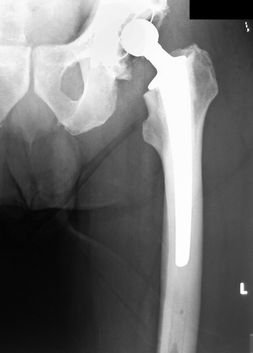
X-rayAnteroposterior radiograph of the left hip: total hip replacement in place five days after surgery, with the femoral stem and acetabular cup visible.
Source : Dramis et al., Cases journal, 2008, figure 1 · CC BY
What questions should be asked to understand the patient and their history properly?
The initial assessment rests on a structured, thorough history, the cornerstone of personalised management. ❓ The aim is not only to catalogue the physical symptoms, but also to understand the patient's expectations and psychosocial context: two major determinants of postoperative satisfaction. The questioning must systematically explore:- The characteristics of the pain : precise site (groin, trochanteric, buttock), type (mechanical, inflammatory, neuropathic), intensity (VAS 0-10), aggravating/relieving factors.
- Functional limitations : walking (distance, speed, limp), going up and down stairs, putting on shoes, getting out of a car, prolonged standing.
- The postoperative history : surgical approach (posterior / lateral / DAA), date of the operation, any complications, the rehabilitation protocol followed.
- The patient's expectations : sporting, occupational and leisure goals, a major predictor of satisfaction.
- Comorbidities : diabetes, cardiovascular disease, depression, the Lan 2022 cohort demonstrates their negative impact on pain and function after THA.²⁷
- Psychosocial flags : kinesiophobia (Morri 2020: an independent predictor of poorer function at 6 months)²⁸, catastrophising, depressive symptoms.
🚩 Red flags specific to the post-THA period
- Suspected periprosthetic joint infection (PJI): fever, chills, discharge from the wound, continuous pain not relieved by rest. ICM 2018 criteria (Parvizi): CRP > 1 mg/dL, D-dimer > 860 ng/mL, ESR > 30 mm/h, score ≥ 6 = infected. PJI incidence: 0.5-2.3 % in primary THA.²¹
- Suspected dislocation: a sense of giving way, acute pain, apparent shortening of the limb, abnormal rotation. Cumulative 30-day incidence: 0.9 % overall (Swedish registry 2024).
- Suspected periprosthetic fracture: trauma, even minor, acute pain with functional incapacity. The Vancouver B1/B2/B3 classification guides management.
- Deep vein thrombosis / pulmonary embolism: pain, swelling, unilateral redness of the calf; sudden breathlessness.
- Acute neurological injury: sudden motor or sensory deficit in the femoral, sciatic or obturator territory.
⚠️ Any combination of signs or clinical doubt → immediate medical referral before rehabilitation continues.
Which clinical tests should be carried out and which other conditions must be ruled out?
Objective assessment combines two families of tools: the PROMs (Patient-Reported Outcome Measures) and the PBMs (Performance-Based Measures). 📋 No single test is sufficient: the combination gives a complete, balanced picture.| Tool | Type | Validated MCID | Source |
|---|---|---|---|
| Oxford Hip Score (OHS) | PROM | ~5 pts (between-group MID) / ~11 pts (within-individual MIC) | Beard 2015²⁵ |
| HOOS-JR | PROM | 18 pts (anchor-based) / 7-13 pts (distribution-based) | Lyman 2018²⁶ |
| Harris Hip Score | Hybrid | Historical (1969), less patient-centred | Harris 1969²³ |
| Timed Up and Go (TUG) | PBM | 13.5 s (general older population), no validated THA-specific cut-off | Shumway-Cook 2000³⁰ |
| 6-Minute Walk Test (6MWT) | PBM | The reference for endurance and cardiovascular capacity | Bohannon 2007⁵⁸ |
| 30-second Chair Stand | PBM | Lower limb strength and overall function | Validated CDC tests |
⚠️ A caution on the TUG after THA
- The cut-off of 13.5 seconds is validated in a community-dwelling older population (Shumway-Cook 2000), not in a THA-specific cohort.
- No prospectively validated TUG cut-off for THA exists to date.
- Recommended use: a tool for tracking progress, not a strict diagnostic threshold.
Should patients be classified into subgroups, and for what benefit?
The modern trend moves away from single « one-size-fits-all » protocols towards a stratified approach. The Sato 2023 longitudinal cohort (n=1,898 patients) confirmed 3 recovery trajectories identifiable early by algorithm.¹⁰🎯 Three post-THA recovery trajectories
Sato 2023: n=1,898 patients, prospective longitudinal cohort
Source: Sato EH, et al. J Arthroplasty. 2023;38(7S):S65-S71.
- The substandard responders (36.1 %) may benefit from more intensive supervision and a prolonged strength programme.
- The late mental responders (13.2 %) need targeted psychological support (kinesiophobia, catastrophising).
- The standard responders (50.8 %) can follow an accelerated programme with guided self-management.
🧪 The rise of wearables
The Natarajan 2023 review (J Orthop Surg Res) shows that IMU sensors (smartwatches, accelerometers) allow continuous monitoring of gait and activity in real life, which is ecological and complementary to one-off tests in clinic.³¹ This technology could transform post-THA follow-up over the next 5 years.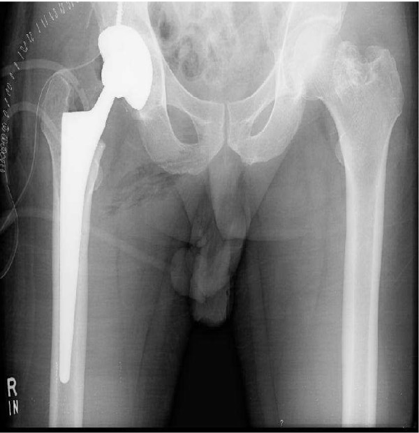
X-rayAnteroposterior pelvic radiograph: total hip replacement in place on one side, with the native contralateral hip visible for comparison.
Source : Rehman et al., Journal of medical case reports, 2007, figure 2 · CC BY
Key points
- Assessment is a triptych: history + red flags + PROMs + PBMs.
- HOOS-JR and the Oxford Hip Score are the validated PROMs; their MCID is now documented (Beard 2015, Lyman 2018).
- No TUG cut-off is validated specifically after THA: use it as a tracking tool, not as a strict diagnostic threshold.
- 3 recovery trajectories exist (Sato 2023), which justifies early stratification of the programme.
- Red flags (PJI 0.5-2.3 %, dislocation 0.9 %, fracture, DVT, nerve injury) require immediate medical referral.
Bibliography
- Adebero T et al. Prehabilitation in orthopaedic surgery: meta-analysis. Disabil Rehabil. 2024;46(24). PMID 38349251.
- Parvizi J, Tan TL, Goswami K, et al. The 2018 Definition of Periprosthetic Hip and Knee Infection. J Arthroplasty. 2018;33(5):1309-1314.e2. PMID 29551303.
- Harris WH. Traumatic arthritis of the hip after dislocation and acetabular fractures (Harris Hip Score). J Bone Joint Surg Am. 1969;51(4):737-755. PMID 5783851.
- Sutton RM, Baker CM, D'Amore T, Krueger CA, Courtney PM. The Appropriateness of Preoperative PROMs as an Indication for THA. J Arthroplasty. 2023;38(7 Suppl 2):S252-S257. PMID 37343279.
- Beard DJ, Harris K, Dawson J, et al. Meaningful changes for the Oxford hip and knee scores after joint replacement surgery. J Clin Epidemiol. 2015;68(1):73-79. PMID 25441700.
- Lyman S, Lee YY, McLawhorn AS, Islam W, MacLean CH. What Are the Minimal and Substantial Improvements in the HOOS and KOOS and JR Versions After Total Joint Replacement? Clin Orthop Relat Res. 2018;476(12):2432-2441. PMID 30179951.
- Lan P, Chen X, Fang Z, et al. Effects of Comorbidities on Pain and Function After Total Hip Arthroplasty. Front Surg. 2022;9:829303. PMID 35647007.
- Morri M, Venturini E, Franchini N, et al. Is kinesiophobia a predictor of early functional performance after total hip replacement? BMC Musculoskelet Disord. 2020;21(1):724. PMID 33160343.
- O'Connor JP, Holden P, Gagnier JJ. Systematic review: preoperative psychological factors and total hip arthroplasty outcomes. J Orthop Surg Res. 2022;17(1):457. PMID 36253795.
- Shumway-Cook A, Brauer S, Woollacott M. Predicting the probability for falls using the Timed Up and Go Test. Phys Ther. 2000;80(9):896-903. PMID 10960937.
- Natarajan P, Fonseka RD, Maharaj MM, Koinis L, Mobbs RJ. Wearable devices for postoperative monitoring in hip, knee and spine procedures. J Orthop Surg Res. 2023;18(1):812. PMID 37907943.
- Sato EH, Stevenson KL, Blackburn BE, et al. Recovery Curves for Patient Reported Outcomes and Physical Function After Total Hip Arthroplasty. J Arthroplasty. 2023;38(7S):S65-S71. PMID 37068568.
Which rehabilitation strategies are scientifically validated?
Where do you start? What is the hierarchy of interventions?
The modern hierarchy places preparing the patient and immediate mobilisation at the top of the priorities. The 2024 data do, however, require the role of prehabilitation in THA to be qualified.📌 2024 update: Prehabilitation and length of stay
- The meta-analysis Adebero 2024 (Disabil Rehabil, orthopaedic RCTs) finds a reduction in length of stay only for TKA (MD ~1 day).
- For isolated THA: MD −0.24 days, p=0.12: NOT SIGNIFICANT.³³
- Prehabilitation nonetheless remains defensible for preparing the patient, managing their expectations and improving satisfaction.
- The Punnoose 2023 review (JAMA Netw Open) confirms an overall benefit in orthopaedic surgery broadly.³⁹
- Very early mobilisation from day 0 (first standing and assisted walking).
- Multimodal analgesia, limiting opioids.
- Structured preoperative education.
- Perioperative nutritional optimisation.
- Regional anaesthesia (spinal or femoral block).
What is the place of exercise, and is there a superior approach?
The literature converges clearly on the superiority of progressive resistance training (PRT) over low-load exercise. 💪 The Coulter 2021 meta-analysis (JAMA Network Open, exercise interventions after THA) confirms a moderate effect on strength and function at 3-12 months.²⁹ Chen 2021 (Asia Pac J Sports Med) demonstrates the feasibility and effectiveness of early PRT within fast-track protocols.³⁵⚙️ GRADE hierarchy of post-THA rehabilitation interventions
Level of evidence (high / moderate / low) from a 2023-2024 critical review
An evidence-based synthesis 2023-2024. GRADE = Grading of Recommendations Assessment, Development and Evaluation.
Manual therapies, telerehabilitation: how effective are they really?
The Zhou 2024 meta-analysis (Disabil Rehabil) on telerehabilitation after THA concludes that the results are equivalent to face-to-face care on function, pain and quality of life. 🏠 This approach supports the patient's independence, reduces logistical constraints and allows asynchronous supervision (video, IMU sensors) or synchronous supervision (video call).³⁶ Similarly, the Cochrane review Khan 2008 (multidisciplinary rehabilitation after arthroplasty) remains the reference on multiprofessional inpatient versus outpatient supervision, with no major difference in long-term outcomes.³⁷ As for manual therapies (joint mobilisation, soft-tissue massage), the evidence of effectiveness as a primary treatment is weak. They can be used as an adjunct for managing pain or stiffness in the short term, but must never replace a programme of active, progressive exercise.Beyond the physical: education and psychological factors
The success of post-THA rehabilitation does not depend on physical recovery alone. 🧠Therapeutic education and the management of psychological factors are now central components.- Preoperative education: Moyer 2017 (JBJS Reviews) confirms the value of preoperative education for satisfaction and anxiety.³⁸ The Cochrane review McDonald 2014 (CD003526) remains the historical reference.⁴⁰
- Kinesiophobia: the Morri 2020 cohort demonstrates that high preoperative kinesiophobia independently predicts poorer functional performance at 6 months.²⁸
- Preoperative psychological factors: the O'Connor 2022 systematic review confirms the impact of depression, anxiety and catastrophising on postoperative PROMs.²⁹
- Self-efficacy: patients who have been taught report more confidence in early weight-bearing, which speeds up recovery.
Key points
- Prehabilitation does NOT significantly reduce length of stay for isolated THA (Adebero 2024: MD −0.24 days, p=0.12).
- The ERAS protocols remain the standard, with mobilisation from day 0 (ERAS Society 2020 consensus).
- And Progressive resistance training (PRT) is the most effective exercise approach, superior to standard low-intensity exercise.
- And Telerehabilitation gives results equivalent to face-to-face care (Zhou 2024 meta-analysis).
- Education and taking account of psychological factors (kinesiophobia) are major levers, often overlooked.
Bibliography
- Adebero T et al. Prehabilitation in orthopaedic surgery: a systematic review and meta-analysis. Disabil Rehabil. 2024;46(24). PMID 38349251.
- Wainwright TW, Gill M, McDonald DA, et al. Consensus statement for perioperative care in total hip and knee replacement (ERAS Society). Acta Orthop. 2020;91(1):3-19. PMID 31663402.
- Stambough JB, Nunley RM, Curry MC, Steger-May K, Clohisy JC. Rapid recovery protocols for primary total hip arthroplasty can safely reduce length of stay without increasing readmissions. J Arthroplasty. 2015;30(4):521-526. PMID 25683296.
- Chen T, Zhang JT, Lin Y, et al. Effects of progressive resistance training for early postoperative fast-track total hip or knee arthroplasty. Asia Pac J Sports Med Arthrosc Rehabil Technol. 2021;25:1-9. PMID 33715964.
- Saueressig T, Owen PJ, Zebisch J, Herbst M, Belavy DL. Evaluation of Exercise Interventions and Outcomes After Hip Arthroplasty: A Systematic Review and Meta-analysis. JAMA Netw Open. 2021;4(2):e210254. PMID 33635329.
- Zhou Z, Zhou X, Cui N, et al. Effectiveness of tele-rehabilitation after total hip replacement: SR and meta-analysis of RCTs. Disabil Rehabil. 2024;46(20):4611-4616. PMID 37990882.
- Khan F, Ng L, Gonzalez S, Hale T, Turner-Stokes L. Multidisciplinary rehabilitation programmes following joint replacement. Cochrane Database Syst Rev. 2008;(2):CD004957. PMID 18425906.
- Moyer R, Ikert K, Long K, Marsh J. The Value of Preoperative Exercise and Education for Patients Undergoing Total Hip and Knee Arthroplasty. JBJS Rev. 2017;5(12):e2. PMID 29232265.
- Punnoose A, Claydon-Mueller LS, Weiss O, et al. Prehabilitation for Patients Undergoing Orthopedic Surgery: A Systematic Review and Meta-analysis. JAMA Netw Open. 2023;6(4):e238050. PMID 37052919.
- McDonald S, Page MJ, Beringer K, Wasiak J, Sprowson A. Preoperative education for hip or knee replacement. Cochrane Database Syst Rev. 2014;(5):CD003526. PMID 24820247.
- O'Connor JP, Holden P, Gagnier JJ. Systematic review: preoperative psychological factors and total hip arthroplasty outcomes. J Orthop Surg Res. 2022;17(1):457. PMID 36253795.
How do you ensure lasting recovery and plan the return to sport?
How do you make the patient an active participant through self-management?
Patient empowerment (patient empowerment) is a strong determinant of long-term results. Moving from the role of passive recipient to that of principal actor in one's own rehabilitation radically changes the prospects of success. That transition should be started as early as possible: ideally before the operation itself. Supervised self-management programmes (education, guided home exercises, SMART goals) have shown they can improve function and quality of life in the short and medium term.³⁶ Telerehabilitation supports that independence while guaranteeing quality professional follow-up.³⁶ One essential point: continuing an exercise programme in the long term, because strength deficits, particularly in the abductors, can persist beyond 24 months and constitute a risk factor for limping, instability and falls.¹¹ 💪When and how should a safe return to sport be planned?
The current trend moves away from a purely chronological approach (« resume at 3 months ») towards progression based on functional criteria. The quantified criteria genuinely validated for THA are rare, however: the ACL criteria must not be extrapolated.⚠️ The « strength ≥ 90 % of the uninjured side » criterion: an unjustified extrapolation
- This threshold is validated for return to sport after ACL reconstruction (Grindem 2016), not for THA.⁴⁵
- No robust quantified recommendation establishes a formal strength cut-off for THA.
- To be used as a pragmatic goal, without treating it as a validated absolute.
- No pain during activities of daily living.
- Near-complete recovery of joint range (particularly extension and external rotation).
- Muscle strength recovering progressively: the trajectory matters more than a cut-off (86.3 % at 6 months → 93.4 % at >24 months, Ismailidis 2021).¹¹
- Passing functional tests (TUG, 30-s chair stand, stairs) with no visible compensation.¹⁰
- The patient's subjective confidence and the absence of marked kinesiophobia.²⁸
⚽ Return-to-sport rates after THA by intensity
A synthesis of Pan 2024 + Hoorntje 2023: % of patients who take it up again
Sources: Pan X 2024 PMID 39811483; Hoorntje A 2023 PMID 37160556. Note: the « <50 % high-impact sports » figure in the earlier version is wrong — the true value is ~56 %.
| Level | Activities | Rationale |
|---|---|---|
| Recommended | Walking, stairs, cycling on the flat, swimming, golf, ballroom dancing, rowing | Low impact, consensus >95 % |
| Allowed (with prior experience) | Hiking, cross-country skiing, doubles tennis, road cycling, gym work | Moderate risk depending on technique |
| Not advised | Running, football, rugby, basketball, volleyball, squash, jumping sports | Repeated impact, risk of wear and dislocation |
🚨 Positions carrying a dislocation risk: by surgical approach
- Posterior approach (the most common): avoid: flexion + adduction + internal rotation. Examples: sitting too low, crossing the legs, bending forwards with rotation.
- Direct anterior approach (DAA): avoid: extension + external rotation. Examples: leg behind with the foot turned outwards, the « sailor » stance.
- Lateral approach: intermediate risk; avoid extreme abduction.
Patient education must be specific to the surgical approach used by the surgeon.⁴⁵ The 2024 Swedish registry confirms a 30-day cumulative incidence of dislocation of 0.9 % on average (posterior 1.1 % vs DAA 0.7 % vs lateral 0.5 %).⁴⁰
Key points
- And Preoperative education and self-management programmes are fundamental to giving the patient responsibility for their recovery.
- 76-85 % return to sporting activity, and about 56 % to high-impact sports (and not <50 %).
- The « strength ≥ 90 % of the uninjured side » criterion is an ACL extrapolation not validated for THA.
- The positions carrying a dislocation risk depend on the surgical approach: education must be personalised.
- Favour low-impact sports (cycling, swimming, golf, walking) to preserve the longevity of the implant.
- And Abductor strength deficits can persist beyond 24 months and require continued strengthening to prevent limping and falls.
Bibliography
- Hailer NP et al. Time to revision after THA — Swedish registry analysis. Acta Orthop. 2024. PMID 38276104.
- Klein GR, Levine BR, Hozack WJ, et al. Return to athletic activity after total hip arthroplasty. Consensus guidelines based on a survey of The Hip Society. J Arthroplasty. 2007;22(2):171-175. PMID 17275629.
- Swanson EA, Schmalzried TP, Dorey FJ. Activity recommendations after total hip and knee arthroplasty: a survey of the AAHKS. J Arthroplasty. 2009;24(6 Suppl):120-126. PMID 19698910.
- Pan X et al. Return to Sports Following Direct Anterior Approach THA vs Other Approaches: SR and Meta-analysis. Orthop Rev. 2024. PMID 39811483.
- Hoorntje A et al. Return to Sport After Hip and Knee Arthroplasty: Counseling Patients. Curr Rev Musculoskelet Med. 2023. PMID 37160556.
- Grindem H, Snyder-Mackler L, Moksnes H, Engebretsen L, Risberg MA. Simple decision rules can reduce reinjury risk by 84 % after ACL reconstruction. Br J Sports Med. 2016;50(13):804-808. PMID 27162233. [The source of the 90 % LSI threshold, in an ACL context — not strictly applicable to THA]
- Brown TD, Elkins JM, Pedersen DR, Callaghan JJ. Impingement and dislocation in total hip arthroplasty: mechanisms and consequences. Iowa Orthop J. 2014;34:1-15. PMID 25328453.
- Ismailidis P et al. Abductor Muscle Strength Deficit After Total Hip Arthroplasty: SR and Meta-analysis. J Arthroplasty. 2021;36(8):3015-3027. PMID 33867208.
- Sato EH, Stevenson KL, Blackburn BE, et al. Recovery Curves for Patient Reported Outcomes and Physical Function After Total Hip Arthroplasty. J Arthroplasty. 2023;38(7S):S65-S71. PMID 37068568.
- Zhou Z et al. Effectiveness of tele-rehabilitation after total hip replacement. Disabil Rehabil. 2024;46(20):4611-4616. PMID 37990882.
What do real clinical cases teach us?
A classic case: the fast-track trajectory
A « classic » case represents the ideal pathway of a patient having a THA and following an accelerated (« fast-track ») rehabilitation protocol. Take the composite example of a 67-year-old man, active and with no major comorbidity, operated on for primary hip osteoarthritis by a posterior approach.📋 Composite typical case: ERAS protocol
- Preoperative: 1 prehabilitation session (education + strength optimisation exercises), managing expectations, planning the return home.⁵¹
- Day of surgery (D0): Surgery under spinal anaesthesia + femoral block, multimodal analgesia, first standing and assisted walking the same evening.⁴⁷
- D1-D3: Walking with a frame over short distances, safe transfers, discharge at D2-D3.
- W2-W6: Outpatient rehabilitation 2-3×/week or telerehabilitation: progressive gluteal/quadriceps strengthening, balance, symmetrical gait.⁴⁸
- M3-M12: Resuming activities of daily living, near-normal function at 3-6 months, gait quality normalised at 12 months.⁴⁹
The diagnostic challenge: when pain after THA hides something else
Postoperative pain is expected, but its persistence or an atypical site should raise the alarm. Case reports are particularly instructive in documenting these diagnostic « traps ».📋 Case report 1: Psoas impingement (Henderson & Lachiewicz 2012, JBJS Br)
- Presentation : persistent groin pain after THA, made worse by active hip flexion (climbing stairs, getting out of a car), sometimes with an audible snapping sensation.
- Mechanism : impingement of the iliopsoas tendon against an acetabular cup that overhangs anteriorly (overhang ≥ 8 mm, trans-acetabular screw or large femoral head).
- Diagnosis : selective anaesthetic block under ultrasound + dynamic MRI.
- Strategy : iliopsoas stretching, ultrasound-guided corticosteroid injection (effective in ~50 %). If that fails: psoas tenotomy or cup revision.⁵²
- PMID 22323676 · Chalmers 2017 JBJS Am: PMID 28375888
📋 Case report 2: Femoral neuropathy (AlGhufaily 2023, Cureus)
- Presentation : a 35-year-old woman with developmental dysplasia (DDH), THA by a posterolateral approach. Immediately after surgery: marked quadriceps weakness with knee instability on weight-bearing, reduced sensation in the anterior femoral territory.
- Diagnosis : nerve conduction studies confirming partial axonal injury to the femoral nerve (surgical stretching or a psoas haematoma).
- Actual incidence : 0.17 % overall (Farrell 2005), 0.21-0.40 % posterior approach, up to 1.1 % direct anterior approach (Brown 2018).⁵³,⁵⁴
- Rehabilitation : an adapted programme including FES (functional electrical stimulation) and very progressive strengthening.
- Prognosis : variable recovery of 6 weeks to 13 months, the majority <2 years, with complete recovery at 6 months common. The idea that recovery systematically takes >12 months is misleading.
- PMID 38116021
Study of complex cases: dislocation, periprosthetic fracture
📋 Case report 3: Recurrent dislocation (Vosinakis 2022, Cureus)
- Presentation : recurrent dislocation after THA in a patient with distorted proximal femoral anatomy. A combination of stabiliser insufficiency and poor cup positioning.
- Management : surgical revision with a dual-mobility component and an anti-dislocation cup.
- Rehabilitation : 12 weeks centred on closed kinetic chain strengthening of the stabilisers and education about at-risk positions (specific to the surgical approach).⁴⁵
- Dislocation incidence 2024 : Swedish registry 30-day CDI = 0.9 % overall, posterior 1.1 % vs DAA 0.7 % vs lateral 0.5 %.⁴⁰
- PMID 36381812
📋 Case report 4: Vancouver B2 periprosthetic fracture (Vergne 2019, PMC6581984)
- Presentation : an 81-year-old man, femoral periprosthetic fractures that were bilateral Vancouver B2, 24 and 21 years after bilateral resurfacing THA (a fall).
- Treatment choice : internal fixation with locking plates (rather than revision) because of comorbidities and frailty. Bone union and stem stability confirmed at 12 months.
- Weight-bearing by Vancouver subtype (2022-2024 data)⁵⁶,⁵⁷:
| Vancouver subtype | Mechanism | Current weight-bearing protocol |
|---|---|---|
| B1 | Stable stem + fracture around it | TTWB 6 wk → PWB 6 wk → FWB |
| B2 | Loose stem + fracture | A trend towards early weight-bearing after adequate revision (2024 study) |
| B3 | Femoral bone loss | Variable: ~36 % immediate FWB, ~29 % PWB 6 wk (EHS) |
The old « 8-12 weeks partial weight-bearing for every type B » protocol is now obsolete. B1/B2/B3 stratification is mandatory.
Key points
- The « classic » clinical cases confirm the effectiveness of structured early rehabilitation protocols (fast-track + ERAS).
- Atypical pain after THA requires a rigorous differential diagnosis : psoas impingement (Henderson 2012), femoral neuropathy (AlGhufaily 2023).
- The actual incidence of femoral nerve injury is 0.17-0.40 % (up to 1.1 % with the direct anterior approach), not 0.08-2 %.
- The prognosis for nerve recovery runs from 6 weeks to 13 months, not systematically >12 months.
- Weight-bearing protocols for a Vancouver B periprosthetic fracture must be individualised by subtype (B1/B2/B3), not fixed at 8-12 weeks.
- Despite their low level of evidence (N=1), clinical cases are irreplaceable for sharpening clinical reasoning and generating hypotheses.
Bibliography
- Wang L et al. Preoperative rehabilitation for TKA/THA: a systematic review and meta-analysis of RCTs. BMJ Open. 2016;6(2):e009857. PMID 26839013.
- Henderson RA, Lachiewicz PF. Groin pain after replacement of the hip: aetiology, evaluation and treatment. J Bone Joint Surg Br. 2012;94(2):145-151. PMID 22323676.
- Chalmers BP, Sculco PK, Sierra RJ, Trousdale RT, Berry DJ. Iliopsoas Impingement After Primary Total Hip Arthroplasty: Operative and Nonoperative Treatment Outcomes. J Bone Joint Surg Am. 2017;99(7):557-564. PMID 28375888.
- AlGhufaily AA, Alshunaifi AI, AlHarbi JS. Femoral Nerve Palsy Post Total Hip Arthroplasty via a Posterolateral Approach. Cureus. 2023;15(11):e50771. PMID 38116021.
- Farrell CM, Springer BD, Haidukewych GJ, Morrey BF. Motor nerve palsy following primary total hip arthroplasty. J Bone Joint Surg Am. 2005;87(12):2619-2625. PMID 16322610.
- Brown GD, Swanson EA, Nercessian OA. Neurologic injuries after total hip arthroplasty. Am J Orthop. 2008;37(4):191-197. (The reference clinical review on femoral nerve palsy incidence by surgical approach.)
- Vosinakis CI, Vossinakis IC. Treatment of Recurrent Total Hip Arthroplasty Dislocation Caused by Distorted Proximal Femoral Anatomy. Cureus. 2022;14(10):e29969. PMID 36381812.
- Vergne G et al. Osteosynthesis of bilateral Vancouver B2 periprosthetic femoral fracture: a case report. Trauma Case Rep. 2019. PMC6581984. PMID 31212096.
- Moreta J, Aguirre U, de Ugarte L, et al. Functional and radiological outcome of periprosthetic femoral fractures after THA. Injury. 2015;46(3):527-532. PMID 25085599.
- Guerra ML, Singh PJ, Taylor NF. Early mobilization of patients who have had a hip or knee joint replacement reduces length of stay. Clin Rehabil. 2015;29(9):844-854. PMID 25452634.
- Nissen T, Wynn R. The clinical case report: a review of its merits and limitations. BMC Res Notes. 2014;7:264. PMID 24758689.
- Wainwright TW et al. ERAS Society consensus statement for THA/TKA. Acta Orthop. 2020;91(1):3-19. PMID 31663402.
How can these recommendations be applied in practice?
When, and to which professionals, should you refer?
The modern physiotherapist in direct access must be able to triage and refer. The system of diagnostic flags structures that decision.🚩 Red flags specific to the post-THA period (Finucane 2020 framework)
- Periprosthetic joint infection (PJI): incidence 0.5-2.3 %; fever, chills, discharge, continuous night pain. ICM 2018 criteria: CRP / D-dimer / ESR.⁶³,⁶⁴
- Dislocation: incidence 0.5-1.1 % depending on approach; deformity, shortening, acute incapacity.⁴⁰
- Periprosthetic fracture: trauma + pain + incapacity; Vancouver B1/B2/B3 classification.
- DVT / pulmonary embolism: unilateral calf pain, sudden breathlessness.
- Acute neurological injury: sudden motor or sensory deficit.
- Cauda equina (where there is associated low back pain): follow the national pathway.⁶⁵
⚠️ Any red flag → prompt medical referral (GP, emergency department, orthopaedic surgeon) before rehabilitation continues.
🟡 Yellow flags (psychological)
Negative beliefs about pain, catastrophising, fear of movement (kinesiophobia), depressive or anxious symptoms. Screening tool: the Örebro Musculoskeletal Pain Screening Questionnaire (Nicholas 2011).⁶⁶ The Morri 2020 cohort validates kinesiophobia as an independent predictor of poorer function at 6 months after THA.²⁸🔵 Blue flags (work-related perceptions)
Work perceived as too demanding, lack of employer support. A Psychologically Informed Practice approach (Keefe, Main, George 2018).⁶⁷ Where these factors predominate and constitute the main obstacle to recovery, interprofessional collaboration (psychologist, occupational health, pain specialist) significantly improves outcomes at 1 year (Cochrane Reeves 2013; Kamper 2015 BMJ multidisciplinary biopsychosocial rehabilitation).⁶⁸,⁶⁹How do you measure outcomes and overcome the barriers to EBP?
Combine PROMs and performance tests with, ideally, a Core Outcome Set (COS) validated for the condition. 📈| Tool | Use | Key reference |
|---|---|---|
| HOOS-JR / OHS | Hip-specific PROMs | Beard 2015; Lyman 2018 |
| Hip/knee OA Core Outcome Set | A standardised minimum set | Smith / OMERACT-OARSI 2019⁷⁰ |
| 6MWT | Endurance, cardiovascular capacity | Bohannon 2007⁷¹ |
| TUG, 30-s Chair Stand, Stair Climb | Mobility, lower limb strength, balance | Validated CDC tests |
| PROMIS-PF | A generic cross-cutting score | NIH Common Data Element |
| Implementing PROMs (guidance) | A methodological framework for implementation | Snyder 2012 (Qual Life Res)⁷² |
🚧 Barriers to EBP in physiotherapy
The Scurlock-Evans 2014 review (Physiotherapy) and the Bernhardsson 2022 cohort identify the common barriers:⁷³,⁷⁴- Lack of time for critical reading (the most-cited barrier).
- Lack of methodological appraisal skills.
- Limited access to databases and to full text.
- The inertia of practice habits.
- Lack of organisational support.
✅ Validated enablers
- Mentoring by experienced peers.
- An institutional subscription to the Cochrane Library / PEDro / PubMed.
- Time set aside for literature searching and case discussion.
- Systematic use of clinical practice guidelines (CPGs) that synthesise the evidence.
- Continuing education with an emphasis on critical reading.
Key points
- Active triage using red flags (Finucane 2020), yellow flags (Nicholas 2011) and blue flags.
- Assessment uses PROMs + PBMs, ideally through a Core Outcome Set (OMERACT-OARSI).
- The barriers to EBP (time, skills, access) can be overcome with mentoring + resources + organisational culture.
- And Interprofessional collaboration is essential for complex psychosocial cases.
Bibliography
- Finucane LM, Downie A, Mercer C, et al. International Framework for Red Flags for Potential Serious Spinal Pathologies. J Orthop Sports Phys Ther. 2020;50(7):350-372. PMID 32438853.
- Parvizi J, Tan TL, Goswami K, et al. The 2018 Definition of Periprosthetic Hip and Knee Infection. J Arthroplasty. 2018;33(5):1309-1314.e2. PMID 29551303.
- Premkumar A, Kolin DA, Farley KX, et al. Projected economic burden of periprosthetic joint infection of the hip and knee in the United States. J Arthroplasty. 2021;36(5):1484-1489. PMID 33422392.
- Williams JT, Lister H, Fakouri B, Panchmatia JR. The National Suspected Cauda Equina Syndrome Pathway: implications for physiotherapists. Physiotherapy. 2024;122:68-69. PMID 38244418.
- Nicholas MK, Linton SJ, Watson PJ, Main CJ. Early identification and management of psychological risk factors ("yellow flags") in patients with low back pain. Phys Ther. 2011;91(5):737-753. PMID 21451099.
- Keefe FJ, Main CJ, George SZ. Advancing Psychologically Informed Practice for Patients With Persistent Musculoskeletal Pain. Phys Ther. 2018;98(5):398-407. PMID 29669084.
- Reeves S, Perrier L, Goldman J, Freeth D, Zwarenstein M. Interprofessional education: effects on professional practice and healthcare outcomes (update). Cochrane Database Syst Rev. 2013;(3):CD002213. PMID 23543515.
- Kamper SJ, Apeldoorn AT, Chiarotto A, et al. Multidisciplinary biopsychosocial rehabilitation for chronic low back pain: Cochrane systematic review and meta-analysis. BMJ. 2015;350:h444. PMID 25694111.
- Smith TO, Hawker GA, Hunter DJ, et al. The OMERACT-OARSI Core Domain Set for Measurement in Clinical Trials of Hip and/or Knee Osteoarthritis. J Rheumatol. 2019;46(8):981-989. PMID 30647185.
- Bohannon RW. The 6-minute walk test: a meta-analysis of data from apparently healthy elders. Top Geriatr Rehabil. 2007;23(2):155-160. doi:10.1097/01.TGR.0000270184.98402.21.
- Snyder CF, Aaronson NK, Choucair AK, et al. Implementing patient-reported outcomes assessment in clinical practice. Qual Life Res. 2012;21(8):1305-1314. PMID 22048932.
- Scurlock-Evans L, Upton P, Upton D. Evidence-based practice in physiotherapy: a systematic review of barriers, enablers and interventions. Physiotherapy. 2014;100(3):208-219. PMID 24780633.
- Bernhardsson S et al. Physiotherapists' opinions, barriers, and enablers to providing evidence-based care: a mixed-methods study. BMC Health Serv Res. 2022;22(1):1382. PMID 36411428.
- Henschke N, Maher CG, Ostelo RW, et al. Red flags to screen for malignancy in patients with low-back pain. Cochrane Database Syst Rev. 2013;(2):CD008686. PMID 23450586.
And after this article?
This article is part of a collection of evidence-based clinical syntheses. A question, a comment, a correction to suggest? Contact us directly through the WhatsApp button at the bottom right of the screen.

