















In brief
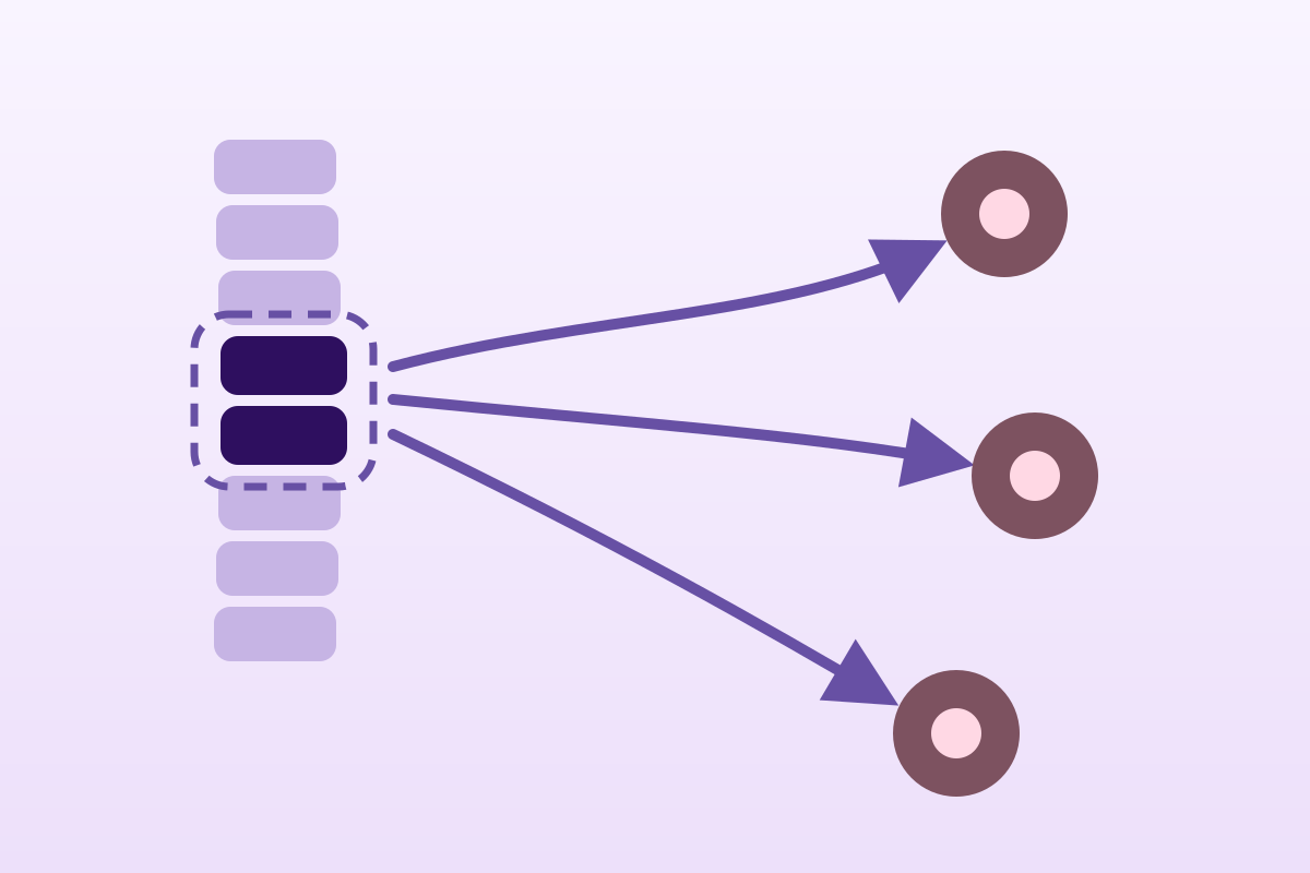
Maigne syndrome means pain whose site of complaint and site of distress do not coincide: the patient points to their iliac crest, their groin, their greater trochanter or their testicle, while the structure at fault sits at the thoracolumbar junction, around T12-L1. The mechanism invoked is the course of the dorsal ramus of T12-L1, whose cutaneous branch crosses the iliac crest 7-8 cm from the midline: a course established by dissection (Maigne 1989). The signs described are the iliac crest point, the skin rolling test and segmental tenderness of the junction. It is an entity with a low level of evidence: no national guideline, no placebo-controlled trial on the named entity, and a single randomised trial (n = 30). Its clinical value is not to provide a definite diagnosis, but to open a hypothesis when crest or groin pain resists well-conducted local treatment.
A clinical synthesis on thoracolumbar junction syndrome: pain that complains far from where it arises. This article deals with the sorting error: why distress at T12-L1 declares itself at the iliac crest, how to recognise it, how not to confuse it with the neighbouring low back conditions, and, a delicate point on this subject, what the literature actually supports.
Clinical summary
- A diagnosis of misdirection. Maigne syndrome is not one more low back pain: it is the hypothesis that pain felt at the iliac crest, in the groin, at the pubis, at the greater trochanter or in the testicle takes its source at the thoracolumbar junction, T11 to L2 depending on the author, T12-L1 most often (Randhawa 2022).
- The anatomical substrate is solid, the syndrome less so. Thirty-seven dissections showed that the skin of the low lumbar region is innervated by the lateral branches of the dorsal rami of T12 and L1 in 60 % of cases, and that the most medial of them crosses the iliac crest through a rigid osteo-aponeurotic opening 7-8 cm from the midline (Maigne 1989). Compression was observed there in 2 cases out of 37.
- Three clinical signs, none validated. The iliac crest point, the skin rolling test (the sign of cellulalgia), segmental tenderness of the junction. None has been the subject of a published inter-examiner reliability study that we could find: their value is one of custom, not of evidence.
- An independent confirmation, through another door. Japanese teams studying the superior cluneal nerve, with no reference to Maigne, find the same point at 70 mm from the midline in 14 % of 834 low back pain patients (Kuniya 2014), and 12 % of 200 chronic low back pains (Kiral 2024).
- But the same anatomy contradicts part of the story. Konno 2017 shows that the branches actually entrapped at the crest come from L3 - L5, not from T12-L1: the opening at 7 cm therefore does not prove the thoracolumbar origin of the pain.
- The treatment must be read with care. Responder rates are high (78.3 %, 95 % CI 72.9-82.9, across 628 patients: Catalano 2026), but they come from observational and non-comparative studies: it is a proportion of people who improved, not an effect size against placebo.
- The real red flag is at the same level. An osteoporotic fracture of T11-L2 produces exactly the same pattern of referral: across 87 fractures, 15 complained away from the site: iliac crest, groin, trochanter (Yang 2014). In someone over 70, age alone multiplies the probability of fracture by 11 (Cochrane 2023).
- What the hypothesis changes in practice: faced with crest or groin pain that does not yield to well-conducted local treatment, examine the thoracolumbar junction before concluding to a tendinopathy, groin pain or a hip condition.
Why can iliac crest pain arise at T12-L1?
The whole syndrome lies in a mismatch: the patient puts their hand in one place, the painful structure is elsewhere. This chapter sets out the anatomy that makes that mismatch possible, and says at the outset how far it has been demonstrated.
A patient comes in with unilateral pain at the iliac crest. They point to it, always at the same spot, three finger-breadths from the midline. Local palpation makes them start. The low lumbar radiographs are normal or unremarkably degenerative. The point is treated: massage, shockwave therapy, injection of the “iliolumbar ligament”, gluteal strengthening. That gives relief for a few days, then it comes back.
The hypothesis formulated by Robert Maigne in 1980 is that this point is not the lesion, but its arrival terminal. The distress would sit two levels higher, at the junction between the thoracic and lumbar spine, and the pain would take a nerve to descend to the crest.
What exactly does the dissection say?
This is the most solid point in the file, and it deserves quoting at the right level of precision. In 1989, Maigne and his team published in Surgical and Radiologic Anatomy a study of 37 dissections. Three results come out of it.
First, the skin of the low lumbar region is innervated by the lateral branches of the dorsal rami of T12 and L1 in 22 cases out of 37 (60 %), of T12, L1 and L2 in 10 cases (27 %), and of T12, L1 and L2 with an L3 anastomosis in 5 cases (13 %). In other words: the skin the patient points to above their iliac crest gets its sensation from levels well above where they are pointing.
Next, the most medial nerve crosses the iliac crest through a rigid osteo-aponeurotic opening, 7-8 cm from the midline. That is a mechanical constraint: a nerve passing through an inextensible ring can, in theory, be compressed there.
Finally, and this is the nuance that secondary accounts often leave out, that compression was actually observed in 2 cases out of 37. Not 37. Two. The course is constant; the conflict is the exception.
What 1991 added: what you palpate is not what you think you are palpating
Two years later, the same team published in Archives of Physical Medicine and Rehabilitation an analysis of the “trigger point” of the posterior iliac crest, the one then attributed to the insertion of the iliolumbar ligament. The reasoning is clear: the iliac insertion of that ligament is inaccessible to palpation, protected by the bony rim of the crest. The dorsal ramus of L1 or L2, for its part, crosses the crest 7 cm from the midline, in a superficial and dorsal position, that is, directly under the examiner's finger.
The authors' conclusion: the painful point palpated there probably corresponds to pain of cutaneous nerve origin, arising from the thoracolumbar junction, rather than to a ligamentous enthesopathy.
A point of method that changes the reading
The 1989 and 1991 publications report the same 37 dissections. They are two readings of a single series, not two independent confirmations. That series remains the anatomical reference for the syndrome; it is also, to this day, the only one of that size devoted specifically to the question.
Why this junction and not another?
The mechanical argument invoked is that of a transition zone. The thoracic spine, constrained by the rib cage, allows rotation; the lumbar spine, with its facets oriented in the sagittal plane, allows little. Passing from one to the other concentrates rotational loading on a few segments. Tsur and Ohry (2024) take up that argument and are a reminder that although the T12-L1 facets take most of that loading, any segment between T9 and L2 may in practice be at fault.
Kozera et al. (2017), in the third part of a Polish anatomical series devoted to the posterior rami, describe the same entity under the name Spinal Dorsal Ramus Mediated Back Pain and give it a precise clinical signature: unilateral pain, triggered by extension and/or rotation, radiating towards the buttock and the posterior iliac crest, and not crossing the midline. They add that, since the posterior ramus and the anterior ramus belong to the same spinal nerve, the complaint may spill over into the anterior territory: pseudo-visceral pain of the hypogastrium, false sciatic neuralgia, tenderness of the pubic symphysis.
The Japanese convergence: the same point, without the same name
An argument often neglected in the French-language literature deserves highlighting, because it is the closest thing to an external validation the syndrome has.
Since the 2010s, Japanese neurosurgery and orthopaedic teams have independently described superior cluneal nerve entrapment (superior cluneal nerve entrapment) as a cause of low back pain. They do not cite Maigne, do not speak of a thoracolumbar junction, and work to criteria of their own. Yet their main diagnostic criterion, in the prospective study by Kuniya et al. (2014) covering 834 patients, is the following: the most painful point lies on the posterior iliac crest 70 mm from the midline, and palpating it reproduces the main complaint.
That is, to the millimetre, the point described by Maigne forty years earlier. One hundred and thirteen patients (14 %) met both criteria. In them, the mean VAS fell from 68.6 ± 19.2 mm to 45.2 ± 28.8 mm one week after an anaesthetic block; 68 % obtained a fall of at least 50 % after three blocks.
Kiral et al. (2024), in Istanbul, repeated the exercise in 200 chronic mechanical low back pains with an ultrasound-guided block and a threshold of 70 % relief at one hour: 24 patients confirmed, that is a frequency of 12 %.
Key points
- The anatomical substrate, a T12-L1 dorsal ramus crossing the crest 7-8 cm from the midline through a rigid opening, is established by dissection.
- Actual compression of that nerve there is rare (2 cases out of 37).
- Two independent teams, with no reference to Maigne, find a painful point at the same place in 12 - 14 % of chronic low back pain patients.
- These figures describe a frequency of a painful point, not a prevalence of disease: they depend entirely on the criterion chosen to define the entity.
How do you recognise Maigne syndrome on clinical examination?
Three signs make up the classic triad. This chapter describes them with their exact manoeuvre, and says frankly what is missing from their validation file, because it is that gap that must govern how they are used.
The diagnosis is described as purely clinical by Maigne himself as early as 1980: the diagnosis is made on purely clinical grounds
. Three signs make it up, to which is added today a test borrowed from the cluneal nerve literature.
Sign 1: The iliac crest point
The manoeuvre: with the patient prone or side-lying, the examiner follows the upper border of the iliac crest starting from the midline and applies perpendicular pressure every centimetre. The point being sought lies 7-8 cm from the midline, on the rim of the crest.
The criterion is not local pain, a bony rim is nearly always tender, but reproduction of the patient's complaint. That is the criterion Kuniya (2014) and Kiral (2024) used to select the patients to be offered a block, and it is the one that gives the 12 to 14 % quoted above.
The difference between “it hurts” and “that is my pain”
A tender point has no diagnostic value; a point that reproduces the complaint has. The wording to use is not “does that hurt?” but “is that your pain?”. Every series that has produced usable figures on this syndrome used the second criterion.
Sign 2: The skin rolling test
The manoeuvre: grasp a fold of skin between thumb and index finger and roll it under the fingers, comparing systematically with the opposite side and with neighbouring levels. The sign is positive when the fold is thickened, doughy and painful on one side only, in the referral territory.
In the original description it corresponds to what French manual medicine calls the cellulo-periosteo-myalgic syndrome: segmental distress would express itself in the three tissues of the metamere: the skin (cellulalgia), the periosteum (painful insertion points) and the muscle (myalgic cords). Janssen and Tomasella (2023), in the Revue Médicale de Liège, make it the core of the examination: a rigorous segmental examination, then the search at a distance for palpable changes in tissue texture
.
The same manoeuvre appears in the English-language literature under the name Kibler fold test, which Kozera et al. (2017) recommend for exploring the peripheral tissues of the iliac crest.
Sign 3: Segmental tenderness of the junction
The manoeuvre: lateral pressure on the spinous processes of T11 to L2, one by one, then direct pressure on the posterior articular pillars a finger-breadth from the midline. Kim et al. (2013) sum the manoeuvre up thus: the pain can be brought out by pressure on the facet joints or on the sides of the spinous processes
.
The sign looked for is tenderness at a single level and on a single side, contrasting with the neighbouring levels. Diffuse tenderness over five levels points to nothing.
Sign 4: The Hip-Knee Flexion Test, the only test with figures
It is the only test in this field for which published diagnostic values exist. Kiral et al. (2024) evaluated it in 200 chronic low back pains, against an ultrasound-guided superior cluneal nerve block as the reference test:
What the examination cannot do
It must be said explicitly: none of these signs confirms the diagnosis. Maigne himself provided, as early as 1980, for confirmation by a periarticular anaesthetic block, and all the modern quantified series (Kuniya, Kiral) rest on a block, never on manual examination alone.
But the anaesthetic block is not itself a perfect arbiter. A single, non-placebo-controlled block carries a false positive rate well documented in the neighbouring field of facet blocks, which means that some of the patients labelled “Maigne syndrome” on a positive block are so labelled in excess.
Imaging: what it brings, and what it does not
Radiography and MRI show, in most of the cases described, only unremarkable degenerative change unrelated to the intensity of the symptoms. Kim et al. (2013) write it: radiological studies show, in most cases, only mild and non-significant degenerative changes
. Their role is therefore to rule out, not to confirm: fracture, spondylodiscitis, tumour, spondyloarthritis.
Musculoskeletal ultrasound has been the subject of recent work. Iudicelli et al. (2026) compared 12 patients with 12 healthy controls and describe a triad of markers: Copeman nodules (91.7 % against 8.3 %), iliac enthesophytes (58.3 % against 0 %), and thickening of the thoracolumbar fascia beyond 3 mm (41.7 % against 0 %). Mean fascia thickness was 3.53 ± 0.46 mm in patients against 2.61 ± 0.28 mm in controls.
These results are interesting and deserve following, but they must be read for what they are: a single-centre pilot study on 24 subjects in total, whose authors themselves call for multicentre validation. You do not make a diagnosis on that basis in 2026.
One secondary result of that work does deserve the physiotherapist's attention: fascia thickness correlated strongly with pain intensity (rho = 0.825) but not with the DN4 score of neuropathic pain. The authors draw from it the hypothesis of a fascial and biomechanical mechanism rather than of direct nerve injury.
Is the pain of this syndrome neuropathic?
The question has a direct practical consequence: it decides whether you should speak of nerve compression to the patient, and whether you should steer them towards neuropathic pain treatments.
Takada et al. (2024) administered the painDETECT questionnaire to 19 patients operated on for confirmed cluneal entrapment. The mean score was 11.8, that is, below the threshold of 12 that classes pain as “neuropathic component unlikely”. Thirteen patients out of 19 were classed as non-neuropathic, only 2 as probably neuropathic. The positive items were shooting pain, radiating pain and pain attacks; the negative items, burning, allodynia to light touch and pain with heat or cold.
Their conclusion is cautious and important: the diagnosis of low back pain due to cluneal entrapment must be made with care, because the symptoms resemble nociceptive pain
. In other words: even when a nerve is at fault and this is verified surgically, the pain does not present as neuropathic. A negative neuropathic screening questionnaire therefore does not rule out the hypothesis.
Key points
- The historical triad (crest point, skin rolling, segmental tenderness) is used to generate a hypothesis, not to conclude.
- The criterion that produced every usable figure is reproduction of the complaint, not local tenderness.
- One test alone has published values: the Hip-Knee Flexion Test, Se 41.7 % / Sp 88.6 %. A negative result rules nothing out.
- Imaging serves to rule out; fascia ultrasound is promising but remains at pilot stage (n = 24).
- The pain does not behave like neuropathic pain, even with confirmed entrapment (mean painDETECT: 11.8).
What is the evidence behind Maigne syndrome really worth?
This chapter is not a polite caveat at the end of an article. On this particular subject, the state of the evidence is a clinical fact of the first order: it governs how cautiously you speak to the patient and the threshold at which you abandon the hypothesis.
Maigne syndrome belongs to an uncomfortable category: that of entities widely taught, clinically plausible, and weakly demonstrated. Saying so clearly is better than dressing it up, for three reasons: the clinician must know what they are promising, the patient must know what is being offered, and a poorly supported entity presented as established ends up producing useless treatments.
What is established
- The anatomical course. Thirty-seven concordant dissections on the cutaneous innervation of the low lumbar region and the passage of the nerve through an osteo-aponeurotic opening 7-8 cm from the midline (Maigne 1989).
- The mechanical possibility of the conflict. Directly observed, though rarely: 2 cases out of 37 (Maigne 1989); 2 medial branches out of 109 specimens with clear entrapment (Kuniya 2013).
- The existence of a clinically identifiable subgroup. Two independent prospective series isolate 12 to 14 % of low back pain patients whose complaint is reproduced by palpation of a precise point and relieved by a block.
- The reality of referred pain from the junction. Supported indirectly but solidly by fractures: 15 patients out of 87 with an osteoporotic fracture of T11-L2 complained away from the site (Yang 2014).
What is consensual without being demonstrated
- That the skin rolling test marks segmental distress. Described since 1980, taken up by every French-language author, taught in manual medicine, and never assessed for inter-examiner reliability in any publication we could identify.
- That manipulation of the junction is the first-line treatment. Recommended by Janssen and Tomasella (2023) and by Kozera (2017), who writes that these patients
respond well to manipulative techniques
, on the basis of series and clinical experience, with no dedicated comparative trial. - That the T12-L1 level is most often at fault. A constant claim in the literature, never measured against a reference distribution.
What remains debated, or even contradicted
The contradiction of Konno 2017, which must be faced
The classic story has it that the superior cluneal nerve, arising from T12-L1, is entrapped at the iliac crest. Konno et al. (2017) dissected 81 cluneal branches in 16 cadavers. They confirm entrapment at the crest: 10 branches out of the 13 crossing the osteofibrous tunnel. But those 13 branches came from L3 (2), L4 (6) and L5 (5). None from T12 or L1.
Their conclusion: it is precisely because the branches at risk come from the lower lumbar spine that cluneal entrapment produces a “pseudo-sciatica” radiating to the thigh and calf. That result explains the leg signs better, but it moves the origin two to four levels, and weakens the mechanical link between the thoracolumbar junction and the crest point.
That tension is not incidental: it suggests that what is called “Maigne syndrome” probably covers several distinct entities brought together by a common palpation point. Randhawa et al. (2022) in fact own this by distinguishing two variants: a central variant, linked to facet arthropathy of the junction, and a peripheral variant, linked to entrapment of the medial branch of the superior cluneal nerve.
The real state of the literature, in one image
Why is this entity mainly a French-language one?
The syndrome bears the name of a French doctor, was described in a Paris department, and its terminology (cellulalgia, skin rolling, minor intervertebral derangement), belongs to the vocabulary of French manual medicine. International take-ups are rare and late: a Canadian review in 2022, an Israeli paper in 2024, a Belgian paper in 2023, a few Korean and Turkish cases.
That invalidates nothing in itself: a sound clinical observation can be born in a single language. But it has two practical consequences. First, the entity has been little exposed to contradiction by teams outside the school that formulated it. Second, the small number of teams working on it explains the absence of trials of sufficient size: there has never been the critical mass of researchers needed.
What that changes in practice
Three concrete consequences, following directly from that level of evidence.
On what is said to the patient. You do not say “you have Maigne syndrome”, you say “your pain might come from a level higher up; that is a hypothesis, and we are going to test it”. The difference is not cosmetic: naming a disease creates an identity of being ill, and the reversibility of a hypothesis is easier to announce than that of a diagnosis.
On the threshold for giving up. A weak hypothesis is tested quickly and dropped quickly. If treatment directed at the junction changes nothing within four to six sessions, the hypothesis is to be revised, not prolonged.
On the choice of treatments. Low-risk modalities with a broad expected benefit (exercise, education, manual therapy) remain proportionate to an uncertain hypothesis. Invasive and irreversible procedures are not: neurotomy and decompression surgery appear in the literature of this field, but with a level of evidence that does not allow them to be offered as the natural end point of a clinical hypothesis.
Key points
- Established: the anatomical course, the rare possibility of the conflict, the existence of a subgroup identifiable by palpation and block.
- Consensual without evidence: the value of skin rolling, the primacy of manipulation, the T12-L1 level.
- Contradicted: the T12-L1 origin of the branches actually entrapped at the crest (Konno 2017 finds them at L3-L5).
- Absent: no guideline, no meta-analysis, a single randomised trial of 30 patients.
- Operational conclusion: a useful hypothesis, to be tested quickly, abandoned quickly, and never presented as a certainty.
How do you avoid confusing Maigne syndrome with the other low back pains?
This is the decisive chapter. A syndrome that shows itself far from its origin is, by construction, an imitator. It imitates a tendinopathy, groin pain, a sacroiliac problem, sciatica, and each of those conditions is far commoner than it is.
The priority rule must be set out at the start: you think of Maigne syndrome only after ruling out the local causes, not before. Greater trochanter pain is a gluteal tendinopathy until proven otherwise; groin pain in a footballer is groin pain of the adductors until proven otherwise. The thoracolumbar hypothesis comes in only when well-conducted local treatment fails, or when local examination does not reproduce the pain.
The eight imitations to rule out, one by one
Each of these conditions shares a territory with Maigne syndrome. For each, the site's article covering it in detail is given: this article does not replace them, it organises the competition between them.
| What the patient describes | Local cause to rule out first | What would tip towards the junction |
|---|---|---|
| Greater trochanter pain, worse lying on that side | Greater trochanteric pain syndrome: gluteal tendinopathy, far commoner | Trochanteric palpation not very painful, abductor loading tests negative, but a crest point reproducing the complaint |
| Pain in the groin crease on prolonged walking | Iliopsoas impingement and hip osteoarthritis | Hip ranges preserved and painless at end of range, normal hip imaging: cf. Ségui 2021, Meadows 2019 |
| Pubic pain in a pivoting athlete | Adductor tendinopathy / groin pain | Adductors not painful on resisted contraction, isolated pubic tenderness: Kozera 2017 describes symphyseal tenderness in this syndrome |
| High buttock pain, one side only, not crossing the midline | Mechanical sacroiliac pain | A negative battery of sacroiliac tests, but a positive crest point at 7-8 cm |
| Pain running down the leg | Sciatica from disc herniation, lumbar stenosis, piriformis syndrome | No neurological deficit signs, negative straight leg raise, but radiation reproduced from the crest. Konno 2017 calls that picture “pseudo-sciatica” |
| Ordinary mechanical low back pain, worse in extension-rotation | Non-specific low back pain, lumbar facet arthropathy | Non-midline pain, strictly unilateral, with asymmetrical skin rolling two levels above the complaint |
| Mid-back pain as well | Thoracic spine pain | A question of boundary more than of diagnosis: the junction belongs to both territories. The criterion is referral at a distance |
| Night waking, prolonged morning stiffness | Axial spondyloarthritis | Nothing: an inflammatory rhythm is never Maigne syndrome. It is a redirection, not a differential diagnosis |
The difference from lumbar facet arthropathy, in one sentence
Lumbar facet arthropathy hurts where it sits: the pain is low lumbar, paramedian, at the level of the affected joint. Maigne syndrome hurts where it does not sit: the distress is at T12-L1, the complaint is at the crest or in the groin. Two articles on the site deal with the first in detail: facet joint osteoarthritis and facet joint injection; reading them does not cover this one.
The trap of the iliolumbar ligament
It deserves a paragraph of its own, because it concerns exactly the same palpation point. Janssen and Tomasella (2023) list three possible causes of posterior iliac crest pain: Maigne syndrome at T12-L1, the iliolumbar ligament syndrome, and the L5-S1 facet syndrome.
But the anatomical study by Maigne and Maigne (1991) argues that the iliac insertion of the iliolumbar ligament is inaccessible to the finger, hidden by the rim of the crest, while the dorsal ramus is superficial there. If that result is correct, some of the “iliolumbar ligament enthesopathies” diagnosed by palpation would be errors of attribution.
That point is not settled: it rests on a single series of dissections, and the iliolumbar ligament also has an established biomechanical role in lumbopelvic stability. But it justifies caution: you cannot conclude to ligament involvement from pain provoked by palpation of the crest.
Three signals that should make you think of the junction
As a synthesis of the concordant descriptions of Kozera 2017, Randhawa 2022, Janssen 2023 and Tsur 2024: none of which has been prospectively validated:
- The pain never crosses the midline. It is strictly unilateral and stays so. Kozera makes it a central descriptive element.
- It is triggered in extension and/or rotation of the trunk, rather than in flexion, which distinguishes it from the discogenic profile.
- The complaint and the examination do not meet. The patient points to one place, and it is elsewhere that the examination finds something. It is the most suggestive sign, and the easiest to miss when you examine only the area pointed to.
Key points
- The thoracolumbar hypothesis is second line: after failure or insufficiency of the local examination.
- Eight pictures imitate it; each has a local cause commoner than it is.
- An inflammatory rhythm does not enter this differential diagnosis: it demands a redirection.
- The “iliolumbar ligament point” and Maigne's crest point are the same point under two names: anatomy does not allow palpation to decide between them.
When is referred pain a red flag?
This is the risk specific to this syndrome: a serious condition of the junction produces exactly the same referral pattern as a benign derangement. The difference cannot be read on the pain map: it is read in the background.
There is one study that ought to appear in every account of this subject, and rarely does.
Yang et al. (2014) followed 87 patients with an osteoporotic compression fracture between T11 and L2. They recorded, for each, the location of the pain relative to the fractured level. Seventy-two had focal pain over the site. But fifteen had pain at a distance: low back near the iliac crest (9 patients), groin (3), trochanteric region (3).
That is, feature for feature, the referral map of Maigne syndrome. Except that the origin is not a reversible functional derangement: it is a fracture.
What that result requires
The consequence is direct: in someone at risk of osteoporosis, new iliac crest pain does not allow you to conclude to a functional cause before having considered a fracture of the junction. The authors in fact conclude exactly so: patients at osteoporotic risk who present with low back pain should not have the possibility of a thoracolumbar fracture overlooked.
The site covers that background in a dedicated article: osteoporosis and the prevention of fragility fractures.
The red flags that really count
The Cochrane review by Han et al. (2023) assessed the diagnostic value of red flags for vertebral fracture. Its general conclusion is severe: meta-analysis was not possible, most signs have poor value, and only one of the 14 studies included was at low risk of bias on the reference standard. A few positive likelihood ratios nevertheless stand out, in primary care:
Red flags with informative value (Cochrane 2023)
- Age over 70: positive likelihood ratio 11.19 (95 % CI 5.33-23.51) for unspecified vertebral fracture, in primary care.
- Trauma: +LR from 1.93 to 12.85 depending on the study; 6.42 (95 % CI 2.94-14.02) for osteoporotic fracture.
- Corticosteroid therapy: +LR from 3.97 (95 % CI 0.20-79.15) to 48.50 (95 % CI 11.48-204.98) depending on the series: the signal is strong but the intervals are very imprecise.
- Older age combined with female sex: +LR 16.17 (95 % CI 4.47-58.43).
These values come from single studies, not pooled: they indicate an order of magnitude, not a precise figure. Source: Han CS et al., Cochrane Database Syst Rev 2023;8(8):CD014461 (PMID 37615643).
The visceral side of the problem
The other risk is symmetrical: attributing pain of visceral origin to the junction.
The syndrome is described as able to produce pseudo-visceral pain: hypogastrium, groin, testicle, sometimes with digestive symptoms. Kozera et al. (2017) mention bowel hypersensitivity and tenderness of the pubic symphysis among the possible manifestations, stressing that these symptoms can lead to diagnostic difficulties
.
The difficulty obviously runs both ways. Testicular pain, flank pain, hypogastric pain can also be what they appear to be. The literature of this field in fact contains an instructive case: a thoracolumbar junction syndrome diagnosed in a patient who had, at the same time, renal artery stenosis.
What never belongs to physiotherapy without a medical opinion
- Testicular or scrotal pain not investigated: urological examination comes before any spinal hypothesis. Aoun 2020, who reports the best manual results on that symptom, included only patients already investigated in urology.
- Flank pain with urinary signs, fever, haematuria.
- Non-mechanical pain: unchanged by position, waking the patient in the second half of the night, with prolonged morning stiffness.
- Deterioration in general condition, weight loss, a history of cancer.
- Any neurological deficit, sphincter disturbance, saddle anaesthesia.
- Sudden onset of pain in an older person or someone on corticosteroids, even with no reported trauma: an osteoporotic fracture often occurs with no identifiable trauma.
One particular point of vigilance: manipulation
The literature of this field widely proposes spinal manipulation of the junction. It must then be recalled what Kozera et al. (2017) state explicitly: in patients with an absolute contraindication to manipulation (advanced osteoporosis, osteogenesis imperfecta), the recommended approach is a posterior ramus block, not the manual procedure.
The remark is not theoretical: the case reported by Kim et al. (2013), often cited as a typical case of junction syndrome, concerns precisely a patient with osteogenesis imperfecta, treated with blocks and not with manipulation.
The conjunction is worth remembering: the population in whom referred pain from the junction is commonest (older people, fragile bone) is also the one in whom the most commonly proposed treatment is the most risky.
Key points
- An osteoporotic T11-L2 fracture can complain only at a distance: 15 patients out of 87 (Yang 2014).
- After 70, age alone carries a likelihood ratio of 11.19 for vertebral fracture.
- Pseudo-visceral pain becomes a spinal hypothesis only after the organ concerned has been investigated.
- Advanced osteoporosis and osteogenesis imperfecta: absolute contraindications to manipulation, explicitly named in the literature of the subject.
What rehabilitation should be offered, and at what level of evidence?
No rehabilitation modality has been tested against placebo in this syndrome. This chapter therefore systematically distinguishes two things: what the literature of the syndrome reports, and what the literature of chronic low back pain allows to be extrapolated from it.
It is a distinction most accounts of this subject skip, and it is decisive. Saying “manual therapy is effective in Maigne syndrome” on the strength of a case series and a meta-analysis covering non-specific low back pain is making solid evidence carry a conclusion it does not concern.
What the literature specific to the syndrome says
It comes down to four pieces of work, all limited.
Alptekin et al. (2017): the only randomised trial. Thirty patients, three arms of ten: exercise alone, corticosteroid injection alone, the two combined. All three groups improve significantly on VAS and Oswestry; the combination does better than each modality alone, with an Oswestry difference of 16.10 points at one month and 22.40 at three months. The cohort's mean age was 23.43 ± 3.75.
The limitations must be stated as clearly as the result: ten patients per arm, no blinding, no no-treatment arm, and a population of young adults that is not the one usually presenting with this picture. That work establishes that a combined treatment can improve these patients; it does not establish reliable superiority or a transferable effect size.
Aoun et al. (2020): 41 patients with chronic testicular pain associated with thoracolumbar dysfunction, treated with one to three sessions of osteopathic manipulation. Thirty-seven completed the protocol: complete disappearance of the pain in 25 (67.5 %), improvement in 7 (18.9 %), failure in 5 (13.5 %), relapse in 2. It is a striking result on a symptom reputed to be stubborn, and it is a single-arm study: no control group, no control for spontaneous course.
Ségui and Ramírez-Moreno (2021): a single case treated with the Mézières method, with complete disappearance of the pain (NRS from 6 to 0) after ten sessions and maintenance at six months.
Özüberk et al. (2024): a single case of cluneal entrapment treated with an eight-week home exercise programme: lumbar stabilisation, gluteal strengthening, thoracolumbar fascia mobilisation, stretching. The authors explicitly point out that no study on exercise existed until then in this field.
What the general literature allows to be extrapolated
Here the evidence is far more robust, but it concerns chronic non-specific low back pain, that is, a population that contains these patients without being defined by them.
Manual therapy. The Cochrane review by de Zoete et al., updated in January 2026, pools 76 randomised trials and 11,866 participants. Against placebo, manipulation produces a pain reduction of 7.01 points out of 100 (95 % CI −12.48 to −1.53), with massive heterogeneity (I² = 94 %) and very low certainty. Against other conservative treatments, the difference is 4.72 points (95 % CI −8.26 to −1.17), which the authors describe as little to no difference
. The adverse effects reported were limited to soreness, stiffness and transient increases in pain, with no serious complication recorded.
The authors' conclusion deserves quoting as it stands: continuing to run trials in the same way will strengthen neither the evidence nor our confidence in it
.
Manual therapy added to exercise. Dos Santos et al. (2026) pooled 5 trials, 260 participants: no significant benefit on short-term pain (SMD −0.87, 95 % CI −1.87 to 0.12, I² = 90 %), but a benefit on short-term disability (SMD −0.73, 95 % CI −1.05 to −0.42, I² = 0 %) and long-term disability (SMD −1.13, 95 % CI −2.06 to −0.19). Certainty judged low to moderate by the authors.
Manual therapy, the overall view. umbrella review The umbrella review by Conde-Vázquez et al. (2026) brings together 21 systematic reviews and 35,711 participants: a short-term benefit on pain (mean difference −10.52, 95 % CI −13.71 to −7.33) and on disability (SMD −0.60, 95 % CI −0.80 to −0.40), fading with time.
The detailed table
| Modality | Direct evidence on the syndrome | Indirect evidence (chronic low back pain) | Certainty |
|---|---|---|---|
| Education, reassurance, staying active | No dedicated study | A pillar of all low back pain guidelines; benefit independent of the topographic diagnosis | Moderate |
| Progressive therapeutic exercise | Alptekin 2017, exercise arm (n = 10); Özüberk 2024 (1 case, 8 weeks) | A broad and consistent base; Dos Santos 2026 confirms exercise as the reference comparator | Low |
| Exercise + manual therapy combined | Alptekin 2017: the best arm (ODI −22.40 at 3 months), but n = 10 | Dos Santos 2026: no gain on pain, gain on disability (SMD −0.73 short term) | Low |
| Mobilisation / manipulation of the junction | Recommended by Janssen 2023, Kozera 2017, Tsur 2024, on clinical experience, with no trial | Cochrane 2026: −7.01/100 vs placebo, I² = 94 %, very low certainty | Very low |
| Osteopathic manipulation (testicular pain) | Aoun 2020: 67.5 % complete disappearance: single arm, n = 37 | Not applicable: too specific a symptom | Very low |
| Soft tissues, thoracolumbar fascia | Özüberk 2024 (1 case); fascial rationale from Iudicelli 2026 (pilot, n = 24) | Included in the broad definitions of manual therapy (Conde-Vázquez 2026) | Very low |
| Global postural approach (Mézières) | Ségui 2021: 1 case, NRS 6 → 0, maintained at 6 months | Not transferable: heterogeneous methods, weak general evidence | Very low |
| Acupuncture | Lee 2023: a series of 6 cases, all improved, with no control | Outside the scope of this article | Very low |
| Injection / posterior ramus block | Alptekin 2017 (n = 10); Kuniya 2014: 68 % with a fall of ≥ 50 % after 3 blocks | Value essentially diagnostic; false positives uncontrolled | Very low |
| Radiofrequency, neurotomy | Catalano 2026: 75.1 % responders (95 % CI 62.1-84.7): studies that are non-comparative | Not applicable | Insufficient |
| Surgical decompression | Alkulli 2026: 236 cases, VAS 73.63 → 33.31, no complications, 4.6 % revisions | Not applicable | Insufficient |
How to read a “responder rate” of 78 %
The meta-analysis by Catalano et al. (2026) gives a pooled responder rate of 78.3 % (95 % CI 72.9-82.9) across 26 arms and 628 patients, rising to 82.9 % at six months. The figure is impressive, and it is accurate. But the authors themselves write that the studies are essentially observational and non-comparative
, with heterogeneous diagnostic criteria and a risk of indication bias.
A single-arm responder rate adds together three inseparable things: the effect of the treatment, the spontaneous course, and regression to the mean: patients are included at the peak of their pain. It is not an effect size. You cannot deduce from it that a treatment does better than doing nothing.
A proposed course of care, owned as such
What follows is not a validated protocol: none exists. It is a reasoned ordering of the modalities by their benefit-to-risk ratio and their level of evidence.
Phase 1, sessions 1 to 2: set the frame. Explain the gap between the site of the complaint and the supposed site of the distress: it is often the patient's first relief, since they have frequently been treated locally without success and have sometimes been told there was “nothing wrong”. Rule out the red flags. Establish staying active.
Phase 2, sessions 2 to 6: test the hypothesis. Work directed at the junction: mobilisation in rotation and extension of the T11-L2 segments, soft tissue work in the referral territory, thoracolumbar fascia mobilisation. Formal reassessment of the crest point and of skin rolling at every session: those are the indicators that must move if the hypothesis is right.
Decision point at 4-6 sessions. If neither the pain nor the local signs have changed, the hypothesis is abandoned. It is the most important point of this whole chapter: a hypothesis with a low level of evidence does not deserve to be pursued against clinical evidence.
Phase 3, beyond: consolidate. If the response is there: progressive exercise, motor control of the deep stabilisers of the thoracolumbar junction, which Janssen and Tomasella (2023) judge essential
, gluteal strengthening, reintroduction of loaded rotations.
Key points
- No modality has been tested against placebo in this syndrome.
- The only dedicated randomised trial has 30 patients and favours the combination of exercise + injection.
- The most solid indirect evidence (Cochrane 2026, 76 trials) gives manipulation a very low certainty and an effect of 7 points out of 100 against placebo.
- Low-risk modalities remain proportionate to an uncertain hypothesis; irreversible procedures do not.
- Decision point at 4-6 sessions: with no change, you change hypothesis.
What do the published clinical cases teach us?
The evidence base of this syndrome is made of case reports. That is its methodological weakness, and, read correctly, its most instructive resource: no other study type shows the mechanism of the sorting error as well.
The six reports that follow are all indexed publications, cited with their identifier. None is reconstructed or composite. They are presented with their limitations: a case report does not demonstrate efficacy, it demonstrates a possibility.
Case 1: Three hip pains treated at the back
Meadows J, Denninger T, Peterson S, Milligan L, Zapanta J. J Orthop Sports Phys Ther 2019;49(8):611-619. PMID 31291797.
Three patients referred to physiotherapy for subacute or chronic hip and groin pain, which had resisted the usual treatments and who had undergone several inconclusive investigations. A complete examination finds in each of them pain and restricted mobility at the thoracolumbar junction.
Treatment is directed at the junction: joint mobilisation and exercises. Marked improvement in pain, thoracic mobility and functional capacity within three to four weeks, after an average of six sessions. All regain their previous activity level.
The most telling detail lies elsewhere: in two of the three patients, hip mobility and strength improved without any treatment being applied to the hip. It is the most direct illustration there is of this syndrome's reasoning: the hip was not the problem, it was the symptom.
Level of evidence: the authors themselves class it as level 4, differential diagnosis. Three patients, no control.
Case 2: Five years of testicular pain
Doubleday KL, Kulig K, Landel R. Arch Phys Med Rehabil 2003;84(12):1903-5. PMID 14669201.
A 36-year-old police officer, five years of low back and testicular pain of undetermined origin. Examination of the thoracolumbar spine reproduces the buttock symptoms on active movement and finds local restrictions of mobility. MRI of the upper lumbar spine shows a midline disc protrusion at T12-L1.
Treatment: passive and active mobilisation of the thoracolumbar region, stretching and hip strengthening. Complete resolution of symptoms, return to full-time work and to leisure activities.
The authors recall the context that gives this case its weight: failure to recognise the origin of testicular pain and the high failure rate of surgery in that indication lead to poor results, psychological distress and increased costs of care.
Case 3: A contraindication that steers the treatment
Kim SR, Lee MJ, Lee SJ, Suh YS, Kim DH, Hong JH. Korean J Fam Med 2013;34(2):152-5. PMID 23560215 / PMC3611104.
A 42-year-old woman with osteogenesis imperfecta, suffering from chronic low back pain. A diagnosis of thoracolumbar junction syndrome is made; treatment combines an epidural block and a sympathetic block, with improvement.
That case is useful for a reason people forget to name: manipulation, the treatment usually offered, was formally contraindicated. It concretely illustrates Kozera et al.'s warning about advanced osteoporosis and osteogenesis imperfecta.
Case 4: Two years of disabling groin pain
Ségui Y, Ramírez-Moreno J. J Bodyw Mov Ther 2021;25:6-15. PMID 33714512.
A 42-year-old woman, chronic non-specific low back pain together with intense right groin crease pain, disabling for two years. Deep, intermittent pain, with difficulty on prolonged walking and on trunk flexion.
Treatment with a global approach of the Mézières type. After ten sessions, the pain disappears completely (NRS from 6 to 0). At six months, the patient is asymptomatic.
To be read with caution: a single case, a method whose general evaluation is weak, and a favourable course that no control allows to be attributed to the treatment. What the case does document solidly is the clinical profile: isolated, chronic groin pain, with no local cause found in two years.
Case 5: An exercise programme, in a field that had none
Özüberk B, Deniz MA, Soyupek FC. J Med Case Rep 2024;18(1):338. PMID 39049100 / PMC11270947.
A 22-year-old man, low back and cervicothoracic pain, with a heavy medical background: osteoporosis, epilepsy, asthma, sarcoidosis, rhythm disturbance. After a diagnosis of cluneal entrapment, a home exercise programme is prescribed for eight weeks: lumbar stabilisation, gluteal strengthening, thoracolumbar fascia mobilisation, stretching. The programme is first taught and checked over three days before the booklet is handed over.
The authors stress the gap they fill: surgery, blocks, prolotherapy and acupuncture had been described in this indication, but no study on exercise. They themselves call for randomised trials of sufficient size.
Case 6: What becomes of a patient when everything has failed
Shokanov T, Anashev T, Shaukhin Y. Int Med Case Rep J 2026;19:590996. PMID 42153128 / PMC13180320.
A 68-year-old woman, chronic unilateral lumbogluteal pain refractory to conservative treatment, with a picture consistent with thoracolumbar junction syndrome and imaging showing no structural abnormality explaining the intensity of the symptoms.
A diagnostic block at the lateral border of the transverse processes of L1-L3 gives more than 60 % temporary relief. Radiofrequency ablation is carried out at the same level. At one month, the VAS falls from 8/10 to 0-1/10 and the Oswestry from 48 % to 10 %; at three months, the benefit is maintained (EQ-5D from 0.54 to 0.88).
To be read with caution: it is a first technique described in one patient, with three months of follow-up, published by the team that devised it. The authors explicitly call for durability and reproducibility studies. This case appears here because it documents the downstream end of the pathway, not because it validates a procedure.
What these cases have in common: lost time
Miki et al. (2019) provide the only comparative element in that list. They compared 35 patients with cluneal entrapment with 33 patients with surgically treated lumbar stenosis. The median duration was significantly longer in the cluneal group: 26 months against 16 (p = 0.012), and the Roland-Morris disability score markedly higher: a median of 13 points (interquartile range 8-15) against 7 (4-9), p < 0.001.
That result must be handled with care: comparing a non-operated population with an operated one introduces obvious selection bias. But the direction of the signal is consistent with every case report in the field: these patients arrive late, and in worse shape than the supposed benignity of the mechanism would suggest.
Key points
- Six published, indexed cases: hip and groin (Meadows), testicle (Doubleday), fragile bone (Kim), chronic groin (Ségui), exercise (Özüberk), radiofrequency (Shokanov).
- Meadows's case contains the strongest argument in the field: the hip improves without being treated.
- The durations before diagnosis are long: 2 years, 5 years, a median of 26 months.
- A case report documents a possibility, never an efficacy: none of these reports has a control.
How do you apply all this in practice?
This chapter converts everything above into actions and decisions. It takes account of the fact that the hypothesis is weak: the approach proposed is therefore designed to be cheap to test and easy to abandon.
The reflex to install
It comes down to one rule: faced with iliac crest, groin, trochanteric or high buttock pain that is not explained by what you find locally, examine T11-L2 before concluding.
Three situations trigger that reflex:
- Well-conducted local treatment has failed at four to six weeks.
- Local examination does not reproduce the patient's pain: it finds things, but not their pain.
- The patient has already consulted several times for the same spot, with different successive diagnoses.
The examination sequence, in five minutes
| Step | Manoeuvre | What you look for |
|---|---|---|
| 1. Ask about laterality | “Show me with one finger”; “does it cross to the other side?” | Strictly unilateral pain, not crossing the midline |
| 2. Provoke in extension-rotation | Trunk extension, then extension combined with ipsilateral rotation | Reproduction of the complaint; the opposite profile to the discogenic picture |
| 3. Palpate the crest | Perpendicular pressure step by step, from the midline outwards | A point at 7-8 cm that reproduces the complaint, not merely tender |
| 4. Comparative skin rolling | Skin fold rolled, side by side, over the referral territory and two levels higher | Clear asymmetry: a thickened, doughy fold, painful on one side only |
| 5. Segmental examination T11-L2 | Lateral pressure on the spinous processes, then on the articular pillars | Tenderness at a single level and on a single side, contrasting with the neighbours |
Three methodological errors that produce false positives
- Palpating too hard. A bony rim becomes painful under enough pressure in anyone. Comparison with the opposite side, at equal pressure, is the only control available.
- Asking “does that hurt?” instead of “is that your pain?”. The two questions do not measure the same thing, and only the second has produced usable figures in the literature.
- Looking for the sign you hope for. In a non-blinded examination, with no known reliability value, the examiner's expectation weighs. Examining both sides in the same order, systematically, limits that bias without removing it.
What you say to the patient
The wording matters particularly here, for two reasons: these patients often have a long journey behind them, and the diagnosis offered is a hypothesis.
What helps: “The nerve that gives sensation to the skin over your iliac crest starts from a level much higher up, at the junction between the back and the lower back. Sometimes distress at that level is felt lower down. That is a hypothesis: we are going to test it by working up there, and within a few sessions we will know whether it was the right track.”
What is better avoided: “you have a trapped nerve”. It is the most spontaneous image, and it is probably the least accurate. Anatomical entrapment is rare (2 dissections out of 37), neuropathic pain scores are negative even in operated patients (mean painDETECT 11.8, Takada 2024), and the available ultrasound study points to a fascial mechanism rather than to direct nerve injury. Announcing a compressed nerve installs lasting anxiety on a fragile basis.
When to refer
- Without delay: any red flag from chapter 5: suspected fracture, inflammatory rhythm, deterioration in general condition, neurological deficit, uninvestigated visceral pain.
- After 4 to 6 sessions with no change: back to the doctor to re-examine the diagnosis, not to request an injection. The right question is not “what more can we do?” but “what if it is not that?”.
- Faced with testicular or pelvic pain: investigation of the organ comes first, systematically.
Frequently asked questions
Is Maigne syndrome a recognised disease?
It is a described and published clinical entity, indexed in PubMed, with a recent review (Randhawa 2022). It has, on the other hand, no diagnostic code of its own and is the subject of no learned society guideline. In practice, it is coded as low back pain (ICD-11 ME84.2) or thoracic spine pain (ME84.1).
Is imaging needed?
Not to confirm the diagnosis: no imaging shows it. It is justified to rule out, in the presence of a red flag, and age over 70 is one on its own, with a likelihood ratio of 11.19 for vertebral fracture.
How many sessions?
The published cases report resolution in 6 sessions (Meadows 2019), 10 sessions (Ségui 2021) or 8 weeks of self-rehabilitation (Özüberk 2024). None of those figures is a norm. The useful marker is the decision point at 4-6 sessions: that is the moment to conclude on whether the hypothesis was right, not to prolong testing it.
Should you manipulate?
The literature of the subject widely recommends it, but on the basis of clinical experience, never of a dedicated trial. The most solid indirect evidence (Cochrane 2026, 76 trials, 11,866 participants) gives manipulation very low certainty and an effect of 7 points out of 100 against placebo in chronic low back pain. Mobilisation offers a more favourable risk profile for a comparable expected benefit. Advanced osteoporosis and osteogenesis imperfecta: contraindication.
And if the patient has already had an injection with no result?
A negative block is more useful information than a positive one: it makes the hypothesis unlikely. Conversely, a single, uncontrolled positive block carries a recognised risk of a false positive: it confirms less than it seems to.
Does it recur?
No long-term follow-up data exists on the named entity. The only published follow-ups are 6 months (Ségui 2021), 3 months (Shokanov 2026) and 12 months (Aoun 2020, with 2 relapses out of 37). Any claim about the long-term prognosis would go beyond what the literature allows.
The six lines to take away from the whole article
- Maigne syndrome is a diagnosis of misdirection: the complaint is at the crest, in the groin or at the trochanter, the supposed distress is at T12-L1.
- The anatomy that makes it possible is established; its real frequency and its clinical signs are not.
- It is a second-line hypothesis, after failure or insufficiency of the local examination.
- The criterion that counts is reproduction of the complaint, never mere tenderness.
- The serious trap is the osteoporotic fracture of the junction, which refers to the same place.
- Decision point at 4-6 sessions: a weak hypothesis is tested quickly and abandoned quickly.
This article specifically covers referred pain from the thoracolumbar junction. For the neighbouring pictures, the site offers dedicated syntheses: non-specific low back pain, lumbar facet arthropathy, sciatica from disc herniation, thoracic spine pain, mechanical sacroiliac pain and greater trochanteric pain syndrome. All the spine syntheses are gathered on the spine and pain hub.

