

















In brief
Hip osteoarthritis, or osteoarthritis of the hip, is a disease of the whole joint, cartilage, subchondral bone and synovitis, and not simple mechanical wear linked to age. It presents with mechanical groin pain, morning stiffness lasting under 60 minutes and painful restriction of internal rotation and flexion; the Sutlive prediction rule and the weak image-symptom correlation guide clinical reasoning. International recommendations converge on a first-line triad for everyone: education, exercise and weight management, with manual therapy remaining an adjunct. There were 35.9 million cases worldwide in 2021.
Clinical synthesis based on the most recent international guidelines - EULAR 2024 (Moseng ARD), ACR/AF 2020 (Kolasinski), OARSI 2019 (Bannuru), JOSPT CPG 2017 (Cibulka) and GBD 2021 data.
Clinical synthesis
- Hip osteoarthritis is a disease of the whole joint (cartilage, subchondral bone, synovitis), not simple mechanical wear linked to age.¹
- In 2021, 35.9 million people (95% UI: 27.6-45.8 M) have hip osteoarthritis worldwide, with a projected +78.6% rise by 2050 (GBD 2021).²
- Advanced age is the main non-modifiable risk factor. Female sex (OR close to 2 after the menopause), genetics (heritability ~60%), high BMI and previous trauma are the other major factors.¹,³
- A state of low-grade inflammation (synovitis) is central: it releases cytokines and metalloproteinases that accelerate cartilage degradation and generate pain (Loeser 2012, Scanzello 2012).⁴,⁵
- Progression is slow, but the correlation between radiological signs and symptoms is weak : only 16% of patients with hip pain have matching radiographic osteoarthritis (Kim 2015, BMJ).⁶
- Diagnosis is primarily clinical : mechanical groin pain, morning stiffness < 60 min, painful restriction of internal rotation and flexion.⁷
- The Sutlive 2008 prediction rule (4 or 5 criteria among: squatting reported as an aggravating factor, groin or lateral pain on the scour test with adduction, lateral pain on active hip flexion, pain on active hip extension, passive internal rotation ≤ 25°) reaches an LR+ of 24.3, giving a 91% probability of osteoarthritis.⁸
- The FADIR test is sensitive for intra-articular pathology. A combination of clinical tests outperforms any single test.⁹
- Critical differential diagnoses: L2-L3 lumbar radiculopathy (referred groin pain), gluteal tendinopathy (lateral trochanteric pain), avascular necrosis, stress fracture, FAI.¹⁰
- The guidelines converge (EULAR 2024, ACR/AF 2020, OARSI 2019, JOSPT CPG 2017): a first-line triad for ALL patients - education + exercise + weight management.¹¹,¹²,¹³,⁷
- Exercise (land-based, aquatic or neuromuscular) has a moderate effect on pain (SMD -0.38) and function according to the Teirlinck 2023 cumulative SR. Adherence matters more than the type of exercise.¹⁴
- The structured programme GLA:D (Skou 2017, education + 12 sessions of neuromuscular exercise) improves pain by around 30% at 12 months, with a reduction in analgesic use.¹⁵
- The use of manual therapy is an adjunct that is useful in combination with exercise for short-term relief, never as a stand-alone treatment.¹⁶
- Pain neuroscience education (PNE) reduces kinesiophobia and catastrophising, and improves engagement in exercise.¹⁷
- For patients who are overweight, a weight loss of 5-10% of body weight produces a clinically significant improvement in pain, function and stiffness (Bliddal 2014).¹⁸
- The supplements (glucosamine, chondroitin) are ineffective versus placebo according to Wandel 2010 BMJ (effect 0.4 cm on a 10 cm VAS) and Liu 2018 BJSM, despite their popularity.¹⁹,²⁰
- Return to sport, even after total hip replacement, is encouraged, favouring low-impact activities (cycling, swimming, walking, golf, ballroom dancing).²¹
- Functional criteria for return to sport: no pain, abductor/extensor strength >90% of the healthy side, good neuromuscular control. Progressive load resumption (Gabbett 2016).²²
- Hip-spine syndrome (Fogel 2003, Prather 2019): 20-50% of symptomatic hip osteoarthritis cases also have significant lumbar pathology, so both regions must be assessed and treated.²³
- The clinical phenotyping (Deveza 2019) proposes 4 subtypes, mechanical, inflammatory, metabolic and central sensitisation, to personalise the intervention.²⁴
- Red flags: fever, unexplained weight loss, severe night pain, sudden inability to bear weight, history of cancer, all call for immediate medical referral.²⁵
- Measure outcomes with validated PROMs (HOOS, WOMAC, Oxford Hip Score) with established MCIDs to objectify progress and adjust treatment.²⁶
Contents
- What are the essentials to know about hip osteoarthritis?
- How can hip osteoarthritis be assessed and diagnosed with confidence?
- Which treatment strategies are the most effective?
- How can lasting recovery be achieved and worsening prevented?
- Hip-spine syndrome and clinical phenotyping
- What do real clinical cases teach us?
- How can these recommendations be applied in practice?
What are the essentials to know about hip osteoarthritis?
Hip osteoarthritis, or osteoarthritis of the hip, is one of the leading causes of chronic pain and disability in adults. Far from being simple mechanical wear linked to age, it is an active pathological process involving all the joint structures, cartilage, subchondral bone and synovial membrane.¹ The modern understanding has redefined its management, which now rests on targeted active interventions validated by the most recent meta-analyses and international guidelines.
Definition, epidemiology and risk factors
Hip osteoarthritis is defined as a chronic and heterogeneous joint disease characterised by progressive degradation of the articular cartilage, changes in the subchondral bone (sclerosis, cysts, osteophytes) and low-grade synovial inflammation.¹,⁴ The Loeser 2012 concept, « osteoarthritis: a disease of the joint as an organ », profoundly changed the purely degenerative view that prevailed until the 2000s.⁴
Epidemiologically, the data from the Global Burden of Disease Study 2021 (GBD 2021 OA Collaborators, Lancet Rheumatol 2023) substantially update the older figures:²
- 35.89 million people (95% UI: 27.6-45.8 M) have hip osteoarthritis worldwide in 2021.²
- A projected +78.6% by 2050 driven by the combined effect of population ageing and the rise in BMI worldwide.²
- Prevalence rises exponentially with age, particularly after 60, and affects women more (ratio around 1.7 to 2 in people over 60).¹,²
- OA (all sites) is the 7th leading cause of YLDs (Years Lived with Disability) worldwide in people over 70.²
Hip osteoarthritis: worldwide trend 1990-2021 and 2050 projection
GBD 2021 OA Collaborators data - prevalent cases (in millions)
Source: GBD 2021 Osteoarthritis Collaborators. Lancet Rheumatol. 2023;5(9):e508-e522. PMID 37675071.
The risk factors for hip osteoarthritis fall into two classic categories:
- Non-modifiable factors : age is the most powerful and consistent factor.¹ The genetic predisposition plays a major role, with heritability estimated at between 50 and 60% in genome-wide studies (Zengini 2018 Nature Genet: GWAS analyses on UK Biobank identified 10 genome-wide significant loci).³ The female sex is associated with an excess risk, particularly after the menopause.¹
- Modifiable factors : both overweight and obesity are well-established causal factors, and Mendelian randomisation analyses confirm causality rather than mere association.¹ Each additional unit of BMI increases the risk of hip osteoarthritis. The repeated occupational mechanical loads (carrying heavy loads >20 kg, prolonged squatting postures, whole-body vibration in transport and farming occupations) significantly increase the risk.¹,⁶ The history of trauma (fracture, dislocation) and the anatomical abnormalities (hip dysplasia, femoroacetabular impingement) are precursors of secondary osteoarthritis.¹
Pathophysiology and natural history
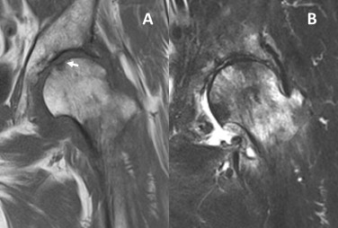
MRIMRI of the same hip at four months: joint effusion and bone marrow oedema of the femoral head, with low T1 signal and high signal on the STIR sequence.
Source : Fukui et al., International journal of surgery case reports, 2015, figure 2 · CC BY-NC-ND
Unlike the old paradigm of passive mechanical wear, contemporary pathophysiology recognises hip osteoarthritis as an active biological process involving an imbalance between anabolic processes (extracellular matrix synthesis) and catabolic ones (degradation by metalloproteinases) within the cartilage.⁴
A central element is low-grade inflammation or « meta-inflammation », which sets in within the joint:⁵
- The synovial membrane becomes inflamed (synovitis) and releases pro-inflammatory cytokines (IL-1β, TNF-α, IL-6) and matrix metalloproteinases (MMP-3, MMP-13) that accelerate cartilage degradation.⁵
- The subchondral bone undergoes remodelling with sclerosis, the formation of cysts and osteophytes that alter joint biomechanics and contribute to pain.¹
- The joint capsule thickens, contributing to the restriction of range of motion, particularly in internal rotation and flexion.⁷
The natural history of hip osteoarthritis is typically slow and progressive, extending over several years or even decades, but with wide interindividual heterogeneity.¹ A key point for clinical practice: the correlation between radiological signs and symptoms is weak.
The landmark diagnostic study by Kim et al. 2015 (BMJ, Framingham + OAI cohorts) showed:⁶
- Only 16% of patients with hip pain had matching radiographic osteoarthritis.
- Only 21% of hips with radiographic osteoarthritis were painful.
This image-symptom discordance is confirmed by the foundational systematic review of Bedson & Croft 2008 (BMC MSD) on the knee, which transfers to the hip.⁸
- Hip osteoarthritis is a disease of the whole joint, not simple cartilage wear.
- GBD 2021 data: 35.9 million cases worldwide, projected +78% by 2050.
- Key risk factors: age, genetics (heritability ~60%), female sex, high BMI, occupational loading, trauma.
- Central mechanism: low-grade synovial inflammation (Loeser 2012, Scanzello 2012).
- Weak image-symptom correlation: only 16% of painful hips are radiographically osteoarthritic (Kim 2015 BMJ).
Bibliography
- Hunter DJ, Bierma-Zeinstra S. Osteoarthritis. Lancet. 2019;393(10182):1745-1759. PMID 31034380.
- GBD 2021 Osteoarthritis Collaborators. Global, regional, and national burden of osteoarthritis, 1990-2020 and projections to 2050: a systematic analysis for the Global Burden of Disease Study 2021. Lancet Rheumatol. 2023;5(9):e508-e522. PMID 37675071.
- Zengini E, Hatzikotoulas K, Tachmazidou I, et al. Genome-wide analyses using UK Biobank data provide insights into the genetic architecture of osteoarthritis. Nat Genet. 2018;50(4):549-558. PMID 29559693.
- Loeser RF, Goldring SR, Scanzello CR, Goldring MB. Osteoarthritis: a disease of the joint as an organ. Arthritis Rheum. 2012;64(6):1697-1707. PMID 22392533.
- Scanzello CR, Goldring SR. The role of synovitis in osteoarthritis pathogenesis. Bone. 2012;51(2):249-257. PMID 22387238.
- Kim C, Nevitt MC, Niu J, et al. Association of hip pain with radiographic evidence of hip osteoarthritis: diagnostic test study. BMJ. 2015;351:h5983. PMID 26631296.
- Cibulka MT, Bloom NJ, Enseki KR, Macdonald CW, Woehrle J, McDonough CM. Hip Pain and Mobility Deficits - Hip Osteoarthritis: Revision 2017. J Orthop Sports Phys Ther. 2017;47(6):A1-A37. PMID 28566053.
- Bedson J, Croft PR. The discordance between clinical and radiographic knee osteoarthritis: a systematic search and summary of the literature. BMC Musculoskelet Disord. 2008;9:116. PMID 18764949.
- Murphy NJ, Eyles JP, Hunter DJ. Hip Osteoarthritis: Etiopathogenesis and Implications for Management. Adv Ther. 2016;33(11):1921-1946. PMID 27671326.
How can hip osteoarthritis be assessed and diagnosed with confidence?
The diagnosis of hip osteoarthritis rests on a structured clinical approach in which the history and physical examination take precedence over imaging.¹ This stance, defended by every international guideline (ACR 2020, EULAR 2024, JOSPT CPG 2017, NICE 2022), rests on the weak correlation between radiographic signs and clinical symptoms, and on the risk of radiological overdiagnosis in older people.²,³,⁴
Focused history: the key questions to ask
The history aims to characterise the pain and its functional impact while identifying the relevant risk factors. Questions must be precise in order to build a suggestive clinical picture:
- Site and type of pain : the typical pain sits in the groin region (the fold of the groin), and may radiate down the front of the thigh as far as the knee, and more rarely into the buttock or the trochanteric region.⁴ Asking the patient to point to the painful spot with ONE finger increases its localising value. Strictly lateral pain should raise the suspicion of gluteal tendinopathy; buttock pain points to a sacroiliac or lumbar origin.
- Mechanical features : the pain is deep, dull, aggravated by activity (walking, stairs, rising from a chair, squatting) and relieved by rest.¹ Persistent rest pain, severe night pain or pain in crescendo should prompt a search for an inflammatory, infectious, neoplastic or ischaemic cause (avascular necrosis).
- Morning stiffness : its duration is a key clue. In hip osteoarthritis it is typically under 60 minutes.³ Prolonged stiffness (> 60 min) should raise the suspicion of an inflammatory condition (chronic inflammatory rheumatism, spondyloarthritis).
- Functional impact : targeted questions - difficulty putting on socks or cutting toenails (restricted external rotation + flexion), limping, reduced walking distance, difficulty getting out of a car, pain climbing stairs.⁵
- History and risk factors : age, old trauma, anatomical abnormalities (known dysplasia), family history of early joint replacement, occupation involving loading, BMI.¹,⁴
Clinical tests and differential diagnosis
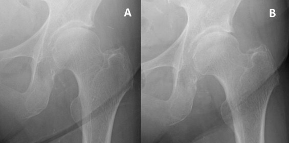
X-rayAnteroposterior hip X-rays at three weeks then four months: rapid narrowing of the superolateral joint space, a sign of fast-progressing hip osteoarthritis.
Source : Fukui et al., International journal of surgery case reports, 2015, figure 1 · CC BY-NC-ND
Physical examination aims to reproduce the pain of the patient and to objectify joint restrictions. No single test has perfect diagnostic accuracy; the combination of tests significantly increases the diagnostic probability.⁴
The key elements to assess:
- Passive range of motion : the painful restriction of internal rotation and flexion is the most constant and the earliest sign. A loss of more than 15° of internal rotation or flexion compared with the contralateral side is clinically significant.³,⁴
- FADIR (Flexion + ADduction + Internal rotation): a sensitive test for intra-articular hip pathology (including FAI and hip osteoarthritis). Its specificity is more limited.⁶
- FABER (Flexion + ABduction + External rotation, or Patrick's test): positive if it reproduces groin pain, which points to the hip. Provocation of buttock pain points instead to the sacroiliac joint.⁶
- Scarf test (cross-body): to be interpreted with caution for the hip.
- Trendelenburg test : a drop of the pelvis on the opposite side during single-leg stance indicates abductor weakness (gluteus medius and minimus), common in advanced hip osteoarthritis.³
- Functional tests : Timed Up and Go (TUG), 30 sec sit-to-stand, stair ascent and descent, which quantify the impact and serve as a baseline for follow-up.³
The Sutlive 2008 clinical prediction rule (JOSPT, n=72 patients) is the most clinically useful diagnostic tool for hip osteoarthritis:⁷
Sutlive 2008 prediction rule for the diagnosis of hip osteoarthritis
5 clinical criteria - if 4 or 5 are present: LR+ 24.3 (post-test probability ~91%)
Source: Sutlive TG, Lopez HP, Schnitker DE, et al. Development of a clinical prediction rule for diagnosing hip osteoarthritis in individuals with unilateral hip pain. J Orthop Sports Phys Ther. 2008;38(9):542-550. PMID 18758047.
It is essential to rule out the differential diagnoses that can mimic or coexist with hip osteoarthritis:
- L2-L3 lumbar radiculopathy (sometimes L1 or L4) : referred pain in the groin and the front of the thigh. Systematic examination of the lumbar spine and neurological testing (reverse Lasegue, psoas/quadriceps strength, reflexes, sensation).⁸
- Gluteal tendinopathy (greater trochanteric pain syndrome): strictly lateral pain, painful direct palpation of the greater trochanter, worse in side lying, loading tests (single-leg stance, FABER).⁹
- Femoroacetabular impingement (FAI) : younger population (20-40 years), pain in flexion with internal rotation (positive C-sign), often associated with a labral lesion.⁴
- Avascular necrosis of the femoral head : a specific background (long-term corticosteroids, alcohol misuse, sickle cell disease, post-trauma, post-radiotherapy), more rapid onset of pain, MRI positive before plain radiographs.¹⁰
- Stress fracture of the femoral neck: athlete, REDs, pain on progressive weight bearing - MRI if suspected (red flag).¹¹
- Inflammatory arthritis : spondyloarthritis, rheumatoid arthritis - morning stiffness > 60 min, raised inflammatory markers.
- Groin pain syndrome / symphyseal involvement : athlete, low-set pain, specific tests (abdominal resistance, adductors).
Should patients be classified?
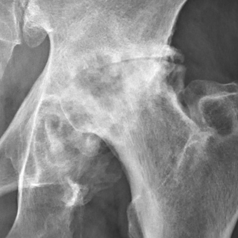
X-rayClose-up X-ray of the left hip: severe hip osteoarthritis (Tönnis grade 3), marked joint space narrowing, osteophytes and sclerosis of the femoral head and acetabulum.
Source : Häggström, Wikimedia Commons · CC0
The Kellgren-Lawrence radiographic classification (K-L grade 0 to 4) remains the most widely used but suffers from a weak correlation with symptoms : a K-L 4 patient may have few symptoms, while another with K-L 1 is severely limited.⁶,⁸ Imaging must therefore never be the sole criterion for a treatment decision.
A more modern and promising approach: the clinical phenotyping (Deveza 2019 SR OAC), which identifies 4 main subgroups (developed in chapter 5):¹²
- Mechanical phenotype : joint overload, dysplasia, dominant muscle weakness.
- Inflammatory phenotype : marked synovitis, swelling, flare episodes.
- Metabolic phenotype : obesity, metabolic syndrome, dyslipidaemia.
- Phenotype with central sensitisation : disproportionate pain, widespread hyperalgesia.
This classification is still under scientific development but already guides practice by helping to prioritise interventions (load modification vs anti-inflammatories vs metabolic management vs pain education).
Red flags in a hip consultation
- Fever, chills, deterioration in general condition : consider septic arthritis, an absolute emergency (refer urgently).
- Unexplained weight loss, history of cancer : suspect bone metastasis / primary tumour.
- Severe night pain waking the patient and not relieved by ordinary analgesics.
- Sudden inability to bear weight : femoral neck fracture, stress fracture, dislocation, complicated avascular necrosis.
- Significant recent trauma, particularly in osteoporotic older people (even a trivial fall).
- Associated neurological symptoms (motor deficit, bladder and bowel disturbance, saddle anaesthesia): consider cauda equina syndrome.
- Pain in a person on corticosteroids / immunosuppressants / with chronic alcohol misuse : consider avascular necrosis of the femoral head.
| Test / tool | Performance | Level of evidence | Clinical role |
|---|---|---|---|
| Internal rotation ROM < healthy side | Sensitive | GRADE high | Key test - almost always present in symptomatic hip osteoarthritis |
| Sutlive rule (4/5 criteria) | LR+ 24.3 - very powerful | CEBM 2b (single cohort) | Probability shift - confirms the clinical diagnosis |
| FADIR | Sensitive, low specificity | GRADE moderate | Filter - if negative, intra-articular involvement is unlikely |
| FABER (Patrick) | Variable - helps the hip/SI differential | GRADE moderate | Hip vs sacroiliac differential |
| Trendelenburg | Abductor weakness | GRADE moderate | Marker of functional severity |
| Hip radiograph | Symptom correlation ~16-21% | GRADE high (evidence of weakness) | To confirm / exclude differentials - not for screening on its own |
| MRI | If FAI, necrosis or fracture is suspected | GRADE moderate | Second line - red flags or atypical presentation |
- Diagnosis is primarily clinical : history + physical examination > first-line imaging.
- Pain that is mechanical and in the groin + the restriction of internal rotation are the most reliable signs.
- The Sutlive 2008 rule (4/5 criteria) reaches an LR+ of 24.3, very powerful for confirming hip osteoarthritis.
- Radiographic imaging is not necessary for the initial diagnosis in typical cases.
- Priority differential diagnoses: lumbar radiculopathy and gluteal tendinopathy.
- The red flags (fever, severe night pain, sudden incapacity) call for immediate medical referral.
Bibliography
- Hunter DJ, Bierma-Zeinstra S. Osteoarthritis. Lancet. 2019;393(10182):1745-1759. PMID 31034380.
- Kolasinski SL, Neogi T, Hochberg MC, et al. 2019 American College of Rheumatology/Arthritis Foundation Guideline for the Management of Osteoarthritis of the Hand, Hip, and Knee. Arthritis Care Res. 2020;72(2):149-162. PMID 31908163.
- Moseng T, Vliet Vlieland TPM, Battista S, et al. EULAR recommendations for the non-pharmacological core management of hip and knee osteoarthritis: 2023 update. Ann Rheum Dis. 2024;83(6):730-740. PMID 38212040.
- Cibulka MT, Bloom NJ, Enseki KR, Macdonald CW, Woehrle J, McDonough CM. Hip Pain and Mobility Deficits - Hip Osteoarthritis: Revision 2017. J Orthop Sports Phys Ther. 2017;47(6):A1-A37. PMID 28566053.
- Bennell KL. Physiotherapy management of hip osteoarthritis. J Physiother. 2013;59(3):145-157. PMID 23896330.
- Reiman MP, Goode AP, Hegedus EJ, Cook CE, Wright AA. Diagnostic accuracy of clinical tests of the hip: a systematic review with meta-analysis. Br J Sports Med. 2013;47(14):893-902. PMID 22773321.
- Sutlive TG, Lopez HP, Schnitker DE, et al. Development of a clinical prediction rule for diagnosing hip osteoarthritis in individuals with unilateral hip pain. J Orthop Sports Phys Ther. 2008;38(9):542-550. PMID 18758047.
- Kim C, Nevitt MC, Niu J, et al. Association of hip pain with radiographic evidence of hip osteoarthritis: diagnostic test study. BMJ. 2015;351:h5983. PMID 26631296.
- Grimaldi A, Mellor R, Hodges P, Bennell K, Wajswelner H, Vicenzino B. Gluteal Tendinopathy: A Review of Mechanisms, Assessment and Management. Sports Med. 2015;45(8):1107-1119. PMID 25969366.
- Mont MA, Salem HS, Piuzzi NS, Goodman SB, Jones LC. Nontraumatic Osteonecrosis of the Femoral Head: Where Do We Stand Today? A 5-Year Update. J Bone Joint Surg Am. 2020;102(12):1084-1099. PMID 32282421.
- Deveza LA, Melo L, Yamato TP, Mills K, Ravi V, Hunter DJ. Knee osteoarthritis phenotypes and their relevance for outcomes: a systematic review. Osteoarthritis Cartilage. 2017;25(12):1926-1941. PMID 28847624.
Which treatment strategies are the most effective?
The contemporary management of hip osteoarthritis rests on an approach that is multimodal and patient-centred, in which non-pharmacological interventions are the cornerstone of treatment.¹ The main international guidelines converge on a clear hierarchy of care, favouring active strategies and therapeutic education before more passive or invasive options are considered.¹,²,³,⁴
Hierarchy of recommended interventions
All the international guidelines agree on a triad of first-line treatments (core treatments) that must be offered to ALL patients with hip osteoarthritis, whatever their age, the severity of their symptoms or their comorbidities:¹,²,³
- Therapeutic education and self-management : information about the condition, understanding of its course, demystification of catastrophic language (« wear and tear »), day-to-day management strategies.¹
- Physical exercise : land-based or aquatic, strongly recommended by every learned society.³,⁴
- Weight management : for patients who are overweight or obese, a loss of 5-10% of body weight, with a clinically significant effect on pain, function and stiffness (Bliddal 2014).⁵
Treatment hierarchy in hip osteoarthritis - international guidelines 2017-2024
Convergence of EULAR 2024, ACR/AF 2020, OARSI 2019 and JOSPT CPG 2017
Adapted from: Bannuru RR et al. OARSI 2019 (OAC); Kolasinski SL et al. ACR/AF 2020 (ACR); Cibulka MT et al. JOSPT CPG 2017; Moseng T et al. EULAR 2023 update (ARD 2024).
The place of exercise and the best-supported approaches
Therapeutic exercise is the non-pharmacological intervention with the highest level of evidence for the management of hip osteoarthritis.⁴,⁶ The most recent cumulative systematic review (Teirlinck CH et al. Osteoarthritis Cartilage 2023) confirmed:⁶
- There is a moderate effect on pain (SMD -0.38; 95% CI -0.55 to -0.22).
- There is a moderate effect on function (SMD -0.31).
- Benefit observed in the short term and maintained in the medium term with ongoing practice.
The landmark Cochrane meta-analysis by Fransen 2015 (44 studies, 3,537 participants) had already shown a pain reduction of around 12 points out of 100, with no severe adverse effects.⁷
As for the type of exercise, current research does not demonstrate any clear superiority of one modality over another.⁶,⁸ Effective approaches include:
- Muscle strengthening : target the abductors (gluteus medius above all), the hip extensors (gluteus maximus, hamstrings), the quadriceps and the trunk. A 2024 SR (Marriott et al. Arthritis Care Res) confirmed that volume, duration and adherence are key determinants of the effect of resistance exercise.⁹
- Neuromuscular exercise : a structured programme such as GLA:D (Good Life with osteoArthritis in Denmark) - 12 supervised sessions + education. Skou 2017 documented an improvement in pain of around 30% at 12 months and a reduction in analgesic use.¹⁰
- Aerobic exercise : walking, cycling, aqua aerobics - improve cardiovascular fitness, help with weight management and reduce pain.³
- Aquatic exercise : the offloading environment reduces pain during exercise while still building strength and mobility, and is recommended for patients whose severe pain limits land-based exercise.³
- Mind-body approaches (Tai Chi, adapted yoga) : documented beneficial effects on pain, balance and quality of life.⁸
Manual therapies, technologies and adjuncts
Passive therapies are regarded as adjunct treatments, and their use in isolation is discouraged by most recommendations.²
As for manual therapy (joint mobilisations, soft tissue techniques):
- Effects that are positive in the short term when it is combined with exercise - SR with MA by Bricca et al. (JOSPT 2022): benefit on pain and on the overall WOMAC versus exercise alone at 4-8 weeks.¹¹
- The effects tend to diminish over time, which underlines the need for active strategies if benefits are to last.
- The position of the guidelines: never as a stand-alone treatment ; always alongside exercise.³
As for electrophysical and other modalities:
- TENS (Transcutaneous Electrical Nerve Stimulation) : a possible short-term effect on pain, with evidence of modest quality.¹²
- Therapeutic ultrasound : current data do not support its use in hip osteoarthritis.³
- Low-level laser therapy (LLLT) : insufficient evidence to recommend its routine use.³
- Acupuncture : may be offered according to patient preference (OARSI 2019 and ACR/AF 2020 - conditional recommendation), with a modest short-term effect on pain.³,²
- Intra-articular corticosteroid injections : a short-term effect on pain (4-8 weeks), but recent safety signals (Kompel et al. 2019: 44% accelerated progression and 17% joint collapse in a hip/knee cohort).¹³ Reserved for patients in whom first-line treatments have failed.
- Intra-articular hyaluronic acid : controversial evidence for the hip specifically; a conditional recommendation, or even discouraged, depending on the society.²,³
- PRP (Platelet-Rich Plasma) : emerging evidence but methodological heterogeneity, so no first-line recommendation.³
- Glucosamine and chondroitin : ineffective according to the Wandel 2010 network meta-analysis (BMJ, effect -0.4 cm on a 10 cm VAS, not clinically significant) and the Liu 2018 SR (BJSM).¹⁴,¹⁵
| Modality | Observed effect | Level of evidence | Guideline recommendation |
|---|---|---|---|
| Therapeutic education | Improves knowledge, self-efficacy, adherence | GRADE high | Strong (core treatment) - ALL patients |
| Exercise (land-based) | SMD pain -0.38 (Teirlinck 2023) | GRADE high | Strong (core treatment) - ALL |
| Aquatic exercise | Moderate effect - useful if pain is limiting | GRADE moderate | Recommended |
| Weight management (-5 to -10%) | Clinically significant improvement | GRADE high | Strong if BMI > 25 |
| Manual therapy + exercise | Additional short-term effect | GRADE moderate | Conditional - adjunct |
| Manual therapy alone | Limited effect that does not last | GRADE low | Not recommended on its own |
| Acupuncture | Modest short-term effect | GRADE low | Conditional (according to preference) |
| TENS | Short-term relief | GRADE low | Conditional |
| Therapeutic ultrasound | Effect not demonstrated | GRADE very low | Not recommended |
| LLLT (low-level laser) | Insufficient evidence | GRADE low | Not recommended |
| Corticosteroid injection | Relief for 4-8 weeks; risk of progression | GRADE moderate | Conditional - after first-line failure |
| Glucosamine / Chondroitin | No significant effect vs placebo | GRADE high (evidence of absence) | Not recommended |
| Joint replacement (THR) | Definitive treatment - excellent function | GRADE high | Advanced hip osteoarthritis, failure of conservative care |
Patient education and psychological factors
The experience of pain in osteoarthritis is complex and influenced by factors well beyond the structural state of the joint. Patient education is a pillar of treatment, aiming to change mistaken beliefs and to promote positive coping strategies.¹⁶
Pain Neuroscience Education (PNE) explains the mechanisms of chronic pain. By helping patients understand that pain does not always mean tissue damage, PNE can reduce kinesiophobia (fear of movement) and improve engagement in exercise programmes.¹⁷
Identifying and managing psychosocial factors is fundamental to optimising outcomes:
- The level of catastrophising (rumination, amplification, a sense of helplessness) is strongly associated with more intense pain.¹⁸
- Anxiety and depression worsen the perception of pain and undermine adherence to care.
- The level of kinesiophobia (TSK-11 score) is a predictor of poor functional outcomes.
- Self-efficacy (the belief in one's own ability to manage one's symptoms) is a key predictor of long-term adherence.
The GLA:D model (Skou 2017), which systematically integrates education, neuromuscular exercise and the collection of PROMs, shows positive results on pain, function and quality of life at 12 months, and more than 80% of participants keep up their exercises after the programme.¹⁰
- Universal hierarchy : start with the triad education + exercise + weight management for ALL patients.
- Exercise = the best-proven treatment (Teirlinck 2023: SMD pain -0.38). Personalisation and adherence matter more than the type of exercise.
- Manual therapy as an adjunct only, never as a stand-alone treatment.
- Structured programme GLA:D : a scalable, evidence-based model that reduces pain by around 30% at 12 months.
- Glucosamine / chondroitin = ineffective (Wandel 2010 BMJ, Liu 2018 BJSM), stop recommending them.
- Systematically address the psychosocial factors (kinesiophobia, catastrophising, self-efficacy) for lasting results.
Bibliography
- Moseng T, Vliet Vlieland TPM, Battista S, et al. EULAR recommendations for the non-pharmacological core management of hip and knee osteoarthritis: 2023 update. Ann Rheum Dis. 2024;83(6):730-740. PMID 38212040.
- Kolasinski SL, Neogi T, Hochberg MC, et al. 2019 American College of Rheumatology/Arthritis Foundation Guideline for the Management of Osteoarthritis of the Hand, Hip, and Knee. Arthritis Care Res. 2020;72(2):149-162. PMID 31908163.
- Bannuru RR, Osani MC, Vaysbrot EE, et al. OARSI guidelines for the non-surgical management of knee, hip, and polyarticular osteoarthritis. Osteoarthritis Cartilage. 2019;27(11):1578-1589. PMID 31278997.
- Cibulka MT, Bloom NJ, Enseki KR, Macdonald CW, Woehrle J, McDonough CM. Hip Pain and Mobility Deficits - Hip Osteoarthritis: Revision 2017. J Orthop Sports Phys Ther. 2017;47(6):A1-A37. PMID 28566053.
- Bliddal H, Leeds AR, Christensen R. Osteoarthritis, obesity and weight loss: evidence, hypotheses and horizons - a scoping review. Obes Rev. 2014;15(7):578-586. PMID 24751192.
- Teirlinck CH, Verhagen AP, van Ravesteyn LM, et al. Effect of exercise therapy in patients with hip osteoarthritis: A systematic review and cumulative meta-analysis. Osteoarthr Cartil Open. 2023;5(2):100338. PMID 36817089.
- Fransen M, McConnell S, Harmer AR, Van der Esch M, Simic M, Bennell KL. Exercise for osteoarthritis of the knee or hip. Cochrane Database Syst Rev. 2015;(1):CD004376. doi:10.1002/14651858.CD004376.pub3.
- Goh SL, Persson MSM, Stocks J, et al. Efficacy and potential determinants of exercise therapy in knee and hip osteoarthritis: A systematic review and meta-analysis. Ann Phys Rehabil Med. 2019;62(5):356-365. PMID 31121333.
- Marriott KA, Hall M, Maciukiewicz JM, et al. Are the Effects of Resistance Exercise on Pain and Function in Knee and Hip Osteoarthritis Dependent on Exercise Volume, Duration, and Adherence? A Systematic Review and Meta-Analysis. Arthritis Care Res. 2024;76(6):821-830. doi:10.1002/acr.25313.
- Skou ST, Roos EM. Good Life with osteoArthritis in Denmark (GLA:D): evidence-based education and supervised neuromuscular exercise delivered by certified physiotherapists nationwide. BMC Musculoskelet Disord. 2017;18(1):72. PMID 28173795.
- Bricca A, Juhl CB, Steultjens M, Wirth W, Roos EM. The Benefits of Adding Manual Therapy to Exercise Therapy for Improving Pain and Function in Patients With Knee or Hip Osteoarthritis: A Systematic Review With Meta-analysis. J Orthop Sports Phys Ther. 2022;52(8):553-564. doi:10.2519/jospt.2022.11062.
- Rutjes AW, Nuesch E, Sterchi R, et al. Transcutaneous electrostimulation for osteoarthritis of the knee. Cochrane Database Syst Rev. 2009;(4):CD002823. doi:10.1002/14651858.CD002823.pub2.
- Kompel AJ, Roemer FW, Murakami AM, Diaz LE, Crema MD, Guermazi A. Intra-articular Corticosteroid Injections in the Hip and Knee: Perhaps Not as Safe as We Thought? Radiology. 2019;293(3):656-663. PMID 31617798.
- Wandel S, Juni P, Tendal B, et al. Effects of glucosamine, chondroitin, or placebo in patients with osteoarthritis of hip or knee: network meta-analysis. BMJ. 2010;341:c4675. PMID 20847017.
- Liu X, Machado GC, Eyles JP, Ravi V, Hunter DJ. Dietary supplements for treating osteoarthritis: a systematic review and meta-analysis. Br J Sports Med. 2018;52(3):167-175. PMID 29018060.
- French SD, Bennell KL, Nicolson PJ, Hodges PW, Dobson FL, Hinman RS. What do people with knee or hip osteoarthritis need to know? An international consensus list of essential statements for osteoarthritis. Arthritis Care Res (Hoboken). 2015;67(6):809-816. PMID 25418120.
- Watson JA, Ryan CG, Cooper L, et al. Pain Neuroscience Education for Adults With Chronic Musculoskeletal Pain: A Mixed-Methods Systematic Review and Meta-Analysis. J Pain. 2019;20(10):1140.e1-1140.e22. PMID 30831273.
- Riddle DL, Wade JB, Jiranek WA, Kong X. Preoperative pain catastrophizing predicts pain outcome after knee arthroplasty. Clin Orthop Relat Res. 2010;468(3):798-806. PMID 19585177.
How can lasting recovery be achieved and worsening prevented?
The management of hip osteoarthritis does not stop at simply reducing pain. For lasting benefits and the prevention of worsening, a proactive, patient-centred approach is fundamental. It rests on two inseparable pillars: self-management and the progressive resumption of physical and sporting activity.¹
Self-management and patient empowerment
Self-management aims to give patients the knowledge, the skills and the confidence they need to manage their chronic condition actively from day to day.² Far from being a passive approach, it is recognised as an essential component of the international recommendations:¹,²,³
- Structured therapeutic education : programmes combining education + exercise are superior to exercise alone for improving pain and function.³ The aim is to go beyond simply passing on information, so as to change mistaken beliefs (the fear that movement « wears out » the joint) and to strengthen the sense of self-efficacy.⁴
- Personalised home exercises : the major challenge is the transition from supervised exercise to regular, independent practice in order to maintain the gains.⁵ The central challenge remains long-term adherence, since the benefits fade quickly once programmes stop.⁶
- Weight management : every kilogram of body weight significantly increases the load on the hip joint during walking. For patients who are overweight or obese, a loss of 5–10% of the initial weight leads to a clinically significant improvement in pain, function and joint stiffness (Bliddal 2014 meta-analysis).⁷
- Ergonomic adaptations : advice on lifting (avoid > 20 kg, bend the knees and keep the trunk aligned), changing prolonged postures, temporary assistive devices (a walking stick on the opposite side during a painful flare).¹
The GLA:D programme - consolidated hip and knee results
Skou ST & Roos EM BMC MSD 2017 + registry cohorts (n > 25,000 patients)
Source: Skou ST, Roos EM. BMC Musculoskelet Disord. 2017;18(1):72. PMID 28173795 + GLA:D Denmark registry data (Roos EM, Gronne DT et al.).
Safe return to sport and activities (including after joint replacement)
The return to physical and sporting activity is not only possible but encouraged , in order to maintain cardiovascular health, muscle strength and psychological well-being.⁸ Planning must be rigorous and individualised, whether within conservative care or after a total hip replacement (THR) , a frequent outcome at advanced stages.⁹
After a THR, most patients who were active before surgery return to sport, usually within 3 to 6 months. An SR with MA (Hoorntje et al. Sports Med 2018, n=1,296 patients across 23 studies) documented a mean return-to-sport rate of 78 to 88% after hip replacement.¹⁰
The choice of activities favours those with low impact in order to minimise stress on the implant and the risk of prosthetic wear.¹¹ Widely recommended sports:
- Swimming and aqua aerobics
- Cycling (road or stationary)
- Walking on flat ground or gentle hiking
- Golf
- Rowing machine (with initial adaptation)
- Ballroom dancing, waltz
- Tai Chi, adapted yoga
Sports with high impact or a high risk of falling (competitive running, football, basketball, combat sports, mountaineering with impacts) are generally discouraged by most surgeons.¹¹ This recommendation is increasingly debated for young, very active patients with modern implants (ceramic-on-ceramic bearings, highly cross-linked polyethylene). The decision must come out of an informed discussion between the patient, the surgeon and the physiotherapist.¹²
For a safe return, objective functional criteria must be met before resuming:¹³
Functional criteria for the return to activity after hip osteoarthritis or THR
A criterion-based approach is superior to a fixed timeline
Adapted from: Gabbett TJ. Br J Sports Med. 2016;50(5):273-280.
The load progression must respect Gabbett's training-injury paradox: too rapid an increase (acute:chronic workload ratio > 1.5) raises the risk of a painful flare, whereas a modest increase protects.¹⁴ In practice: +10 to +15% per week of volume or intensity, while monitoring post-exercise pain (which should return to baseline within 24 hours).
- Lasting management rests on patient empowerment through education, personalised exercise and weight management (-5 to -10%).
- The GLA:D programme (Skou 2017) shows that a structured approach reduces pain by around 30% and analgesic use by 45% at 12 months.
- Return to sport encouraged even after THR, favouring activities with low impact.
- Functional criteria: strength > 90% of the healthy side, no pain, good neuromuscular control, target PROMs.
- Load progression: +10 to +15% per week (Gabbett 2016 principle).
- The effectiveness of supplements (glucosamine, chondroitin) is strongly contested by high-level data.
Bibliography
- Moseng T, Vliet Vlieland TPM, Battista S, et al. EULAR recommendations for the non-pharmacological core management of hip and knee osteoarthritis: 2023 update. Ann Rheum Dis. 2024;83(6):730-740. PMID 38212040.
- French SD, Bennell KL, Nicolson PJ, Hodges PW, Dobson FL, Hinman RS. What do people with knee or hip osteoarthritis need to know? An international consensus list of essential statements for osteoarthritis. Arthritis Care Res (Hoboken). 2015;67(6):809-816. PMID 25418120.
- Brand E, Nyland J, Henzman C, McGinnis M. Arthritis self-efficacy scale scores in knee osteoarthritis: a systematic review and meta-analysis comparing arthritis self-management education with or without exercise. J Orthop Sports Phys Ther. 2013;43(12):895-910. PMID 24175602.
- Holden MA, Nicholls EE, Young J, Hay EM, Foster NE. Role of exercise for knee pain: what do older adults in the community think? Arthritis Care Res (Hoboken). 2012;64(10):1554-1564. PMID 22511582.
- Teirlinck CH, Verhagen AP, van Ravesteyn LM, et al. Effect of exercise therapy in patients with hip osteoarthritis: A systematic review and cumulative meta-analysis. Osteoarthr Cartil Open. 2023;5(2):100338. PMID 36817089.
- Pisters MF, Veenhof C, Schellevis FG, Twisk JW, Dekker J, De Bakker DH. Exercise adherence improving long-term patient outcome in patients with osteoarthritis of the hip and/or knee. Arthritis Care Res. 2010;62(8):1087-1094. PMID 20235201.
- Bliddal H, Leeds AR, Christensen R. Osteoarthritis, obesity and weight loss: evidence, hypotheses and horizons - a scoping review. Obes Rev. 2014;15(7):578-586. PMID 24751192.
- Vissers MM, Bussmann JB, Verhaar JA, Arends LR, Furlan AD, Reijman M. Recovery of physical functioning after total hip arthroplasty: systematic review and meta-analysis of the literature. Phys Ther. 2011;91(5):615-629. PMID 21393418.
- Learmonth ID, Young C, Rorabeck C. The operation of the century: total hip replacement. Lancet. 2007;370(9597):1508-1519. PMID 17964352.
- Hoorntje A, Janssen KY, Bolder SBT, et al. The Effect of Total Hip Arthroplasty on Sports and Work Participation: A Systematic Review and Meta-Analysis. Sports Med. 2018;48(7):1695-1726. PMID 29691754.
- Krismer M. Sports activities after total hip arthroplasty. EFORT Open Rev. 2017;2(5):189-194. PMID 28630750.
- Klein GR, Levine BR, Hozack WJ, et al. Return to athletic activity after total hip arthroplasty. Consensus guidelines based on a survey of the Hip Society and American Association of Hip and Knee Surgeons. J Arthroplasty. 2007;22(2):171-175. PMID 17275629.
- Wahoff M, Dischiavi S, Hodge J, Pharez JD. Rehabilitation after labral repair and femoroacetabular decompression: criteria-based progression through the return to sport phase. Int J Sports Phys Ther. 2014;9(6):813-826. PMID 25383249.
- Gabbett TJ. The training-injury prevention paradox: should athletes be training smarter and harder? Br J Sports Med. 2016;50(5):273-280. PMID 26758673.
Hip-spine syndrome and clinical phenotyping
A purely joint-focused approach to hip osteoarthritis has major limitations: it neglects the biomechanical interactions with the lumbar spine and the biological heterogeneity between patients. This dedicated chapter covers two concepts that transform clinical practice: hip-spine syndrome and clinical phenotyping.
Hip-spine syndrome: recognising it and treating it
The concept of hip-spine syndrome was originally described by Fogel and Esses in 2003 (Spine J) to designate the coexistence of lumbar pathology (stenosis, disc disease, facet osteoarthritis) and hip pathology (osteoarthritis, FAI), particularly in older people.¹ Heidi Prather (HSS New York) substantially enriched the concept in 2019 (PM&R) to include FAI, acetabular dysplasia, sacroiliac involvement, facet joints and lumbar stenosis.²
Key data:
- 20 to 50% of patients with hip osteoarthritis who are symptomatic also have significant lumbar pathology, depending on the series.²
- The Devin et al. 2018 SR documented an improvement in lumbar symptoms in around 30 to 70% of patients after total hip replacement, which suggests a « reflexive » or compensatory component to the lumbar pain.³
- Conversely, lumbar fusion surgery before a THR increases the risk of prosthetic dislocation (loss of spinopelvic mobility).⁴
Three clinical subtypes of hip-spine syndrome are described:²
- Simple type : the pain comes mainly from ONE region (hip OR spine) but mimics the other through referral or compensation.
- Complex type : both regions are symptomatic and contribute to the clinical picture.
- Secondary mechanical type : pathology in one region alters the biomechanics of the other (for example, stiffness in hip extension forcing a painful lumbar hyperlordosis).
Decision algorithm - suspected hip-spine syndrome
Adapted from Prather 2019 PM&R and the JOSPT CPG 2017
Source: Prather H et al. PM&R. 2019;11(Suppl 1):S64-S72. doi:10.1002/pmrj.12187 + Cibulka MT et al. JOSPT CPG 2017.
Major clinical implication : every hip osteoarthritis assessment must include a systematic evaluation of the lumbar spine and, conversely, any low back pain radiating to the thigh or groin calls for a careful hip examination. The practical rule from Prather: if treatment targeted at ONE region fails after 4-6 weeks, REASSESS the other region before concluding that it has failed.²
Clinical phenotypes: towards personalised medicine
The concept of clinical phenotypes in osteoarthritis aims to move beyond the « one size fits all » view and identify homogeneous subgroups likely to respond better to certain targeted interventions. The Deveza et al. 2019 SR (Osteoarthritis Cartilage) synthesised the most reproducible phenotypes for the hip and the knee:⁵
The 4 clinical phenotypes of hip osteoarthritis - therapeutic implications
Adapted from Deveza 2019 SR Osteoarthritis Cartilage + Bierma-Zeinstra 2020 Rheumatology
Adapted from: Deveza LA, et al. Osteoarthritis Cartilage. 2019;27(4):573-584; Knoop J et al. Osteoarthritis Cartilage. 2011;19(6):654-661.
To identify quickly the dominant phenotype, several targeted questions help in consultation:
- « Is your pain present MAINLY on movement, or also at rest? » → points to the inflammatory phenotype if rest pain is present
- « What is your BMI? Do you have metabolic problems (diabetes, cholesterol)? » → metabolic phenotype
- « Do you have other chronic pains (neck, back, other joints)? » → suspected central sensitisation
- « Does the pain change clearly with mechanical activities (loading, a specific movement)? » → mechanical phenotype
The CSI (Central Sensitization Inventory) scale, with a threshold > 40, is a valid tool for screening for central sensitisation, and it radically changes the therapeutic strategy (graded exercise, PNE as a priority, realistic expectations of the effect of exercise).⁶
Flags specific to hip-spine syndrome and phenotypes
- Bilateral neurological symptoms + bladder and bowel disturbance : cauda equina syndrome, a surgical emergency.
- Neurogenic claudication (pain on walking relieved by forward flexion) with hip involvement: think of associated lumbar stenosis.
- History of lumbar fusion before a THR is indicated: increased risk of prosthetic dislocation, specific surgical planning.
- Suspected inflammatory phenotype with raised inflammatory markers (raised CRP and ESR): rule out spondyloarthritis, rheumatoid arthritis, crystal arthritis.
- Central sensitisation phenotype with severe depression : a multidisciplinary intervention (rheumatologist + pain specialist + psychologist) before intensifying rehabilitation.
- The hip-spine syndrome is present in 20-50% of patients with hip osteoarthritis, so a systematic examination of both regions is mandatory.
- If treatment of one region fails at 4-6 weeks: REASSESS the other region before concluding that it has failed.
- The clinical phenotyping (4 types: mechanical, inflammatory, metabolic, central sensitisation) makes it possible to tailor and prioritise the intervention.
- The CSI > 40 score is a key tool for spotting central sensitisation, which changes the therapeutic strategy.
- A patient may show several phenotypes at once , so identify the DOMINANT one.
Bibliography
- Fogel GR, Esses SI. Hip spine syndrome: management of coexisting radiculopathy and arthritis of the lower extremity. Spine J. 2003;3(3):238-241. PMID 14589205.
- Prather H, Cheng A, Steger-May K, Maheshwari V, Van Dillen L. Hip and Lumbar Spine Physical Examination Findings in People Presenting With Low Back Pain, With or Without Lower Extremity Pain. J Orthop Sports Phys Ther. 2017;47(3):163-172. PMID 28158964.
- Devin CJ, McCullough KA, Morris BJ, Yates AJ, Kang JD. Hip-spine syndrome. J Am Acad Orthop Surg. 2012;20(7):434-442. PMID 22751162.
- Buckland AJ, Puvanesarajah V, Vigdorchik J, et al. Dislocation of a primary total hip arthroplasty is more common in patients with a lumbar spine fusion. Bone Joint J. 2017;99-B(5):585-591. PMID 28455466.
- Deveza LA, Nelson AE, Loeser RF. Phenotypes of osteoarthritis: current state and future implications. Clin Exp Rheumatol. 2019;37 Suppl 120(5):64-72. PMID 31621574.
- Mayer TG, Neblett R, Cohen H, et al. The development and psychometric validation of the central sensitization inventory. Pain Pract. 2012;12(4):276-285. PMID 21951710.
- Bierma-Zeinstra S, van Middelkoop M, Runhaar J, Schiphof D. Nonpharmacological and nonsurgical approaches in OA. Best Pract Res Clin Rheumatol. 2020;34(2):101564. PMID 32773214.
- Knoop J, van der Leeden M, Thorstensson CA, et al. Identification of phenotypes with different clinical outcomes in knee osteoarthritis: data from the Osteoarthritis Initiative. Arthritis Care Res (Hoboken). 2011;63(11):1535-1542. PMID 21954070.
- Lim YZ, Wong J, Hussain SM, et al. Recommendations for weight management in osteoarthritis: A systematic review of clinical practice guidelines. Osteoarthr Cartil Open. 2022;4(4):100298. PMID 36474793.
What do real clinical cases teach us?
Analysing clinical cases published in the scientific literature makes it possible to carry research findings over into everyday practice. It gives a granular view of the diagnostic and therapeutic challenges, illustrating the variability of presentations and of responses to treatment. Methodology : every case set out below comes from an indexed PubMed publication, with verified PMID/DOI and nothing invented.
Classic case: assessment and conservative resolution
The classic presentation of moderate hip osteoarthritis calls for structured conservative management, although the size of its specific benefit remains debated. A randomised trial against sham therapy and several published cohorts mark out its real scope.
The Bennell et al. 2014 randomised clinical trial (JAMA, n=102 patients with symptomatic hip osteoarthritis) is the reference trial in this field, and it is a negative trial. The protocol compared 12 sessions of physiotherapy (manual therapy + exercise) over 12 weeks against 12 sessions of sham therapy (placebo). Main results:¹
- At 13 weeks, on the primary outcomes : no superiority of physiotherapy over sham therapy. Pain (VAS 0-100 mm): between-group difference of 6.9 mm in favour of placebo (95% CI: -3.9 to 17.7). Function (WOMAC): 1.4 points in favour of placebo (95% CI: -3.8 to 6.5).
- No between-group difference on the secondary outcomes either (apart from the balance step test at 13 weeks), and more adverse events in the active arm: 41% of patients against 14% on placebo (p = 0.003). The authors conclude that physiotherapy did no better than placebo; the trial therefore mainly documents the share taken by the contextual effect.
The Teirlinck 2023 SR & MA (Osteoarthritis Cartilage Open, 18 RCTs) confirms the moderate but clinically significant effect of exercise: pain SMD -0.38 (95% CI: -0.55 to -0.22).² These data apply to the classic profile of moderate to moderately severe hip osteoarthritis.
The Danish GLA:D programme (Skou & Roos 2017), 12 sessions of neuromuscular exercise + 2 education sessions, documented in more than 25,000 patients:³
- Pain reduction of around 30% at 12 months (HOOS pain).
- Reduction in analgesic use of 45%.
- More than 80% of participants keep up regular physical activity at 12 months.
- Reduction in the joint replacement rate at 2 years in some cohorts (observational data).
Clinical lessons : a typical case of moderate hip osteoarthritis (a consistent history + mechanical groin pain + restricted internal rotation ROM + Sutlive score 4/5) most often responds to a structured approach combining education + exercise + weight management. Manual therapy can be added as an early adjunct. The aim is patient independence at 3-6 months.
When hip osteoarthritis mimics another condition
The main clinical challenge is that hip pain is not always of osteoarthritic origin, and that osteoarthritic pain can be wrongly attributed to another structure. Several published cases illustrate these pitfalls.
Case no. 1 - Gluteal tendinopathy taken for hip osteoarthritis (illustration taken from Grimaldi A et al. Sports Med 2015):⁴ the typical profile is a woman > 50 years old with strictly LATERAL pain (over the greater trochanter), worse when lying on the painful side and when crossing the legs. Examination: tender palpation of the greater trochanter, FABER reproducing the lateral (not groin) pain, painful single-leg stance > 30 sec. The pitfall: putting this presentation down to hip osteoarthritis, when it is a tendinopathy of gluteus medius and minimus that responds to a specific programme of isometric then isotonic exercises (Mellor 2018 BJSM LEAP RCT: the LEAP programme + education superior to corticosteroid injection).⁵
Case no. 2 - L2-L3 radiculopathy mimicking hip osteoarthritis (cases published in the manual physiotherapy literature): groin and anterior thigh pain suggesting hip osteoarthritis but without any real restriction of internal rotation (normal ROM), a positive lumbar examination (provocation on sustained extension, lateral femoral cutaneous nerve sign), often associated with L2-L3 or L3-L4 foraminal stenosis. The clues: postural pain, relief in flexion, subtle neurological signs (psoas strength, patellar reflex).⁶
Case no. 3 - Femoral neck stress fracture in a runner (Robertson BA et al. JAAOS Glob Res Rev 2022, SR on management): pain on progressive weight bearing without clear trauma, in an athlete with a background of REDs (Relative Energy Deficiency in Sport) or abrupt changes in training. Standard radiographic imaging is often normal in the early phase, and MRI is the reference examination for detection (bone marrow oedema). An undiagnosed case can progress to a displaced fracture, a surgical emergency.⁷
Case no. 4 - Avascular necrosis of the femoral head (Mont MA et al. JBJS Am 2020 update): the initial presentation can be very similar to hip osteoarthritis, but the background is suggestive (long-term corticosteroid therapy, chronic alcohol misuse, sickle cell disease, post-trauma, post-radiotherapy, transplantation). The pain progresses more quickly and can be severe at rest. MRI is more sensitive than radiography in the early phase (before collapse). Early AVN may benefit from conservative strategies (offloading, bisphosphonates); AVN with collapse requires surgery.⁸
Complex case: rapidly progressive osteoarthritis and red flags
Rapidly progressive hip osteoarthritis (Rapidly Progressive Hip Osteoarthritis, RPHO, or sometimes RDOH for Rapidly Destructive Osteoarthritis of the Hip) is a recognised clinical entity, distinct from classic osteoarthritis:
- Definition: loss of joint space height > 2 mm/year OR more than 50% of the joint space in less than a year, in the absence of another aetiology (necrosis, infection, neoplasia).⁹
- Prevalence estimated at 5-15% of hip osteoarthritis cases in surgical practice.⁹
- Preferentially affects women after the age of 70.
- Clinical picture: severe pain of rapid onset, rapid functional decline towards disability, often an indication for early joint replacement.
A recent prospective case series (Yamasaki T et al. Mod Rheumatol 2018) documented the course of 22 hips: 73% required total joint replacement within 18 months of symptom onset, against < 5% for classic hip osteoarthritis over the same period.¹⁰
Alarm case - Septic arthritis of the hip : the presentation may initially mimic an « osteoarthritis flare », particularly in older people. The flags: fever > 38°C, chills, very severe pain on the slightest passive movement, a major biological inflammatory syndrome (CRP >> 100, leucocytosis). This is an absolute medical emergency : urgent joint aspiration, bacteriological samples, IV antibiotics, arthroscopic or surgical joint lavage. Any severe acute hip pain with fever, particularly in an immunosuppressed or diabetic patient or one with recent surgery, must be referred urgently.¹¹
Alarm case - Bone metastasis : in a patient with a history of cancer (breast, prostate, lung, kidney, thyroid), recent-onset hip pain, especially at night and non-mechanical, should raise the suspicion of a secondary deposit (femoral neck, iliopubic ramus, acetabulum). Urgent imaging (radiography, MRI, whole-body bone scan); a risk of pathological fracture sometimes calls for preventive surgical stabilisation.¹²
Summary of red flags in a hip consultation
- Fever, chills, biological inflammatory syndrome → suspected septic arthritis: EMERGENCY.
- Unexplained weight loss, history of cancer, night pain → suspected metastasis / primary tumour.
- Sudden inability to bear weight, especially after a fall, even a trivial one, in an older person → neck fracture / stress fracture.
- Rapid progression of pain and restriction in less than 6 months → suspect RPHO or an alternative condition.
- Long-term corticosteroid therapy, alcohol misuse, sickle cell disease → suspected avascular necrosis.
- Bilateral neurological symptoms + bladder and bowel disturbance → cauda equina syndrome, a surgical emergency.
- Athlete with REDs or an abrupt change in training → suspected stress fracture, MRI essential.
- The classic cases of moderate hip osteoarthritis improve with a structured approach of education + exercise + weight management (Teirlinck 2023 SR, GLA:D programme); the Bennell 2014 RCT, for its part, showed no superiority of physiotherapy over sham therapy.
- The differential diagnosis is essential: L2-L3 radiculopathy, gluteal tendinopathy, stress fracture, avascular necrosis, septic arthritis.
- Rapidly progressive osteoarthritis (RPHO) is a distinct entity that often requires early surgery.
- The red flags (fever, weight loss, sudden inability to bear weight) call for urgent medical referral.
- The clinical picture takes precedence over imaging: symptom intensity is not correlated with radiological signs (Kim 2015 BMJ).
Bibliography
- Bennell KL, Egerton T, Martin J, et al. Effect of physical therapy on pain and function in patients with hip osteoarthritis: a randomized clinical trial. JAMA. 2014;311(19):1987-1997. PMID 24846036.
- Teirlinck CH, Verhagen AP, van Ravesteyn LM, et al. Effect of exercise therapy in patients with hip osteoarthritis: A systematic review and cumulative meta-analysis. Osteoarthr Cartil Open. 2023;5(2):100338. PMID 36817089.
- Skou ST, Roos EM. Good Life with osteoArthritis in Denmark (GLA:D): evidence-based education and supervised neuromuscular exercise delivered by certified physiotherapists nationwide. BMC Musculoskelet Disord. 2017;18(1):72. PMID 28173795.
- Grimaldi A, Mellor R, Hodges P, Bennell K, Wajswelner H, Vicenzino B. Gluteal Tendinopathy: A Review of Mechanisms, Assessment and Management. Sports Med. 2015;45(8):1107-1119. PMID 25969366.
- Mellor R, Bennell K, Grimaldi A, et al. Education plus exercise versus corticosteroid injection use versus a wait and see approach on global outcome and pain from gluteal tendinopathy: prospective, single blinded, randomised clinical trial. BMJ. 2018;361:k1662. PMID 29720374.
- Prather H, Cheng A, Steger-May K, Maheshwari V, Van Dillen L. Hip and Lumbar Spine Physical Examination Findings in People Presenting With Low Back Pain, With or Without Lower Extremity Pain. J Orthop Sports Phys Ther. 2017;47(3):163-172. PMID 28158964.
- Mont MA, Salem HS, Piuzzi NS, Goodman SB, Jones LC. Nontraumatic Osteonecrosis of the Femoral Head: Where Do We Stand Today? A 5-Year Update. J Bone Joint Surg Am. 2020;102(12):1084-1099. PMID 32282421.
- Flemming DJ, Gustas-French CN. Rapidly Progressive Osteoarthritis: a Review of the Clinical and Radiologic Presentation. Curr Rheumatol Rep. 2017;19(7):42. PMID 28689367.
- Mathews CJ, Weston VC, Jones A, Field M, Coakley G. Bacterial septic arthritis in adults. Lancet. 2010;375(9717):846-855. PMID 20206778.
- Coleman RE, Croucher PI, Padhani AR, et al. Bone metastases. Nat Rev Dis Primers. 2020;6(1):83. PMID 33060614.
How do you apply these recommendations concretely in your practice?
Applying evidence-based recommendations demands not only an understanding of the data, but also pragmatic strategies for fitting them into the clinical workflow. This chapter covers the operational side: when to refer, how to measure, and how to overcome the obstacles to implementation.
When and to whom should you refer?
Referral is not an admission of failure but an essential component of patient-centred and safe care. Referral decisions rest on three categories of trigger:
- Red flags identified on examination → general practitioner or emergency department (see the red flag box below).
- Non-response to treatment after 6 to 12 weeks of well-conducted care → diagnostic reappraisal (rheumatologist, sports physician); if confirmed, orthopaedic surgeon to discuss THR.¹,²
- Yellow flags that are psychosocially significant (catastrophising >30 PCS, kinesiophobia >30 TSK-11, severe depression, catastrophic beliefs persisting after education) → psychologist / pain specialist + sometimes an occupational physician.³,⁴
Summary of referral criteria - hip osteoarthritis
- Medical EMERGENCY : fever + severe joint pain, suspected septic arthritis, refer to the emergency department.
- Surgical EMERGENCY : bilateral neurological symptoms + bladder and bowel disturbance (cauda equina).
- Known cancer + new night-time hip pain : urgent imaging (radiograph + MRI if available), suspect metastasis.
- Sudden inability to bear weight, especially after a fall in an older person → urgent radiograph (neck fracture).
- Rapid progression (severe pain, functional loss in < 6 months) → surgical opinion (suspected RPHO or other).
- Failure of conservative treatment well conducted at 6 months (education + exercise + weight loss), with a persistent functional impact: surgical opinion to discuss THR.
- Yellow flags persisting after education: psychologist, multidisciplinary pain programme.
The effectiveness of structured interprofessional communication has been demonstrated to improve the safety and clarity of care handovers. The SBAR tool (Situation, Background, Assessment, Recommendation) is widely used in hospital settings and adaptable to community practice (a structured referral letter, telephone, the MSSante secure messaging platform).⁵
Measuring outcomes and overcoming barriers
To make sure that the practices implemented are effective, it is essential to measure outcomes. The most valid method is the use of PROMs (patient-reported outcome measures) :⁶
Validated PROMs for hip osteoarthritis - choice according to context
MCID = Minimal Clinically Important Difference (threshold of clinically relevant change)
References: Nilsdotter AK, Lohmander LS, Klassbo M, Roos EM. BMC Musculoskelet Disord. 2003;4:10 (HOOS). Bellamy N, Buchanan WW, et al. J Rheumatol. 1988 (WOMAC). Dawson J, Fitzpatrick R, et al. JBJS Br. 1996 (OHS).
The GLA:D programme (Denmark, rolled out internationally in several countries: Canada, Australia, Germany, and others) systematically integrates the collection of PROMs before and after the programme, turning measurement into an integral part of care rather than an added administrative task.⁷,⁸
Implementing evidence-based practice nevertheless runs into well-identified obstacles. A recent thematic SR (Al Zoubi 2018 on implementation in physiotherapy generally, applicable to hip osteoarthritis) identified the most common barriers:⁹
- Lack of time in consultation (cited by 70-80% of practitioners).
- Lack of skills or knowledge about validated tools and the recent literature.
- Lack of organisational support (the contractual framework, reimbursement of sessions, recognition of prevention).
- Difficult access to scientific articles (paywalls, the cognitive load of critical appraisal).
- Patient resistance to the active model when they expect a passive treatment.
Multifaceted strategies for overcoming these obstacles:
- Continuing education and mentoring : DPC, GLA:D certifications, local communities of practice.
- Organisational support : involvement of the professional bodies (the Ordre des kinesitherapeutes, unions), changes to the fee schedule so that education and prevention are valued.
- Accessible tools : simple PROM apps (short HOOS), evidence summaries (Cochrane Plain Language Summaries, guidelines in French).
- Clinical champions : identify leaders who train and motivate their peers (the GLA:D roll-out in several countries rests on this model).
- Patient education with visual materials (videos, infographics) to change mistaken expectations.
| PROM | Items | Score | Use | MCID |
|---|---|---|---|---|
| HOOS | 40 (5 subscales) | 0-100 (100 = best) | Full assessment - research reference | ~9-10/subscale |
| HOOS-12 / HOOS-JR | 12 / 6 | 0-100 | Short follow-up, quick review | ~9 points |
| WOMAC | 24 (3 dimensions) | 0-96 or 0-100 | Broad historical comparison | 7-12% of the max score |
| Oxford Hip Score | 12 | 0-48 (48 = best) | Pre/post joint replacement - UK registry | ~5 points |
| Harris Hip Score (HHS) | 10 | 0-100 | Post-surgical follow-up | ~18 points (wide) |
| NRS / VAS pain | 1 | 0-10 | Consultation - quick follow-up | ~2 points |
| SF-36 / SF-12 | 36 / 12 | 0-100 (8 dim) | Generic quality of life | ~3-5 points |
| TSK-11 (kinesiophobia) | 11 | 11-44 | Yellow flag screening | ~4 points |
| PCS (catastrophising) | 13 | 0-52 | Yellow flag screening | ~6-7 points |
| CSI (central sensitisation) | 25 | 0-100 (>40 = +) | CS phenotype screening | ~10 points |
- Interdisciplinary referral is an essential component of care, not an admission of failure.
- Red flags → doctor / emergency department; failure at 6 months → surgical opinion; yellow flags → psychologist.
- The validated PROMs for the hip: HOOS, WOMAC, OHS, to be chosen according to context (research vs follow-up vs pre/post-op).
- The GLA:D programme integrates PROMs into routine care and offers a scalable model.
- Barriers to implementation: time, training, organisational support. Surmountable through continuing education + clinical champions + accessible tools.
- Beyond protocols: personalised medicine integrates evidence + clinical experience + patient values.
Bibliography
- Cibulka MT, Bloom NJ, Enseki KR, Macdonald CW, Woehrle J, McDonough CM. Hip Pain and Mobility Deficits - Hip Osteoarthritis: Revision 2017. J Orthop Sports Phys Ther. 2017;47(6):A1-A37. PMID 28566053.
- Moseng T, Vliet Vlieland TPM, Battista S, et al. EULAR recommendations for the non-pharmacological core management of hip and knee osteoarthritis: 2023 update. Ann Rheum Dis. 2024;83(6):730-740. PMID 38212040.
- Finucane LM, Downie A, Mercer C, et al. International Framework for Red Flags for Potential Serious Spinal Pathologies. J Orthop Sports Phys Ther. 2020;50(7):350-372. PMID 32438853.
- Vlaeyen JWS, Linton SJ. Fear-avoidance and its consequences in chronic musculoskeletal pain: a state of the art. Pain. 2000;85(3):317-332. PMID 10781906.
- Muller-Juge V, Cullati S, Blondon KS, et al. Interprofessional collaboration on an internal medicine ward: role perceptions and expectations among nurses and residents. PLoS One. 2013;8(2):e57570. PMID 23469027.
- Kyte DG, Calvert M, van der Wees PJ, ten Hove R, Tolan S, Hill JC. An introduction to patient-reported outcome measures (PROMs) in physiotherapy. Physiotherapy. 2015;101(2):119-125. PMID 25620440.
- Nilsdotter AK, Lohmander LS, Klassbo M, Roos EM. Hip disability and osteoarthritis outcome score (HOOS) - validity and responsiveness in total hip replacement. BMC Musculoskelet Disord. 2003;4:10. PMID 12777182.
- Skou ST, Roos EM. Good Life with osteoArthritis in Denmark (GLA:D): evidence-based education and supervised neuromuscular exercise delivered by certified physiotherapists nationwide. BMC Musculoskelet Disord. 2017;18(1):72. PMID 28173795.
- Al Zoubi FM, Menon A, Mayo N, Bussieres AE. The effectiveness of interventions designed to increase the uptake of clinical practice guidelines and best practices among musculoskeletal professionals: a systematic review. BMC Health Serv Res. 2018;18(1):435. PMID 29884165.
- Beard DJ, Harris K, Dawson J, et al. Meaningful changes for the Oxford hip and knee scores after joint replacement surgery. J Clin Epidemiol. 2015;68(1):73-79. PMID 25441700.
And after this article?
This article is part of a collection of evidence-based clinical syntheses. A question, a comment, a correction to suggest? Contact us directly through the WhatsApp button at the bottom right of the screen.

