














On-siteDPCFIFPL

Lower limb
Simplifying hip rehabilitation and empowering your patients
Mehmet Gemthe.hip.physio
Go further
Avascular necrosis of the femoral head: a normal radiograph defines stage I, it rules nothing out. MRI, the Kerboul angle and what offloading is worth.

A normal radiograph does not reduce the probability of osteonecrosis: it defines stage I, it does not rule it out.
50.4years, mean age at diagnosis in the Japanese national registry
What follows covers each of these points in detail, with sources. It is there if you need it.
This topic is taught in a course: Simplifying hip rehabilitation and empowering your patients, on-site with Mehmet Gem.
1 / 10
9 article chapters · 43 min in total
Suspect◔ 7 min
One patient in five first receives a wrong diagnosis, often disc herniation.
Risk factors◔ 8 min
Corticosteroid therapy and alcohol together cover eight cases in ten.
Prognosis◔ 5 min
An asymptomatic hip progresses in 59 % of cases, but a small medial lesion rarely.
Offload◔ 6 min
No trial has compared offloading with free weight bearing: the benefit is not established.
Medications◔ 5 min
Two randomised trials on alendronate give opposite results.
Physiotherapy◔ 4 min
Physiotherapy does not revascularise the bone and does not change the Kerboul angle.
Case studies◔ 2 min
The physiotherapist has a lookout role, and a functional role, not a structural one.
In practice◔ 3 min
Explicitly request an MRI of both hips: bilateral involvement is common.
Questions◔ 3 min
Contralateral risk stays high for two years, then becomes unlikely.
Train on this
Lower limb
Mehmet Gemthe.hip.physio
Next comes practice: the course that teaches this topic, with Mehmet Gem.
Our courses on this topic may be covered by DPC or FIFPL, the French continuing-education funding schemes, depending on your profession and situation.
Osteonecrosis · Hip · Lower limb
A painful hip in a patient of forty on corticosteroids, with a radiograph read as normal. It is the most ordinary presentation of osteonecrosis, and it is also the one that costs the most: at that stage the disease is reversible, and the investigation that sees it is not the one that was requested.
Osteonecrosis of the femoral head is the death of bone tissue in a sector of the head, through interruption of its blood supply. It strikes a young patient: the mean age in the Japanese national registry is 50.4 years, with a ratio of two men to one woman1. Two exposures dominate, and they are the ones the history should cover: corticosteroid therapy (47.4 % of cases) and alcohol (30.5 %)1.
The problem is not rarity, it is delay. Across 1,471 patients in a Chinese database, 303 (20.6 %) had received an incorrect first diagnosis : 118 as lumbar disc herniation, 86 as hip synovitis, 48 as hip osteoarthritis, 11 as piriformis syndrome. The strongest risk factor for getting it wrong has been measured, and it is technical: having done only a radiograph at the first examination multiplies the risk of error by 4.748.
What the radiograph cannot do. Stage I of the ARCO classification is defined precisely as follows: normal radiograph, positive MRI9. A normal radiograph is therefore not a reassuring investigation in a patient at risk, it is a non-conclusive one. The German S3 guideline is explicit: radiograph first, and if it is normal, MRI11. The 2026 update in the Journal of Bone and Joint Surgery adds an instruction that is forgotten even more often: involvement is frequently bilateral, and MRI of the contralateral hip is recommended10.
The window of risk is short and known. In 22 patients caught at ARCO stage I on corticosteroid therapy, the interval between the start of treatment and the MRI diagnosis ranged from 1 to 16 months, mean 5.3, and 21 out of 22 were diagnosed in the first twelve months4. That is where vigilance matters, not five years later.
The prognosis is read from the size, not from the pain. The Kerboul combined necrotic angle, measured on MRI, separates clearly: none of the hips at 190 degrees or less collapsed, all 25 hips at 240 degrees or more collapsed12. And an asymptomatic hip is not a quiet hip: 59 % of the 664 asymptomatic hips in a systematic review progress, except the small medial lesions, of which fewer than 10 % collapse13.
Offloading, for its part, has never been proved. This is the point this article defends most firmly, because it is the one most often reversed in practice. No trial has compared protocolised offloading with free weight bearing. What is known is indirect and harsh: the historical non-operative treatment, of which offloading is the mainstay, gives 22.7 % satisfactory results against 63.5 % for core decompression15. And in the only randomised trial that has compared a programme of physiotherapy alone with surgery, physiotherapy holds its own: hip survival at three years of 86 % against 82 %16.
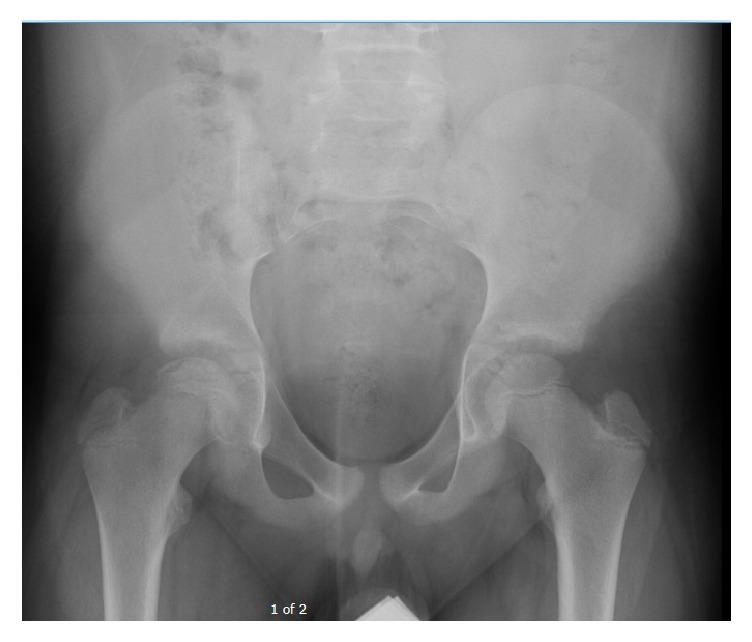
X-rayAnteroposterior pelvic X-ray showing subchondral cystic lucency of the femoral head, an early sign of osteonecrosis, without femoral head collapse.
Source : Egger et al., Case Reports in Pediatrics, 2017, figure 1 · CC BY
This chapter sets out the central problem of the disease, which is not therapeutic but diagnostic. Osteonecrosis is distinguished from referred low back pain, early hip osteoarthritis or gluteal tendinopathy neither by the site of the pain nor by its quality. It is distinguished by the background, and by an investigation that is not requested.
The opening pain of osteonecrosis is a mechanical hip pain, most often in the groin, sometimes in the buttock or referred to the front of the thigh and the knee. There is nothing specific about it. What is specific, on the other hand, is the combination of three elements: a patient younger than the osteoarthritic one, an identifiable exposure, and first-line imaging that shows nothing.
The most useful work on this question is not a case series but a case-control study on the diagnosis itself. Li and colleagues extracted 1,471 patients from the China Osteonecrosis of Femoral Head Database, recruited between July 2016 and December 2018, and separated them according to whether or not they had received an incorrect first diagnosis, defined by a different second diagnosis within six months with unchanged symptoms, or by disagreement with the expert group8.
Three hundred and three patients, that is 20.6 %, had been mislabelled. The detail of the errors is worth more than a long discussion: 118 as lumbar disc herniation, 86 as hip synovitis, 48 as hip osteoarthritis, 32 as rheumatoid arthritis, 11 as piriformis syndrome, 5 as sciatica, 3 as soft tissue injury8.
| Diagnosis made in error | Number | What probably misled |
|---|---|---|
| Lumbar disc herniation | 118 | Pain referred to the thigh, and a lumbar MRI that always finds something in an adult |
| Hip synovitis | 86 | The joint effusion, real but not specific |
| Hip osteoarthritis | 48 | Mechanical groin pain and limited internal rotation, common to both |
| Rheumatoid arthritis | 32 | The background: it is often the disease that justified the corticosteroids |
| Piriformis syndrome | 11 | Buttock pain in a young, active person |
| Sciatica | 5 | The downward path of the pain |
| Soft tissue injury | 3 | A minor trauma found in the history |
The multivariate analysis in that same study is the result that should change practice. Having done only a radiograph at the first examination multiplies the risk of getting it wrong by 4.74 (95 % CI 3.16 to 7.12). Conversely, having performed an MRI straight away is protective (OR 0.65, 95 % CI 0.43 to 0.99)8.
Two other factors are protective, and they say something about the history: a known history of corticosteroid therapy (OR 0.30) and documented alcohol consumption (OR 0.31)8. In other words, when the clinician knows the exposure, they think of the diagnosis. The delay does not come from ignorance of the disease, it comes from a history that has not asked the two questions.
Two factors finally increase the risk of error: subtle symptoms (OR 1.55) and the grade of the practitioner at the first examination, a trainee or registrar rather than a senior (OR 3.32)8.
This is not a nuance, it is the definition. The 2019 revision of the ARCO classification, obtained by a four-round Delphi bringing together 29 international experts with response rates of 93.1 to 100 %, states stage I thus: “the radiograph is normal, but MRI or bone scan is positive”9.
A normal radiograph therefore does not reduce the probability of osteonecrosis in a patient at risk: it only excludes stages II to IV. That is exactly the opposite of the spontaneous reasoning, which treats normal imaging as a reassuring element.
What each investigation can see, stage by stage
The four ARCO 2019 stages, and the minimum investigation that reveals them
Source: Yoon BH, Mont MA, Koo KH, et al. J Arthroplasty. 2020;35(4):933-940. PMID 31866252. A four-round Delphi consensus, 29 experts, agreement threshold set at 70 % of scores of 4 or 5 out of 5.
From those three elements (the background, the definition of stage I, and the measured risk factor for error) follows a simple course of action, which adds no unnecessary investigation but moves one.
Faced with a painful hip: when MRI becomes the investigation to request
A tree built on the ARCO definition of stage I and on the measured risk factors for diagnostic error
Sources: Yoon BH, et al. J Arthroplasty. 2020;35(4):933-940 (PMID 31866252) for the definition of stage I; Li WL, et al. Orthop Surg. 2020;12(6):1792-1798 (PMID 33063422) for the odds ratio of 4.74; Roth A, et al. Arch Orthop Trauma Surg. 2016;136(2):165-174 (PMID 26667621) for the radiograph-then-MRI sequence; Mont MA, et al. J Bone Joint Surg Am. 2026 (PMID 42475623) for the 50-year threshold and the contralateral MRI. This tree is a SYNTHESIS of those recommendations; it has not been validated prospectively as a decision rule.
Two exposures dominate, and they do not carry the same weight. This chapter gives the figures that allow the history to be prioritised, and the result that surprises most: in a patient already on corticosteroids, alcohol adds almost nothing.
Japan registers non-traumatic osteonecrosis nationally, which produces data that few countries have. Ikeuchi and colleagues examined over three years the records and images of Aichi prefecture, 7.4 million inhabitants. Of 327 registration applications, 285 met the diagnostic criteria : 42 records, that is 12.8 %, were not osteonecroses1.
In those 285 patients: mean age 50.4, male-to-female ratio of 2.1 to 1. The aetiological breakdown is the figure to remember.
What causes non-traumatic osteonecrosis
285 patients from the Japanese national registry, Aichi prefecture, over three years
Source: Ikeuchi K, Hasegawa Y, Seki T, et al. Mod Rheumatol. 2015;25(2):278-281. PMID 25036228. Estimated annual incidence for Japan: 1.91 per 100,000. Be careful about transposing: this breakdown is Japanese, and prescribing and drinking habits vary between countries.
The reference demonstration dates from 1987 and has not been contradicted. Felson and Anderson brought together 22 publications giving both the mean corticosteroid dose of a cohort and the percentage of patients in it who developed osteonecrosis, then correlated the two at the level of the studies3.
The result comes down to two lines. The total daily dose is strongly correlated with the osteonecrosis rate (r from 0.61 to 0.80), and the oral dose more so still (r from 0.70 to 0.86). By contrast, the dose given as a bolus is not associated with risk3.
The authors themselves point out why the individual studies saw nothing: within a given centre, cases and controls receive similar protocols and therefore barely differ in dose. Comparison had to be made across studies for the relationship to appear. That is a methodological limitation to keep in mind: this correlation is ecological, it concerns cohort means, not individuals. It establishes that a dose-effect relationship exists; it provides no individual threshold below which a patient would be safe.
Koo and colleagues reviewed, across four university hospitals, the records of patients whose corticosteroid-induced osteonecrosis had been caught at ARCO stage I, that is with a positive MRI and normal radiographs. Twenty-two patients, aged 17 to 60, mean 334.
The cumulative dose at the time of detection ranged from 1,800 to 15,505 mg of prednisolone equivalent, mean 5,928 mg. The interval between the start of treatment and the MRI diagnosis ranged from 1 to 16 months, mean 5.3 months. And above all: 21 of the 22 patients were diagnosed in the first twelve months4.
When osteonecrosis appears under corticosteroid therapy
Interval between the start of treatment and the MRI diagnosis, 22 patients caught at ARCO stage I
Source: Koo KH, Kim R, Kim YS, et al. Clin Rheumatol. 2002;21(4):299-303. PMID 12189457. A retrospective series of 22 patients: the window described is the one in which these patients were DETECTED, not necessarily the one in which the necrosis formed.
Yoon and colleagues conducted a dose-response meta-analysis on five Japanese case-control studies, 1,251 subjects, modelling the relationship with restricted cubic splines5.
Current consumption is associated with risk: OR 3.63 in the occasional drinker, 5.90 in the daily drinker. And the relationship is indeed graded: risk increases by 35.3 % per 100 g a week (95 % CI 1.24 to 1.47) and by 44.1 % per 500 g-years (95 % CI 1.295 to 1.601). The authors specify that the shape of the relationship is non-linear5.
Fukushima and colleagues conducted a Japanese multicentre hospital-based case-control study, 71 cases of idiopathic osteonecrosis against 227 matched controls, to look specifically for an interaction between the two exposures6.
The result is clear-cut and counter-intuitive. Across all subjects, consumption of at least 3,032 drink-years gives an OR of 3.93 (95 % CI 1.18 to 13.1). But after stratification: that OR is 11.1 in those who have never taken corticosteroids (95 % CI 1.30 to 95.5), and 1.10 in those who have (95 % CI 0.21 to 4.79). On a two-way table, corticosteroids alone give an OR of 31.5, and the two combined an OR of 31.6, that is, no additional increase. Neither multiplicative interaction (p = 0.19), nor additive (synergy index 0.95)6.
The respective weight of the two exposures
Adjusted odds ratios, 71 cases and 227 matched controls
Source: Fukushima W, Yamamoto T, Takahashi S, et al. Bone Joint J. 2013;95-B(3):320-325. PMID 23450014. The confidence intervals are wide (for example 1.30 to 95.5 for the OR of 11.1): these are orders of magnitude, not precise measurements. The authors conclude that “the added effect of alcohol may be trivial against the overwhelming effect of steroids”.
The massive corticosteroid courses used in severe COVID-19 raised fears of a wave of osteonecroses. The most solid data comes from the Japanese multicentre database, which compared 20 patients (32 hips) who developed osteonecrosis after COVID with 693 patients with corticosteroid-associated osteonecrosis not linked to COVID over the same period7.
The COVID group had a much shorter course of corticosteroids (2.3 months against 44.9), at a lower maximum dose (28.1 against 53.2 mg a day), and less often as a bolus (15.0 % against 49.4 %). It had more men, and a higher proportion of type C2 in the Japanese classification, that is of laterally extensive lesions7.
The authors' conclusion is cautious and deserves to be taken up as it stands: it must be recognised that osteonecrosis can occur even when the corticosteroid course has been brief and moderate, in a patient who has had COVID, and therefore that a musculoskeletal symptom in that context should be investigated early. Twenty patients out of a database of 5,371 do not constitute an epidemic; it is a signal, described as such.
The question of systematic screening is settled by no trial, and that has to be said from the outset. None of the sources in this base establishes that systematically imaging patients started on corticosteroid therapy improves their outcome. What the data do allow is to delimit a population and a window where the yield of an MRI would be highest if it were decided to request one.
The three elements overlap. The window is that of the first twelve months of corticosteroid therapy, where 21 of the 22 diagnoses made at ARCO stage I are concentrated4. The population is that of prolonged high daily doses, the dose-effect relationship being established on cumulative oral dose and not on boluses3. And the expected benefit is concentrated on hips that are still round, since joint preservation is mainly effective before structural collapse10.
Where the reasoning tightens markedly is in a patient already diagnosed on one side. That is no longer population screening but the investigation of a patient at known risk, and two sources support it: contralateral MRI is explicitly recommended by the 2026 update10, and the S3 guideline specifies that contralateral risk is concentrated in the first two years and then becomes unlikely11. An MRI at diagnosis and two years of clinical surveillance are therefore enough to cover most of the risk, without repeated imaging indefinitely.
Two orders of magnitude, from two national registries, place the problem.
In Japan, the estimated annual incidence is 1.91 per 100,000 inhabitants1. In South Korea, analysis of the health insurance database over 2013-2022 shows a crude prevalence starting from 74.1 per 100,000 adults, a fall in 2020 attributed by the authors to restricted access to care during the pandemic, then a rise again. Total medical cost went from 26.7 to 41.2 billion won over the period2.
Two cautions before transposing. First, these two countries register the disease nationally, which is not the case everywhere: the gap between 1.91 (incidence) and 74.1 (prevalence) does not measure a difference in frequency between Japan and Korea, but two different indicators. Second, no French incidence data exists in this base, and corticosteroid prescribing and alcohol consumption habits vary enough from one country to another to forbid a direct extrapolation.
The figure from the Aichi registry that speaks most to the clinician is elsewhere, and it is humbling: of 327 records submitted for registration, 42 (12.8 %) did not meet the diagnostic criteria1. The diagnosis is missed in both directions: by default in one patient in five8, by excess in one record in eight.
This is the question every decision depends on, and the answer is not single. Some hips collapse inexorably, others stop along the way, and what separates them is measured on the initial MRI. This chapter gives the two series that make sorting possible.
An asymptomatic hip with osteonecrosis is most often discovered as the contralateral side of a painful hip. Should it be treated? Mont and colleagues conducted a systematic review to answer: 16 studies, 664 asymptomatic hips13.
Overall, 394 hips out of 664, that is 59 %, progressed to symptoms or collapse. But the stratification is what counts: small medial lesions have a prevalence of collapse below 10 %. Conversely, medium and above all large, or lateral, lesions progress in a substantial proportion of patients. The background modulates it too: sickle cell disease gives the most frequent progression, lupus the most benign course13.
The counterpart is just as important, and it is less well known. Nishii and colleagues followed for at least five years 54 hips with no initial collapse or with a simple crescent sign14.
Twenty-eight hips, that is 52 %, collapsed. But among those 28, the collapse stopped in 15 cases, that is 54 %, and notably in 8 of the 9 hips with a small necrotic lesion (89 %), that is occupying less than the medial two-thirds of the weight-bearing area. Of those 15 stabilised hips, 11 stayed under 2 mm of collapse and 10 became asymptomatic again with no surgery at all14.
Two series, two ways of looking at the same disease
What becomes of unoperated hips, depending on whether you start from the absence of symptoms or the absence of collapse
Sources: Mont MA, Zywiel MG, Marker DR, et al. J Bone Joint Surg Am. 2010;92(12):2165-2170 (PMID 20844158); Nishii T, Sugano N, Ohzono K, et al. Clin Orthop Relat Res. 2002;(400):149-157 (PMID 12072757). The two series do not start from the same population: one starts from hips without symptoms, the other from hips without collapse. The percentages do not add up and cannot be compared term for term.
Ha and colleagues tested the hypothesis that the combined necrotic angle, measured no longer on radiographs but on mid-coronal and mid-sagittal MRI, predicts collapse. Thirty-seven hips at an early stage in 33 consecutive patients, followed until collapse or for a minimum of five years12.
The separation is clear-cut. By grade: none of the 5 grade 1 hips (less than 200 degrees) collapsed; 6 of the 9 grade 2 hips did; all 16 grade 3 hips and all 7 grade 4 hips collapsed before 36 months. Expressed as continuous thresholds: none of the 4 hips at 190 degrees or less collapsed, all 25 hips at 240 degrees or more collapsed, and 4 of the 8 hips between the two12.
The combined necrotic angle, and the risk of collapse
37 hips at an early stage, angle measured on coronal and sagittal MRI, minimum 5-year follow-up
Source: Ha YC, Jung WH, Kim JR, et al. J Bone Joint Surg Am. 2006;88 Suppl 3:35-40. PMID 17079365. Prognostic level of evidence I. A series of 37 hips: the thresholds are robust in their direction, the numbers in each cell remain small.
The German S3 guideline, built on 3,715 publications screened of which 159 were retained under SIGN criteria, gives two useful time markers. Untreated, the subchondral fracture commonly occurs within two years. And it is during that same period that the risk of contralateral involvement is high: beyond it, it becomes unlikely11.
That second piece of information has a direct practical consequence: it bounds the surveillance. A patient whose second hip has stayed healthy two years after the diagnosis of the first should not be imaged indefinitely.
This is the pivotal chapter of this article, and the one where the gap between practice and evidence is greatest. Offloading is prescribed almost systematically before collapse, on mechanical reasoning that seems obvious. It has never been tested.
The reasoning is simple: the necrotic femoral head has lost its mechanical strength, weight bearing loads it, so relieving the weight must delay the subchondral fracture. It is coherent. It has nevertheless never been the subject of a trial comparing protocolised offloading with free weight bearing, on hips otherwise treated the same way.
That void is not an oversight of the literature search: it is an observation that must be stated explicitly, because the existing literature is regularly read as though it had answered that question. It answered a different one.
The review by Mont, Carbone and Fairbank is the source cited to condemn non-operative treatment. It brings together 42 publications, 2,025 hips : 1,206 treated by core decompression, 819 by non-operative treatment, excluding electrical stimulation and isolated cases15.
Satisfactory clinical results are 63.5 % for core decompression against 22.7 % for non-operative treatment. Taking only the pre-collapse hips, the gap remains wide: 71 % against 34.5 %. And the authors themselves correct their own result: excluding the four centres that perform the most decompressions and report the best results from them, the surgical success rate falls to 53 %, that of non-operative treatment staying at 22.7 %15.
What this review does not say deserves to be written down in black and white. “Non-operative treatment” there aggregates heterogeneous protocols gathered over thirty years, of which offloading is the historical mainstay but whose exact nature, duration and adherence are not controlled. It is not a randomised comparison, it is a gathering of series. The authors themselves in fact conclude that only large prospective randomised trials would clarify the effect.
What can therefore be stated honestly: surgical abstention accompanied by offloading gives, historically, fewer than one satisfactory result in four. What cannot be stated: that offloading is responsible for that, nor that it contributes anything at all.
Neumayr and colleagues conducted a prospective multicentre randomised trial in patients with sickle cell disease and osteonecrosis of Steinberg stage I, II or III. Forty-six patients randomised, eight withdrawals, 38 analysed : 17 in core decompression followed by a physiotherapy programme, 21 in physiotherapy alone16.
At a mean of three years, hip survival is 82 % in the surgical arm and 86 % in the physiotherapy-alone arm. The functional gain, measured with a modified Harris score, is 18.1 points against 15.7. None of those differences is significant. The authors' conclusion is explicit: physiotherapy alone appeared as effective as core decompression followed by physiotherapy in improving function and delaying the need for further intervention16.
Two reservations, which do not cancel it but bound its scope. The population has sickle cell disease, and the paper is a reminder in its introduction that in that disease collapse occurs in 90 % of patients within five years of diagnosis: it is a particularly unfavourable background. And above all, there was no untreated arm : that trial establishes that physiotherapy holds its own against surgery, it does not establish that it does better than nothing.
Two synthesis texts decide, and in the same direction.
The German S3 guideline writes that conservative treatment with iloprost and alendronate may be tried, but that the other pharmacological or physical treatments are inappropriate11. It is harsh wording, and it targets physical treatments explicitly.
The JBJS update of 2020 is of the same order, with one exception worth keeping: “non-operative modalities have generally been ineffective at halting progression. Non-operative treatment is therefore not appropriate at the early stages when the aim is to preserve the native joint, except possibly, on rare occasions, for small, medially located lesions, which may heal without surgery”17.
That exception matches exactly what the two natural history series show: a small medial lesion, fewer than 10 % collapsing in Mont13, collapse stopping in 8 cases out of 9 in Nishii14.
From all this, a position can be drawn that invents nothing.
The conversation about crutches often comes down to two sentences, and those two sentences commit months of behaviour. Here is what the data allow.
What can be said. “The crutches are there to relieve your pain. Use them for as long as they relieve you, and put them down when they no longer make any difference.” That wording is accurate: it assigns to offloading the only role the literature grants it, an analgesic role, identical to the one it holds in any painful joint.
What is better avoided. “Do not put your foot on the ground, or your femoral head will crush.” That sentence is very widespread and has no support. It produces three measurable and all harmful effects: a loss of strength that will lengthen the rehabilitation, a fear of movement that will outlive the indication, and adherence that collapses at the first lapse, since the patient who has walked without harm concludes that the advice was false.
What you say about the prognosis. This is where information has real value, because it is quantified and depends on a parameter the patient can understand: the size of the affected area. A patient whose Kerboul angle is below 190 degrees can be told that none of the hips in that category collapsed in the available series12 ; a patient whose lesion exceeds 240 degrees should know that collapse is very likely and that the surgical question will arise, which is better than a brutal discovery six months later.
What you say about the timetable. Two years. That is the period in which the subchondral fracture commonly occurs, and the one in which the contralateral risk is concentrated11. A patient who knows that limit accepts close surveillance, and accepts it all the better for knowing that it will stop.
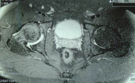
MRIPreoperative hip MRI showing aseptic necrosis of the femoral head.
Source : Moga et al., Journal of Medicine and Life, 2015, figure 1 · CC BY
The physiotherapist prescribes neither bisphosphonate nor core decompression, but does look after patients who receive them, and is the first to be asked what they are worth. Two randomised trials on alendronate give opposite answers: how they differ is instructive.
Lai and colleagues, in 2005, randomised 40 patients with osteonecrosis of Steinberg stage II or III with a necrotic area of more than 30 % (class C). Twenty received 70 mg of alendronate a week for 25 weeks, twenty received nothing. Minimum 24-month follow-up. A spectacular result: collapse of 2 heads out of 29 in the alendronate arm against 19 out of 25 in the control arm (p less than 0.001), and one arthroplasty against sixteen (p less than 0.001)18.
Chen and colleagues, in 2012, redid the trial differently: multicentre, randomised, double-blind, against placebo, over two years. Sixty-four patients included, 52 analysed (65 hips), University of Pennsylvania stages IIC or IIIC. Result: 4 arthroplasties out of 32 hips on alendronate against 5 out of 33 on placebo (p = 0.837). No radiographic, MRI, Harris score or SF-36 difference. The authors' conclusion: “alendronate has no obvious effect”19.
| Lai 2005 | Chen 2012 | |
|---|---|---|
| Design | Randomised, single-centre, no placebo, no blinding | Randomised, multicentre, double-blind against placebo |
| Number analysed | 54 hips | 65 hips |
| Duration | Minimum 24-month follow-up, 25 weeks of treatment | 2 years |
| Collapse | 2/29 against 19/25 (p < 0.001) | No radiographic or MRI difference |
| Arthroplasty | 1 against 16 (p < 0.001) | 4/32 against 5/33 (p = 0.837) |
| Conclusion | Prevents early collapse | No obvious effect |
Two trials of comparable size, on comparable stages, reach opposite conclusions. The most visible difference is methodological: the first had neither placebo nor blinding, the second had both. That is not proof that Lai is wrong, but it is the hierarchy of evidence that critical reading imposes. What should be said to a patient on alendronate for this indication: the evidence is contradictory, and the only double-blind placebo-controlled trial is negative.
The S3 guideline recommends no conservative procedure in particular, but holds that core decompression may be discussed at early stages if the necrosis occupies less than 30 % of the head. At ARCO stages IIIc or IV, total arthroplasty should be considered, with results comparable to those obtained in osteoarthritis. The main factor for revision is young age11.
The JBJS update of 2020 confirms it: joint preservation procedures should be attempted at the early stages, and the results of total arthroplasty in osteonecrosis are excellent, similar to those in osteoarthritis17. The 2026 update puts the same thing differently: preservation procedures are mainly effective before structural collapse, and total arthroplasty remains the most reliable treatment once it has occurred10.
That last sentence is the hinge of the whole management: collapse of the head is the boundary. Before it, preservation can be hoped for. After it, you replace. That is why early diagnosis is not an academic nicety.
What supports each modality, from the most solid to the emptiest
Cards stacked by level of evidence, with the study type that underpins it
Sources, in the order of the cards: Mont 2020 (PMID 32282421) and Roth 2016 (PMID 26667621); Mont 2010 (PMID 20844158) and Nishii 2002 (PMID 12072757); Mont 1996 (PMID 8595753) and Neumayr 2006 (PMID 17142406); Lai 2005 (PMID 16203877) and Chen 2012 (PMID 22127729). The levels shown are an editorial reading of these sources, not a published formal GRADE rating.
| Modality | What the evidence establishes | Level |
|---|---|---|
| Total arthroplasty after collapse | Excellent results, similar to those obtained in osteoarthritis. Young age is the main factor for revision1711 | High |
| Core decompression before collapse | 63.5 % satisfactory results against 22.7 %, but in a non-randomised review whose authors themselves call for trials15. Holds its own against physiotherapy alone in the only randomised trial16 | Moderate |
| Active physiotherapy | Holds its own against surgery at 3 years in a randomised trial of 38 sickle cell patients, with no control arm16. Functional gains maintained at 2 years in a published case21 | Low |
| Alendronate | One positive open-label trial18, one negative double-blind placebo-controlled trial19. Retained as worth trying by the S311 | Contradictory |
| Offloading to preserve the head | No trial. The historical fate of non-operative treatment, of which it is the mainstay, is poor15 ; the S3 judges physical treatments inappropriate11 | None |
| Abstention for a small medial lesion | Fewer than 10 % collapse13 ; collapse stops in 8 cases out of 914 ; an exception accepted by the JBJS17 | Moderate |
One has to be exact here, because there are two temptations: overestimating what rehabilitation can do to bone, or giving up on management that held its own against surgery in the only trial available. Neither.
It does not revascularise a necrotic sector, does not close a subchondral fracture, does not change the Kerboul angle. No data suggests that an exercise programme changes the structural natural history of the disease, and the S3 guideline goes so far as to judge physical treatments inappropriate in this indication11. Claiming the opposite to a patient means making them miss a surgical window.
The physiotherapy arm of the Neumayr trial is the best data that exists: 21 patients treated by physiotherapy alone, 86 % hip survival at three years, 15.7 points gained on the modified Harris score, with no significant difference from surgery16. It is a level I randomised trial by its design, and it is also a trial of 38 patients in a particular population: both things are true.
To that is added a documented clinical case with unusually long follow-up. Jayaseelan and colleagues describe a 56-year-old man with bilateral osteonecrosis, managed with impairment-based manual therapy, therapeutic exercise and education: eight sessions over ten weeks, clinically significant improvements in pain, in the Lower Extremity Functional Scale and in the Harris score, maintained at two years21. The authors themselves write that non-surgical interventions in this disease “are not well described”. One case is not proof of efficacy; it is the demonstration that functional management is possible and that it can hold.
What follows is not a validated protocol: none exists for this disease, and writing otherwise would be lying. It is a reasoned transposition of what the physiotherapy arm of the only randomised trial contained16 and of what the case followed for two years describes21, presented as such to the patient.
First session: establish the baseline and the contract. Assessment of abductor and quadriceps strength, of ranges, of walking distance, and of a functional score that will serve as a reference. The Harris score and the Lower Extremity Functional Scale are the ones the two available sources use, which allows comparison with them. The contract comes down to one sentence: rehabilitation targets function, surveillance targets structure, and the two run in parallel.
Strengthening, and the question of load. The practical problem is real: the external load that would build strength is often the one that wakes the pain. That is exactly the configuration in which strengthening under blood flow restriction finds its indication, since it produces a strength gain at low intensities. There is no study of that modality in osteonecrosis, and that has to be said to the patient: a tool is being transposed, no evidence is being followed.
Walking and endurance. Walking distance is the parameter most meaningful to the patient and the easiest to follow. It also allows weight bearing to be set by the symptom rather than by a protocol, which is the defensible position set out above.
Education, which is not an extra. Three messages: what physiotherapy can do (function) and cannot do (bone); the analgesic and not protective role of offloading; and the two-year timetable, with surveillance of the contralateral hip. The patient in the published case obtained his gains in eight sessions over ten weeks, and held them for two years21 : a modest volume, which suggests that the added value lies as much in the direction taken as in the number of sessions.
Two published reports, both from the physiotherapy literature, frame the two sides of the subject: the one where the disease escapes diagnosis, and the one where it is managed without surgery. They are cited with their PubMed identifier.
Signorino, Jayaseelan and Brindle report in the Journal of Orthopaedic and Sports Physical Therapy the case of a 51-year-old man presenting to unscheduled care two weeks after the onset of left lateral hip and buttock pain. The radiographs are unhelpful. An orthopaedic surgeon refers him to physiotherapy. Faced with worsening of the clinical picture, he is sent back to the orthopaedic surgeon for further assessment. The MRI then requested shows an abnormal contour of the femoral head and acetabulum, extensive marrow oedema and a complex joint effusion: osteonecrosis of the femoral head20.
What this case teaches. First, that the presentation can be atypical, here lateral and gluteal rather than in the groin, which spontaneously points towards a gluteal tendinopathy. Next, that the physiotherapist occupies a particular position: they are the only clinician who sees the patient several times a week, and it is that repetition that makes the worsening visible. Finally, that the right decision here was not to change the treatment but to send the patient back for imaging.
The same authors, in the Journal of Bodywork and Movement Therapies, report a 56-year-old man with bilateral osteonecrosis of the femoral heads. Management combines impairment-based manual therapy, therapeutic exercise and education, over eight sessions spread across ten weeks. The improvements, clinically significant in pain, in the Lower Extremity Functional Scale and in the Harris score, are maintained at two-year follow-up21.
What this case teaches, and what it does not. It shows that functional independence can be obtained and held without surgery, on a modest volume of care. It does not show that physiotherapy changed the bone: no follow-up imaging data is reported in the abstract, and a single case allows no causal inference. The authors say so: work is needed to describe the optimal methods.
In both, the physiotherapist sees the patient for a long time and often. In the first, that position lets them detect the failure; in the second, run continuous care. That is exactly the double role the literature allows us to claim: a lookout role, and a functional role. Not a structural role.
This chapter translates all the above into actions. It comes down to three moments: what you ask at the first assessment, what you write in the letter, and what you monitor afterwards.
Faced with any painful hip, two questions must be asked systematically, because they are the ones that protect against diagnostic error8 :
To that are added a history of sickle cell disease, of transplantation, of chemotherapy, of pelvic radiotherapy, of lupus, and of severe hospitalised COVID7. And age: under 50 with persistent hip pain and normal radiographs, the 2026 update asks that the diagnosis be suspected10.
The wording counts, because it is what triggers the MRI or not. A letter that says “hip pain, physiotherapy with no improvement” gets nothing. A letter that says the following gets one:
First error: treating the normal radiograph as a reassuring result. It defines stage I. It is the only fully reversible stage, and the one where joint preservation has the best chance10.
Second error: prescribing long offloading in the belief of protecting the head. The benefit is not established, the functional cost is. Offload to relieve, yes; offload to preserve, no.
Third error: looking at only one hip. Bilateral involvement is frequent, contralateral MRI is recommended, and the risk is concentrated in the first two years1011.
| Entity | What brings it close | What distinguishes it |
|---|---|---|
| Hip osteoarthritis | Mechanical groin pain, limited internal rotation | An older patient, abnormal radiographs, no exposure to corticosteroids or to alcohol. It is the diagnosis made in error in 48 of the 303 mislabelled patients8 |
| Transient osteoporosis of the hip | Marrow oedema on MRI, subacute onset of pain | A spontaneously resolving course over a few months, diffuse oedema without the geometry of necrosis. Cited as a differential by the S311 |
| Bone bruise | Marrow oedema, normal radiograph | A dated and proportionate trauma, expected resolution. A differential explicitly listed by the S311 |
| Subchondral insufficiency fracture | Sudden pain, oedema, sometimes an older person | An osteoporotic background, absence of proportionate trauma. A differential listed by the S311 |
| Labral tear and femoroacetabular impingement | Groin pain in a young person, catching | Positional mechanics, bone morphology, no systemic exposure |
| Greater trochanteric pain syndrome | Lateral pain, difficulty standing on one leg | Pain reproduced on trochanteric palpation and on lying on that side. It is the trap in the 51-year-old case published in the JOSPT20 |
No, and it is the most important point of this article. Stage I of the ARCO 2019 classification is literally defined by a normal radiograph together with a positive MRI or bone scan9. In a patient at risk, a normal radiograph does not change the probability of the diagnosis: it only excludes stages II to IV.
Yes, in a newly diagnosed patient. The 2026 JBJS update explicitly recommends MRI of the contralateral hip, bilateral involvement being frequent10. And an asymptomatic hip is not a hip free of progression: 59 % progress in the systematic review of 664 hips13.
The S3 guideline gives the marker: the risk of contralateral involvement is high during the following two years, and becomes unlikely afterwards11. It is also the period in which the subchondral fracture of the affected hip commonly occurs.
Nothing demonstrates it. No trial has compared protocolised offloading with free weight bearing. What is known is indirect: the historical non-operative treatment, of which offloading is the mainstay, gives 22.7 % satisfactory results15, and the S3 guideline judges physical treatments inappropriate in this indication11. Offloading keeps an analgesic value, and that is how it should be presented.
Yes, in one precise configuration: small, medially located lesions. They collapse in fewer than 10 % of cases13, the collapse stops in 8 cases out of 9 when it occurs14, and the JBJS update explicitly recognises that exception17.
There is no individual threshold. Felson and Anderson established a dose-effect relationship at the level of studies, between the mean daily dose of a cohort and its osteonecrosis rate3. In Koo's series, the cumulative dose at detection ranged from 1,800 to 15,505 mg of prednisolone equivalent4 : an eight-fold spread says enough that no threshold protects.
The available data says the opposite: in the analysis by Felson and Anderson, the dose given as a bolus was not associated with risk, whereas the oral dose strongly was3. It is the cumulative daily dose that counts.
Stopping remains desirable for other reasons, but its benefit on the risk of osteonecrosis is probably small in that configuration: in corticosteroid users, the odds ratio of alcohol falls to 1.10, and the combination of the two exposures gives no more than corticosteroids alone6. In a patient who has never received corticosteroids, on the other hand, alcohol is the dominant factor.
Function, not structure. The only randomised trial available shows that a programme of physiotherapy alone holds its own against core decompression at three years, in 38 sickle cell patients16, and a published case documents gains maintained at two years after eight sessions21. No data shows an effect on the necrosis itself.
No. The 2020 and 2016 updates agree: the results are excellent and comparable to those in osteoarthritis17. The main factor for revision is not the disease, it is the patient's young age at the time of implantation11. What follows is described in our article on rehabilitation after total hip replacement.
Twenty-one references, verified one by one through the NCBI E-utilities: a real PMID, journal, issue year, complete author list, and abstract read to check that the source does establish what is attributed to it. The working base is versioned in the repository, with the list of what it does not support.
This page deals with osteonecrosis of the femoral head as a disease in its own right. The commonest differential diagnosis in the older person is the subject of a separate article: hip osteoarthritis. Bone marrow oedema and its non-necrotic causes are set out in bone bruise. The same disease in another bone is described in Kienböck's disease, avascular necrosis of the lunate. And the end of the trajectory, when the head has collapsed, is dealt with in rehabilitation after total hip replacement.
Behind this article
An author who explains, a reviewer who checks.
How we write and check our content

Physiotherapist · co-founder of Physio Learning
Marked for life by his first four-hour lecture without a single image, he took a master’s in instructional design so that it would never happen to anyone again. He hunts down publication bias and unreadable slides with the same intransigence.

Head of scientific content
Physiotherapist specialising in neuro-musculoskeletal practice and holder of a master’s in public health. He checks the methodological rigour of every article: primary sources, levels of evidence, no exceptions.
