


















In brief
A labral tear is visible on MRI in 54 % of hips that are not painful, against 62 % of those that are: eight points apart. A report announcing a "labral tear" therefore makes no diagnosis, it describes a population trait. This article deals with the labral tear itself: what it is, what it means, how to tell whether it is the source of the pain, and what to do about it. For the morphological cause cam or pincer and the impingement syndrome, the dedicated article is femoroacetabular impingement.
Clinical review based on the meta-analysis by Heerey (BJSM 2018) on the comparative prevalence of intra-articular lesions with and without pain, the meta-analysis by Reiman (BJSM 2015) on the diagnostic values of the tests, the series by Vahedi (CORR 2019) on the fate of silent labral tears, and Kemp's PhysioFIRST trial (BJSM 2026): 37 references checked one by one on PubMed.
Clinical review
We have to start with the figure that makes everything else legible. Heerey's meta-analysis, which pooled 29 hip imaging studies, finds a labral tear in 62 % (95 % CI 47 to 75) of people who have pain and in 54 % (95 % CI 41 to 66) of people who do not (PMID 29540366). The confidence intervals overlap widely. In other words: learning that an MRI shows a labral tear barely shifts the probability that this hip is the painful hip.
The comparison with cartilage is what makes the finding unanswerable. In the same meta-analysis, a cartilage defect is present in 64 % of symptomatic people and in only 12 % of asymptomatic ones. Cartilage discriminates; the labrum does not. So the point is not that "hip imaging is uninformative", it is something more precise and more useful: that particular sign is uninformative, and other signs from the same scan are far more informative.
The figure varies enormously depending on who reads which machine. In asymptomatic volunteers, the published prevalence ranges from 38.6 % (Lee, 70 volunteers with a mean age of 26, 3 tesla, PMID 25922455) – 85.7 % (Schmitz, 42 hips with a mean age of 34, 1.5 tesla with an optimised protocol, PMID 22422932). These two populations are close to one another. It is the protocol and the reader that separate 38 from 86, not the hips. A prevalence that doubles depending on the machine is not a property of the patients: it is a property of the measurement.
A silent labral tear usually stays silent. Vahedi followed the contralateral asymptomatic hips of patients operated on for femoroacetabular impingement: 41 to 43 % carried a labral tear, and over two years, only 9 % became symptomatic (PMID 30444756). It is the most direct natural history evidence available, and it says that the lesion is not a time bomb.
The FADIR is a screening test, and nothing more. Reiman's meta-analysis gives a pooled sensitivity of 0.94 to 0.99 depending on the studies included, but diagnostic odds ratios whose confidence interval crosses 1 (5.71; 95 % CI 0.84 to 38.86, and 7.82; 95 % CI 1.06 to 57.84). The authors conclude in so many words that these tests "possess only screening accuracy" (PMID 25515771). A negative FADIR helps; a positive FADIR teaches almost nothing, because it is positive in almost everyone who has hip pain, whatever the cause.
Rehabilitation achieves a great deal, and not where one expects. The PhysioFIRST trial compared targeted, individualised strengthening with a standardised stretching programme in 154 people: no difference in hip-related quality of life at six months, but the two groups improved by 19 to 21 iHOT-33 points, well beyond the clinically relevant change (PMID 42082318). The message is not "it does not matter what we do", it is this: structured follow-up by a physiotherapist delivers most of the benefit, and the detail of the content delivers less than was hoped.
The three surgery versus rehabilitation trials do not say the same thing, and that is instructive. UK FASHIoN finds +6.8 iHOT-33 points for arthroscopy at 12 months (PMID 29893223), FAIT finds +10.0 HOS-ADL points at 8 months (PMID 30733197), and Mansell's trial in military personnel finds no difference at two years, with 28 patients from the rehabilitation arm operated on along the way (PMID 29443538). None of these three trials randomised on "labral tear": all randomised on femoroacetabular impingement syndrome. That is a limitation to be stated before drawing any management conclusion from them.
What this changes in the clinic. The diagnosis of a symptomatic labral tear is not made on an image. It is made on a compatible history, an examination that points to the joint, an image confirming a lesion and the absence of a better explanation, and, when doubt persists, on the response to an intra-articular anaesthetic injection. That is the logic of the Warwick agreement on femoroacetabular impingement, which requires the triad symptoms + clinical signs + imaging, and never imaging alone (PMID 27629403).
- An MRI showing a labral tear in someone who has hip pain is compatible with the diagnosis. It does not make it.
- The right reflex in front of the report is not "how many lesions?" but "is there a cartilage defect?": that is the sign that really separates painful hips from the rest (64 % against 12 %).
- An asymptomatic labral tear found incidentally justifies neither close surveillance nor activity restriction: only 9 % become painful within two years.
- The word "tear" does damage. After the age of 40, radiologists propose speaking of a fissure, precisely to prevent the drift towards surgery (Amber 2018).
- Rehabilitation is the first line, and its benefit is broad and reproducible in every "physiotherapy" arm of the trials, including those that served as the comparator for surgery.
What does the labrum do, and what does "tear" mean?
Before discussing an image, one has to know what the structure does. The labrum is not a meniscus of the hip, and that misconception explains a good part of the decisions taken about it.
A seal, not a shock absorber
The acetabular labrum is a fibrocartilaginous ring attached to the rim of the acetabulum, which it extends and deepens. Its dominant mechanical function is not to absorb load: it is to seal the synovial fluid inside the joint. Ferguson's poroelastic model showed that under a load of 1,200 N, an intact labrum maintains a pressurised layer of fluid between the femoral head and the acetabulum, so that load is transmitted by pressurisation of the interstitial fluid rather than by direct contact between the cartilage surfaces (PMID 10771126). Without this seal, the model predicts far higher stresses in the solid matrix of the cartilage: 20 % strain against 3 %. The labrum does not protect the joint by cushioning it; it protects it by stopping the fluid from escaping.
This did not remain a theoretical model. Philippon measured intra-articular pressure directly in eight cadaveric hips compressed to 2.7 times body weight, in six successive labral states. The result: compared with the intact state, a labral tear leaves pressurisation at 75 %, partial resection at 53 %, and complete resection collapses it to 24 % (PMID 24519614). The companion paper, on stability to distraction, finds the same order of magnitude: complete resection brings the maximum distraction force down to 27 % of the intact state (PMID 24509878).
Two practical consequences follow from these figures, and they run in a direction that the word "tear" does not suggest. First, a labral tear does not abolish the function of the labrum: it leaves three quarters of it. Second, the act that really damages the seal is the surgical resection, not the lesion itself. That is also why modern surgery repairs or reconstructs rather than resects, a point we return to in chapter 7.
What the word "tear" leads people to believe
In French as in English, "déchirure du bourrelet" or labral tear suggests an accident: something ruptured, at a given instant, through a movement. Yet the vast majority of labral lesions seen on MRI are of gradual onset, degenerative, and without any identifiable triggering episode. In Burnett's series, of 66 hips arthroscoped for a confirmed labral tear, 40 had an insidious onset, 20 a low-energy trauma and only 6 a major trauma (PMID 16818969).
Two radiologists went as far as proposing a change of vocabulary. Amber and Mohan, in Academic Radiology, argue for speaking of a "fissure" rather than a "tear", fissure rather than tear , in patients over 40, on the grounds that the high prevalence and the lack of specificity of this sign make it an area particularly exposed to overdiagnosis, which in turn exposes patients to unnecessary surgery and its complications (PMID 29199059). They explicitly draw the parallel with what happened in the spine, where reframing the vocabulary of reports measurably reduced the cascade of investigations and procedures.
The physiotherapist does not write the reports, but is almost always the first to translate them for the patient. That is real power: the sentence "you have a tear" and the sentence "your MRI shows wear of the labrum, which is found in one person in two who has no pain anywhere" describe the same DICOM file and produce two different patients.
What the mechanics of the labrum establish, in four measurements
Three cadaveric studies and one computational model. Level of evidence: basic research, not directly transferable to the patient.
Where the lesions sit, and why always in the same place
The location is remarkably constant. Tresch assessed six clock positions of the labrum in 63 asymptomatic volunteers and 63 matched patients with femoroacetabular impingement: in all groups, the majority of cartilage and labral defects sat in the anterosuperior, superior and posterosuperior (PMID 27981665). The authors' conclusion deserves to be quoted in its exact order: the topographical predilection is the same in volunteers and in patients.
This detail carries a diagnostic significance that is underestimated. A report noting an anterosuperior lesion does not describe a peculiarity of this patient: it describes the place where the labrum wears out in everyone. Topography alone therefore does not distinguish a lesion that speaks from one that stays silent.
- The labrum is a seal. Its function is hydraulic before it is mechanical.
- A lesion leaves 75 % of the pressurisation; it is resection that brings it down to 24 %. The lesion and its treatment do not carry the same mechanical cost.
- The vast majority of lesions are of insidious onset (40 cases out of 66 in Burnett), not traumatic.
- The anterosuperior location is everyone's, symptomatic or not: it has no discriminating value.
- The word used in front of the patient is part of the treatment. "Fissure" is more accurate than "tear" for most degenerative lesions.
Why does a positive MRI not make the diagnosis?
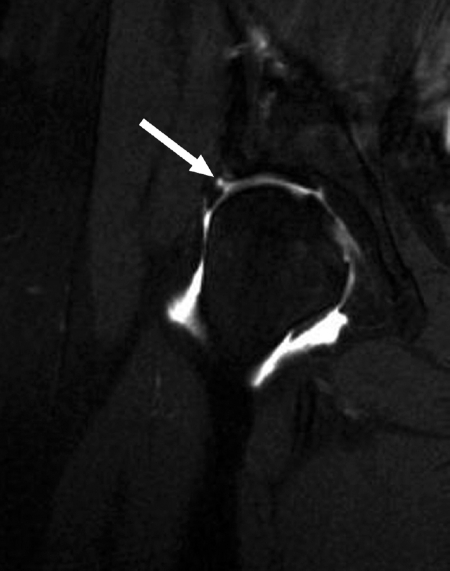
MR arthrographyCoronal MR arthrography of the hip: acetabular labral tear, arrow on the contrast medium infiltrating the base of the labrum.
Source : Troelsen et al., Acta orthopaedica, 2009, figure 4 · CC BY-NC
This is the chapter that governs all the others. If we accept the idea that a labral tear seen on imaging is a diagnosis, then the tests, the injection, rehabilitation and surgery no longer have any ordered meaning. So we must start by dismantling that idea, with the figures and their intervals.
The central figure, and the interval no one quotes
Heerey's meta-analysis pooled 29 studies reporting the prevalence of intra-articular hip lesions on MRI, MR arthrography or CT, in people with and without pain. For the labrum:
- 62 % (95 % CI 47 to 75) in symptomatic people, a level of evidence judged limited by the authors;
- 54 % (95 % CI 41 to 66) in asymptomatic people, a level of evidence judged moderate (PMID 29540366).
The two intervals overlap over almost their whole length, from 47 to 66 %. And a detail that matters: it is the asymptomatic estimate that rests on the more solid level of evidence. We know the prevalence of the lesion better in people who have no pain than in those who do.
The labrum does not discriminate, cartilage does
The same meta-analysis measured, in the same populations, the prevalence of cartilage defects: 64 % in symptomatic people against 12 % in asymptomatic ones. Five times as many. It is this juxtaposition that makes the finding unassailable, because it closes off the usual escape route.
Faced with this kind of data, one often hears that "imaging shows things in everybody" and that it should therefore be distrusted across the board. That is not what these figures say. They say that within the same scan, in the same patients, read by the same radiologists, one sign strongly separates painful hips from the rest and another barely separates them at all. The problem is not the MRI: it is the use made of one of its lines.
Two signs from the same scan, two opposite discriminating powers
Heerey 2018 meta-analysis, 29 studies. Prevalence in people with and without hip pain.
There is a methodological lesson in this chart, beyond the hip. An imaging sign is not "reliable" or "unreliable" in the absolute; it has a discriminating power, which is measured by comparing its frequency in patients and in everyone else. No radiology report carries that information, and it is for the clinician to know it for every line read.
Prevalence rises with age, but it depends above all on the machine
This is where reading demands caution, and where most reviews oversimplify. One commonly reads that the prevalence of asymptomatic labral tears increases with age. That is true in broad terms, and false as soon as the studies are examined one by one, because the scatter at equal age is as large as the effect of age itself.
No published study stratifies labral prevalence by age band within a single cohort. The only honest chart is therefore this one: one study, one point, placed according to the mean age of its cohort.
Prevalence of labral tears in people without hip pain, by mean age of the cohort
Each bar is a separate study, with its own protocol and its own readers. These are not age bands within a single population: no study stratifies by age internally.
What this chart forbids one to write is the sentence "X % of asymptomatic hips have a labral tear" without saying which of the bars was chosen. The difference between announcing 38.6 % and 85.7 % to a 30-year-old patient is considerable, and it comes down to nothing other than the reference selected.
One methodological detail deserves to be noted, because it runs against intuition. Register's cohort was read by three specialist radiologists, with a lesion counted as present as soon as two of them agreed, and the volunteers' scans were mixed with those of 19 symptomatic patients to blind the readers (PMID 23104610). It is the most careful protocol of the lot, and it produces 69 %. Methodological rigour does not bring the figure down: it confirms it.
| Study | Population | Imaging | Labral prevalence | What it establishes in its own right |
|---|---|---|---|---|
| Georgiadis 2016 PMID 26907862 | 108 subjects, 216 hips, aged 2 to 18, with no hip complaint | Pelvic MRI, blinded reading | 1.4 % (3/216 hips) | In children, a labral tear is not background noise. A positive image keeps its value there. |
| Jones 2025 PMID 39881446 | Athletes aged 9 to 18 (mean 14.9), painless contralateral hip | 3 T, two independent radiologists | 18 % (against 30 % in symptomatic ones, p = 0.16) | From sporting adolescence onwards, the symptomatic / asymptomatic gap is no longer significant. |
| Frank 2015 PMID 25636988 | Systematic review, 2,114 asymptomatic hips, mean age 25.3 years | MRI without contrast (7 of 26 studies report the labrum) | 68.1 % | The broadest estimate, but resting on only seven of the twenty-six studies included. |
| Lee 2015 PMID 25922455 | 70 young volunteers, aged 19 to 41 (mean 26), 67 % women | 3 T, non-arthrographic | 38.6 % (27/70) | Separates pure labral tears (22.9 %) from those associated with another lesion (15.7 %). |
| Schmitz 2012 PMID 22422932 | 42 hips, aged 27 to 43 (mean 34) | 1.5 T, optimised protocol without contrast, 2 readers | 85.7 % and 80.9 % depending on the reader | The highest figure in the corpus, obtained with the weakest magnet: it is the protocol that decides. |
| Register 2012 PMID 23104610 | 45 volunteers, aged 15 to 66 (mean 37.8) | 3 T, 3 radiologists, threshold of 2 opinions out of 3, blinded readers | 69 % (73 % of abnormalities across all categories) | The most rigorous protocol. There, age predicts cartilage and subchondral cysts, not labral tears. |
| Tresch 2017 PMID 27981665 | 63 volunteers aged 20 to 50, matched with 63 femoroacetabular impingement patients | 1.5 T, 2 radiologists, 6 clock positions | 44 % against 61 % in patients (p ≤ 0.12) | The only matched comparison: the labral difference does not reach the significance threshold, whereas the acetabular cartilage difference does (14 % against 47 %). |
The table contains one observation that deserves to be singled out, because it contradicts what everyone repeats, including this article two paragraphs above. Register explicitly tested the association between age and each of the lesions found. Subjects over 35 were 13.7 times more likely to have a cartilage defect (95 % CI 2.4 to 80) and 16.7 times more likely to have a subchondral cyst (95 % CI 1.8 to 158). But the authors write: "no other joint lesion was associated with age". In the only study that tested the association directly, age predicted cartilage and not the labrum.
What can therefore be stated without straining: asymptomatic labral prevalence is very high from the twenties onwards, and it is low before puberty. Between the two, the slope is poorly established, and the literature that asserts it relies on between-study comparisons that the scatter of protocols does not allow.
In athletes, the gap reverses
Heerey repeated the same work in the sporting population alone, in a second meta-analysis of twenty studies. The result is the most counter-intuitive in the whole review, and it must be read slowly. In asymptomatic athletes, the prevalence of labral tears is 54 % per person (limited evidence) and 33 % per hip (moderate evidence). In symptomatic athletes, it is 20 % per hip (moderate evidence) (PMID 30972659).
At hip level, and on the estimates the authors judge the most solid, labral tears are more frequent in athletes who have no pain than in those who do. This must not be over-interpreted: these estimates come from different studies, with different definitions and protocols, and the authors rate the whole body of evidence at moderate to high risk of bias. But the reversal is enough to destroy the idea that this sign, in athletes, carries diagnostic information.
The same meta-analysis adds a reassuring and often ignored piece of data: hip osteoarthritis is rare in athletes, with or without pain (0 to 17 % in asymptomatic athletes, 2 % in symptomatic ones). The argument "it must be repaired now so as not to develop osteoarthritis later", frequently put forward in consultation, finds no support there.
What becomes of a labral tear left alone
The question every patient asks when told that nothing will be done is: "what if it gets worse?". There is a numerical answer, and it comes from a particularly clean observational design. Vahedi studied the asymptomatic contralateral hips of patients operated on for femoroacetabular impingement on one side: same patient, same morphological background, same exposure, but one side that does not hurt.
The two-year fate of asymptomatic labral tears
100 painless contralateral hips of patients operated on for femoroacetabular impingement, two independent assessors.
Nine per cent over two years, in patients who nevertheless carry every risk factor: the morphology that made the other hip symptomatic, the age, the activity. It is the best available argument for not treating an incidental finding, and it should be given to the patient in that form, because it answers exactly the question asked.
Red flags in hip pain attributed to the labrum
- Constant night pain, not eased by lying down, with impaired general condition or a history of cancer → a labral tear does not explain pain that is independent of position. Bone imaging before any rehabilitation.
- Adolescent, hip or knee pain with a limp and spontaneous external rotation → slipped capital femoral epiphysis until proven otherwise. In children, remember that asymptomatic labral tears are rare (1.4 %): a positive labral image before puberty should not reassure, it should prompt a search for what goes with it.
- Rapidly worsening groin pain in a distance runner, especially in a context of low energy availability or amenorrhoea → femoral neck stress fracture. A report of "labral tear" must never rule out this hypothesis: it is present in more than one painless hip out of two and excludes nothing.
- Fever, effusion, sudden loss of function → septic arthritis. Emergency.
- Hip pain in a patient on prolonged corticosteroid therapy, with sickle cell disease or with alcohol dependence → osteonecrosis of the femoral head, whose early stages can coexist with a labral tear and be eclipsed by it in the report.
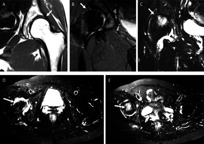
MRIHip MRI, five compared sequences: normal acetabular labrum on T1 (A), an area of increased signal on STIR indicating a labral tear (B and C), joint effusion (D) and bone marrow oedema of the femoral head (E).
Source : Shang et al., Archives of medical science : AMS, 2013, figure 4 · CC BY-NC-ND
- Labral tears are present in 54 % of painless hips (CI 41 to 66) against 62 % of painful ones (CI 47 to 75): the intervals overlap almost entirely.
- Within the same scan, the cartilage defect separates strongly (64 % against 12 %). Look for that line of the report, not the labral one.
- Depending on the study chosen, asymptomatic prevalence ranges from 38.6–85.7 % in populations of similar age. It is the measurement that varies, not the hips.
- In athletes, labral tears are more frequent per hip in asymptomatic people (33 %) than in symptomatic ones (20 %).
- A silent labral tear carries about a 9 % risk of becoming painful within two years, even in patients with proven impingement morphology.
What are hip clinical tests really worth?
If the image does not settle the matter, we turn to the examination. That is the right reflex, but one has to know what the examination can do: point the way, yes; confirm, almost never.
The FADIR: what the meta-analysis establishes, word for word
The flexion-adduction-internal rotation test, or FADIR, is the manoeuvre most taught for anterior hip pain. Reiman reviewed it in the British Journal of Sports Medicine: of 21 articles identified, only one of high quality, and nine eligible for meta-analysis. The results:
- pooled FADIR sensitivity of 0.94 (95 % CI 0.90 to 0.97) to 0.99 (95 % CI 0.98 to 1.00) depending on the subset of studies;
- diagnostic odds ratio of 5.71 (95 % CI 0.84 to 38.86) – 7.82 (95 % CI 1.06 to 57.84) ;
- flexion-internal rotation alone: pooled sensitivity 0.96 (95 % CI 0.81 to 0.99), odds ratio 8.36 (95 % CI 0.41 to 171.3) (PMID 25515771).
The authors' conclusion is unambiguous: these tests "possess only screening accuracy" (possess only screening accuracy). It is worth pausing on the confidence intervals of the odds ratios, because that is where all the information lies and it is the line that is never quoted. A diagnostic odds ratio whose interval crosses 1 means that, given the imprecision of the data, one cannot rule out that the test adds no information at all. That is the case for two of the three estimates above.
The arithmetic is mechanical: for a test with a sensitivity of 0.94-0.99 to have such a low odds ratio, its specificity has to be poor. Reiman does not pool specificity, which is a limitation of the meta-analysis rather than an oversight in reading, but the arithmetic of the odds ratio constrains it. Dhillon's systematic review, published in 2025 on 15 studies and 1,378 hips, reaches the same conclusion by the descriptive route: the sensitivity and the specificity of physical examination tests for prearthritic intra-articular hip pathology are "highly variable" (PMID 40692936).
Why a specificity of 100 % should worry, not reassure
Spectacular specificities for hip physical examination turn up regularly, including in very recent publications. A retrospective comparative series published in 2026 in Arthroscopy reports, on 224 patient records that went on to arthroscopy, a sensitivity of 94 % and a specificity of 100 % for physical examination alone, against 67 % and 83 % for MR arthrography (PMID 41838562).
This figure must not be taken at face value, and the reason is instructive. Specificity is calculated in subjects free of the lesion. In a series of patients all referred for arthroscopy for suspected femoroacetabular impingement, almost all have a labral tear: the denominator of "unaffected" subjects is tiny, and a specificity of 100 % may rest on a handful of hips. This is spectrum bias in its purest form, the same mechanism that long made meniscal tests look excellent, because they were only evaluated in patients already scheduled for knee arthroscopy.
The general rule for reading such figures fits in one sentence: a diagnostic value only means something in the population in which it was measured. Figures obtained in a surgical waiting list do not transfer to the community clinic, where the prevalence of symptomatic lesions is far lower and the spectrum of differential diagnoses far broader.
What a negative test contributes, and it is considerable
It would be wrong to conclude that the FADIR is useless. A pooled sensitivity of 0.94 to 0.99 makes it an excellent exclusion test: a clearly negative FADIR makes an intra-articular origin very unlikely, and that is worth saying, because it is information that changes management. Faced with hip pain and a negative FADIR, one should stop looking for the labrum and look elsewhere: gluteal tendinopathy, lumbar or sacroiliac dysfunction, groin pathology, referred pain.
Burnett's series gives the mirror image. Of 66 hips with a labral tear confirmed at arthroscopy, 95 % had a positive impingement sign, 92 % predominant groin pain, 91 % activity-related pain, 71 % night pain, 39 % a limp and 38 % a positive Trendelenburg sign (PMID 16818969). These figures describe the clinical picture well when the lesion really is the culprit, but they are frequencies among patients, not diagnostic values: they say nothing about what is observed in those whose pain has another cause.
The same series measures the cost of diagnostic uncertainty, and the figure is striking: between symptom onset and diagnosis, 21 months on average elapsed, during which these patients consulted 3.3 healthcare professionals on average. Surgery at another anatomical site had been proposed to 17 % of them, and four had undergone an unsuccessful operation before the labral tear was identified. The wandering does not come from a lack of tests: it comes from the fact that none of them concludes on its own.
Sensitivity and specificity: what is pooled, and what is not
Two meta-analyses. The values for clinical examination and for imaging do not come from the same populations and are not comparable term by term.
The table of tests, with what can be asked of them
| Examination | How it is performed | Measured value | What can be asked of it |
|---|---|---|---|
| FADIR flexion-adduction-internal rotation | Hip at 90° of flexion, adduction then passive internal rotation. Positive if it reproduces the familiar groin pain. | Pooled sensitivity 0.94 to 0.99. Odds ratio 5.71 to 7.82, intervals crossing 1 (Reiman 2015). | Excluding an intra-articular origin when it is negative. Nothing more when it is positive. |
| FABER flexion-abduction-external rotation | Heel on the opposite knee, measurement of the knee-to-table distance. | Sensitivity and specificity "highly variable" according to Dhillon's 2025 review (15 studies, 1,378 hips). | Pointing towards the joint or the sacroiliac joint. Useful as a right-left comparison, not as an absolute value. |
| Flexion-internal rotation | Range of internal rotation measured with the hip flexed to 90°. | Pooled sensitivity 0.96 (CI 0.81 to 0.99), odds ratio 8.36 (CI 0.41 to 171.3). | Screening. The odds ratio interval is so wide that it allows no firm conclusion. |
| Location of the pain | History taking. The "C" sign, hand cupped around the greater trochanter. | 92 % predominant groin pain in 66 patients with a confirmed lesion (Burnett 2006). | Redirecting straight away: strictly lateral or gluteal pain makes the labral hypothesis unlikely. |
| Conventional MRI | 1.5 or 3 T, dedicated hip protocol. | Sensitivity 66 %, specificity 79 % (Smith 2011, 19 studies, 881 hips). | Seeing the other structures, cartilage, bone, tendons, some of which discriminate far better than the labrum. |
| MR arthrography | Intra-articular injection of gadolinium under guidance, then MRI. | Sensitivity 87 %, specificity 64 % (Smith 2011; identical in Huang 2023). | Clarifying the anatomy before a surgical procedure. Not a first-line examination in a patient who is being rehabilitated. |
- The FADIR is an exclusion test. Negative, it shifts the search; positive, it concludes nothing.
- No meta-analysis provides a pooled specificity for the FADIR: when an article announces one, check in which population it was calculated.
- A specificity of 100 % measured in a surgical waiting list is an artefact of spectrum bias, not a performance.
- MR arthrography sees more (87 % sensitivity) and is wrong more often (64 % specificity). It prepares a procedure, it does not establish an indication.
Does the diagnostic injection settle what imaging cannot?
Since neither the image nor the examination identifies the culprit, one logical avenue remains: anaesthetise the joint and see whether the pain disappears. It is appealing, it is used everywhere, and the result is more nuanced than its reputation.
The reasoning, and what it assumes
The intra-articular local anaesthetic injection rests on a simple syllogism: if the inside of the joint is anaesthetised and the pain stops, the pain was coming from inside the joint. The test does not say which intra-articular structure hurts, labrum, cartilage, capsule, ligamentum teres, but it settles the prior question, which is often the real one: articular or not.
The syllogism assumes three things: that the needle really is in the joint, that the product has not diffused to neighbouring structures, and that the response is not an expectancy effect. None of the three is guaranteed, which is why the procedure must be done under ultrasound or fluoroscopic guidance, and interpreted with a threshold defined in advance.
The figure that matters: four lesions in ten do not respond
Martin studied 105 subjects, including 49 potential arthroscopy candidates who received an anaesthetic injection. All had MR arthrography: 18 with a definite labral tear, 29 with a possible one, 2 with none. The result is blunt:
- among the definite lesions, 39 % (7 of 18) did not obtain more than 50 % relief ;
- among the possible lesions, 45 % (13 of 29) did not either (PMID 18760208).
In other words: in selected patients, referred for arthroscopy, with a labral tear documented on the most sensitive examination available, about four in ten do not have their pain inside the joint. The authors' conclusion is explicit: not all labral tears seen on MR arthrography are major contributors to the patient's complaint, and other causes have to be sought.
Response to the intra-articular anaesthetic injection, by what MR arthrography shows
49 hip arthroscopy candidates, mean age 42 years. Threshold used: more than 50 % reduction in pain.
Martin's second result is even more useful to the physiotherapist than the first. None of the classic clinical signs, groin pain, clicking, pain on prolonged sitting, lateral thigh pain, FABER, FADIR, trochanteric tenderness, distinguished the patients who would respond to the injection from those who would not. Clinical examination does not predict where the pain source lies, even in highly selected patients.
What the response really predicts: the outcome of surgery
Chinzei followed 49 patients operated on for femoroacetabular impingement with a labral tear, after a preoperative anaesthetic injection classified as a poor response (0 to 50 % relief) or a good one (51 to 100 %). Two independent findings:
- All the patients had a labral tear, and yet neither the classification of the lesion nor the extent of the cartilage damage was associated with the quality of the response to the injection. The severity of the image does not predict sensitivity to anaesthesia of the joint.
- One year after surgery, patients with a good preoperative response had a significantly better outcome (p < 0.01) than those with a poor response (PMID 31960159).
The diagnostic injection therefore changes status. It is not an examination that confirms a labral tear, nothing does that, but a test of where the pain source lies and, incidentally, one of the few elements that predicts the benefit of arthroscopy. That is valuable, and it is different from what is usually attributed to it.
When to propose it, and how to read it
In a sensible care pathway, the diagnostic injection does not come early. It has its place when: the pain is disabling and persistent; imaging shows a labral tear but the examination has not settled matters; well-conducted rehabilitation has not been enough; and a surgical decision is genuinely at stake. In a patient who is going to be rehabilitated for three months anyway, it adds nothing that changes management.
Three cautions when reading it:
- Define the threshold before the injection, and write it down. "More than 50 % relief" is the threshold used in the literature cited here; the patient must be asked to rate the pain before and within the hour that follows, not to recall it afterwards.
- A partial response is an ambiguous response, not a weakened positive one. It is compatible with mixed pain, articular and extra-articular, which is common when a hip has been painful for a long time.
- A negative response is strong information, stronger than a positive one: it redirects the whole assessment, and it should.
- In arthroscopy candidates with a documented labral tear, 39 to 45 % do not have their pain in the joint.
- No classic clinical sign predicts who will respond to the injection: the test provides information that the examination does not contain.
- The severity of the lesion on imaging is not associated with the response to the injection (Chinzei 2020).
- In practice, a good preoperative response predicts a better surgical outcome at one year: that is its most solid use.
- It is proposed when a surgical decision is at stake, not as a routine step in the assessment.
How can the diagnosis be made without overdiagnosing?
Three chapters to dismantle the tools one by one, and now the practical question: what do we do, in what order, given what we know of their limits?
The rule of three requirements
The Warwick international consensus, drawn up by 22 panel members and one patient, from 9 countries and 5 different specialties, set out for femoroacetabular impingement a rule that applies word for word to labral tears: the diagnosis requires appropriate symptoms, positive clinical signs and imaging abnormalities, all three together (PMID 27629403). The very term "syndrome" was introduced in that text to recall the central role of the patient's symptoms in the disorder.
This is not a verbal precaution. It is the direct translation of the figures from the previous chapters: since imaging alone is positive in more than one painless hip out of two, and since the FADIR alone is positive in almost everything that hurts in the hip, neither has any value in isolation. It is their conjunction, on a coherent clinical picture, that builds the probability.
The negative consequence of this rule is the one that is least often applied: positive imaging in a patient whose examination does not fit does not tip the diagnosis. It should prompt a search for something else, not reassurance that something has been found.
The order of operations
Decision tree for hip pain suggesting intra-articular involvement
Built from the Warwick consensus (2016), the diagnostic values of Reiman (2015) and the injection data of Martin (2008).
What must be ruled out before settling on the labrum
A symptomatic labral tear is a diagnosis of partial exclusion: it is retained only after ruling out whatever explains things better. The table below lists the diagnoses that, in practice, get eclipsed by an MRI report mentioning the labrum.
| Diagnosis | What points towards it | What rules it out | Pitfall |
|---|---|---|---|
| Greater trochanteric pain syndrome | Lateral pain, unable to lie on that side, pain on palpation of the trochanter and on single-leg stance. | Strictly groin pain, clearly positive FADIR, restricted internal rotation range. | The two often coexist. See the dedicated article. |
| Early hip osteoarthritis | Morning stiffness, global and non-selective loss of range, age, radiographic joint space narrowing. | Ranges preserved apart from internal rotation, no radiographic sign. | The cartilage defect is the MRI sign that really discriminates: its presence points here rather than to the labrum. See hip osteoarthritis. |
| Adductor tendinopathy, groin pain syndrome | Pain at the pubis or at the adductor insertion, pain on resisted adduction. | Deep pain reproduced by the FADIR and not by resisted contraction. | Frequently associated in athletes: one of the case reports in chapter 8 illustrates exactly this overlap. See athletic groin pain. |
| Iliopsoas impingement, bursitis | Groin crease pain on resisted active flexion, internal snapping sometimes audible. | No pain on resisted flexion, purely passive pain on the FADIR. | The tendon can irritate the anterior labrum. See iliopsoas impingement. |
| Intra-articular snapping | A clear, reproducible click, sometimes with a sensation of true locking. | No mechanical phenomenon; continuous rather than triggered pain. | This is the presentation in which a labral tear is most often responsible. See the snapping hip. |
| Lumbar or sacroiliac origin | High gluteal pain, posterior radiation, reproduction on spinal provocation tests. | Isolated groin pain, normal spinal examination. | A negative intra-articular injection should bring one back here first. |
| Femoral neck stress fracture | Distance runner, rapid worsening, pain on weight bearing, context of low energy availability. | Long-standing, stable pain, not aggravated by running. | A relative emergency. A report of "labral tear" never rules it out, since it is positive in more than one painless hip out of two. |
The words used in front of the patient
Something has to be said about the lesion, because the patient has read the report. Three formulations that hold up:
- "Your MRI shows wear of the labrum. It is a sign found in about one person in two who has no pain anywhere, and in a little more than one in two who does. It is compatible with your pain; it does not explain it on its own."
- "This is not a tear in the sense of an accident. In the great majority of cases it is progressive wear, and it leaves the labrum with most of its function."
- "The question is not whether the image is abnormal, it is whether it is it that is causing your pain. That is what we are going to test, with the treatment itself."
The third formulation is the most important, because it turns rehabilitation into a diagnostic trial and gives the patient a reason to commit that does not rest on authority.
- The diagnosis requires the triad: symptoms + signs + imaging. Two out of three are not enough, and imaging alone even less so.
- Positive imaging with a discordant examination should prompt one to look for something else.
- The trial of rehabilitation is a diagnostic tool as much as a therapeutic one: it costs nothing and it informs.
- Imaging is only usefully requested when a surgical decision becomes plausible.
- The vocabulary used in front of the patient is part of the treatment. "Wear" and "tear" do not induce the same behaviour.
What first-line rehabilitation, and what does it really achieve?
Rehabilitation is the first line, and nobody disputes that. What is disputed, and what has to be faced, is exactly what it achieves: a great deal on function, distinctly less on pain.
What a rehabilitation programme that has been tested contains
The best described programme in the literature is Personalised Hip Therapy, developed to serve as the comparator for surgery in the UK FASHIoN trial. Wall published the full protocol: four components, delivered by a physiotherapist (PMID 27629405).
- In practice, a a detailed assessment of the patient, before anything else.
- Some education and advice: explaining the condition, what the imaging means, what makes it worse and what does not.
- In practice, a help with pain relief, with no dogma about the means.
- An individualised, supervised and progressive exercise programme, delivered over 12 to 26 weeks in 6 to 10 contacts, supplemented by a home programme.
Two figures from this protocol are worth remembering, because they set the minimum format of what is called "rehabilitation" in the trials: three to six months, and six to ten supervised sessions. An assessment and three sessions are not what was compared with surgery: it is not the same intervention, and it is not legitimate to expect the same results from it.
Targeted strengthening versus stretching: the result nobody expected
The PhysioFIRST trial, published in 2026, randomised 154 people aged 18 to 50 with femoroacetabular impingement syndrome between targeted, individualised strengthening and a standardised stretching programme, over six months, with blinded assessors. The results (PMID 42082318) :
- on hip-related quality of life (iHOT-33), no difference: 0.2 points apart (95 % CI −5.9 to 6.3);
- on global perceived improvement, no difference either (GROC-pain 0.2; CI −0.2 to 0.7; p = 0.23);
- but the two groups improved by 19.2 and 20.8 iHOT-33 points, well beyond the clinically relevant change;
- in secondary analysis, 72 % of the strengthening group reported themselves improved for pain against 52 % of the stretching group (odds ratio 2.36; 95 % CI 1.15 to 4.84), and the strengthening group had gained more strength.
Two lazy readings of this trial must be resisted. The first: "stretching is as good as strengthening". The second: "strengthening is superior, look, 72 % against 52 %". The correct reading is that the primary outcome did not separate the two contents, and that the secondary analysis, which is exploratory, favours strengthening on perceived pain and on strength. A secondary analysis does not overturn a negative primary outcome; it guides the next study and, for want of anything better, practice.
What rehabilitation achieves on the labral tear itself
The previous trials dealt with impingement syndrome. One study looked directly at symptomatic labral tears treated without surgery: Quinlan followed 52 patients with a mean age of 38.9 years, with or without associated femoroacetabular impingement, for a minimum of one year. The functional scores all improve significantly: mHHS from 72.6 to 81.8, HOS-activities of daily living from 78.6 to 86.4, HOS-sport from 56.0 to 71.1, iHOT-33 from 47.5 to 67.9 (PMID 30557033).
And then come the figures that the title of the paper announces honestly, "improvement in functional outcome scores despite persistent pain" :
- 48.1 % report no improvement in their pain ;
- 69.2 % remain limited in their activities ;
- 40.4 % are still considering surgery at one year;
- and yet 71.2 % report themselves satisfied with the non-surgical treatment.
This combination of figures is the most useful in the whole article for the consultation, because it makes it possible to tell the patient what will probably happen, without overselling it: you will very probably regain function; there is close to a one in two chance that the pain will not disappear; and despite that, seven patients in ten in your situation are glad to have avoided the operation.
The overall level of evidence remains modest and that has to be said. Theige's review of non-surgical treatment of labral tears in athletes found only four studies, all of low quality, the best level reached being 4 on the Oxford scale, for a strength of recommendation of level 3. The authors conclude that conservative management is effective, but that it should not be applied to every athlete on the basis of such weak research (PMID 28253054).
The modalities, and what the evidence allows one to say about them
Management modalities and level of evidence
Grading adapted from the GRADE system. The "high" and "moderate" levels are those assigned by Ramadanov's meta-analysis (2025), which applies GRADE explicitly; the others are derived from the type of study available.
- What was compared with surgery is 3 to 6 months and 6 to 10 supervised sessions. Below that, it is not the same intervention.
- PhysioFIRST: no superiority of strengthening over stretching on the primary outcome, but +20 iHOT-33 points in both arms. Structured follow-up carries most of the benefit.
- On the labral tear specifically, at one year: function improves in everyone, but 48 % have no improvement in pain and 40 % are still considering surgery, for 71 % satisfied all the same.
- Announcing these three figures to the patient at the start of treatment is better than discovering them at three months.
- The corticosteroid injection given for therapeutic purposes has no demonstrated efficacy beyond the short term.
When does arthroscopy add something more?
This is the question the patient asks first and that we deal with last, because it only makes sense after everything else. Randomised trials exist, which is rare in orthopaedic surgery, and one has to read what they actually compared.
The limitation to be stated before the figures
None of the randomised trials cited below randomised on "labral tear". All randomised on femoroacetabular impingement syndrome, that is, on the morphology and its syndrome, and labral repair is only one of the procedures performed during the arthroscopy. Transposing their results to an isolated labral tear without associated impingement is an extrapolation, and it must be named as such.
This limitation is structural, not incidental: it follows from everything above. One cannot easily randomise on a diagnosis that no examination reliably establishes.
The three trials, and the meta-analysis that pools them
Arthroscopy versus rehabilitation: mean differences and confidence intervals
The iHOT-33 and HOS-ADL scores are both rated from 0 to 100, but they do not measure the same thing: the lines are not comparable term by term.
Ramadanov's meta-analysis, published in 2025, is the most rigorous synthesis to date: 7 randomised trials, 489 patients in conservative treatment and 484 in arthroscopy, risk of bias assessed with Cochrane RoB 2 and levels of evidence assigned by GRADE. Its results deserve to be quoted with their nuance:
- iHOT at 12 months or less: +10.74 points (95 % CI 7.06 to 14.42) favouring arthroscopy, high level of evidence, zero heterogeneity;
- proportion of patients reaching the threshold of clinically important change: difference of 0.85 (CI 0.53 to 1.17) favouring arthroscopy, high level of evidence ;
- HOS-ADL at 8 months or less: no difference in a random effects model (+4.10; CI −12.31 to 20.50; I² = 69 %), moderate level of evidence (PMID 40616355).
Two measures, two answers. On the iHOT, surgery does better, with a high level of evidence and zero heterogeneity: that is solid. On the HOS-ADL, heterogeneity rises to 69 % and the effect disappears. There is no contradiction, since the two scores do not measure the same thing, the iHOT being more centred on quality of life and the HOS-ADL on daily activities, but there is enough here to refuse a one-sided conclusion.
What longer follow-up changes
Mansell's trial is the only one at two years, and it is the only negative one. In 80 military personnel, no significant difference on any of the three scores, and a median perceived change corresponding to "I feel about the same". A third of the patients, operated on or not, had been medically discharged at two years (PMID 29443538).
This trial has to be read with its limitations, which the authors state themselves: a single hospital, a single surgeon, and above all 28 patients from the rehabilitation arm ended up being operated on (out of 80 randomised in all). Such a crossover rate mechanically brings the two arms closer together in an intention-to-treat analysis and pushes the result towards no difference. A sensitivity analysis of "operated versus not operated" did not, however, change the conclusion.
The most cautious reading these data allow: the advantage of arthroscopy is clear at one year on the iHOT, and its persistence at two years is not established. That is exactly what should be said to the patient who asks whether the operation "sorts the problem out".
Which procedure, if surgery is needed
Once the indication has been set, the question of what to do to the labrum arises. Biomechanics points one way and the clinical data follow: resection brings intra-articular pressurisation down to 24 % of the intact state, repair with through-and-through sutures restores it distinctly better than looped sutures, and reconstruction brings pressurisation back to a level close to intact (PMID 24519614).
Clinically, Akhtar's meta-analysis, on 14 comparative cohorts and 2,290 hips, compares repair and reconstruction in the primary setting. The functional scores and the risk of revision arthroscopy do not differ convincingly, but repair is associated with a significantly lower risk of conversion to total hip replacement than reconstruction (PMID 41456810). None of these cohorts is randomised, which leaves ample room for indication bias: the most damaged labra are reconstructed, in the most affected hips.
Return to sport, and why the figure is misleading
Elwood's systematic review covers 22 studies and 1,146 hips in elite athletes with a mean age of 28.4 years. 93.9 % (95 % CI 90.5 to 96.6) returned to competition, on average 6.8 months after the operation, and 9.6 % needed a further procedure during the mean follow-up of 35.8 months (PMID 34148120).
This figure is real and it is good, but it does not answer the question it is made to answer. There is no control group: we do not know how many of these athletes would have returned without surgery. The studies included are case series, and the sample is 99 % male, which limits extrapolation to female athletes. Announcing "94 % return to sport" to a patient without saying that there is no comparator is presenting a case series as a trial.
- No randomised trial has compared surgery and rehabilitation on an isolated labral tear: all randomised on femoroacetabular impingement syndrome.
- Most recent meta-analysis: +10.7 iHOT points at 12 months for arthroscopy, high level of evidence, but no difference on the HOS-ADL at 8 months.
- The only trial at two years is negative, with 28 patients from the rehabilitation arm eventually operated on.
- If surgery is performed: repair rather than resect. Resection brings the sealing function down to a quarter.
- The 94 % return to sport comes from series with no control group, 99 % male.
What do published clinical cases teach us?
Three cases reported in the indexed literature, chosen because they cover three situations that really are encountered: the child, the young adult in whom the injection served as a test, and the patient in whom the labral tear was not the only explanation. None is invented; each carries its identifier.
Case 1: a 12-year-old figure skater, isolated labral tear, no surgery
Liem reports the case of a 12-year-old figure skater, premenarcheal, in whom an isolated acetabular labral tear was diagnosed, with no associated morphological deformity of the hip. Treated without surgery, she returned to competition pain-free four months after the onset of symptoms (PMID 24713178).
Two lessons. First, in children, a labral tear on imaging is a sign that counts: asymptomatic prevalence is only 1.4 % before the age of 18 in Georgiadis's series, so we are not in the adult situation where the image is background noise. Second, the absence of an associated bony deformity is notable: it is a reminder that a labral tear can exist without femoroacetabular impingement, which is precisely the reason for this separate article.
The authors conclude that labral tears in the skeletally immature athlete can respond to a trial of non-operative treatment. It is a single case, level of evidence 4: it proves nothing, it establishes that it is possible.
Case 2: an 18-year-old woman, diagnostic injection then neuromuscular retraining
Narveson describes the pathway of an 18-year-old woman with recent-onset right groin pain, with an acute labral tear and femoroacetabular impingement. The sequence is exactly the one argued for in chapter 4: the intra-articular injection was performed for diagnostic as much as for analgesic purposes, before rehabilitation (PMID 27686411).
The detail that makes this case instructive is what the initial assessment showed after the injection: pain improved, but persistent kinaesthetic deficits, stiffness, muscle imbalances and reproduction of symptoms at end of range. In other words, the injection answered the question "is the pain articular?" without settling anything else, and it was the rest that was treated, with a neuromuscular retraining programme.
The results at discharge and at six months are marked on pain, strength and function, measured with the patient-specific functional scale, the global rating of change and the iHOT-33. Level of evidence 4.
Case 3: a 45-year-old runner, in whom the labrum was not the whole story
Moran reports the case of a 45-year-old female runner with a dual diagnosis: athletic pubalgia and a hip labral tear. She was followed over 14 sessions, from the initial examination to return to sport, with a review 12 months after discharge (PMID 33344033).
Treatment did not target the labrum. It consisted of hip, pelvis and lumbar spine stability work, and above all of gait retraining. The changes obtained are described precisely: increased cadence, reduced pelvic drop, reduced overstriding, better knee control with less dynamic valgus in stance. At discharge and at 12 months, no pain on running or in recreational activities.
This is the case closest to everyday practice, and the most demonstrative of this article's argument. The labral tear was in the file; it was neither repaired, nor injected, nor even directly treated; and the patient became asymptomatic again. It is impossible to know retrospectively whether that lesion contributed to the pain or whether it was one of the 54 % of silent findings. That is precisely the point: we did not need to know in order to treat her properly.
| Case | Profile | What was done | Result | Level of evidence |
|---|---|---|---|---|
| Liem 2014 PMID 24713178 | Figure skater, 12 years old, premenarcheal. Isolated labral tear with no bony deformity. | Non-operative treatment. | Return to competition pain-free 4 months after symptom onset. | 4 (single case) |
| Narveson 2016 PMID 27686411 | Woman, 18 years old. Acute labral tear with femoroacetabular impingement. | Intra-articular injection for diagnostic and analgesic purposes, then neuromuscular retraining. | Marked improvements at discharge and at 6 months (patient-specific functional scale, global rating of change, iHOT-33). | 4 (single case) |
| Moran 2020 PMID 33344033 | Runner, 45 years old. Athletic pubalgia and labral tear. | 14 sessions: hip, pelvis and lumbar stability and gait retraining. | No pain on running at discharge and at 12 months. Cadence and knee control modified. | 4 (single case) |
- In children, a labral tear on imaging retains diagnostic value (1.4 % in asymptomatic children) and can respond to non-operative treatment.
- The diagnostic injection fits in before rehabilitation when one wants to know what is being treated, and it removes none of the deficits to be corrected afterwards.
- A labral tear can exist without a bony deformity: that is what justifies treating it as an entity in its own right.
- The third case is the commonest in practice: we treat what we can change, the load, the running, the strength, without having resolved the question of whether the labrum was the culprit.
- Three single cases at level 4 found no recommendation. They illustrate plausible pathways, nothing more.
How is this applied in practice in the consultation?
What follows is not a validated protocol: it is the translation, into clinic-room actions, of what the previous chapters establish. Each element refers back to its source; anything without one is flagged as an organisational choice.
The first session
The patient almost always arrives with a report. The order of operations matters: examine first, read the report afterwards. It is the only way to stop the image from steering the examination, and it takes the same amount of time.
- Locate the pain before naming it. Predominant groin pain: an intra-articular hypothesis is admissible (92 % of confirmed lesions in Burnett). Isolated lateral or gluteal pain: look elsewhere first.
- FADIR, FABER, internal and external rotation ranges, strength in abduction, adduction and flexion, with systematic right-left comparison. A negative FADIR makes the intra-articular hypothesis unlikely, and that information is worth recording.
- Actively look for the competing diagnosis: gluteal, adductor or iliopsoas tendinopathy, lumbar spine, sacroiliac joint. The table in chapter 5 serves as a checklist.
- Record a score. The iHOT-33 is the one the trials use; the HOS is shorter. It matters little which, provided it is repeated at the same time points.
What to say about the report, and what not to say
Imaging has to be addressed explicitly, otherwise the patient will conclude that it has been ignored. What works: give the figure, then the consequence, then the plan. "A labral lesion is visible in about one person in two who has no pain anywhere. Yours is therefore compatible with your pain, without proving it. What we are going to do is treat, and the treatment will also tell us whether it really was the labrum."
What to avoid, and it is tempting, is saying that the imaging "means nothing". That is false, and the patient can tell that it is false. The lesion exists, it is visible, and it is more frequent in those who have pain. It is simply far less specific than it appears.
The programme, and its milestones
The format that was compared with surgery in the trials: 12 to 26 weeks, 6 to 10 supervised sessions, plus a home programme (Wall 2016). That is the minimum benchmark. The milestones that follow are an organisation of that framework, not trial data:
| Period | Content | What is checked | Decision at the end |
|---|---|---|---|
| Weeks 1 to 3 | Education, explanation of the imaging, identification and modification of provocative factors. Analytical strengthening in pain-free ranges (abductors, extensors, external rotators). | The patient can say what makes things worse and what does not. Load tolerance established. | If pain clearly increases: revisit the diagnosis, not the dose. |
| Weeks 4 to 8 | Progressive strengthening under load, lumbopelvic control, work on useful ranges. Strengthening is preferred to stretching, on the basis of the PhysioFIRST secondary analysis. | Abduction and adduction strength compared with the sound side. Score repeated at 6 weeks. | Score improving: continue. Complete stagnation: reassess the diagnosis. |
| Weeks 9 to 12 | Reintroduction of sport-specific or work-specific movements. For runners, gait retraining has a reported basis (Moran 2020): cadence, pelvic drop, stride length. | Effective resumption of target activities. Score repeated at 12 weeks. | Clinically useful improvement: continue without imaging. Failure: imaging and discussion of the diagnostic injection. |
Three costly mistakes
- Treating the report. An asymptomatic labral tear found incidentally during another investigation is not treated, not monitored and justifies no restriction: 9 % become painful within two years (Vahedi 2019).
- Stopping too soon. Three sessions are not a trial of rehabilitation. The trials used six to ten sessions over three to six months, and it is that format that obtains +20 iHOT-33 points.
- Promising that the pain will disappear. In symptomatic labral tears treated without surgery, 48 % report no improvement in pain at one year, whereas function does improve (Quinlan 2019). Announcing both from the outset avoids a sense of failure at three months.
When to hand over
Referral to a hip surgeon is justified when: well-conducted rehabilitation over three months has achieved nothing; imaging shows a lesion consistent with the clinical picture; the intra-articular diagnostic injection gave more than 50 % relief; and the patient has understood the true order of magnitude of the expected benefit, that is, about ten iHOT points at twelve months, with no guarantee that it holds at two years.
Refer urgently, before any rehabilitation, when a red flag from chapter 2 is present.
- Examine before reading the report. The reverse order contaminates the examination.
- Record a score at inclusion, at 6 and at 12 weeks. Without measurement, the decision to continue or stop is taken on impression.
- The minimum format of a trial of rehabilitation: 12 weeks, 6 to 10 supervised sessions.
- Announce Quinlan's three figures from the start: function improves, pain persists one time in two, seven patients in ten are satisfied all the same.
- An incidental finding is not treated.
Frequently asked questions
Can a labral tear heal on its own?
The labrum has a poor blood supply in its inner portion, which makes complete spontaneous healing unlikely. But the question actually being asked is one of pain, not of the image: and on that point, the data show that function improves markedly without anything having healed (Quinlan 2019, PMID 30557033). A hip can become painless again with an unchanged lesion: that is in fact the commonest situation among people who have never had pain.
Should a repeat MRI be done to check that things are better?
No. There are no data linking the change in labral appearance to the change in symptoms, and the starting point of this whole review is precisely the mismatch between the two. A follow-up MRI will not change management; it does, on the other hand, risk reporting an unchanged lesion to a patient who is doing well, with the effect one can imagine.
Does sport make a labral tear worse?
The available data do not show that. In athletes, hip osteoarthritis is rare with or without pain (0 to 17 % in asymptomatic people), and labral tears are even more frequent per hip in painless athletes than in symptomatic ones (Heerey 2019, PMID 30972659). The practical question is not "should I stop?" but "what dose is this hip tolerating today?".
My MRI says "tear". Is that serious?
The word is more alarming than the thing. In the great majority of cases it is progressive wear, with no traumatic episode: 40 of the 66 patients in Burnett's series had an insidious onset. Radiologists have even proposed speaking of a "fissure" rather than a "tear" after the age of 40, precisely to prevent the drift towards surgery (Amber 2018, PMID 29199059). Mechanically, a tear leaves 75 % of the labrum's sealing function intact.
The FADIR is positive: does that confirm the diagnosis?
No. The FADIR has a pooled sensitivity of 0.94 to 0.99, which makes it a good test for exclusion, but diagnostic odds ratios whose confidence interval crosses 1. The meta-analysis concludes that it has only "screening accuracy" (Reiman 2015, PMID 25515771). When positive, it indicates that the search must go on; it points to no structure.
How long before we know whether rehabilitation is working?
The trials use programmes of 12 to 26 weeks and assess their primary outcome at six months. In practice, one review at six weeks and another at twelve weeks are enough to guide the decision: a complete absence of progress at twelve weeks, with a correctly dosed programme, justifies revisiting the diagnosis rather than persisting.
Is the injection for treatment or for information?
Both exist, and they must be distinguished. The injection of local anaesthetic for diagnostic purposes answers the question "does the pain come from the joint?": that is its best founded use, and a good response predicts a better surgical result (Chinzei 2020, PMID 31960159). The injection of corticosteroid for therapeutic purposes, on the other hand, has no demonstrated efficacy beyond the short term (Dancy 2025, PMID 40238927).
If I have surgery, will I get everything back?
The trials give an order of magnitude, not a promise: about ten iHOT points more than rehabilitation at twelve months, with a high level of evidence (Ramadanov 2025, PMID 40616355). At two years, the only available trial no longer finds any difference (Mansell 2018, PMID 29443538). In elite athletes, 93.9 % return to competition at 6.8 months on average, but these series have no control group, and we do not know how many would have returned without surgery.
What is the difference with femoroacetabular impingement?
Femoroacetabular impingement refers to the bony morphology (cam or pincer) and to the syndrome it causes; the labral tear is its commonest consequence, but it can exist without impingement, as the case of the 12-year-old figure skater in chapter 8 illustrates. The two are read together: our article on femoroacetabular impingement deals with the morphological cause and the syndrome, this one deals with the labral lesion itself.
And what if the hip clicks?
A click changes the approach. Intra-articular snapping is the only one of the three types of snapping hip that calls for imaging and a surgical opinion, and it is the one in which a labral tear is most often responsible. Sorting between internal, external and intra-articular snapping is the subject of a separate article: the snapping hip.
References
Thirty-seven references, resolved one by one through the PubMed E-utilities API. For each of them, the abstract was read and the figures cited in the article were checked against the source.
Prevalence of labral tears in asymptomatic subjects (11)
- Heerey JJ, Kemp JL, Mosler AB, et al. What is the prevalence of imaging-defined intra-articular hip pathologies in people with and without pain? A systematic review and meta-analysis. Br J Sports Med. 2018;52(9):581-593. PMID 29540366.
- Heerey JJ, Kemp JL, Mosler AB, et al. What is the Prevalence of Hip Intra-Articular Pathologies and Osteoarthritis in Active Athletes with Hip and Groin Pain Compared with Those Without? A Systematic Review and Meta-Analysis. Sports Med. 2019;49(6):951-972. PMID 30972659.
- Register B, Pennock AT, Ho CP, Strickland CD, Lawand A, Philippon MJ. Prevalence of abnormal hip findings in asymptomatic participants: a prospective, blinded study. Am J Sports Med. 2012;40(12):2720-4. PMID 23104610.
- Schmitz MR, Campbell SE, Fajardo RS, Kadrmas WR. Identification of acetabular labral pathological changes in asymptomatic volunteers using optimized, noncontrast 1.5-T magnetic resonance imaging. Am J Sports Med. 2012;40(6):1337-41. PMID 22422932.
- Lee AJ, Armour P, Thind D, Coates MH, Kang AC. The prevalence of acetabular labral tears and associated pathology in a young asymptomatic population. Bone Joint J. 2015;97-B(5):623-7. PMID 25922455.
- Tresch F, Dietrich TJ, Pfirrmann CWA, Sutter R. Hip MRI: Prevalence of articular cartilage defects and labral tears in asymptomatic volunteers. A comparison with a matched population of patients with femoroacetabular impingement. J Magn Reson Imaging. 2017;46(2):440-451. PMID 27981665.
- Frank JM, Harris JD, Erickson BJ, et al. Prevalence of Femoroacetabular Impingement Imaging Findings in Asymptomatic Volunteers: A Systematic Review. Arthroscopy. 2015;31(6):1199-204. PMID 25636988.
- Georgiadis AG, Seeley MA, Chauvin NA, Sankar WN. Prevalence of acetabular labral tears in asymptomatic children. J Child Orthop. 2016;10(2):149-54. PMID 26907862.
- Jones H, Chang IJ, Chen D, et al. Prevalence of Asymptomatic Acetabular Labrum Abnormalities in the Active Pediatric Population. J Pediatr Orthop. 2025;45(5):e468-e472. PMID 39881446.
- Vahedi H, Aalirezaie A, Azboy I, Daryoush T, Shahi A, Parvizi J. Acetabular Labral Tears Are Common in Asymptomatic Contralateral Hips With Femoroacetabular Impingement. Clin Orthop Relat Res. 2019;477(5):974-979. PMID 30444756.
- Amber I, Mohan S. Preventing Overdiagnosis of Acetabular Labral "Tears" in 40-Plus-year-old Patients: Shouldn't these be called Labral "Fissures" Instead? Acad Radiol. 2018;25(3):387-390. PMID 29199059.
Diagnostic values: clinical examination, imaging, injection (7)
- Reiman MP, Goode AP, Cook CE, Hölmich P, Thorborg K. Diagnostic accuracy of clinical tests for the diagnosis of hip femoroacetabular impingement/labral tear: a systematic review with meta-analysis. Br J Sports Med. 2015;49(12):811. PMID 25515771.
- Dhillon J, Hernandez EJ, Keeter C, Kraeutler MJ. Sensitivity and Specificity for Physical Examination Tests in Diagnosing Prearthritic Intra-Articular Hip Pathology Are Highly Variable: A Systematic Review. Arthrosc Sports Med Rehabil. 2025;7(3):101117. PMID 40692936.
- Halliwell C, Moyer R, Konstantinidis G, Amar E, Wong I. Clinical Physical Exam Shows High Sensitivity for Detecting Labral Tears in Individuals With Femoroacetabular Impingement Syndrome. Arthroscopy. 2026;42(4):745-752. PMID 41838562.
- Smith TO, Hilton G, Toms AP, Donell ST, Hing CB. The diagnostic accuracy of acetabular labral tears using magnetic resonance imaging and magnetic resonance arthrography: a meta-analysis. Eur Radiol. 2011;21(4):863-74. PMID 20859632.
- Huang Z, Liu W, Li T, Liu Z, Zhao P. Diagnostic value of magnetic resonance imaging and magnetic resonance arthrography for assessing acetabular labral tears: A systematic review and meta-analysis. Medicine (Baltimore). 2023;102(9):e32963. PMID 36862877.
- Martin RL, Irrgang JJ, Sekiya JK. The diagnostic accuracy of a clinical examination in determining intra-articular hip pain for potential hip arthroscopy candidates. Arthroscopy. 2008;24(9):1013-8. PMID 18760208.
- Chinzei N, Hashimoto S, Hayashi S, Takayama K, Matsumoto T, Kuroda R. Prediction of intra-articular pathology and arthroscopic outcomes for femoroacetabular impingement and labral tear based on the response to preoperative anaesthetic hip joint injections. Eur J Orthop Surg Traumatol. 2020;30(4):737-742. PMID 31960159.
Anatomy and biomechanics of the labrum (3)
- Ferguson SJ, Bryant JT, Ganz R, Ito K. The acetabular labrum seal: a poroelastic finite element model. Clin Biomech (Bristol). 2000;15(6):463-8. PMID 10771126.
- Philippon MJ, Nepple JJ, Campbell KJ, et al. The hip fluid seal--Part I: the effect of an acetabular labral tear, repair, resection, and reconstruction on hip fluid pressurization. Knee Surg Sports Traumatol Arthrosc. 2014;22(4):722-9. PMID 24519614.
- Nepple JJ, Philippon MJ, Campbell KJ, et al. The hip fluid seal--Part II: The effect of an acetabular labral tear, repair, resection, and reconstruction on hip stability to distraction. Knee Surg Sports Traumatol Arthrosc. 2014;22(4):730-6. PMID 24509878.
Clinical presentation, consensus and treatment (13)
- Burnett RS, Della Rocca GJ, Prather H, Curry M, Maloney WJ, Clohisy JC. Clinical presentation of patients with tears of the acetabular labrum. J Bone Joint Surg Am. 2006;88(7):1448-57. PMID 16818969.
- Griffin DR, Dickenson EJ, O'Donnell J, et al. The Warwick Agreement on femoroacetabular impingement syndrome (FAI syndrome): an international consensus statement. Br J Sports Med. 2016;50(19):1169-76. PMID 27629403.
- Wall PD, Dickenson EJ, Robinson D, et al. Personalised Hip Therapy: development of a non-operative protocol to treat femoroacetabular impingement syndrome in the FASHIoN randomised controlled trial. Br J Sports Med. 2016;50(19):1217-23. PMID 27629405.
- Kemp JL, Scholes MJ, Smith AJ, et al. Physiotherapist-led treatment for femoroacetabular impingement syndrome (the PhysioFIRST study): an assessor-blinded, limited disclosure randomised controlled trial. Br J Sports Med. 2026;60(13):951-961. PMID 42082318.
- Quinlan NJ, Alpaugh K, Upadhyaya S, Conaway WK, Martin SD. Improvement in Functional Outcome Scores Despite Persistent Pain With 1 Year of Nonsurgical Management for Acetabular Labral Tears With or Without Femoroacetabular Impingement. Am J Sports Med. 2019;47(3):536-542. PMID 30557033.
- Theige M, David S. Nonsurgical Treatment of Acetabular Labral Tears. J Sport Rehabil. 2018;27(4):380-384. PMID 28253054.
- Dancy ME, Oladipo V, Boadi P, et al. Femoroacetabular Impingement: Critical Analysis Review of Current Nonoperative Treatments. JBJS Rev. 2025;13(4). PMID 40238927.
- Griffin DR, Dickenson EJ, Wall PDH, et al. Hip arthroscopy versus best conservative care for the treatment of femoroacetabular impingement syndrome (UK FASHIoN): a multicentre randomised controlled trial. Lancet. 2018;391(10136):2225-2235. PMID 29893223.
- Palmer AJR, Ayyar Gupta V, Fernquest S, et al. Arthroscopic hip surgery compared with physiotherapy and activity modification for the treatment of symptomatic femoroacetabular impingement: multicentre randomised controlled trial. BMJ. 2019;364:l185. PMID 30733197.
- Mansell NS, Rhon DI, Meyer J, Slevin JM, Marchant BG. Arthroscopic Surgery or Physical Therapy for Patients With Femoroacetabular Impingement Syndrome: A Randomized Controlled Trial With 2-Year Follow-up. Am J Sports Med. 2018;46(6):1306-1314. PMID 29443538.
- Ramadanov N, Lettner J, Voss M, et al. Minimal Clinically Important Differences in Conservative Treatment Versus Hip Arthroscopy for Femoroacetabular Impingement Syndrome: A Frequentist Meta-Analysis of RCTs. Orthop Surg. 2025;17(9):2514-2528. PMID 40616355.
- Akhtar M, Razick D, Dhaliwal A, et al. Clinical outcomes of arthroscopic hip labral reconstruction versus repair in the primary setting: A systematic review and meta-analysis. J ISAKOS. 2026;16:101058. PMID 41456810.
- Elwood R, El-Hakeem O, Singh Y, Shoman H, Weiss O, Khanduja V. Outcomes and rate of return to play in elite athletes following arthroscopic surgery of the hip. Int Orthop. 2021;45(10):2507-2517. PMID 34148120.
Published case reports (3)
- Liem BC, Loveless MS, Apple EL, Krabak BJ. Nonoperative management of acetabular labral tear in a skeletally immature figure skater. PM R. 2014;6(10):951-5. PMID 24713178.
- Narveson JR, Haberl MD, Grabowski PJ. Management of a Patient With Acute Acetabular Labral Tear and Femoral Acetabular Impingement With Intra-articular Steroid Injection and a Neuromotor Training Program. J Orthop Sports Phys Ther. 2016;46(11):965-975. PMID 27686411.
- Moran MW, Rogowski KR. HIP AND PELVIC STABILITY AND GAIT RETRAINING IN THE MANAGEMENT OF ATHLETIC PUBALGIA AND HIP LABRAL PATHOLOGY IN A FEMALE RUNNER: A CASE REPORT. Int J Sports Phys Ther. 2020;15(6):1174-1183. PMID 33344033.
Going further on the hip
Knowing what an image does not prove is a start. Hip rehabilitation itself has to be worked through in full.

