

















In brief
Snapping hip is not a diagnosis: it is a noise, and the only question that matters is where it comes from. Three origins, three incompatible management pathways: the iliopsoas tendon at the front (internal snapping), the iliotibial band or the gluteus maximus tendon over the greater trochanter (external snapping), an intra-articular lesion, most often labral (intra-articular snapping). Among 87 elite ballet dancers, 91% report a snapping hip and most live perfectly well with it: a painless snap is not treated, it is explained. When it does hurt, rehabilitation is first line, and iliopsoas tenotomy, often presented as harmless, leaves a hip flexion strength deficit measured at 32% at three years.
Clinical review based on the landmark study by Winston (AJSM 2007), the ultrasound description of the mechanisms by Deslandes (AJR 2008), the arthroscopic series by Yamamoto on intra-articular snapping (Arthroscopy 2005) and the 2026 systematic review of iliopsoas-related groin pain: 45 references verified one by one on PubMed.
Clinical review
A patient describes a snapping noise in the hip. Before looking for what to do, it is worth remembering what is not known: nobody knows how many people have snapping hips. The most cited reviews put forward "5 to 10% of the population" for asymptomatic snapping (Sugrañes, JBJS Reviews 2023, PMID 37289915; Giai Via, 2024, PMID 38874780), but neither of the two refers back to a population survey. The only systematic review devoted to the subject says the opposite, in so many words: the prevalence of asymptomatic snapping is unknown, and the incidence of symptomatic forms poorly defined (Potalivo, Hip International 2017, PMID 28222210). Everything written below is to be read with that uncertainty in mind.
Snapping is a sign, not a disease. What is called snapping hip syndrome in the English-language literature does not mean the noise, but the noise that is painful: the distinction is explicit in Randelli (KSSTA 2021, PMID 33064193), for whom lateral snapping is "usually asymptomatic". Making "coxa saltans" the diagnosis means stopping at the symptom and leaving the patient with a Latin word instead of an explanation.
Three origins, three management pathways. The internal snap arises from the iliopsoas tendon; it is triggered in the groin, on coming out of the flexion-abduction-external rotation position. The external snap arises from the iliotibial band or from the anterior border of the gluteus maximus tendon crossing the greater trochanter; it is visible, often spectacular, and sometimes reproducible in standing by the patient themselves. The intra-articular snap is the only one that must urgently change the plan: in Yamamoto's series (32 operated hips, PMID 16171638), MRI identified the cause in none of the 31 cases investigated, whereas arthroscopy found a labral tear in 80% of cases.
The classic mechanism of internal snapping is probably wrong. Textbooks describe the iliopsoas tendon crossing the iliopectineal eminence. On dynamic ultrasound, across 18 snapping hips, Deslandes found in 14 cases a sudden flip of the tendon around the iliacus muscle, which then strikes the pubic bone, not the eminence (AJR 2008, PMID 18287424). Three cases involved a two-headed tendon whose heads flip over one another, and one a paralabral cyst. This has a direct practical consequence: stretches aimed at "the psoas over the eminence" target a geometry that imaging does not confirm.
Rehabilitation is first line, but the evidence is thin. The 2026 systematic review of iliopsoas-related groin pain reports 77 to 100% success with conservative treatment in idiopathic and athletic populations, against 16 to 50% after total hip arthroplasty (doi:10.3390/jcm15155912). For external snapping, only one prospective rehabilitation study exists: nine women, a third of dropouts, no control group (Kjeldsen, Physiotherapy Theory and Practice 2022, PMID 32643987). That is not a reason not to rehabilitate; it is a reason not to promise.
Surgery works, and it has a price. Across 875 hips, arthroscopic iliopsoas tenotomy abolishes the snap in 93% of cases, with muscle atrophy visible on imaging in 92.4% of the hips checked (Gouveia, AJSM 2021, PMID 32628861). The hip flexion strength deficit reaches 32% at three years after tenotomy for impingement on a prosthesis, and a symmetry index of 40% at 30° of flexion in another series. In the native hip, four matched cohorts show no clear benefit of iliopsoas release beyond treating the associated intra-articular lesion. In other words: when internal snapping accompanies femoroacetabular impingement, the tendon is not what should be cut first.
- A snap that is painless is not an indication for treatment. It is an indication for explanation.
- The clinical question is never "is this a coxa saltans?" but "internal, external or intra-articular?", and the three answers share neither the same work-up nor the same management.
- The only one of the three that requires imaging and a surgical opinion is the intra-articular snap, and it is also the one that standard MRI misses most often.
- The "5-10% of the population" figure circulates with no population study behind it. Do not quote it to a patient as a fact.
In this article
- Why is snapping hip not a diagnosis?
- Which structures produce the snap, and by exactly what mechanism?
- How do you tell internal, external and intra-articular snapping apart?
- Which snaps must above all not be treated?
- What rehabilitation should be offered when the snap hurts?
- What are injection and surgery worth when rehabilitation fails?
- What changes in a population where almost every hip snaps?
- What do published clinical cases teach us?
- How is this applied in practice in the consulting room?
Why is snapping hip not a diagnosis?
The word, and what it does not say
Three vocabularies coexist for the same clinical observation. Coxa saltans is the historical Latin term, still used in France and in the radiological literature; it literally means "a palpable or audible snapping accompanying movement of the hip joint".⁹ Snapping hip is the descriptive English term. Snapping hip syndrome (SHS) means something narrower, and that is where the misunderstanding lies.
Randelli et al. state the distinction unambiguously in their review of treatments for external snapping: when the snap is felt on the lateral aspect of the hip, it is called external snapping or lateral coxa saltans, "which is usually asymptomatic"; the term snapping hip syndrome refers instead to a snap that is painful.³ In other words, the literature reserves the word "syndrome" for patients who hurt, and most of the people who snap are not among them.
This precision is not semantics. It decides management: the patient who consults for a painless noise does not have the same request, nor the same need, as the one whose noise comes with a functional limitation. Confusing the two leads to treating hips that have nothing wrong with them.
The most cited figure in the field has no source
Most recent reviews open with the same sentence: asymptomatic snapping hip is said to affect 5 to 10% of the population.¹ A 2024 systematic review of endoscopic treatment takes up the upper bound: "up to 10% of the general population".⁶ The figure is convenient, it is everywhere, and neither of the two publications refers back to a population survey that measured it.
Yet a dedicated systematic review says exactly the opposite. Potalivo and Bugiantella, in Hip International, write that "the prevalence of asymptomatic snapping in the population is unknown and the incidence of symptomatic cases is not well defined".² They add an observation that is itself clinical and reliable: painless snapping is common in the general population, whereas the symptomatic, disabling form is seen mainly in dancers and hurdlers.
Practical consequence: this figure has no place in an explanation given to a patient. Saying "one person in ten has this" lends statistical authority to an estimate that has none. Saying "it is very common, so common that nobody knows how to count it" is both more honest and just as reassuring.
What is really known comes from 87 dancers
The landmark study in the field is cross-sectional, published in the American Journal of Sports Medicine in 2007, and covers 87 unselected elite ballet dancers from two institutions.⁴ All of them completed a questionnaire; the 26 dancers able to trigger their snap voluntarily (50 hips) were then examined by two clinicians and scanned with ultrasound.
The results are unusually dense for this subject, and they feed almost every figure met elsewhere:
What 87 elite ballet dancers report
Cross-sectional study, level of evidence 3: extreme population, not generalisable to a general caseload
Source: Winston P, Awan R, Cassidy JD, Bleakney RK. Clinical examination and ultrasound of self-reported snapping hip syndrome in elite ballet dancers. Am J Sports Med 2007;35(1):118-126. PMID 17021311. Professional dancer population: these proportions do not transfer to a clinic caseload.
A figure that changes denominator when it changes article
The systematic review by Smith et al. on ballet injuries reports, among the few prevalence data available, "58% of painful snapping hips".⁷ That 58% comes from Winston. But in Winston it does not apply to the whole cohort: it applies to the dancers who report a snap, that is 91% of the 87. Referred to the whole group, the order of magnitude falls to around 53%.
The gap is modest, the mechanism is not: a percentage lost its denominator when it changed publication, and now reads as a raw prevalence. This is the commonest form of bibliographic drift, and it shows up only by going back to the primary source. We therefore write the figure this way: 58% of the dancers who snap have pain associated with the snap, which is exactly what the original article says.
Painful snapping exists, and it is costly
It would be just as wrong to conclude that every snap is benign. Again in Winston, 7% of the dancers had to stop dancing because of their snap.⁴ In a cross-sectional cohort of 486 injuries in Spanish professional dancers, the lateral snap emerges as significantly associated with the classical and Spanish disciplines (p = 0.02).⁸ And Walker et al. remind us that treatment is recommended only for symptomatic forms, most of them resolving after 6 to 12 months of conservative management.⁵
The right reading is therefore neither "it is benign, forget it" nor "it is a syndrome, act on it". It has two steps: first establish whether the snap hurts, and only then look for where it comes from.
Red flags in a snapping hip
- Non-mechanical night pain, at rest, progressive, or a palpable groin mass → a tenosynovial giant cell tumour has been reported as a cause of internal snapping, with nine months of evolution before the diagnosis (Vervaecke 2022).
- Growing adolescent, hip or knee pain with a limp → slipped capital femoral epiphysis until proven otherwise: pain referred to the knee is a classic cause of diagnostic delay.
- Rapidly worsening groin pain in an endurance athlete, especially in a context of low energy availability → femoral neck stress fracture; the snap must not capture the attention.
- Fever, inflammatory syndrome, weight loss, history of cancer → infection or secondary lesion; the Zurich consensus requires non-musculoskeletal and serious causes to be ruled out before accepting mechanical hip pain.¹⁰
- True locking (the hip locks and has to be unlocked), rather than a simple snap → suspect a loose body or an unstable labral tear; this is the intra-articular picture, developed in chapter 3.
- Coxa saltans = the noise. Snapping hip syndrome = the painful noise. The two are not managed in the same way.
- The "5-10% of the population" comes from reviews that cite no primary study; the systematic review of the field says the prevalence is unknown.
- Almost all the available epidemiology comes from a study of 87 ballet dancers: an extreme population, figures that do not transfer to the clinic.
- The snap can cost a career: 7% of the dancers in that cohort had to stop dancing.
Bibliography, chapter 1
- Sugrañes J, Jackson GR, Warrier AA, Allahabadi S, Chahla J. Snapping Hip Syndrome: Pathoanatomy, Diagnosis, Nonoperative Therapy, and Current Concepts in Operative Management. JBJS Rev. 2023;11(6). PMID 37289915.
- Potalivo G, Bugiantella W. Snapping hip syndrome: systematic review of surgical treatment. Hip Int. 2017;27(2):111-121. PMID 28222210.
- Randelli F, Mazzoleni MG, Fioruzzi A, Giai Via A, Calvisi V, Ayeni OR. Surgical interventions for external snapping hip syndrome. Knee Surg Sports Traumatol Arthrosc. 2021;29(8):2386-2393. PMID 33064193.
- Winston P, Awan R, Cassidy JD, Bleakney RK. Clinical examination and ultrasound of self-reported snapping hip syndrome in elite ballet dancers. Am J Sports Med. 2007;35(1):118-126. PMID 17021311.
- Walker P, Ellis E, Scofield J, Kongchum T, Sherman WF, Kaye AD. Snapping Hip Syndrome: A Comprehensive Update. Orthop Rev (Pavia). 2021;13(2):25088. PMID 34745476.
- Giai Via R, Elzeiny A, Pantè S, De Vivo S, Massè A, Giachino M. Can we encourage the endoscopic treatment for external snapping hip (ESH)? A systematic review of current concepts. Eur J Orthop Surg Traumatol. 2024;34(6):2835-2844. PMID 38874780.
- Smith PJ, Gerrie BJ, Varner KE, McCulloch PC, Lintner DM, Harris JD. Incidence and Prevalence of Musculoskeletal Injury in Ballet: A Systematic Review. Orthop J Sports Med. 2015;3(7):2325967115592621. PMID 26673541.
- Sobrino FJ, de la Cuadra C, Guillén P. Overuse Injuries in Professional Ballet: Injury-Based Differences Among Ballet Disciplines. Orthop J Sports Med. 2015;3(6):2325967115590114. PMID 26665100.
- Yen YM, Lewis CL, Kim YJ. Understanding and Treating the Snapping Hip. Sports Med Arthrosc Rev. 2015;23(4):194-199. PMID 26524554.
- Reiman MP, Agricola R, Kemp JL, et al. Consensus recommendations on the classification, definition and diagnostic criteria of hip-related pain in young and middle-aged active adults from the International Hip-related Pain Research Network, Zurich 2018. Br J Sports Med. 2020;54(11):631-641. PMID 31959678.
Which structures produce the snap, and by exactly what mechanism?
The map of the three snaps
The classification has been stable throughout the literature since the 1980s and has not been challenged: the snap is extra-articular, internal or external, or intra-articular.²·⁶ What has changed is how finely the mechanisms are described, thanks to dynamic ultrasound.
The three sites of snapping hip
Three topographies, three structures, three management pathways: what the patient points to is on its own enough to steer towards one of the three families
Pictograms produced for this article. Structures and topographies after Piechota 2016 (PMID 27679733), Randelli 2021 (PMID 33064193) and Yamamoto 2005 (PMID 16171638). Simplified representation, not to scale.
Internal snapping: the textbook mechanism does not match the image
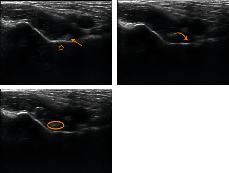
UltrasoundDynamic hip ultrasound in three stages: the iliopsoas tendon moves away from the bone then snaps abruptly back against it on return to the neutral position, the mechanism of internal snapping hip.
Source : Piechota et al., Journal of Ultrasonography, 2016, figure 2 · CC BY-NC-ND
The classic description is the one still taken up by the radiological literature: the internal form is "attributed to a sudden movement of the iliopsoas tendon against the iliopectineal eminence".² That is the taught version, the one that justifies most psoas stretching protocols.
In 2008, Deslandes et al. did something simple and rare: they reviewed the video recordings of the dynamic ultrasound scans used to make the diagnosis in 18 snapping iliopsoas tendons in 14 patients (9 women, 5 men, 13 to 50 years old), with the probe placed obliquely transverse just above the joint, parallel to the pubis. The triggering movement was the same in all of them: bringing the hip back from flexion-abduction-external rotation towards neutral.¹
What dynamic ultrasound actually films in internal snapping
18 snapping iliopsoas tendons in 14 patients: mechanisms identified in real time
Source: Deslandes M, Guillin R, Cardinal E, Hobden R, Bureau NJ. The snapping iliopsoas tendon: new mechanisms using dynamic sonography. AJR Am J Roentgenol 2008;190(3):576-581. PMID 18287424. Series of 14 patients referred for snapping: small numbers, but a direct measurement of the mechanism.
The authors' conclusion is clear-cut: "sudden flipping of the iliopsoas tendon over the iliacus muscle was the most frequent cause of snapping hip".¹ The tendon does not cross a fixed bony prominence: it changes side relative to the muscle belly of the iliacus, then strikes the pubic bone.
This nuance changes two things in practice. First, the most reliable provocation manoeuvre is not pure extension, it is the return from the flexion-abduction-external rotation position: the very one used in every patient of the series. Second, a stretching programme designed to "lift the psoas off the eminence" targets a geometry that the image does not confirm; the reasoning has to move towards control of the movement at the critical passage rather than towards the length of the tendon.
The iliopsoas tendon is not one tendon: it is often two, sometimes three
The Deslandes series found three snaps out of eighteen caused by two tendinous heads flipping over one another.¹ This is not a curiosity: it is the anatomical norm.
A cadaveric study published in 2022 dissected the iliopsoas complex unilaterally in 28 specimens (13 men, 15 women, mean age 85.6 years). It found 12 single, 12 double and 4 triple tendinous insertions, that is multiple tendons in more than half of the subjects.⁴ When they exist, these tendons insert separately on the lesser trochanter. The total width of the psoas tendon decreases as their number rises: 14.6 ± 2.2 mm for a single tendon, 8.2 ± 3.0 mm for two, 5.9 ± 1.1 mm for three (p < 0.001). The iliopsoas is, moreover, the structure whose groin pathology is the most polymorphic: tendinopathy, bursitis, impingement and snapping all belong to the same musculotendinous complex.⁸ The authors also describe a muscular, non-tendinous insertion of the most lateral fibres of the iliacus on the anterior surface of the lesser trochanter and of the shaft: a variant not described until then.
Two reservations to keep in mind: this is level V evidence, and a mean age of 85.6 years is not that of the patients who consult for snapping. But the surgical consequence is real and documented elsewhere: a release that finds only one head leaves the other intact, and the snap comes back; that is exactly the case reported by Shu and Safran, developed in chapter 8.
External snapping: two structures, not one
External snapping is classically attributed to the greater trochanter being crossed by the iliotibial band.³ But surgery has shown that a second structure has to be counted in: the anterior border of the gluteus maximus tendon. Modern endoscopic series release one, the other, or both, and report complete resolution in both configurations.⁶
The consequence for palpation matters: depending on the structure involved, the ridge that jumps is felt more anteriorly and higher (band) or more posteriorly and lower (gluteus maximus). And a third situation exists, independent of the previous two: gluteal muscle contracture, an entity described mainly in the Asian literature, often a sequela of repeated intramuscular injections in childhood, which produces external snapping through a different mechanism, the band is not over-tensioned by the sporting movement, it is permanently retracted.¹⁰
This is also where the clinical neighbourhood tightens. The iliotibial band that rubs over the greater trochanter is the same structure that compresses the gluteus medius and minimus tendons against the bone, the central mechanism of gluteal tendinopathy described by Grimaldi et al.⁷ External snapping and greater trochanteric pain syndrome share a topography, a structure and an aggravating position: relative hip adduction. They can coexist, and persistent lateral hip pain in a patient who also snaps is not necessarily explained by the snapping.
Intra-articular snapping: rare, but the one that changes management
Yamamoto et al. studied 32 hips (30 patients, mean age 26 years) in whom the pain associated with the snap was transiently relieved by an intra-articular injection of local anaesthetic: a selection criterion that guarantees an articular origin. Radiography, arthrography, MRI and arthroscopy were compared.⁵
The result has two halves, and both of them count:
- Arthroscopy finds an acetabular labral tear in 80% of cases, in the 11 hips with a normal radiograph as well as in 15 of the 19 osteoarthritic hips. The other causes are an intra-articular loose body (2 hips), incongruity between the labrum and a deformed femoral head (2 hips), and a sequela of Perthes disease.
- MRI identified the cause of the snap in none of the 31 cases investigated. Zero out of thirty-one.
Two reading precautions: the study dates from 2005 and MRI sequences have progressed, notably with MR arthrography; and selection by the anaesthetic test mechanically enriches the series in articular pathology. But the clinical lesson holds: a normal MRI does not rule out intra-articular snapping, and faced with a deep picture, with locking, in a young subject, the absence of any abnormality on MRI must not close the work-up.
This is the direct link with femoroacetabular impingement: the labral tear is its commonest associated lesion, and a hip that snaps deep in a young athlete with signs of impingement calls for the same diagnostic approach.
The rare causes, and why they have to be known
They are met only now and then, but each one caught out the team that reported it:
- Ischiofemoral impingement. Narrowing of the space between the ischium and the lesser trochanter, with quadratus femoris oedema on MRI, can produce an audible snap.¹¹ The measurement thresholds are established by meta-analysis: ischiofemoral space ≤ 15 mm (Se 76.9%, Sp 81.0%), quadratus femoris space ≤ 10 mm (Se 78.7%, Sp 74.1%).¹²
- Calcific tendinitis of the rectus femoris, reported as a cause of snapping hip.⁹
- Constitutional hypermobility. Coxa saltans has been described in patients with Ehlers-Danlos syndrome type III¹³: the question of hypermobility is worth asking in the face of a bilateral, voluntary, early-onset snap.
- After total hip arthroplasty, the picture changes in nature: the iliopsoas tendon can catch on an overhanging cup. This is no longer an athlete's snap, it is an implant-tendon impingement, developed in our article on iliopsoas impingement.
- Internal snapping does not cross the iliopectineal eminence: in 14 cases out of 18 filmed, the tendon flips around the iliacus muscle and strikes the pubic bone.
- The iliopsoas has several tendons in more than half of subjects: the first explanation for a snap that persists after release.
- External snapping implicates the iliotibial band and the gluteus maximus tendon; it is the same structure that compresses the gluteal tendons in greater trochanteric pain syndrome.
- In intra-articular snapping, the cause is labral 4 times out of 5, and MRI did not see it once in 31 cases in the landmark series.
Bibliography, chapter 2
- Deslandes M, Guillin R, Cardinal E, Hobden R, Bureau NJ. The snapping iliopsoas tendon: new mechanisms using dynamic sonography. AJR Am J Roentgenol. 2008;190(3):576-581. PMID 18287424.
- Piechota M, Maczuch J, Skupiński J, Kukawska-Sysio K, Wawrzynek W. Internal snapping hip syndrome in dynamic ultrasonography. J Ultrason. 2016;16(66):296-303. PMID 27679733.
- Sugrañes J, Jackson GR, Warrier AA, Allahabadi S, Chahla J. Snapping Hip Syndrome: Pathoanatomy, Diagnosis, Nonoperative Therapy, and Current Concepts in Operative Management. JBJS Rev. 2023;11(6). PMID 37289915.
- Lin B, Bartlett J, Lloyd TD, Challoumas D, Brassett C, Khanduja V. Multiple iliopsoas tendons: a cadaveric study and treatment implications for internal snapping hip syndrome. Arch Orthop Trauma Surg. 2022;142(6):1147-1154. PMID 34347120.
- Yamamoto Y, Hamada Y, Ide T, Usui I. Arthroscopic surgery to treat intra-articular type snapping hip. Arthroscopy. 2005;21(9):1120-1125. PMID 16171638.
- Randelli F, Mazzoleni MG, Fioruzzi A, Giai Via A, Calvisi V, Ayeni OR. Surgical interventions for external snapping hip syndrome. Knee Surg Sports Traumatol Arthrosc. 2021;29(8):2386-2393. PMID 33064193.
- Grimaldi A, Mellor R, Hodges P, Bennell K, Wajswelner H, Vicenzino B. Gluteal Tendinopathy: A Review of Mechanisms, Assessment and Management. Sports Med. 2015;45(8):1107-1119. PMID 25969366.
- Anderson CN. Iliopsoas: Pathology, Diagnosis, and Treatment. Clin Sports Med. 2016;35(3):419-433. PMID 27343394.
- Pierannunzii L, Tramontana F, Gallazzi M. Case report: calcific tendinitis of the rectus femoris: a rare cause of snapping hip. Clin Orthop Relat Res. 2010;468(10):2814-2818. PMID 20054675.
- Dai Z, Chen Z, Liao Y, Tang Z, Cui J. Comparison of arthroscopic versus open surgery on external snapping hip caused by gluteal muscle contracture. Hip Int. 2018;28(2):173-177. PMID 29890911.
- Ali AM, Whitwell D, Ostlere SJ. Case report: imaging and surgical treatment of a snapping hip due to ischiofemoral impingement. Skeletal Radiol. 2011;40(5):653-656. PMID 21207021.
- Singer AD, Subhawong TK, Jose J, Tresley J, Clifford PD. Ischiofemoral impingement syndrome: a meta-analysis. Skeletal Radiol. 2015;44(6):831-837. PMID 25672947.
- Kaalund S, Høgsaa B, Grevy C. Coxa saltans in patients with Ehlers-Danlos syndrome, type III. Scand J Rheumatol. 1988;17(3):229-230. PMID 3175552. Case report with no indexed abstract: only the description carried by the title can be verified.
How do you tell internal, external and intra-articular snapping apart?
Four questions that do most of the diagnosis
1. Where exactly? Topography is the first discriminator, and the patient gives it spontaneously if asked to put a finger on it. At the front, in the groin crease: internal snapping. On the lateral aspect, over the bony prominence: external snapping. "Deep down", without the patient being able to point to it: intra-articular snapping or referred pain.
2. Does it hurt, or does it make a noise? The question sounds naive; it is the most useful one. A painless snap does not call for treatment (chapter 4). Pinning down the real limitation (sporting, occupational, social), avoids treating a worry with a protocol.
3. Can you trigger it yourself? In Winston, 60% of the dancers could trigger their snap voluntarily, and one or more of three dance movements provoked it in 81%.³ A snap reproducible on command is almost always extra-articular: the patient then becomes the best examiner in the room.
4. Does it snap, or does it lock? A snap that does not stop the movement points to the extra-articular. True locking (the hip stays stuck and has to be freed), points to the intra-articular: loose body, unstable labral tear, incongruity.⁴
The tests, and what they are really worth
The most important point first: there is no study of the diagnostic value of clinical tests for "snapping hip" as such. The available figures concern iliopsoas-related groin pain, a neighbouring but broader entity, and they are fragile.
The 2026 systematic review of iliopsoas-related groin pain retained three diagnostic accuracy studies. Three. All based on guided injection as the reference standard, and all at high risk of bias according to QUADAS-3.¹ In those conditions, the hip-external rotation-flexion-ceiling (HEC) test and resisted hip flexion in sitting come out with the best performance: Se 94%, Sp 88% for HEC. The authors themselves call these estimates preliminary. The supine provocation tests, for their part, have high sensitivity and mediocre specificity: they rule out, they do not confirm.
| Test | How it is done | What it brings | Level of evidence |
|---|---|---|---|
| FABER manoeuvre → neutral | From flexion-abduction-external rotation, bring the hip back to neutral, actively; this is the manoeuvre described for examining the dancer⁹ | It is the movement that triggered the snap in all 14 patients of the landmark ultrasound series.⁶ Reproduction of the snap in the groin = internal snapping | Moderate (documented mechanism, diagnostic value not quantified) |
| HEC test (hip-external rotation-flexion-ceiling) | Provocation in external rotation, flexion, against the ceiling | Se 94% / Sp 88% for iliopsoas-related groin pain: 3 studies, all at high risk of bias | Low (estimate called preliminary by the authors)¹ |
| Resisted hip flexion, sitting | Resisted isometric contraction in the sitting position | Best performance along with HEC in the same review; reproduces the tendon pain more than the snap itself | Low (same 3 studies)¹ |
| Snap triggered by the patient | Standing, the patient reproduces their own snap | 60% of dancers manage it; 46 of the 50 self-reported snaps were palpable by the clinician³ | Moderate (dancer cohort, n = 50 hips) |
| Palpation of the greater trochanter during standing flexion-extension | Hand on the trochanteric prominence, hip in flexion-extension, patient weight-bearing | External snapping: the band or the gluteus maximus tendon is felt crossing the bone. External snapping is "obvious on physical examination and ultrasound"³ | Moderate |
| Intra-articular anaesthetic test | Guided intra-articular injection of local anaesthetic | Inclusion criterion of the landmark series on intra-articular snapping⁴; recommended before any tendon release, to avoid a surgical failure¹⁰ | Moderate (reference standard by default, for want of better) |
| Femoroacetabular impingement tests (FADIR) | Flexion-adduction-internal rotation | A negative FADIR helps rule out intra-articular hip pain; positive, it designates no structure⁷ | Moderate (Zurich 2018 consensus) |
The decision tree
Sorting out a snapping hip
The first fork is not anatomical: it is symptomatic
Tree built for this article from Winston 2007 (PMID 17021311), Deslandes 2008 (PMID 18287424), Yamamoto 2005 (PMID 16171638), Walker 2021 (PMID 34745476) and Frans-Jozef 2026 (doi:10.3390/jcm15155912). No prospectively validated algorithm exists for this sorting: this tree organises the data, it does not replace them.
Imaging: the right one, in the right order
A reflex has to be reversed. Faced with a painful hip, the spontaneous request is MRI. Yet for the extra-articular forms, dynamic ultrasound is the reference examination, and MRI sees nothing of what happens in movement.²
What each examination brings, and what it misses
Reported performance, with the level of evidence that goes with it
Sources: Winston 2007 PMID 17021311 · Yamamoto 2005 PMID 16171638 · Frans-Jozef 2026 doi:10.3390/jcm15155912. The four rows do not measure the same thing: the first is a rate of aetiological identification, the second a sensitivity, the third a rate of palpatory agreement, the fourth a rate of identification as well.
Dynamic ultrasound. It is the standard for the two extra-articular forms.² It films the mechanism in real time, without radiation, and allows associated lesions to be detected: bursitis, tendinopathy. In Winston, it showed a snapping iliopsoas tendon in 59% of hips and a band in 4%, but in a third of cases it did not allow the cause to be identified.³ It is operator-dependent, and it requires the patient to be able to trigger their snap on the couch.
MRI. Its role is to look for something else: concomitant intra-articular pathology, an anatomical variant liable to change a surgical technique, guidance for an injection.⁵ It is not the examination that makes the diagnosis of the snap: the history and physical examination most often suggest the right diagnosis, and imaging only confirms it and rules out the others.⁵·¹¹ And it missed 31 intra-articular snaps out of 31 in the landmark series.⁴
Radiographs. Normal in the internal form.² They keep their place for what they rule out: dysplasia, impingement morphology, osteoarthritis, slipped epiphysis in the adolescent. The Zurich consensus recommends an anteroposterior pelvic radiograph and a lateral view of the head and neck as first-line imaging for hip pain in the young subject, advanced imaging coming in only for an additional detail.⁷
Differential diagnosis: what snaps is not always what hurts
The central trap of this subject is attribution. A patient can have a perfectly genuine snap and pain that comes from elsewhere. The Doha consensus recalls that groin pain in the athlete is distributed between several entities defined by their structure (adductors, iliopsoas, inguinal region, pubis, hip), which frequently coexist.⁸
| To consider | What should raise it | How it is settled |
|---|---|---|
| Greater trochanteric pain syndrome | Lateral pain, waking on lying on that side, pain on single-leg stance, in a patient who also snaps | Compression of the gluteal tendons by the band is the mechanism; see our dedicated article. The snap does not necessarily explain the pain |
| Femoroacetabular impingement | Deep groin pain, C sign, limited internal rotation, young athlete | Radiographs and impingement tests; the associated labral tear is also the leading cause of intra-articular snapping |
| Iliopsoas tendinopathy without snapping | Groin pain on resisted flexion, without any snap | This is the pathology of the tendon, not the mechanical phenomenon; dedicated article on iliopsoas impingement |
| Ischiofemoral impingement | Low buttock pain, discomfort in extension-adduction, middle-aged woman | MRI: ischiofemoral space ≤ 15 mm (Se 76.9% / Sp 81.0%), quadratus femoris oedema¹³ |
| Adductor pathology, groin pain | More medial pain, positive squeeze test | Doha classification; palpation of the pubic insertion⁸ |
| Tumoural or synovial lesion | Snap of recent onset with no triggering factor, a mass, a course over several months | Dynamic ultrasound then MRI: one case of localised tenosynovial giant cell tumour took 9 months to be diagnosed¹² |
| Slipped capital femoral epiphysis | Adolescent, limp, hip or knee pain | Urgent radiographs; never attribute hip pain in an adolescent to a snap without having them |
- Four questions do the sorting: where, does it hurt or make a noise, can you trigger it, does it snap or does it lock.
- There is no study of the diagnostic value of the tests for the snap itself; the figures quoted (HEC: Se 94%, Sp 88%) concern iliopsoas-related pain and come from 3 studies at high risk of bias.
- The reference examination for the extra-articular forms is dynamic ultrasound, not MRI, but it stays silent in a third of cases.
- A normal MRI does not rule out intra-articular snapping.
Bibliography, chapter 3
- Frans-Jozef V, Abner S, Stijn R, Annick T, Kristoff C. Clinical Diagnosis and Treatment of Iliopsoas-Related Groin Pain: A Systematic Review. J Clin Med. 2026;15(15):5912. doi:10.3390/jcm15155912. PMID 42590015.
- Piechota M, Maczuch J, Skupiński J, Kukawska-Sysio K, Wawrzynek W. Internal snapping hip syndrome in dynamic ultrasonography. J Ultrason. 2016;16(66):296-303. PMID 27679733.
- Winston P, Awan R, Cassidy JD, Bleakney RK. Clinical examination and ultrasound of self-reported snapping hip syndrome in elite ballet dancers. Am J Sports Med. 2007;35(1):118-126. PMID 17021311.
- Yamamoto Y, Hamada Y, Ide T, Usui I. Arthroscopic surgery to treat intra-articular type snapping hip. Arthroscopy. 2005;21(9):1120-1125. PMID 16171638.
- Le J. Snapping Hip Syndrome: Imaging Diagnosis. Magn Reson Imaging Clin N Am. 2025;33(1):75-82. PMID 39515962.
- Deslandes M, Guillin R, Cardinal E, Hobden R, Bureau NJ. The snapping iliopsoas tendon: new mechanisms using dynamic sonography. AJR Am J Roentgenol. 2008;190(3):576-581. PMID 18287424.
- Reiman MP, Agricola R, Kemp JL, et al. Consensus recommendations on the classification, definition and diagnostic criteria of hip-related pain in young and middle-aged active adults, Zurich 2018. Br J Sports Med. 2020;54(11):631-641. PMID 31959678.
- Weir A, Brukner P, Delahunt E, et al. Doha agreement meeting on terminology and definitions in groin pain in athletes. Br J Sports Med. 2015;49(12):768-774. PMID 26031643.
- Nolton EC, Ambegaonkar JP. Recognizing and Managing Snapping Hip Syndrome in Dancers. Med Probl Perform Art. 2018;33(4):286-291. PMID 30508831.
- Coulomb R, Nougarede B, Maury E, Marchand P, Mares O, Kouyoumdjian P. Arthroscopic iliopsoas tenotomies: a systematic review of surgical technique and outcomes. Hip Int. 2022;32(1):4-11. PMID 33226846.
- Sugrañes J, Jackson GR, Warrier AA, Allahabadi S, Chahla J. Snapping Hip Syndrome: Pathoanatomy, Diagnosis, Nonoperative Therapy, and Current Concepts in Operative Management. JBJS Rev. 2023;11(6). PMID 37289915.
- Vervaecke AJ, Declercq H, Wetzels K, van den Broek M. Localized tenosynovial giant cell tumor: a rare case of snapping hip. Skeletal Radiol. 2022;51(11):2205-2210. PMID 35536359.
- Singer AD, Subhawong TK, Jose J, Tresley J, Clifford PD. Ischiofemoral impingement syndrome: a meta-analysis. Skeletal Radiol. 2015;44(6):831-837. PMID 25672947.
Which snaps must above all not be treated?
A painless snap is not a pathology
The literature is explicit, and has been for a long time. Randelli et al. write that external snapping "is usually asymptomatic", and reserve the term syndrome for the painful snap.¹ Walker et al. state that "treatment is recommended for symptomatic snapping".³ Sugrañes et al. make pain the tipping criterion: "when pain becomes the main symptom, it is called snapping hip syndrome".⁷
In other words: the very definition of the disease includes pain. A patient who snaps and does not hurt does not have the disease. They have a hip that makes a noise.
What the patient is really coming for
A hip that snaps loudly is worrying, and the worry is the real reason for consulting in a good share of cases. Three fears recur, and they can be answered:
- "Is it wearing out?" No data link extra-articular snapping to joint wear. The mechanism of internal snapping happens outside the joint, between a tendon, a muscle and the pubic bone.
- "Is it going to get worse?" In symptomatic patients, most cases resolve after 6 to 12 months of conservative management.³ Painless snapping is otherwise described as common in the general population, the disabling form remaining the preserve of very demanding activities such as ballet or hurdling.² For asymptomatic patients, no natural history study exists, which, said as such, is more honest than promising one thing or the other.
- "Do I have to stop sport?" No, in the absence of pain. In dancers, stopping the activity concerns 7% of the cohort, those whose snap was painful and disabling.⁴
Management is then a consultation of explanation: name the structure, show the movement that triggers it, explain that the noise comes from an abrupt change of position and not from destructive rubbing, and set a criterion for coming back, "if it becomes painful, if it locks, if it changes in nature, we will look at it again". Nothing more.
The opposite trap: treating the snap instead of the pain
A visible, noisy snap captures the attention of the patient and of the clinician alike. The risk is to attribute to it a pain that comes from elsewhere, and to rehabilitate, or operate on, a structure that is not guilty.
The 2026 systematic review of iliopsoas-related groin pain is blunt on this point: in the native hip, four matched comparative cohorts show that arthroscopic iliopsoas release brings no clear benefit beyond treating the associated intra-articular pathology, while being accompanied by measurable atrophy and a loss of flexion strength.⁵ That atrophy is not anecdotal: it is found on imaging in 92.4% of the hips checked.⁸ The tendon was snapping, it was cut, and the improvement came from elsewhere.
The Zurich consensus sets the same requirement in a broader frame: faced with hip pain in the young and middle-aged subject, non-musculoskeletal and serious causes must be ruled out, then competing conditions, the lumbar spine in particular, before a local explanation is accepted.⁶ The snap exempts from nothing.
When a hitherto ordinary snap stops being ordinary
- It changes in nature: a snap that becomes a catch, or a hip that truly locks → intra-articular reassessment.
- It appears for no reason in an adult who has changed nothing in their activity, and settles in over several months → think of the rare lesional causes before concluding that it is mechanical.
- It comes with night pain, pain at rest, or systemic repercussions → step outside the mechanical frame.
- It occurs after a total hip arthroplasty → this is no longer the same problem: the tendon can catch on an overhanging cup, and management goes through assessment of the implant position.
- It comes with a strength deficit that is progressing, rather than simple discomfort → look for a neurological lesion or a tendon rupture.
- Pain is part of the definition of the syndrome. No pain, no syndrome, no treatment.
- The useful consultation for a painless snap is a consultation of explanation, with a clear criterion for coming back.
- The symmetrical risk is to attribute to the snap a pain that came from elsewhere: four matched cohorts show that cutting the tendon adds nothing to the treatment of the associated intra-articular lesion.
- No data link extra-articular snapping to joint degradation, and none has followed the natural history of asymptomatic snaps either.
Bibliography, chapter 4
- Randelli F, Mazzoleni MG, Fioruzzi A, Giai Via A, Calvisi V, Ayeni OR. Surgical interventions for external snapping hip syndrome. Knee Surg Sports Traumatol Arthrosc. 2021;29(8):2386-2393. PMID 33064193.
- Potalivo G, Bugiantella W. Snapping hip syndrome: systematic review of surgical treatment. Hip Int. 2017;27(2):111-121. PMID 28222210.
- Walker P, Ellis E, Scofield J, Kongchum T, Sherman WF, Kaye AD. Snapping Hip Syndrome: A Comprehensive Update. Orthop Rev (Pavia). 2021;13(2):25088. PMID 34745476.
- Winston P, Awan R, Cassidy JD, Bleakney RK. Clinical examination and ultrasound of self-reported snapping hip syndrome in elite ballet dancers. Am J Sports Med. 2007;35(1):118-126. PMID 17021311.
- Frans-Jozef V, Abner S, Stijn R, Annick T, Kristoff C. Clinical Diagnosis and Treatment of Iliopsoas-Related Groin Pain: A Systematic Review. J Clin Med. 2026;15(15):5912. doi:10.3390/jcm15155912.
- Reiman MP, Agricola R, Kemp JL, et al. Consensus recommendations on the classification, definition and diagnostic criteria of hip-related pain in young and middle-aged active adults, Zurich 2018. Br J Sports Med. 2020;54(11):631-641. PMID 31959678.
- Sugrañes J, Jackson GR, Warrier AA, Allahabadi S, Chahla J. Snapping Hip Syndrome: Pathoanatomy, Diagnosis, Nonoperative Therapy, and Current Concepts in Operative Management. JBJS Rev. 2023;11(6). PMID 37289915.
- Gouveia K, Shah A, Kay J, Memon M, Simunovic N, Cakic JN. Iliopsoas Tenotomy During Hip Arthroscopy: A Systematic Review of Postoperative Outcomes. Am J Sports Med. 2021;49(3):817-829. PMID 32628861.
What rehabilitation should be offered when the snap hurts?
The real state of the evidence
All the reviews recommend conservative treatment first.³·⁴·⁸ The recommended content is stable throughout: anti-inflammatories, stretching and avoidance of the triggering movement.⁹ This unanimity must not be confused with a high level of evidence: it reflects an expert consensus in a field that has been little studied.
For external snapping, the only prospective study identified is a Danish feasibility trial published in 2022. Nine women with symptomatic external snapping followed a progressive strengthening programme. Three dropped out, that is a third of the sample. Among those who finished, adherence was 97 ± 4%, pain stayed acceptable (VAS ≤ 50 mm) in 76% of sessions, overall pain fell during the programme (−10 mm, 95% CI [−3; −18]), and every HAGOS subscale, the Forgotten Joint Score, strength and a loaded stair test improved.¹ The authors conclude that progressive strengthening "seems feasible in some patients". There is no control group: what time alone would have given is not known.
For internal snapping, no study isolates it. The closest data come from the 2026 systematic review of iliopsoas-related groin pain, which reports conservative treatment success rates of 77 to 100% in idiopathic and athletic populations, against 16 to 50% after total hip arthroplasty.⁵ The methodological quality of the 71 therapeutic studies included is judged "overall low to moderate" (MINORS 5-14/16 for the non-comparative studies).
Treatment modalities and certainty of the evidence
GRADE reading grid applied by us: no published GRADE assessment exists on this subject
Ranking established for this article by applying GRADE logic to the verified sources: Kjeldsen 2022 PMID 32643987 · Frans-Jozef 2026 doi:10.3390/jcm15155912 · Gouveia 2021 PMID 32628861 · Giai Via 2024 PMID 38874780 · Walker 2021 PMID 34745476. No formal GRADE assessment has been published on this subject: this grid is a reading, not a citation.
| Modality | What the source says | Sample and study type | Certainty |
|---|---|---|---|
| Progressive weight-bearing strengthening (external snapping) | Feasible; improvements in HAGOS, the Forgotten Joint Score, strength and the loaded stair test; pain down by 10 mm during the programme | 9 women, feasibility, 33% dropouts, no control group¹ | Low |
| Conservative treatment (iliopsoas-related pain) | Success 77-100% in idiopathic and athletic populations; 16-50% after total hip arthroplasty | 71 therapeutic studies, low to moderate quality (MINORS)⁵ | Low |
| Activity adaptation, active rest | "Active rest with training modification should be tried to limit worsening" | Narrative review, dancers² | Very low |
| Education and explanation | Not evaluated on its own; the corollary of chapter 4 is that it is sometimes the only treatment needed | No direct study | Very low |
| Iliopsoas stretching | Recommended by the review articles; the geometry they target is not the one dynamic ultrasound films | Narrative reviews; mechanism contradicted by a series of 18 hips⁸ | Very low |
| Manual therapy and mobilisations | High-grade joint mobilisation + exercise: clinically important improvements maintained at 6 months | 1 case, man of 72, after a failed tenotomy⁷ | Very low |
| Shockwave therapy, PRP | No study identified on snapping hip | Not applicable | No data |
What is actually done, and on what reasoning
For want of a controlled trial, the content of the programme rests on an explicit mechanical rationale, which has to be owned as such in front of the patient.
Phase 1: reduce the provocation, not the activity. Identify precisely the movement that triggers the painful snap: in the dancer, one or more of three movements is enough to provoke it in 81% of cases.⁴ Remove it temporarily, or modify its end range, without stopping the overall activity. Complete rest is recommended nowhere.
Phase 2: load the tendon concerned. For internal snapping, that means working the iliopsoas in resisted flexion over a controlled range, including beyond 90° where the muscle is most demanded and where the post-surgical deficits are most marked. For external snapping, the Danish programme worked on progressive strengthening of the hip muscles, with a loaded stair test as the functional criterion.¹
Phase 3: take the triggering movement back under control. The documented mechanism of internal snapping is an abrupt flip of the tendon on the return from flexion-abduction-external rotation.⁸ The work therefore bears on the speed and control of that passage rather than on lengthening the tendon: slow it down, break it up, then restore the speed.
What is measured. The HAGOS (Copenhagen Hip and Groin Outcome Score) was the tool of the Danish study and exists in French; the Forgotten Joint Score targets precisely the awareness of the joint, which suits a patient bothered by a noise. A loaded functional test (stairs, step-up), gives an objective marker that the patient understands. For comparison, the neighbouring pathology of the greater trochanter does have a randomised controlled trial showing the superiority of education combined with exercise over corticosteroid injection and over watchful waiting⁶: that is the level of evidence snapping hip does not have.
How long before concluding failure
The reviews converge: most symptomatic cases resolve after 6 to 12 months of conservative management.³ It is also the delay after which surgery is considered in the published series.
Three months is a reasonable delay to reassess the trajectory, not to conclude. A patient improving on their functional scores without the snap having disappeared is not a failure: the snap is not the target, pain and function are. Many patients keep their snap and stop suffering from it.
- Rehabilitation is recommended as first line everywhere, on an evidence base that is very thin: a single prospective study (n = 9, a third of dropouts) for external snapping, none isolating internal snapping.
- Conservative treatment success rates differ radically according to context: 77-100% in the native hip, 16-50% after arthroplasty.
- The target of treatment is pain and function, never the disappearance of the noise.
- Reference delay before considering anything else: 6 to 12 months, not six weeks.
Bibliography, chapter 5
- Kjeldsen T, Reimer LU, Drejer SM, Hvid LG, Mechlenburg I, Dalgas U. Is progressive resistance training feasible in patients with symptomatic external snapping hip? Physiother Theory Pract. 2022;38(5):704-716. PMID 32643987.
- Nolton EC, Ambegaonkar JP. Recognizing and Managing Snapping Hip Syndrome in Dancers. Med Probl Perform Art. 2018;33(4):286-291. PMID 30508831.
- Walker P, Ellis E, Scofield J, Kongchum T, Sherman WF, Kaye AD. Snapping Hip Syndrome: A Comprehensive Update. Orthop Rev (Pavia). 2021;13(2):25088. PMID 34745476.
- Winston P, Awan R, Cassidy JD, Bleakney RK. Clinical examination and ultrasound of self-reported snapping hip syndrome in elite ballet dancers. Am J Sports Med. 2007;35(1):118-126. PMID 17021311.
- Frans-Jozef V, Abner S, Stijn R, Annick T, Kristoff C. Clinical Diagnosis and Treatment of Iliopsoas-Related Groin Pain: A Systematic Review. J Clin Med. 2026;15(15):5912. doi:10.3390/jcm15155912.
- Mellor R, Bennell K, Grimaldi A, et al. Education plus exercise versus corticosteroid injection use versus a wait and see approach on global outcome and pain from gluteal tendinopathy: prospective, single blinded, randomised clinical trial. BMJ. 2018;361:k1662. PMID 29720374.
- Peterson S. Physical therapy management of a patient with persistent groin pain after total hip arthroplasty and iliopsoas tenotomy: a case report. Physiother Theory Pract. 2022;38(3):481-491. PMID 32544015.
- Deslandes M, Guillin R, Cardinal E, Hobden R, Bureau NJ. The snapping iliopsoas tendon: new mechanisms using dynamic sonography. AJR Am J Roentgenol. 2008;190(3):576-581. PMID 18287424.
- Yen YM, Lewis CL, Kim YJ. Understanding and Treating the Snapping Hip. Sports Med Arthrosc Rev. 2015;23(4):194-199. PMID 26524554.
What are injection and surgery worth when rehabilitation fails?
The injection: a diagnostic tool before it is a therapeutic one
Its main value is not to relieve, it is to designate the structure. The systematic review by Coulomb et al. explicitly recommends a diagnostic injection test before any tendon release, so as not to operate on an innocent tendon.⁴ It is also that criterion, transient relief of the pain by an intra-articular anaesthetic, that was used to assemble the landmark series on intra-articular snapping.
On the therapeutic side, the results are modest and brief: the 2026 systematic review reports short-term improvement in 50 to 70% of patients, with limited lasting efficacy, particularly in the mechanical cases after arthroplasty.¹ Injection does not settle a mechanical impingement; it buys time and provides information.
Iliopsoas tenotomy: what it settles, and what it costs
Let us start with what works. The systematic review by Gouveia et al. brings together 21 studies, 824 patients and 875 hips, of whom 82.5% were women, mean age 28.1 years, with a mean follow-up of 32.1 months. The success rate for resolution of the snap is 93.0% (266/286), and every study reports an improvement in functional scores.²
The comparison between approaches leans clearly towards arthroscopy. Khan et al., across 11 studies and 248 patients: resolution of the snap in 100% of patients operated on arthroscopically against 77% with open surgery, and complications 2.3% against 21%.³ The authors add, and this matters, that "in the absence of high-quality data or of a direct comparison, these results must be interpreted with caution".³ The most recent review article makes the same observation, specifying that the advantage of endoscopy over the open approach is clear for internal snapping and much less marked for external snapping.¹³
Now the price.
What tenotomy leaves behind
Flexion strength on the operated side, relative to the healthy side (symmetry index): the shorter the bar, the greater the deficit
Sources: Twardy 2026 PMID 41205716 · Portet 2024 PMID 38833166 · Gouveia 2021 PMID 32628861 · Loppini 2026 PMID 41669375 · Randelli 2021 PMID 34757530.
Atrophy is almost constant. In Gouveia, iliopsoas atrophy was assessed radiologically in 3 studies (66 hips) and found in 92.4% of them.² Only six studies, covering 93 hips, measured flexion strength: four report full recovery, two moderate falls.
The most recent series, which measure with a dynamometer, are less reassuring. Twardy et al. tested 31 patients operated on with endoscopic tenotomy for iliopsoas impingement after arthroplasty: pain collapses (VAS 8.3 → 2.9), but flexion strength on the operated side is reduced by 32.4%, that is a symmetry index of 67.4% at 37 months of follow-up.⁷ In Portet et al., across 36 tenotomies, the symmetry index is 63% at 90° of flexion and 40% at 30°.⁸ At ten years of follow-up, Loppini et al. find 15% flexion weakness with atrophy on MRI, and 15% persistent snapping despite the tenotomy.⁶
The remarkable fact is that satisfaction remains high: 26 of Twardy's 31 patients would have the operation again. The strength deficit does not necessarily translate into perceived discomfort, which does not license keeping quiet about it: the authors conclude that it "should be discussed during preoperative counselling".
The difficult question: what if the snap was not the problem?
This is the most disturbing result in the recent literature, and it comes from two independent places.
Perets et al. followed 57 patients operated on with fractional lengthening of the iliopsoas during arthroscopy for femoroacetabular impingement and labral tear, with a mean follow-up of 69.3 months. The painful snap disappeared in 80.7% of them, the scores improved, but the results do not differ from those of a matched control group operated on without lengthening, because it had no snapping.⁵ In other words, those patients got better like the others, and the improvement cannot be attributed to the procedure on the tendon.
The 2026 systematic review goes further by pooling four matched comparative cohorts in the native hip: arthroscopic iliopsoas release brings no clear benefit beyond treating the associated intra-articular pathology, and is accompanied by measurable atrophy and loss of strength.¹
The practical consequence is direct: faced with painful internal snapping associated with femoroacetabular impingement or a labral tear, the surgical discussion must bear on the articular lesion; that is the subject of our article on femoroacetabular impingement, where the FASHIoN trial compared arthroscopy with personalised conservative care.¹⁴ The tendon is not the priority target.
External snapping: simpler surgery, better tolerated
The contrast is stark. The systematic review by Giai Via et al. brings together 9 studies, 403 patients and 689 hips treated endoscopically: recurrence 1.02%, revision 0.15%, minimal complications.⁹ The techniques consist in releasing the iliotibial band, performing a gluteus maximus tenotomy, or both.
Randelli et al. measured what release of the gluteus maximus tendon costs in 22 patients: resolution of the snap in 100%, VAS from 6.8 to 0.6, modified Harris hip score from 48.6 to 88.2, and above all no significant difference in gluteus maximus strength, despite a 3.7% reduction in thigh circumference whose significance the authors themselves say remains to be determined.¹⁰ The historical series by Ilizaliturri, with endoscopic diamond-shaped release of the band in 11 hips, reported at two years a single residual painless snap, the other patients having returned to their previous level.¹¹
When painful external snapping coexists with femoroacetabular impingement, release of the gluteus maximus and of the band during arthroscopy gives 100% resolution of the snap and two-year results comparable to a matched control group without snapping.¹²
| Procedure | Resolution of the snap | Documented functional cost | Certainty |
|---|---|---|---|
| Arthroscopic iliopsoas tenotomy | 93.0% (266/286 hips)² | Atrophy in 92.4% of the hips imaged; strength symmetry index 63-67% in the recent dynamometric series⁷·⁸ | Low (systematic reviews of level IV studies) |
| Open approach for internal snapping | 77%³ | Complications 21% against 2.3% with arthroscopy³ | Low, and unfavourable |
| Endoscopic release, external snapping | Recurrence 1.02%, revision 0.15% across 689 hips⁹ | No loss of gluteus maximus strength found; thigh circumference −3.7%¹⁰ | Low (level IV series, no randomised trial) |
| Iliopsoas lengthening combined with arthroscopy for impingement | 80.7% at 5 years⁵ | Results identical to the control group without snapping: no benefit of its own demonstrated⁵ | Low: additional benefit not established |
| Tenotomy after total hip arthroplasty | Significant improvement in scores⁷·⁸ | Flexion strength deficit of 32% at 37 months; cup overhang > 8 mm: consider acetabular revision instead⁴ | Low |
- The injection serves first to designate the structure; recommended before any release, it relieves 50 to 70% of patients, in the short term only.
- Iliopsoas tenotomy abolishes the snap in 93% of cases, at the price of atrophy in 92% of the hips imaged and a third of flexion strength lost in the dynamometric series.
- When an intra-articular lesion coexists, the benefit of tendon release in its own right is not demonstrated.
- The external snap is treated better: 1% recurrence across 689 hips, with no documented loss of strength.
Bibliography, chapter 6
- Frans-Jozef V, Abner S, Stijn R, Annick T, Kristoff C. Clinical Diagnosis and Treatment of Iliopsoas-Related Groin Pain: A Systematic Review. J Clin Med. 2026;15(15):5912. doi:10.3390/jcm15155912.
- Gouveia K, Shah A, Kay J, Memon M, Simunovic N, Cakic JN. Iliopsoas Tenotomy During Hip Arthroscopy: A Systematic Review of Postoperative Outcomes. Am J Sports Med. 2021;49(3):817-829. PMID 32628861.
- Khan M, Adamich J, Simunovic N, Philippon MJ, Bhandari M, Ayeni OR. Surgical management of internal snapping hip syndrome: a systematic review evaluating open and arthroscopic approaches. Arthroscopy. 2013;29(5):942-948. PMID 23510943.
- Coulomb R, Nougarede B, Maury E, Marchand P, Mares O, Kouyoumdjian P. Arthroscopic iliopsoas tenotomies: a systematic review of surgical technique and outcomes. Hip Int. 2022;32(1):4-11. PMID 33226846.
- Perets I, Chaharbakhshi EO, Mansor Y, et al. Midterm Outcomes of Iliopsoas Fractional Lengthening for Internal Snapping as a Part of Hip Arthroscopy for Femoroacetabular Impingement and Labral Tear: A Matched Control Study. Arthroscopy. 2019;35(5):1432-1440. PMID 31000391.
- Loppini M, Minelli M, Chiappetta K, La Camera F, Grappiolo G, Della Rocca F. Outcomes of endoscopic transcapsular iliopsoas tenotomy for snapping hip syndrome: Minimum 10-year follow-up. J Exp Orthop. 2026;13(1):e70543. PMID 41669375.
- Twardy V, Rösch MBJ, Pohlig F, von Eisenhart-Rothe R, Banke IJ. High Satisfaction Despite 32% Persistent Hip Flexor Weakness After Endoscopic Tenotomy for Iliopsoas Impingement Following Total Hip Arthroplasty. J Arthroplasty. 2026;41(7):2184-2191. PMID 41205716.
- Portet A, Repellin M, Lambrey PJ, Benhenneda R, Vieira TD, Thaunat M. Assessment of function and muscle strength after endoscopic iliopsoas tenotomy to treat iliopsoas impingement after total hip arthroplasty. Int Orthop. 2024;48(9):2375-2382. PMID 38833166.
- Giai Via R, Elzeiny A, Pantè S, De Vivo S, Massè A, Giachino M. Can we encourage the endoscopic treatment for external snapping hip (ESH)? A systematic review of current concepts. Eur J Orthop Surg Traumatol. 2024;34(6):2835-2844. PMID 38874780.
- Randelli F, Fioruzzi A, Magnani M, Mazzoleni M, Elhiny M, Via AG. Endoscopic gluteus maximus tendon release for external snapping hip syndrome: a functional assessment. J Orthop Traumatol. 2021;22(1):45. PMID 34757530.
- Ilizaliturri VM Jr, Martinez-Escalante FA, Chaidez PA, Camacho-Galindo J. Endoscopic iliotibial band release for external snapping hip syndrome. Arthroscopy. 2006;22(5):505-510. PMID 16651159.
- Maldonado DR, Glein RM, Lee MS, et al. Patients With Concomitant Painful External Snapping Hip and Femoroacetabular Impingement Syndromes Reported Complete Snapping Resolution With Release of the Gluteus Maximus and Iliotibial Band. Arthroscopy. 2022;38(6):1890-1899. PMID 34920010.
- Sugrañes J, Jackson GR, Warrier AA, Allahabadi S, Chahla J. Snapping Hip Syndrome: Pathoanatomy, Diagnosis, Nonoperative Therapy, and Current Concepts in Operative Management. JBJS Rev. 2023;11(6). PMID 37289915.
- Griffin DR, Dickenson EJ, Wall PDH, et al. Hip arthroscopy versus best conservative care for the treatment of femoroacetabular impingement syndrome (UK FASHIoN): a multicentre randomised controlled trial. Lancet. 2018;391(10136):2225-2235. PMID 29893223.
What changes in a population where almost every hip snaps?
A sign present in 9 out of 10 no longer sorts anything
Let us go back to Winston's data: of 87 unselected elite ballet dancers, 91% report a snapping hip, 80% of them bilaterally, 60% know how to trigger it voluntarily.¹ In this population, finding a snap conveys no information: it is the norm.
What the clinician has to look for is therefore not the snap, but what distinguishes this particular dancer: the associated pain (58% of the dancers who snap), the real impact on practice (7% had to stop dancing), and above all everything that does not fit an ordinary mechanical snap.
Why dance produces this sign
The documented mechanism of internal snapping, the flip of the iliopsoas tendon on the return from flexion-abduction-external rotation, describes precisely the path of a développé à la seconde brought back to first position, or of a rond de jambe. In Winston, one or more of three dance movements triggered the snap in 81% of the dancers concerned.¹
Nolton and Ambegaonkar describe the dancer's picture: a snap resulting from the musculotendinous tension produced by repeated hip flexion-extension, accompanied by abduction or external rotation. They stress two points that clinical practice confirms: the syndrome is under-reported and often misdiagnosed, because it resembles other hip conditions; and what begins as a harmless snap can become painful enough to limit activity.⁴
The training load of the setting adds its own weight: the RED-D consensus recalls that an environment where physical activity can exceed 30 hours a week and where the culture can instil the idea that one must be thin exposes dancers to an increased risk of relative energy deficiency.⁶
The hip in the epidemiology of dance
The systematic review by Smith et al. covers more than 1,365 amateur and 900 professional dancers. The injury incidence is 0.97 injuries per 1,000 hours of dance in amateurs and 1.24 in professionals. The lower limbs account for 66 to 91% of all injuries. Most are overuse lesions: 75% in amateurs, 64% in female professionals against 50% in male professionals (p < 0.001).² It is in this review that the figure of "58% of painful snapping hips" appears, whose origin we traced in chapter 1.
Sobrino et al., across 486 injuries in Spanish professional dancers between 2005 and 2010, add a useful detail: the lateral snap emerges as significantly associated with the classical and Spanish disciplines (p = 0.02), with 75.9% of injuries occurring in women and 83.6% in classical ballet, the most technically demanding discipline.³ The commonest overuse injury there remains patellofemoral syndrome, ahead of Achilles tendinopathy.
In the non-dancing athlete
The literature is poorer. Potalivo and Bugiantella note that painful, disabling snapping is met mainly in those who take part in activities such as ballet and hurdling.⁵ Surgical series give an indirect but consistent profile: in Gouveia, 824 patients operated on with an iliopsoas tenotomy, 82.5% women, mean age 28.1 years.⁷ In Khan, 248 patients, mean age 26.5 years.⁸
This profile (young woman, sporty, mobile hip) is that of symptomatic internal snapping taken as far as the operating theatre. It says nothing about the population that snaps without suffering, which is never counted.
In the dancer, what must not be missed behind a snap
- Progressive groin pain with loss of performance, amenorrhoea or repeated fractures → relative energy deficiency: the RED-D consensus proposes a specific pathway for screening, risk stratification and return to dance.⁶
- Pain that persists at rest and on weight-bearing in a dancer under heavy load → stress fracture, notably of the femoral neck; a pre-existing snap must not be reassuring.
- Hip pain in a growing adolescent dancer, with or without a limp → slipped epiphysis and apophysitis; radiographs come before mechanical attribution.
- True locking, giving way → intra-articular snapping, on already hypermobile ground.
- Bilateral, voluntary, early-onset snapping, with generalised hypermobility → coxa saltans has been reported in patients with Ehlers-Danlos syndrome type III⁹; this 1988 case report has no indexed abstract, only its description appears in the title. The question of hypermobility is nonetheless worth asking.
Management that takes the context into account
Three principles emerge from the above, and only one of them is founded on direct data.
Do not screen for the snap. Systematically looking for a snap in a company would amount to labelling nine dancers out of ten. What is usefully screened for is pain, loss of function, and markers of energy availability.
Adapt, rather than stop. Active rest with training modification is the explicit recommendation of the review devoted to dancers.⁴ Complete cessation is recommended nowhere, and for a dancer it carries a disproportionate professional and identity cost.
Treat the movement, not the noise. Since the snap is triggered at a precise passage, it is that passage that is reworked (speed, control, end range), rather than the tendon itself. The exit criterion is resuming the movement without pain, not silencing the hip.
- In elite ballet dancers, 91% report a snap: this sign no longer discriminates anything in that population.
- What is looked for is the associated pain, the impact, and above all what does not fit an ordinary mechanical snap.
- The lower limbs account for 66–91% of injuries in ballet, with a majority of overuse lesions.
- A load that can exceed 30 hours a week and pressure on weight expose dancers to relative energy deficiency: that is the diagnosis not to be missed behind hip pain in a dancer.
Bibliography, chapter 7
- Winston P, Awan R, Cassidy JD, Bleakney RK. Clinical examination and ultrasound of self-reported snapping hip syndrome in elite ballet dancers. Am J Sports Med. 2007;35(1):118-126. PMID 17021311.
- Smith PJ, Gerrie BJ, Varner KE, McCulloch PC, Lintner DM, Harris JD. Incidence and Prevalence of Musculoskeletal Injury in Ballet: A Systematic Review. Orthop J Sports Med. 2015;3(7):2325967115592621. PMID 26673541.
- Sobrino FJ, de la Cuadra C, Guillén P. Overuse Injuries in Professional Ballet: Injury-Based Differences Among Ballet Disciplines. Orthop J Sports Med. 2015;3(6):2325967115590114. PMID 26665100.
- Nolton EC, Ambegaonkar JP. Recognizing and Managing Snapping Hip Syndrome in Dancers. Med Probl Perform Art. 2018;33(4):286-291. PMID 30508831.
- Potalivo G, Bugiantella W. Snapping hip syndrome: systematic review of surgical treatment. Hip Int. 2017;27(2):111-121. PMID 28222210.
- Allen N, Kelly S, Lanfear M, Reynolds A, Clarke R, Mountjoy ML. Relative energy deficiency in dance (RED-D): a consensus method approach to REDs in dance. BMJ Open Sport Exerc Med. 2024;10(1):e001858. PMID 38463191.
- Gouveia K, Shah A, Kay J, Memon M, Simunovic N, Cakic JN. Iliopsoas Tenotomy During Hip Arthroscopy: A Systematic Review of Postoperative Outcomes. Am J Sports Med. 2021;49(3):817-829. PMID 32628861.
- Khan M, Adamich J, Simunovic N, Philippon MJ, Bhandari M, Ayeni OR. Surgical management of internal snapping hip syndrome: a systematic review evaluating open and arthroscopic approaches. Arthroscopy. 2013;29(5):942-948. PMID 23510943.
- Kaalund S, Høgsaa B, Grevy C. Coxa saltans in patients with Ehlers-Danlos syndrome, type III. Scand J Rheumatol. 1988;17(3):229-230. PMID 3175552. Case report with no indexed abstract.
What do published clinical cases teach us?
The tendon thought to be single: Shu and Safran, 2011
A softball player aged 18 years presents with painful internal snapping. She undergoes arthroscopic release of the iliopsoas in the peripheral compartment. Postoperatively, the painful snap persists. A revision arthroscopy, with a wider capsulotomy, reveals a bifid iliopsoas tendon: the first procedure had released only one of the two heads. The revision release resolves the snap.¹
The authors point out that a bifid tendon as a cause of persistent snapping had not then been reported in the orthopaedic literature, and that prevention goes through a capsulotomy wide enough to identify the tendon and check that it is not bifid. It is now known, from cadaveric anatomy, that multiple insertions concern more than half of subjects.⁷ A second case, published in 2020, finds the same mechanism, this time identified on ultrasound before any procedure.²
What this case teaches the rehabilitation clinician: faced with a snap that persists after surgery, the first hypothesis is not failure of postoperative rehabilitation, it is anatomy.
The mass taken for a tendon: Vervaecke, 2022
A man of 40 years consults for a right snapping hip present for nine months. Dynamic ultrasound, performed while the patient triggers his own snap, shows an anterior mass passing behind the psoas tendon, which reproduces the pain and the sensation of snapping. MRI finds a peripheral hypointense rim of haemosiderin around the synovial mass, the signature of a localised tenosynovial giant cell tumour. Arthroscopic resection abolishes the snap immediately.³
It is the only reported case of this aetiology in extra-articular internal snapping. It counts for three reasons that the authors list: dynamic ultrasound sorts between the types of snap; some MRI signs are suggestive and make it possible to avoid a diagnostic delay; and a peri-articular synovial lesion can present as an ordinary internal snap.
What this case teaches: nine months of snapping in an adult of 40 who has changed nothing in their activity is not an athlete's picture. The delay and the absence of a triggering factor are the signal.
The snap that came from behind: Ali, 2011
A girl of 17 years consults for post-traumatic hip pain. She then develops an audible snap, and MRI shows narrowing of the ischiofemoral space with quadratus femoris oedema. Resection of the lesser trochanter removes both the pain and the snap. The authors present this case as the first surgically confirmed non-iatrogenic ischiofemoral impingement, and conclude that the diagnosis should be considered in patients complaining of a snapping hip.⁴
What this case teaches: the posterior topography exists, in addition to the three classic ones. Faced with a snap that localises neither at the front nor on the outside, the MRI thresholds for ischiofemoral impingement are available and measurable.
The patient operated on for nothing, and relieved by physiotherapy: Peterson, 2022
A man of 72 years has been suffering for four years from persistent groin pain after a right total hip arthroplasty and an iliopsoas tenotomy that had no effect. On examination, pain and restricted range in flexion, abduction and external rotation, active as well as passive.
Treatment comprises six sessions over three weeks: high-grade joint mobilisations intended to recover range, and an exercise programme. The improvements are clinically important on the numeric pain rating scale and on the Lower Extremity Functional Scale, with a gain in range and strength, and they are maintained at six months.⁵
What this case teaches: it is the only published physiotherapy case report in this field, and it says two things. That the failure of a tenotomy does not condemn the patient. And that pain attributed to the tendon could stem from joint stiffness, which chimes, at the scale of one patient, with the result of the matched cohorts in chapter 6.
The snap that was not mechanical: Pierannunzii, 2010
Even a calcific tendinitis of the rectus femoris has been reported as a rare cause of snapping hip.⁶ The case appears here for what it stands for: every structure that passes close to the joint can produce a snap, and a tendon calcification is visible on a plain radiograph, the one that was not requested because "radiographs are normal in internal snapping".
| Case | Background | What tipped the diagnosis | Lesson |
|---|---|---|---|
| Shu 2011 PMID 20593254 | Woman, 18, softball | Revision arthroscopy with widened capsulotomy | A snap persisting after release suggests a multiple tendon before anything else |
| Liebeskind 2020 PMID 32329518 | Internal snapping | Ultrasound identifying the bifid tendon before any procedure | Dynamic ultrasound can anticipate what surgery would discover |
| Vervaecke 2022 PMID 35536359 | Man, 40, 9 months of evolution | Mobile mass visible on dynamic ultrasound, haemosiderin on MRI | A long course with no triggering factor steps outside the mechanical frame |
| Ali 2011 PMID 21207021 | Woman, 17, post-traumatic | Narrowed ischiofemoral space, quadratus femoris oedema | The snap can come from behind; MRI thresholds available |
| Peterson 2022 PMID 32544015 | Man, 72, post-arthroplasty and post-tenotomy | Response to joint mobilisation and exercise | Pain attributed to the tendon can stem from joint stiffness |
| Pierannunzii 2010 PMID 20054675 | Snapping hip | Calcific tendinitis of the rectus femoris | A radiograph remains useful for what it rules out |
- A snap that persists after surgery: think multiple tendon before thinking insufficient rehabilitation.
- A snap of slow onset, with no triggering factor, in an adult: step outside the mechanical frame and image.
- Even a failed tenotomy is not a dead end: the only published physiotherapy case in this field reports gains maintained at six months after six sessions.
- All the cases cited here are indexed publications, cited with their identifier: that is the minimum required of a case report.
Bibliography, chapter 8
- Shu B, Safran MR. Case report: Bifid iliopsoas tendon causing refractory internal snapping hip. Clin Orthop Relat Res. 2011;469(1):289-293. PMID 20593254.
- Liebeskind B, Olinger K. Bifid iliopsoas tendon as a cause for internal snapping hip syndrome: A case report. J Clin Ultrasound. 2020;48(6):346-349. PMID 32329518.
- Vervaecke AJ, Declercq H, Wetzels K, van den Broek M. Localized tenosynovial giant cell tumor: a rare case of snapping hip. Skeletal Radiol. 2022;51(11):2205-2210. PMID 35536359.
- Ali AM, Whitwell D, Ostlere SJ. Case report: imaging and surgical treatment of a snapping hip due to ischiofemoral impingement. Skeletal Radiol. 2011;40(5):653-656. PMID 21207021.
- Peterson S. Physical therapy management of a patient with persistent groin pain after total hip arthroplasty and iliopsoas tenotomy: a case report. Physiother Theory Pract. 2022;38(3):481-491. PMID 32544015.
- Pierannunzii L, Tramontana F, Gallazzi M. Case report: calcific tendinitis of the rectus femoris: a rare cause of snapping hip. Clin Orthop Relat Res. 2010;468(10):2814-2818. PMID 20054675.
- Lin B, Bartlett J, Lloyd TD, Challoumas D, Brassett C, Khanduja V. Multiple iliopsoas tendons: a cadaveric study and treatment implications for internal snapping hip syndrome. Arch Orthop Trauma Surg. 2022;142(6):1147-1154. PMID 34347120.
How is this applied in practice in the consulting room?
The sequence, in order
1. Pin down the complaint before touching the hip. A noise, a pain, or both? The patient who comes for a noise and leaves with a protocol has been badly listened to.
2. Have them show, not describe. The patient puts a finger on the area, then reproduces the snap themselves if able: six patients out of ten are, in the dancer population. The clinician's hand goes on the patient's finger: 46 of the 50 self-reported snaps in the landmark series were palpable.¹
3. Classify. Groin and inguinal crease, triggered on the return from flexion-abduction-external rotation: internal snapping. Lateral aspect, over the trochanteric prominence, often visible standing: external snapping. Deep, poorly localised, with true locking: intra-articular snapping.
4. Look for what does not fit. Night pain, a long course with no trigger, a growing skeleton, an arthroplasty context, systemic repercussions. It is the only step where something other than the snap is looked for.
5. Decide on imaging, or on its absence. No imaging for a typical painless snap. Dynamic ultrasound if the extra-articular form remains uncertain or if the clinical picture is not enough: it is the reference examination for the two extra-articular forms.⁴ Pelvic radiographs for any hip pain in the young subject, if only for what they rule out. MRI to look for something else, knowing that it does not make the diagnosis of the snap, and that it missed 31 intra-articular snaps out of 31 in the landmark series.²
6. Treat the pain and the function, never the noise. Six to twelve months is the reference delay for judging a conservative treatment.⁵ Success is not silence of the hip.
Where to go on according to what has been found
After the sorting: the rest of the reasoning is elsewhere
This article deals with the sign and its orientation. Each form identified points to the pathology that carries it
Editorial orientation map built for this article. The three destinations correspond to published articles of this journal, each with its own verified bibliography.
What is said to the patient, word for word
Faced with a painless snap: "What you can hear is a tendon changing position all at once instead of sliding gradually. It makes a noise because the movement is abrupt, not because something is being damaged. We know of no data showing that it wears the joint out. If one day it becomes painful, if the hip really locks, or if it changes in nature, we will look at it again."
Faced with a painful snap: "We know where the noise comes from, and we are going to treat what hurts, not the noise. It is quite possible that you will go on snapping and no longer be in pain; that is even the commonest scenario. The reference delay is six to twelve months, and we are going to measure progress on what you manage to do, not on silencing the hip."
Faced with a request for surgery: "The operation abolishes the snap in more than nine cases out of ten. On the groin tendon, on the other hand, it leaves muscle atrophy in almost all the patients checked, and the series that measure strength with a dynamometer find about a third less flexion strength several years later. Most patients are satisfied with it, but that has to be known beforehand, not afterwards."
Frequently asked questions
Is a hip that cracks serious?
No, in the vast majority of cases. Snapping hip is so common that the systematic review of the field writes that its exact prevalence is unknown⁹, and among elite ballet dancers, 91% report one. The literature reserves the term syndrome for the painful forms. A painless, stable, reproducible snap calls for no treatment.
What is the difference between a hip that snaps and a hip that locks?
It is decisive. A snap that does not stop the movement points to an extra-articular cause: iliopsoas tendon at the front, iliotibial band or gluteus maximus on the outside. True locking, where the hip stays stuck and has to be freed, points to an intra-articular cause: labral tear in 80% of cases in the landmark series, more rarely a loose body. Beware, though, of what the image will then say: a labral lesion is visible in more than one painless hip out of two, so finding one does not prove that it produces the snap. The detail is dealt with in the article devoted to labral tears.
Should an MRI be done for a snapping hip?
Not first line. The reference examination for the extra-articular forms is dynamic ultrasound, the only one able to film the mechanism in movement. MRI is used to look for something else: intra-articular pathology, an anatomical variant, a rare lesion. And it is not reliable for this particular diagnosis: in Yamamoto's series, it identified the cause of the snap in none of the 31 intra-articular cases investigated.
Does psoas stretching make the snap go away?
It is recommended by the review articles, but its mechanical justification is fragile. Dynamic ultrasound shows that the tendon flips around the iliacus muscle before striking the pubic bone, not that it crosses a bony prominence that lengthening would clear.³ Working on movement control at the critical passage is better aligned with the observed mechanism, without this having been compared in a trial.
How long before it goes away?
The reviews converge on 6 to 12 months of conservative treatment for most symptomatic forms. Three months is a delay for reassessing the trajectory, not for concluding failure.
Is the operation risky?
Complications are rare with the arthroscopic approach: 2.3% against 21% with open surgery in Khan's systematic review.⁶ The real issue is not the operative risk, it is the functional cost: iliopsoas atrophy in 92.4% of the hips checked on imaging,⁷ and a flexion strength deficit of the order of a third in the series measured with a dynamometer. For external snapping, this cost has not been found.
Can I go on dancing or running?
Yes in the absence of pain. If there is pain, the explicit recommendation is active rest with training adaptation,⁸ never complete cessation: the point is to remove the provoking movement temporarily or to reduce its end range, not to interrupt the practice.
My snapping is bilateral, is that more worrying?
No. Among the dancers who report a snap, 80% have it on both sides. Bilaterality is the rule rather than the exception. It does, however, invite a question about constitutional hypermobility if it comes with other signs.
- Have the patient show the snap rather than describe it: the patient is often the best examiner.
- Do not image a typical painless snap. When imaging is done, the dynamic ultrasound comes before MRI.
- The success criterion is pain and function; many patients keep their noise and do well.
- Once the sorting is done, the rest of the reasoning is in the article on the corresponding pathology: the snap was only the way in.
Bibliography, chapter 9
- Winston P, Awan R, Cassidy JD, Bleakney RK. Clinical examination and ultrasound of self-reported snapping hip syndrome in elite ballet dancers. Am J Sports Med. 2007;35(1):118-126. PMID 17021311.
- Yamamoto Y, Hamada Y, Ide T, Usui I. Arthroscopic surgery to treat intra-articular type snapping hip. Arthroscopy. 2005;21(9):1120-1125. PMID 16171638.
- Deslandes M, Guillin R, Cardinal E, Hobden R, Bureau NJ. The snapping iliopsoas tendon: new mechanisms using dynamic sonography. AJR Am J Roentgenol. 2008;190(3):576-581. PMID 18287424.
- Piechota M, Maczuch J, Skupiński J, Kukawska-Sysio K, Wawrzynek W. Internal snapping hip syndrome in dynamic ultrasonography. J Ultrason. 2016;16(66):296-303. PMID 27679733.
- Walker P, Ellis E, Scofield J, Kongchum T, Sherman WF, Kaye AD. Snapping Hip Syndrome: A Comprehensive Update. Orthop Rev (Pavia). 2021;13(2):25088. PMID 34745476.
- Khan M, Adamich J, Simunovic N, Philippon MJ, Bhandari M, Ayeni OR. Surgical management of internal snapping hip syndrome: a systematic review evaluating open and arthroscopic approaches. Arthroscopy. 2013;29(5):942-948. PMID 23510943.
- Gouveia K, Shah A, Kay J, Memon M, Simunovic N, Cakic JN. Iliopsoas Tenotomy During Hip Arthroscopy: A Systematic Review of Postoperative Outcomes. Am J Sports Med. 2021;49(3):817-829. PMID 32628861.
- Nolton EC, Ambegaonkar JP. Recognizing and Managing Snapping Hip Syndrome in Dancers. Med Probl Perform Art. 2018;33(4):286-291. PMID 30508831.
- Potalivo G, Bugiantella W. Snapping hip syndrome: systematic review of surgical treatment. Hip Int. 2017;27(2):111-121. PMID 28222210.
Going further on the hip
Sorting out the snap is only a way in. Hip rehabilitation, for its part, is worked through in full.

