

















In brief
Iliopsoas impingement is a painful mechanical syndrome of the groin caused by repeated compression of the iliopsoas tendon against the underlying structures (the acetabular rim, the femoral head, a prosthetic cup); iliopsoas bursitis is its reactive inflammatory consequence. It affects mainly young athletes who load hip flexion repeatedly and patients with a total hip replacement. The diagnosis is essentially clinical (Doha agreement 2015: palpation, pain on stretch or on resisted flexion). First line is conservative: activity modification, a short course of NSAIDs and physiotherapy targeting the glutes and lumbopelvic control. Its prevalence after total hip replacement is around 4.3 %.
A clinical synthesis based on the Doha agreement (Weir 2015), the 2023-2025 meta-analyses and the systematic reviews on impingement after total hip arthroplasty.
Clinical synthesis
- The iliopsoas impingement syndrome is a painful mechanical condition caused by repeated compression of the tendon against underlying structures (the acetabular rim, the femoral head, a prosthetic cup). The bursitis of the iliopsoas is often its reactive inflammatory consequence.
- Two main populations: young athletes doing activities with repetitive hip flexion (dance, football, gymnastics, running) and patients with a total hip replacement (prevalence 0.4 to 8.3 %, Risk Factors SR 2025).
- The risk factors include anatomical abnormalities (femoroacetabular impingement, anterior overhang of the prosthetic cup), muscle imbalances (weakness of the glutes and external rotators) and functional overload (sport, repeated movements).
- The diagnosis is essentially clinical, based on the Doha agreement 2015 (Weir et al.): pain on palpation of the tendon, pain on stretch OR pain on resisted hip flexion.
- The FADIR test has very high sensitivity (close to 99 %) but low specificity (around 7 %) for the associated femoroacetabular impingement (Reiman 2015); no single test is enough, it is the convergence of signs that establishes the suspicion.
- The differential diagnosis is critical: femoroacetabular impingement (FAI), labral tear, adductor involvement, athletic pubalgia, referred lumbar pain, inguinal hernia.
- Imaging (MRI, dynamic ultrasound) is a supporting tool to rule out other conditions but does not establish the diagnosis, because abnormalities are frequent in asymptomatic people.
- Conservative treatment is the near-universal first line : activity modification, a short course of NSAIDs, and active physiotherapy centred on strengthening the glutes and external rotators, lumbopelvic control and progressive management of tendon load (Cook & Purdam 2014).
- Aggressive stretching of the psoas should be avoided in compressive impingement because it can worsen the compression against the underlying bony structures.
- The image-guided injections significantly improve pain (NRS 7.3 → 2.5) and function (HHS 58 → 90), with 28.9 % of patients going on to surgery (SR JBJS Rev 2025).
- For impingement after THR, the therapeutic cascade is: conservative care for 3-6 months → a diagnostic test injection → endoscopic tenotomy (80-90 % success but 32 % residual weakness in flexion).
- Return to sport must be guided by objective functional criteria (strength ≥ 90 % of the sound side on a dynamometer, painless hop tests, psychological readiness on the I-PRRS) and not by a fixed timetable.
- Self-management (Silbernagel's pain-monitoring model, the 10 % rule from the Soligard 2016 IOC consensus) is the mainstay of preventing recurrence.
- Medical referral is required in the presence of red flags (Finucane 2020): fever, a pulsatile mass, unexplained weight loss, night pain waking the patient, a history of cancer.
- The barriers to implementing EBP (lack of time, organisational resources) remain the main limiting factor in turning recommendations into everyday practice.
Contents
- What are the fundamentals to know about iliopsoas impingement and bursitis?
- How can iliopsoas impingement be assessed and diagnosed with certainty?
- Which treatment strategies are the most effective?
- What are the specific considerations after total hip replacement?
- How is durable recovery ensured and recurrence prevented?
- What do real clinical cases teach us?
- How do you apply these recommendations concretely in your practice?
What are the fundamentals to know about iliopsoas impingement and bursitis?
How is this condition defined, who is affected and what are the risk factors?
The iliopsoas impingement (IPI) is defined as a painful syndrome caused by abnormal mechanical compression or irritation of the iliopsoas tendon against underlying anatomical structures.² The impingement is generally extra-articular (the tendon rubs against the acetabular rim, the femoral head or an overhanging prosthetic cup), but it can also be intra-articular in some rare configurations. The iliopsoas bursitis is inflammation of the serous bursa lying between the tendon and the joint capsule; it is very often a consequence of the repeated mechanical impingement and not an independent entity.¹·³ The tendinopathy of the iliopsoas corresponds to the degenerative structural changes of the tendon (collagen disorganisation, neovascularisation) that set in when the overload persists.⁴- 🤸♀️ Young athletes : the condition is prevalent in sports involving repetitive, large-amplitude hip flexion, classical dance, football, athletics (hurdles, sprint), martial arts and gymnastics.⁵·⁶ In the reference cohort of Winston et al. (87 elite ballet dancers), 91 % report a snapping hip and 58 % associated pain; on dynamic ultrasound, abnormal movement of the iliopsoas tendon is objectified in only 59 % of the hips examined, a third of the cases remaining without an identified cause.⁷ An MRI study reports around 21 % of iliopsoas pathology in athletes consulting for groin pain.⁸
- 🔩 Patients with a total hip replacement (THR) : iliopsoas impingement is a recognised cause of persistent groin pain after arthroplasty. The 2025 systematic review (J Clin Med) consolidates a prevalence of between 0.4 % and 8.3 % depending on the diagnostic methods, with a weighted mean of around 4.3 %.⁹·¹⁰ The impingement is typically due to anterior overhang of the acetabular cup beyond the bony rim by more than 10-12 mm, creating a point of direct friction with the tendon on active flexion.¹¹
- Anatomical factors : a cam- or pincer-type femoroacetabular impingement (FAI) alters hip mechanics and increases the stress on the iliopsoas tendon.¹² A prominent anterior inferior iliac spine can also create a secondary point of impingement.
- Biomechanical factors : the weakness of the gluteal muscles (medius, maximus) and of the external rotators is documented in symptomatic FAI patients (a deficit of 0.34 to 0.38 Nm/kg in flexion-extension vs controls, Casartelli 2011).¹³ This weakness leads to compensatory overuse of the iliopsoas complex as an anterior stabiliser, which the biomechanical models of Lewis and Sahrmann confirm: reduced force in the glutes or the iliopsoas increases the anterior joint force under load.¹⁴
- Factors related to sporting load : the systematic review of Whittaker 2015 (BJSM) identifies older age, a history of groin injury and low adductor strength as robust predictors of groin injury.¹⁵ The IOC consensus 2016 (Soligard) stresses the « training-injury paradox »: too little load exposes the athlete as much as too much: it is the abrupt variation in load that is the main risk.¹⁶·¹⁷
- Surgical factors (after THR) : cup size, anterior overhang, low anteversion, the direct anterior approach and a large prosthetic head are the main determinants of iatrogenic impingement.⁹·¹⁰·¹¹
📊 Iliopsoas impingement after THR: prevalence range across the series (SR 2025)
Methodology and population strongly influence the reported prevalence
Source: Risk Factors for Iliopsoas Impingement Following Total Hip Arthroplasty: A Systematic Review (J Clin Med 2025). The variability of the reported rate (0.4 → 8.3 %) reflects the heterogeneity of the diagnostic criteria (clinical alone vs imaging vs test injection). The direct anterior approach and anterior acetabular over-coverage are the most reproducible determinants.
What happens in the body and how does the condition evolve naturally?
The pathophysiology is mainly mechanical. 🦴 The iliopsoas tendon, as it passes in front of the hip joint capsule, can be compressed against the acetabular rim or the femoral head during flexion, adduction and internal rotation: the FADIR combination.² This phenomenon is often accompanied by an audible or palpable snap (internal snapping hip, coxa saltans interna) corresponding to the abrupt passage of the tendon over the bony prominence.⁶ The snap is a sign, not a diagnosis: to tell the internal, external and intra-articular forms apart, see Snapping hip (coxa saltans): this article deals with the tendon pathology itself. 📈 The natural course depends on the persistence of the mechanical impingement. The repetitive microtrauma leads to progressive structural changes in the tendon: an initially inflammatory response (tendinitis) evolves, if the stimulus persists, towards a tendinopathy characterised by disorganisation of the collagen fibres, neovascularisation and tendon thickening without genuine inflammatory cells.⁴ This degeneration lowers the tolerance of the tendon to load.- The pathological iliopsoas complex comes in three often overlapping entities: impingement (mechanical), bursitis (reactive inflammation) and tendinopathy (chronic degeneration).
- Two target populations: young athletes with repetitive hip flexion (dance, football, gymnastics) and patients after THR (prevalence 0.4–8.3 %, mean ≈ 4.3 %).
- Key risk factors: FAI, anterior acetabular overhang, gluteal weakness, abrupt sporting overload (Soligard 2016 IOC consensus).
- Natural course without treatment: chronicity through an impingement → bursitis → impingement loop. Aggressive stretching is to be avoided in the compressive forms.
Bibliography, chapter 1
- Weir A, Brukner P, Delahunt E, et al. Doha agreement meeting on terminology and definitions in groin pain in athletes. Br J Sports Med. 2015;49(12):768-774. PMID 26031643.
- Kiel J, Kaiser K. Psoas Syndrome. In: StatPearls. Treasure Island (FL): StatPearls Publishing; 2024. NBK551701.
- Johnston CA, Wiley JP, Lindsay DM, Wiseman DA. Iliopsoas bursitis and tendinitis. A review. Sports Med. 1998;25(4):271-283. PMID 9587184.
- Cook JL, Purdam CR. The challenge of managing tendinopathy in competing athletes. Br J Sports Med. 2014;48(7):506-509. PMID 23666020.
- Walker P, Ellis E, Scofield J, et al. Snapping Hip Syndrome: A Comprehensive Update. Orthop Rev (Pavia). 2021;13(2):25088. PMID 34745476.
- Ilizaliturri VM Jr, Villalobos FE Jr, Chaidez PA, Valero FS, Aguilera JM. Internal snapping hip syndrome: treatment by endoscopic release of the iliopsoas tendon. Arthroscopy. 2005;21(11):1375-1380. PMID 16325091.
- Winston P, Awan R, Cassidy JD, Bleakney RK. Clinical examination and ultrasound of self-reported snapping hip syndrome in elite ballet dancers. Am J Sports Med. 2007;35(1):118-126. PMID 17021311.
- Tibor LM, Sekiya JK. Differential diagnosis of pain around the hip joint. Arthroscopy. 2008;24(12):1407-1421. PMID 19038713.
- Pisano A, Cipollaro L, Cusano A, et al. Risk Factors for Iliopsoas Impingement Following Total Hip Arthroplasty: A Systematic Review. J Clin Med. 2025;14(18):6376. PMC 12471251.
- Cipollaro L, et al. Iliopsoas Impingement After Total Hip Arthroplasty: A Review of Diagnosis and Management. Cureus. 2025. PMC 12128184.
- Chalmers BP, Sculco PK, Sierra RJ, Trousdale RT, Berry DJ. Iliopsoas Impingement After Primary Total Hip Arthroplasty: Operative and Nonoperative Treatment Outcomes. J Bone Joint Surg Am. 2017;99(7):557-564. JBJS Am 2017.
- Griffin DR, Dickenson EJ, Wall PDH, et al. Hip arthroscopy versus best conservative care for the treatment of femoroacetabular impingement syndrome (UK FASHIoN): a multicentre randomised controlled trial. Lancet. 2018;391(10136):2225-2235. PMID 29893223.
- Casartelli NC, Maffiuletti NA, Item-Glatthorn JF, et al. Hip muscle weakness in patients with symptomatic femoroacetabular impingement. Osteoarthritis Cartilage. 2011;19(7):816-821. PMID 21515390.
- Lewis CL, Sahrmann SA, Moran DW. Effect of position and alteration in synergist muscle force contribution on hip forces when performing hip strengthening exercises. Clin Biomech (Bristol). 2009;24(1):35-42. PMID 19028000.
- Whittaker JL, Small C, Maffey L, Emery CA. Risk factors for groin injury in sport: an updated systematic review. Br J Sports Med. 2015;49(12):803-809. PMID 25833903.
- Soligard T, Schwellnus M, Alonso JM, et al. How much is too much? (Part 1) International Olympic Committee consensus statement on load in sport and risk of injury. Br J Sports Med. 2016;50(17):1030-1041. PMID 27535989.
- Gabbett TJ. The training-injury prevention paradox: should athletes be training smarter and harder? Br J Sports Med. 2016;50(5):273-280. PMID 26758673.
How can iliopsoas impingement be assessed and diagnosed with certainty?
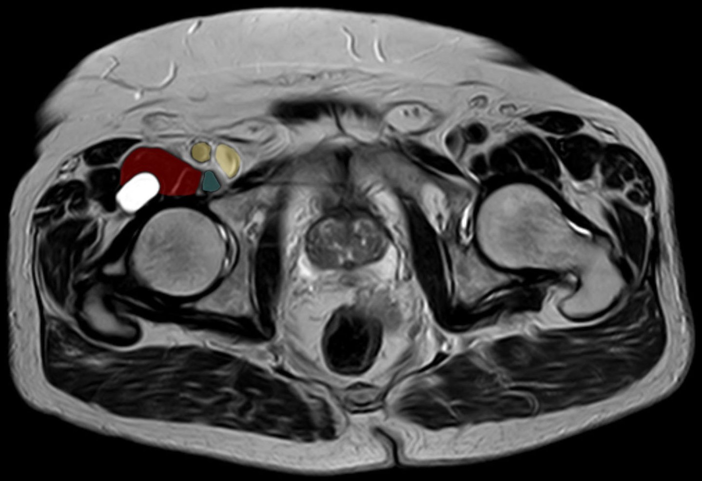
MRIAxial T2 hip MRI, colour-annotated: enlarged iliopectineal bursa (red) behind the iliacus muscle, psoas tendon (green) and femoral vessels (yellow) marked nearby.
Source : Hellerhoff, Wikimedia Commons · CC BY-SA
Which questions should you ask to understand the patient and their history?
🧐 A structured history makes it possible to characterise the pain and the contributing factors. Several items must be collected systematically:- Precise site : pain specifically localised to the anterior part of the groin, at the inguinal crease, over the femoral triangle, is a key indicator.¹ Patients often describe it as a deep pain, sometimes hard to point to.
- Triggering activities and movements : symptoms typically exacerbated by activities with repeated or resisted hip flexion, running (especially uphill), kicking in football, climbing stairs, moving from sitting ↔ standing, getting out of a car.⁴·⁵ Patients after THR often describe pain arising when putting on socks or rising from a low chair.⁶
- Mechanical phenomena : the presence of an audible or palpable snap or click in the groin during hip movements (flexion → active extension against resistance) strongly suggests an internal snapping hip related to the iliopsoas.⁷·⁸ The patient should be asked whether they can reproduce the phenomenon voluntarily.
- History : record any history of hip arthroplasty (date, approach, type of implant), of hip surgery or of a recent joint procedure. Iliopsoas impingement after THR can appear several months after the operation, with a median delay of 5 to 8 months across the series.⁹·¹⁰
- Characteristics of the pain : mode of onset (sudden vs gradual), intensity (NRS 0-10), behaviour over the day (mechanical vs inflammatory), functional impact (a short HAGOS or the EQ-5D to quantify the disability).¹¹
- Red flags : fever, unexplained weight loss, night sweats, a history of cancer, night pain waking the patient, a pulsatile inguinal mass (femoral artery aneurysm), any such signal calls for prompt medical referral.¹²
Which clinical tests should you run and which other conditions must be ruled out?
The physical examination aims to reproduce the patient's familiar pain through palpation and specific provocation tests, while carrying out a rigorous differential diagnosis. The diagnosis rests on a body of evidence and never on an isolated test.¹·³ Provocation tests specific to the iliopsoas complex:- Direct palpation : palpation of the tendon over the lesser trochanter (in the femoral triangle, lateral to the artery). Reproduction of the familiar pain is a positive sign, but the test is technically difficult and not very specific.¹
- Resisted hip flexion (the Stinchfield test) : patient supine, hip flexed to 30° with the knee extended, against the clinician's resistance. Reproduction of the anterior pain is a positive sign.¹·⁴
- Modified Thomas test : beyond the measurement of flexibility, the reproduction of the anterior pain when the psoas is put under tension (passive hip extension with the pelvis held in posterior tilt) is the relevant diagnostic element, not the mere sensation of stretch.⁵
- Passive stretch in prone : can also provoke the pain by stretching the musculotendinous complex.⁵
- Femoroacetabular impingement (FAI) : the main differential diagnosis. The FADIR test (Flexion, Adduction, Internal Rotation) has high sensitivity (close to 99 %) but very low specificity (≈ 7 %) in the meta-analysis of Reiman 2015: it loads multiple anterosuperior structures and a positive test does not designate a single structure.³ The « C-sign » (a C-shaped hand around the hip) is typical of the FAI patient.
- Labral tears : often associated with FAI, they can cause pain, locking or intra-articular snapping.³
- Hip osteoarthritis : mechanical pain on weight-bearing, reduced range (internal rotation in particular).⁸
- Adductor-related pain : more medial pain, reproduced by palpation of the pubic insertion and by the squeeze test (isometric adduction).¹·¹³
- Athletic pubalgia (sports hernia) : peri-pubic pain, linked to the deep abdominal muscles, often in footballers.¹
- Non-musculoskeletal origins : L1-L2 radiculopathy, inguinal or femoral hernia (hence the importance of examining the patient standing and straining), urogenital disease, femoral aneurysm in the older atheromatous patient.
📊 Diagnostic performance of the clinical tests for the painful hip
Sensitivity (%) and specificity (%) for femoroacetabular impingement and labral tear: meta-analysis Reiman 2015 BJSM (9 studies retained)
Sources: Reiman MP, Goode AP, Cook CE, Hölmich P, Thorborg K. Br J Sports Med. 2015;49(12):811 (PMID 25515771). This meta-analysis pools only sensitivities and diagnostic odds ratios (FADIR: pooled sensitivity 0.94 to 0.99; DOR 5.71 to 7.82), and its target condition is femoroacetabular impingement and labral tear, never iliopsoas impingement. Clinical implication: the FADIR serves to rule out (high sensitivity) more than to confirm, and the authors conclude that it has only screening value; no quantified diagnostic value is published for resisted hip flexion or for palpation of the iliopsoas tendon, whose usefulness remains that of a body of evidence (the Doha criteria) and not of an isolated test.
🚩 Red flags specific to groin pain
- Night pain waking the patient + unexplained weight loss + a history of cancer → a bone metastasis work-up (breast, prostate, lung).
- Fever + groin pain + raised CRP/ESR → suspicion of septic arthritis of the hip, pubic osteomyelitis or a psoas abscess (an emergency).
- A pulsatile inguinal mass → femoral artery aneurysm (a vascular emergency).
- Pain reproduced on coughing + a reducible mass → inguinal or femoral hernia (surgical referral).
- Haematuria + lumbar and groin pain → renal colic, urological disease.
- A recent limp in a child or adolescent → slipped capital femoral epiphysis, Perthes disease (Legg-Calvé-Perthes).
- Focal bone pain + inability to weight-bear after trauma → stress fracture of the femoral neck (endurance athletes, REDs).
⚠️ Any red flag → prompt medical referral before any physiotherapy management.
Should patients be classified according to the Doha agreement, and for what benefit?
Yes. Classification is an essential step towards better clinical communication, more homogeneous research and better targeted treatment. ✅ The Doha agreement, published in 2015 (Weir et al. BJSM), is the international reference. It proposes a clinical classification of groin pain in athletes based on the history and the physical examination, organised into three broad categories:¹- Defined clinical entities for groin pain : pain related to the adductors, à the iliopsoas, to the inguinal region, to the pubic region.
- Hip-related groin pain : intra-articular pathology (FAI, labral, osteoarthritis, osteochondral).
- Other causes of groin pain in the athlete (neurogenic, visceral, etc.).
- Diagnostic clarity : it forces the clinician to identify the main structure involved, avoiding the vague diagnosis of « pubalgia ».
- Standardised communication between physiotherapists, sports physicians and surgeons.
- Better research through more homogeneous cohorts: essential to evaluate the effectiveness of interventions reliably.
- Targeted treatment direction : specific exercises according to the dominant structure, without falling into « one size fits all ».
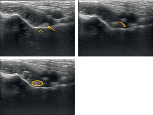
UltrasoundDynamic hip ultrasound: the iliopsoas tendon moves and then snaps against the iliopectineal eminence on return to neutral position, the mechanism of iliopsoas snapping.
Source : Piechota et al., Journal of Ultrasonography, 2016, figure 2 · CC BY-NC-ND
- Diagnosis essentially clinical, based on the history and the reproduction of the familiar pain by the provocation tests.
- Three Doha criteria for the iliopsoas: palpation, stretch and/or resisted flexion: the convergence of signs prevails.
- The FADIR has high sensitivity but low specificity: useful to rule out FAI, not to confirm the iliopsoas.
- Compulsory differential diagnosis: FAI, labral tear, adductors, pubalgia, inguinal hernia, L1-L2 radiculopathy.
- Imaging serves to rule out serious conditions, not to confirm the impingement. Asymptomatic abnormalities are frequent.
- The anaesthetic test injection under ultrasound remains the confirmatory gold standard in doubtful or pre-surgical cases.
Bibliography, chapter 2
- Weir A, Brukner P, Delahunt E, et al. Doha agreement meeting on terminology and definitions in groin pain in athletes. Br J Sports Med. 2015;49(12):768-774. PMID 26031643.
- Register B, Pennock AT, Ho CP, Strickland CD, Lawand A, Philippon MJ. Prevalence of abnormal hip findings in asymptomatic participants: a prospective, blinded study. Am J Sports Med. 2012;40(12):2720-4. PMID 23104610.
- Reiman MP, Goode AP, Cook CE, Hölmich P, Thorborg K. Diagnostic accuracy of clinical tests for the diagnosis of hip femoroacetabular impingement/labral tear: a systematic review with meta-analysis. Br J Sports Med. 2015;49(12):811. PMID 25515771.
- Kiel J, Kaiser K. Psoas Syndrome. StatPearls. 2024. NBK551701.
- Johnston CA, Wiley JP, Lindsay DM, Wiseman DA. Iliopsoas bursitis and tendinitis. A review. Sports Med. 1998;25(4):271-283. PMID 9587184.
- Chalmers BP, Sculco PK, Sierra RJ, Trousdale RT, Berry DJ. Iliopsoas Impingement After Primary Total Hip Arthroplasty: Operative and Nonoperative Treatment Outcomes. J Bone Joint Surg Am. 2017;99(7):557-564.
- Walker P, Ellis E, Scofield J, et al. Snapping Hip Syndrome: A Comprehensive Update. Orthop Rev (Pavia). 2021;13(2):25088. PMID 34745476.
- Tibor LM, Sekiya JK. Differential diagnosis of pain around the hip joint. Arthroscopy. 2008;24(12):1407-1421. PMID 19038713.
- Pisano A, Cipollaro L, Cusano A, et al. Risk Factors for Iliopsoas Impingement Following Total Hip Arthroplasty: A Systematic Review. J Clin Med. 2025;14(18):6376. PMC 12471251.
- Iliopsoas Injections: A Systematic Review of Patient Outcomes and Progression to Surgery. JBJS Reviews. 2025;13(1):e24.00162. PMID 39813362.
- Thorborg K, Hölmich P, Christensen R, Petersen J, Roos EM. The Copenhagen Hip and Groin Outcome Score (HAGOS): development and validation according to the COSMIN checklist. Br J Sports Med. 2011;45(6):478-491. PMID 21478502.
- Finucane LM, Downie A, Mercer C, et al. International Framework for Red Flags for Potential Serious Spinal Pathologies. J Orthop Sports Phys Ther. 2020;50(7):350-372. PMID 32438853.
- Whittaker JL, Small C, Maffey L, Emery CA. Risk factors for groin injury in sport: an updated systematic review. Br J Sports Med. 2015;49(12):803-809. PMID 25833903.
What are the most effective treatment strategies for iliopsoas impingement?
Where do you start? What is the recommended hierarchy of interventions?
The initial approach to managing psoas syndrome is almost universally conservative treatment.¹ The systematic review of the rate of response to non-operative treatment for hip-related pain (Probst 2023 JOSPT, 26 studies, 1,153 patients) reports an overall response rate of 54 % (95 % CI 32–76 %), with a mean improvement of 11.3 points on 100-point functional scales (moderate evidence).² For FAI patients specifically, the UK FASHIoN trial (Griffin 2018 Lancet, 348 patients randomised across 23 NHS centres) shows that arthroscopy is statistically superior to « personalised hip therapy » at 12 months (a difference of 6.8 points on the iHOT-33), but with a modest effect size clinically: 51 % of the patients in the conservative arm still reach the MCID, which justifies always starting with well-conducted conservative care.³🔀 Recommended therapeutic cascade for iliopsoas impingement / bursitis
A staged approach: move to the next step only after a well-conducted trial has failed (3-6 months)
A synthesis based on Probst 2023 (JOSPT, rate of response), Iliopsoas Injections SR 2025 (JBJS Rev), Griffin 2018 (UK FASHIoN, Lancet) and the practice recommended by the learned societies (IOC, ESSKA).
- Activity modification + load management : relative rest (never complete), temporary avoidance of the most provocative activities (sprinting, steep climbs, kicking), maintenance of painless cross-training (cycling, swimming), pain education.⁴
- Initial pharmacology (if needed) : NSAIDs over 7-14 days to manage the acute inflammatory phase, in the absence of contraindications. Paracetamol and tramadol can complement them.⁵
- Structured active physiotherapy : the cornerstone (see 3.2). Focused on progressive strengthening, lumbopelvic motor control and load management.
- Image-guided injections : after 8-12 weeks of failed physiotherapy, an injection of corticosteroid ± local anaesthetic under ultrasound guidance. It serves both as a diagnostic tool (confirmation) and a therapeutic one (an analgesic window in which to intensify rehabilitation). The 2025 JBJS Rev SR (12 studies, 235 patients) shows an improvement in NRS from 7.33 → 2.47 and in HHS from 58.5 → 89.9, with 28.9 % going on to surgery.⁶
- Surgery : reserved for persistent failures. In the athlete: endoscopic release of the tendon (Ilizaliturri 2005) with return to sport at 4-6 months. In the patient after THR: see chapter 4.⁷
Where does exercise fit in, and is there a superior approach? 🏋️♂️
Exercise is the best-validated component of conservative management. There is no single protocol superior to all the others, but a solid consensus around several principles: The modern approach moves away from aggressive, direct stretching of the psoas, which can increase the compression of the tendon in compressive impingement and worsen the symptoms.⁸ The strategy focuses instead on:- Strengthening the antagonist and synergist muscles : glutes (maximus, medius) and external rotators. Strong glutes improve the control of extension and rotation, which can reduce the overuse of the iliopsoas as an anterior stabiliser.⁹·¹⁰ Typical exercises: glute bridge (then single-leg), clamshells, side-lying abduction, standing sequences (mini-band side step, monster walk).
- Lumbopelvic motor control : core stability, lumbopelvic dissociation, control of pelvic anteversion, to reduce the compensatory stresses on the hip flexors.¹¹
- Progressive management of tendon load : after the initial phase, progressive reloading of the iliopsoas tendon. Isometric work in hip flexion first (analgesia), then eccentric and concentric as tolerated, with the pain-monitoring model of Silbernagel: pain acceptable up to 5/10 if it settles quickly after the exercise and does not worsen the next day.¹²·¹³
- Hip hinge and functional patterns : restoring the deep squat, the lunge and stair climbing without lumbar compensation.
| Modality | GRADE level | Key source | Recommendation |
|---|---|---|---|
| Progressive gluteal and pelvic strengthening | High | Casartelli 2011 OAC, Probst 2023 JOSPT | First line, the basis of any programme |
| Load management / pain-monitoring | High | Silbernagel 2007 AJSM, Cook & Purdam 2014 | Allows continued sporting activity (pain ≤ 5/10 acceptable) |
| Lumbopelvic motor control | Moderate | Lewis & Sahrmann 2009 Clin Biomech | Relevant when a deficit is identified |
| Aggressive stretching of the psoas | Low / to be avoided if compressive | Cook & Purdam 2014, biomechanics | Avoid in the compressive forms (FAI, after THR) |
| Manual therapy (mobilisations) | Moderate (adjunct) | Probst 2023 JOSPT | Useful short term, does not replace exercise |
| Dry needling on trigger points | Low | Limited data | A symptomatic adjunct option |
| Extracorporeal shockwave therapy | Low | Extrapolated from other tendinopathies | No data specific to the psoas |
| Ultrasound-guided corticosteroid injection | Moderate | Iliopsoas Injections SR 2025 JBJS Rev | Second line, after 3 months of physiotherapy |
| Endoscopic tenotomy | Moderate | Ilizaliturri 2005, Coulomb 2022 SR | Third line, after failed injections |
Manual therapies and technologies: how effective are they really?
Manual therapies and technologies are frequently used as an adjunct to the exercise programme, but their effectiveness as a stand-alone treatment is limited. Their main role is to modulate pain in the short term and to prepare the patient to tolerate the active exercise programme better.²- Manual therapy : soft-tissue mobilisation of the iliopsoas complex, the quadriceps and the adductors; joint mobilisation of the hip, the sacroiliac joints and the lumbar spine. Symptomatic relief is often real, but the evidence of a long-term effect is weak without an associated active programme.²
- Dry needling : can reduce the pain of trigger points in the psoas and the glutes, making participation in rehabilitation easier. Data specific to the psoas are scarce.
- Extracorporeal shockwave therapy : studied mainly for calcific tendinopathies; extrapolation to the psoas in chronic tendinopathy, without solid specific data.
- TENS / electrotherapy : transient analgesic effect, an adjunct role.
Beyond the physical: how do you educate the patient and address psychological factors? 🧠
Modern management incorporates the biopsychosocial model. For pain persisting beyond 3 months, the non-physical factors become major obstacles. The therapeutic education of the patient is non-negotiable. It aims to:- Demystify the diagnosis : explain the mechanical nature of the pain in simple terms, without catastrophising language. Reassure the patient that pain does not mean serious structural damage.¹⁴
- Explain load management : give tools to modulate activities. The concept of optimal load is more useful than complete rest.¹³
- Promote self-efficacy : give the patient responsibility through adherence to the home programme.¹⁴
- Kinesiophobia (fear of movement): graded exposure to movement in a safe setting is the best therapy.¹⁵
- Catastrophising beliefs : reframe negative beliefs (« my body is damaged », « I am going to make things worse »).¹⁴
- Stress and anxiety : they increase the sensitivity of the CNS and amplify the perception of pain, stress management techniques are relevant.
- ✅ Management always begins with conservative treatment centred on physiotherapy (response rate 54 %, CI 32-76 %, Probst 2023 JOSPT).
- 🏋️♂️ Exercise is the key: strengthening the glutes and lumbopelvic control, progressive load management, and not aggressive stretching of the psoas.
- 🤲 Manual therapies, dry needling and shockwave are useful adjuncts in the short term but they never replace an active programme.
- 🧠 The education of the patient and the management of psychological factors (kinesiophobia, catastrophising) are essential to prevent chronicity.
- 💉 The guided corticosteroid injections (NRS 7.3 → 2.5) are a useful second line to confirm the diagnosis and unblock rehabilitation.
- 🔪 The surgery (endoscopic tenotomy) is a third line reserved for persistent failures after ≥ 6 months of well-conducted treatment.
Bibliography, chapter 3
- Kiel J, Kaiser K. Psoas Syndrome. StatPearls. 2024. NBK551701.
- Probst DT, Sookochoff MF, Harris-Hayes M, Prather H, Lipsey KL, Cheng AL. What is the Rate of Response to Nonoperative Treatment for Hip-Related Pain? A Systematic Review With Meta-analysis. J Orthop Sports Phys Ther. 2023;53(5):286–306. PMID 36892224.
- Griffin DR, Dickenson EJ, Wall PDH, et al. Hip arthroscopy versus best conservative care for the treatment of femoroacetabular impingement syndrome (UK FASHIoN): a multicentre randomised controlled trial. Lancet. 2018;391(10136):2225-2235. PMID 29893223.
- Soligard T, Schwellnus M, Alonso JM, et al. How much is too much? (Part 1) International Olympic Committee consensus statement on load in sport and risk of injury. Br J Sports Med. 2016;50(17):1030-1041. PMID 27535989.
- Johnston CA, Wiley JP, Lindsay DM, Wiseman DA. Iliopsoas bursitis and tendinitis. A review. Sports Med. 1998;25(4):271-283. PMID 9587184.
- Iliopsoas Injections: A Systematic Review of Patient Outcomes and Progression to Surgery. JBJS Reviews. 2025;13(1):e24.00162. PMID 39813362.
- Ilizaliturri VM Jr, Villalobos FE Jr, Chaidez PA, Valero FS, Aguilera JM. Internal snapping hip syndrome: treatment by endoscopic release of the iliopsoas tendon. Arthroscopy. 2005;21(11):1375-1380. PMID 16325091.
- Cook JL, Purdam CR. The challenge of managing tendinopathy in competing athletes. Br J Sports Med. 2014;48(7):506-509. PMID 23666020.
- Casartelli NC, Maffiuletti NA, Item-Glatthorn JF, et al. Hip muscle weakness in patients with symptomatic femoroacetabular impingement. Osteoarthritis Cartilage. 2011;19(7):816-821. PMID 21515390.
- Hoit G, Whelan DB, Dwyer T, Ajrawat P, Chahal J. Physiotherapy as an Initial Treatment Option for Femoroacetabular Impingement: A Systematic Review of the Literature and Meta-analysis of 5 Randomized Controlled Trials. Am J Sports Med. 2020;48(8):2042-2050.
- Lewis CL, Sahrmann SA, Moran DW. Effect of position and alteration in synergist muscle force contribution on hip forces when performing hip strengthening exercises. Clin Biomech (Bristol). 2009;24(1):35-42. PMID 19028000.
- Silbernagel KG, Thomeé R, Eriksson BI, Karlsson J. Continued sports activity, using a pain-monitoring model, during rehabilitation in patients with Achilles tendinopathy: a randomized controlled study. Am J Sports Med. 2007;35(6):897-906. PMID 17307888.
- Gabbett TJ. The training-injury prevention paradox. Br J Sports Med. 2016;50(5):273-280. PMID 26758673.
- Louw A, Zimney K, Puentedura EJ, Diener I. The efficacy of pain neuroscience education on musculoskeletal pain: A systematic review of the literature. Physiother Theory Pract. 2016;32(5):332-355.
- Leeuw M, Goossens ME, Linton SJ, Crombez G, Boersma K, Vlaeyen JW. The fear-avoidance model of musculoskeletal pain: current state of scientific evidence. J Behav Med. 2007;30(1):77-94.
What are the specific considerations after total hip replacement?
Epidemiology and iatrogenic risk factors of impingement after THR
The 2025 systematic review (J Clin Med, Pisano et al.) brings together the data of 23 studies to estimate the prevalence of iliopsoas impingement after THR at between 0.4 % and 8.3 % depending on the diagnostic definition (strictly clinical, imaging or a confirmatory test injection) and the surgical approach.¹ The weighted mean is around 4.3 %, with a peak of 6-8 % for the direct anterior approach.- Anterior overhang of the acetabular cup : the most powerful factor. Beyond 10-12 mm of overhang relative to the anterior bony rim (measured on CT or MRI), the risk of impingement increases significantly.¹·² A threshold of 8 mm of anterior cup protrusion is sometimes used in practice.
- Low acetabular anteversion : anteversion below 10° (ideal target 15-25°) increases the tension on the tendon in hip flexion.
- Direct anterior approach (DAA) : a higher rate of clinical impingement than with posterior or lateral approaches, possibly related to the change in the tendon's path caused by the approach.²·³
- A large prosthetic head (≥ 36 mm): increases the lever arm and the friction on the tendon.
- Anterior bone deficiency of the native acetabulum : limits the possibility of burying the cup without overhang.
- Long fixation screws protruding anteriorly (a screw-fixed cup).
- Dual-mobility cup : in certain designs it can itself come into contact with the tendon.
Specific diagnosis: imaging and the test injection
🩺 The typical clinical picture combines:- Anterior groin pain of secondary onset (often 3-9 months after the surgery, sometimes later).
- Pain reproduced by active resisted flexion of the hip (straight leg raise, putting on socks, rising from a low chair, getting in and out of a car).
- Pain relieved by passive flexion, worsened when walking up stairs.
- No signs of infection (normal CRP/ESR, no fever) and no loosening (stable radiographs).
- Chronic prosthetic infection (CRP, ESR, joint aspiration at the slightest doubt).
- Aseptic loosening (comparative radiographs, bone scan).
- Periprosthetic stress fracture (mechanical pain, CT).
- Particle disease (granuloma).
- Referred lumbar pain (L1-L2 radiculopathy).
- Visceral disease (inguinal hernia, urological disease).
- Standard radiographs anteroposterior + lateral: looking for anterior overhang of the cup, checking the positioning (target inclination 40°, anteversion 15-25°).
- Low-dose 3D CT with acetabular reconstruction: quantifies the anterior overhang precisely in millimetres and the actual anteversion.
- Dynamic ultrasound : visualises the tendon in movement, looks for dynamic impingement against the cup and makes it possible to guide the test injection.
- MRI (METAL-suppressed sequences, MARS or SEMAC type) : looking for peri-tendinous oedema, bursitis and tendinopathy without major metal artefact.
The therapeutic cascade: from conservative care to endoscopic tenotomy
Management is staged, with progression to the invasive options only after documented failure of the previous one:- Conservative treatment (3-6 months) : education, activity modification (avoiding abrupt active resisted flexion), a short course of NSAIDs, physiotherapy focused on lumbopelvic motor control and strengthening of the glutes. Stretching the psoas is clearly contraindicated here (direct compression against the cup).
- Ultrasound-guided injections : 1 to 3 injections 3 to 6 weeks apart, with intensive rehabilitation in the analgesic window. The 2025 JBJS Rev SR (12 studies, 235 patients) reports 28.9 % going on to surgery, which means that ~71 % avoid surgery thanks to this strategy.⁴
- Endoscopic tenotomy of the iliopsoas tendon : a minimally invasive procedure carried out at the lesser trochanter or at the insertion. Reported success rate of 80 to 90 % on pain, but with 32 % residual weakness in hip flexion detectable on a dynamometer, often well tolerated functionally but to be explained before surgery.⁵·⁶
- Acetabular revision : reserved for cases with a clearly malpositioned cup (major overhang ≥ 12-15 mm, very deficient anteversion). A major procedure, whose indication must be weighed carefully.²
🔀 Therapeutic cascade for iliopsoas impingement after THR
A staged approach based on the 2025 JBJS Rev SR and reference practice
Sources: Iliopsoas Injections SR 2025 (PMID 39813362), Risk Factors IPI after THA SR 2025 (PMC 12471251), Chalmers 2017 JBJS Am, Ilizaliturri 2005 Arthroscopy. The tenotomy can be done on the tendon at the lesser trochanter or at the femoral insertion; the choice depends on the procedure and on surgical preference.
📌 Key points: iliopsoas impingement after THR
- Affects 0.4–8.3 % of patients after THR (mean ≈ 4.3 %, SR 2025), with a peak for the direct anterior approach.
- Main risk factor: anterior overhang of the cup (≥ 10-12 mm on CT) ± insufficient anteversion.
- Typical clinical picture: pain of secondary onset (5-8 months after surgery) reproduced by active resisted flexion (« can no longer put on their socks »).
- Always rule out infection, loosening and periprosthetic fracture before accepting the diagnosis.
- Gold standard for diagnosis: a test injection under ultrasound (NRS ↓ ≥ 50 % at 30 min).
- Cascade: conservative care for 3-6 months → injection (71 % avoid surgery) → endoscopic tenotomy (80-90 % success but 32 % residual weakness) → acetabular revision if the cup is clearly malpositioned.
- Stretching the psoas is contraindicated in this compressive indication.
Bibliography, chapter 4 (after THR)
- Pisano A, Cipollaro L, Cusano A, et al. Risk Factors for Iliopsoas Impingement Following Total Hip Arthroplasty: A Systematic Review. J Clin Med. 2025;14(18):6376. PMC 12471251.
- Cipollaro L, et al. Iliopsoas Impingement After Total Hip Arthroplasty: A Review of Diagnosis and Management. Cureus. 2025. PMC 12128184.
- Iliopsoas Impingement After Direct Anterior Approach Total Hip Arthroplasty: Epidemiology, Risk Factors, and Treatment Options. J Arthroplasty. 2021. PMID 33414039.
- Iliopsoas Injections: A Systematic Review of Patient Outcomes and Progression to Surgery. JBJS Reviews. 2025;13(1):e24.00162. PMID 39813362.
- Chalmers BP, Sculco PK, Sierra RJ, Trousdale RT, Berry DJ. Iliopsoas Impingement After Primary Total Hip Arthroplasty: Operative and Nonoperative Treatment Outcomes. J Bone Joint Surg Am. 2017;99(7):557-564.
- Ilizaliturri VM Jr, Villalobos FE Jr, Chaidez PA, Valero FS, Aguilera JM. Internal snapping hip syndrome: treatment by endoscopic release of the iliopsoas tendon. Arthroscopy. 2005;21(11):1375-1380. PMID 16325091.
- Coulomb R, Nougarède B, Maury E, Marchand P, Mares O, Kouyoumdjian P. Arthroscopic iliopsoas tenotomies: a systematic review of surgical technique and outcomes. Hip Int. 2022;32(2):144-152. Hip Int 2022.
- Guicherd W, Bonin N, Gicquel T, et al. Endoscopic or arthroscopic iliopsoas tenotomy for iliopsoas impingement following total hip replacement. A prospective multicenter 64-case series. Orthop Traumatol Surg Res. 2017;103(8S):S207-S214. PMID 28917519.
- Nikou S, et al. Arthroscopic iliopsoas tenotomy after total hip arthroplasty: safe method for the right patient. J Exp Orthop. 2023;10:42. JEO 2023.
How is durable recovery ensured and recurrence prevented?
How do you make the patient an actor in their own recovery through self-management?
Self-management (self-management) is a collaborative approach in which the therapist guides the patient towards autonomy in managing their condition. This shift of responsibility correlates directly with better functional outcomes and a lower risk of recurrence.² Therapeutic education of the patient is its mainstay and must cover several fundamental aspects:- Understanding the condition and the risk factors : explain the mechanisms, the interaction between training load, personal biomechanics (lumbopelvic control, strength of the stabilisers) and anatomical factors. Pain is often a signal of overload rather than of damage, which can reduce kinesiophobia and encourage a return to activity.³
- Pain monitoring: the traffic-light model 🚦: pain 0-3/10 during the activity that settles quickly afterwards = green light; pain 4-5/10 settling within a few hours = amber light (adapt the volume); pain > 5/10 that persists or worsens the next day = red light (reduce sharply). An approach taken directly from the pain-monitoring model of Silbernagel 2007.⁴
- Adherence to a home exercise programme : not an endless list but a programme that is concise, targeted and progressive , combining progressive strengthening of the glutes, the external rotators, the flexors (respecting pain) and the core. This programme must be reviewed and adapted every 4 to 6 weeks.
- Progressive management of the training load : one of the greatest predictors of recurrence is an abrupt increase in volume or intensity.⁵ The patient must learn the principles of gradual progression: the 10 % rule (do not increase the weekly volume by more than 10 %), monitoring of the acute:chronic workload ratio, the importance of recovery days.⁶·⁷
- Recognising situations at risk : a rushed return after a break, an abrupt increase in load at the start of the season, dehydration, accumulated fatigue, all situations known as precipitating factors, identified by the Whittaker 2015 SR.⁸
When and how do you plan a safe return to sport and to activity?
Return to sport (RTS) must never be based on time elapsed since the start of treatment alone. Current research strongly supports an approach based on objective functional criteria which guarantee that the patient has regained the physical capacities needed to withstand the demands of their sport without compensation or risk of re-injury.⁹🎯 Functional criteria for return to sport: a multidimensional checklist
All the criteria must be met before an unrestricted return to training
A conceptual framework adapted from Soligard 2016 (IOC consensus), Thorborg 2018 (groin pain management), and the FASHIoN protocols for athletic hip pathology. Strength ≥ 90 % of the contralateral side is ideally measured on a hand-held dynamometer (HHD), which is more objective than manual testing.
- Criterion 1. Pain-free : no pain on palpation, on resisted flexion, or during activities of daily living for at least 14 days.
- Criterion 2. Symmetrical strength : strength of the key hip muscles (flexion, extension, abduction, adduction, rotations) ≥ 90 % of the unaffected side on a hand-held dynamometer (HHD).⁹·¹⁰
- Criterion 3. Functional tests passed : able to perform hop tests (single, triple, crossover), changes of direction (cutting) and sport-specific movements without pain, without apprehension and with good movement quality.
- Criterion 4. Psychological readiness : confidence regained, no kinesiophobia. Validated scales such as the Injury-Psychological Readiness to Return to Sport (I-PRRS) can quantify this crucial aspect.¹¹
- Criterion 5. Sport-specific tests : reproduction of the movements of the sport without pain (kicking in football, the splits in dance, sprinting with changes of direction in athletics).
- Preventing recurrence rests on the education and empowerment of the patient (self-management), not on passive treatments.
- Return to sport is guided by objective functional criteria (strength ≥ 90 % of the sound side on the HHD, painless hop tests, I-PRRS) and not by a fixed timetable.
- An effective exercise programme targets the whole lumbo-pelvi-femoral complex, not only the psoas.
- The progressive load management (the 10 % rule, the acute:chronic ratio, the Soligard 2016 IOC consensus) is essential to avoid recurrence.
- The traffic-light model allows the patient to modulate their activities without stopping completely (Silbernagel 2007).
Bibliography, chapter 5
- Probst DT, Sookochoff MF, Harris-Hayes M, Prather H, Lipsey KL, Cheng AL. What is the Rate of Response to Nonoperative Treatment for Hip-Related Pain? A Systematic Review With Meta-analysis. J Orthop Sports Phys Ther. 2023;53(5):286–306. PMID 36892224.
- Thorborg K, Reiman MP, Weir A, et al. Clinical examination, diagnostic imaging, and testing of athletes with groin pain: an evidence-based approach to effective management. J Orthop Sports Phys Ther. 2018;48(4):239-249. JOSPT 2018.
- Louw A, Zimney K, Puentedura EJ, Diener I. The efficacy of pain neuroscience education on musculoskeletal pain. Physiother Theory Pract. 2016;32(5):332-355.
- Silbernagel KG, Thomeé R, Eriksson BI, Karlsson J. Continued sports activity, using a pain-monitoring model, during rehabilitation in patients with Achilles tendinopathy: a randomized controlled study. Am J Sports Med. 2007;35(6):897-906. PMID 17307888.
- Gabbett TJ. The training-injury prevention paradox: should athletes be training smarter and harder? Br J Sports Med. 2016;50(5):273-280. PMID 26758673.
- Soligard T, Schwellnus M, Alonso JM, et al. How much is too much? (Part 1) IOC consensus statement on load in sport and risk of injury. Br J Sports Med. 2016;50(17):1030-1041. PMID 27535989.
- Cook JL, Purdam CR. The challenge of managing tendinopathy in competing athletes. Br J Sports Med. 2014;48(7):506-509. PMID 23666020.
- Whittaker JL, Small C, Maffey L, Emery CA. Risk factors for groin injury in sport: an updated systematic review. Br J Sports Med. 2015;49(12):803-809. PMID 25833903.
- Thorborg K, Hölmich P, Christensen R, Petersen J, Roos EM. The Copenhagen Hip and Groin Outcome Score (HAGOS): development and validation according to the COSMIN checklist. Br J Sports Med. 2011;45(6):478-491. PMID 21478502.
- Hoit G, Whelan DB, Dwyer T, Ajrawat P, Chahal J. Physiotherapy as an Initial Treatment Option for Femoroacetabular Impingement: Systematic Review and Meta-analysis of 5 RCTs. Am J Sports Med. 2020;48(8):2042-2050.
- Webster KE, Feller JA, Lambros C. Development and preliminary validation of a scale to measure the psychological impact of returning to sport. Phys Ther Sport. 2008;9(1):9-15. (The I-PRRS scale, relevant to RTS in general.)
- Lewis CL, Sahrmann SA, Moran DW. Effect of position and alteration in synergist muscle force contribution on hip forces when performing hip strengthening exercises. Clin Biomech. 2009;24(1):35-42. PMID 19028000.
What do real clinical cases teach us?
Analysis of a classic case: the ballet dancer cohort (Winston 2007)
The « classic » case of iliopsoas impingement, often described as an internal snapping hip, typically affects athletes in disciplines that demand repetitive hip flexion, such as classical dance.¹ The reference cohort of Winston et al. (2007 AJSM, 87 elite ballet dancers) remains the most cited observational study.² Of the 87 dancers questioned, 91 % reported a snapping hip, of whom 80 % bilaterally and 58 % with associated pain; 60 % could trigger it voluntarily, and 26 of them (50 hips) were selected from among the latter for clinical examination by 2 independent clinicians and dynamic ultrasound. The clinicians palpated the snap in 46 of the 50 hips, and ultrasound showed a snapping iliopsoas tendon in 59 % of the hips (iliotibial band in 4 %), with no identifiable cause in a third of the cases. The diagnosis is essentially clinical, ultrasound serving to objectify the mechanism and to rule out other causes (labral tear, isolated bursitis).² Typical management in the symptomatic dancer:- Activity modification (temporary reduction of the movements that provoke the snap: développé, aerial splits).⁴
- Specific strengthening of the glutes and external rotators to improve the dynamic stability of the pelvis.⁵
- Education about the « asymptomatic » snap: the click is not pathognomonic in itself: many dancers have an audible snap without pain; it is the associated pain that defines the condition.²
- Proprioceptive and motor control work over the lumbo-pelvi-femoral chain.
- Progressive return to technique with criteria of strength ≥ 90 % and being pain-free for 14 days.
The diagnostic challenge: when bursitis mimics an inguinal hernia
Iliopsoas bursitis can present in atypical forms that delay the diagnosis. Several case reports describe presentations as an inguinal mass initially raising the suspicion of a femoral or inguinal hernia.⁶ The typical case is that of a palpable, non-pulsatile inguinal mass, sometimes painful on hip movement, without clear local inflammatory signs. The clinical examination alone is often insufficient to tell bursitis from hernia. It is imaging that plays the crucial role:- Ultrasound : visualises a hypoechoic fluid collection communicating with the hip joint in 15 % of cases, typical of iliopsoas bursitis. It also allows a femoral aneurysm to be told apart (Doppler essential).
- MRI : confirms the bursitis with T2 hyperintensity, shows any intra-articular communication, looks for an associated tendinopathy and rules out a tumour mass.⁷
- CT : useful if an associated hernia is suspected or in a traumatic context.
Study of a complex case: impingement after arthroplasty treated by tenotomy
The most complex cases are often iatrogenic, arising after a total hip replacement. The typical case reported in the literature concerns a patient in their fifties, operated on through a direct anterior approach, who develops 6 to 9 months after the operation a disabling groin pain preventing them from putting on their socks and climbing the stairs.⁸ 🤖 The standardised diagnostic approach comprises:- Ruling out the emergencies : no fever, normal CRP, normal ESR → infection unlikely. Stable radiographs → no loosening and no periprosthetic fracture.
- Targeted imaging : 3D CT quantifying the anterior overhang of the cup at 13 mm (above the critical threshold of 10-12 mm). MRI with METAL-suppressed sequences showing peri-tendinous oedema and tendinopathy of the iliopsoas.
- Diagnostic test injection under ultrasound : 8 mL of 1 % lidocaine + 40 mg of methylprednisolone, with a fall in the NRS from 8/10 to 2/10 at 30 min, confirming the diagnosis.
Critique and controversy: where do clinical cases sit in the hierarchy of evidence?
Illuminating though clinical cases are, it is crucial to recognise their limits. 🧠 The main criticism is their low level of evidence in the pyramid of scientific evidence. A single case illustrates a possibility, not a generality; it is impossible to conclude a cause-and-effect relationship because placebo effects, regression to the mean and natural history are not controlled.¹¹ Published cases often tend to report successes (publication bias), which can overestimate the effectiveness of an intervention.📐 Hierarchy of scientific evidence: where does each type of study belong?
Strength of evidence decreasing from the top (meta-analyses) towards the bottom (isolated cases)
A simplified GRADE / Oxford CEBM hierarchy. The length of the bar on the right illustrates the relative strength of evidence. Practical implication: when an appealing clinical case and a meta-analysis diverge, the decision must follow the meta-analysis. Clinical cases remain valuable for generate hypotheses, signal rare presentations or illustrate a piece of clinical reasoning.
📌 Key points: lessons from the clinical cases
- The classic case of the ballet dancer (Winston 2007) shows that many snaps are asymptomatic: it is the associated pain that makes the condition.
- Bursitis can mimic an inguinal hernia : ultrasound is essential to the differential diagnosis.
- Impingement after THR is a complex cause of groin pain that may require an endoscopic tenotomy (80-90 % success, 32 % loss of flexion strength) after conservative care and injections have failed.
- Always ask about the underlying cause of the overuse (FAI, cup overhang, instability, motor deficit) rather than treating the symptom alone.
- ⚠️ A case report = level 5 (the weakest). When they diverge, follow the meta-analyses (level 1a), not the isolated case.
Bibliography, chapter 6
- Walker P, Ellis E, Scofield J, et al. Snapping Hip Syndrome: A Comprehensive Update. Orthop Rev (Pavia). 2021;13(2):25088. PMID 34745476.
- Winston P, Awan R, Cassidy JD, Bleakney RK. Clinical examination and ultrasound of self-reported snapping hip syndrome in elite ballet dancers. Am J Sports Med. 2007;35(1):118-126. PMID 17021311.
- Ilizaliturri VM Jr, Villalobos FE Jr, Chaidez PA, Valero FS, Aguilera JM. Internal snapping hip syndrome: treatment by endoscopic release of the iliopsoas tendon. Arthroscopy. 2005;21(11):1375-1380. PMID 16325091.
- Whittaker JL, Small C, Maffey L, Emery CA. Risk factors for groin injury in sport: an updated systematic review. Br J Sports Med. 2015;49(12):803-809. PMID 25833903.
- Casartelli NC, Maffiuletti NA, Item-Glatthorn JF, et al. Hip muscle weakness in patients with symptomatic femoroacetabular impingement. Osteoarthritis Cartilage. 2011;19(7):816-821. PMID 21515390.
- Johnston CA, Wiley JP, Lindsay DM, Wiseman DA. Iliopsoas bursitis and tendinitis. A review. Sports Med. 1998;25(4):271-283. PMID 9587184.
- Polster JM, Elgabaly M, Lee H, Klika A, Drake R, Barsoum W. MRI and gross anatomy of the iliopsoas tendon complex. Skeletal Radiol. 2008;37(1):55-8. PMID 17938916.
- Chalmers BP, Sculco PK, Sierra RJ, Trousdale RT, Berry DJ. Iliopsoas Impingement After Primary Total Hip Arthroplasty: Operative and Nonoperative Treatment Outcomes. J Bone Joint Surg Am. 2017;99(7):557-564.
- Coulomb R, Nougarède B, Maury E, Marchand P, Mares O, Kouyoumdjian P. Arthroscopic iliopsoas tenotomies: a systematic review of surgical technique and outcomes. Hip Int. 2022;32(2):144-152.
- Nikou S, et al. Arthroscopic iliopsoas tenotomy after total hip arthroplasty: safe method for the right patient. J Exp Orthop. 2023;10:42.
- Nissen T, Wynn R. The clinical case report: a review of its merits and limitations. BMC Res Notes. 2014;7:264. PMID 24758689.
- Cook JL, Purdam CR. The challenge of managing tendinopathy in competing athletes. Br J Sports Med. 2014;48(7):506-509. PMID 23666020.
How do you apply these recommendations concretely in your practice?
When, and to which other health professionals, should you refer?
One of the fundamental skills of the modern physiotherapist is to recognise the limits of their scope and to identify the situations that call for interprofessional collaboration. This process of triage is crucial to the safety and the effectiveness of care.¹ Identifying the red flags is the first non-negotiable step. These signs suggest a serious underlying condition (fracture, infection, tumour, vascular syndrome) and call for immediate or urgent medical referral. It should be noted, however, that many traditional red flags, taken in isolation, have poor diagnostic accuracy and must be interpreted in the context of a complete examination.² The Cochrane review of Han et al. (2023) on red flags for vertebral fracture shows this limit and stresses the importance of combining several signs to increase the predictive value.³🚩 Red flags specific to groin pain: prompt medical referral
- Fever + acute groin pain + raised CRP/ESR → suspicion of septic arthritis, psoas abscess, pubic osteomyelitis (an emergency).
- A pulsatile inguinal mass → femoral artery aneurysm (a vascular emergency).
- Night pain waking the patient + unexplained weight loss + a history of cancer → bone metastases, primary tumour.
- Pain reproduced on coughing + a mass that reduces on exertion → inguinal or femoral hernia.
- Haematuria + lateralised colic → renal colic, urological disease.
- A recent limp in a child → slipped capital femoral epiphysis, Legg-Calvé-Perthes, juvenile idiopathic arthritis.
- Recent THR + severe pain + fever / inflammatory signs → prosthetic infection (a surgical emergency).
- A history of THR + abrupt mechanical pain → periprosthetic fracture, acute loosening.
⚠️ Any red flag → prompt medical referral before any physiotherapy management.
- Sports physician : for prescribing targeted imaging (MRI, dynamic ultrasound) and advising on injections.
- Interventional radiologist : for the diagnostic and therapeutic injections under ultrasound.
- Orthopaedic surgeon (hip) : advice on tenotomy or acetabular revision after prolonged conservative failure.
- Rheumatologist : if an inflammatory cause is suspected (spondyloarthritis, rheumatoid arthritis).
- Psychologist / psychiatrist : for significant yellow flags.
How do you measure outcomes and overcome the barriers to implementation?
Measuring outcomes objectively and in a patient-centred way is the foundation of evidence-based practice. The systematic use of validated PROMs is essential.⁶ For groin pain and the iliopsoas complex, the reference PROMs are:- HAGOS (Copenhagen Hip and Groin Outcome Score, Thorborg 2011): 37 items, 6 subscales: pain, symptoms, daily living, sport/recreation, participation, quality of life. COSMIN-validated, available in French.⁷
- iHOT-33 (international Hip Outcome Tool): 33 items, validated for athletic hip pathology.
- HOOS (Hip disability and Osteoarthritis Outcome Score): for patients after THR and with hip osteoarthritis.
- NRS or the pain VAS: an unavoidable minimum, simple and quick.
- I-PRRS (Injury-Psychological Readiness to Return to Sport): for the psychological dimension at the time of return to sport.⁸
- Active continuing education : interactive workshops, peer mentoring, more effective than passive reading of articles.¹⁰
- Audit and feedback : analysing one's own practice data and comparing it against standards (Ivers Cochrane 2012).¹¹
- Organisational support : the most powerful enabler, a work culture that values learning, protected time for training, access to the databases.⁹
- Technological integration : patient records with PROMs built in, telehealth platforms, pain-monitoring apps.
- Referral is crucial: the red flags require prompt medical assessment, the yellow flags justify collaboration with a psychologist.
- For the iliopsoas complex: collaboration physiotherapist ↔ sports physician ↔ interventional radiologist ↔ surgeon according to the stage.
- Measure with validated PROMs : HAGOS (Thorborg 2011), iHOT-33, HOOS, NRS, I-PRRS.
- The barriers to implementation (time, training, organisational support) call for multi-faceted strategies; the organisational support is the most powerful enabler.
- Recognise the tension between standardisation (PROMs, recommendations) and personalisation (the patient's unique goals): this is the art of clinical science.
Bibliography, chapter 7
- Marks D, et al. The effectiveness of first contact physiotherapy in primary care: a systematic review. Physiotherapy. 2020;107:133-146.
- Finucane LM, Downie A, Mercer C, et al. International Framework for Red Flags for Potential Serious Spinal Pathologies. J Orthop Sports Phys Ther. 2020;50(7):350-372. PMID 32438853.
- Han CS, Hancock MJ, Downie A, et al. Red flags to screen for vertebral fracture in people presenting with low back pain. Cochrane Database Syst Rev. 2023;8:CD014461. PMID 37615643.
- Nicholas MK, Linton SJ, Watson PJ, Main CJ. Early identification and management of psychological risk factors ("yellow flags") in patients with low back pain. Phys Ther. 2011;91(5):737-753.
- Marks D, et al. First contact physiotherapy in primary care: systematic review. Physiotherapy. 2020;107:133-146.
- Kyte DG, et al. The routine use of patient-reported outcome measures (PROMs) in clinical practice. BMC Health Serv Res. 2019;19:147.
- Thorborg K, Hölmich P, Christensen R, Petersen J, Roos EM. The Copenhagen Hip and Groin Outcome Score (HAGOS): development and validation according to the COSMIN checklist. Br J Sports Med. 2011;45(6):478-491. PMID 21478502.
- Webster KE, Feller JA, Lambros C. Development and preliminary validation of a scale to measure the psychological impact of returning to sport (I-PRRS). Phys Ther Sport. 2008;9(1):9-15.
- Scurlock-Evans L, Upton P, Upton D. Evidence-based practice in physiotherapy: a systematic review of barriers, enablers and interventions. Physiotherapy. 2014;100(3):208-219.
- van der Wees PJ, et al. Multifaceted strategies to improve evidence-based practice in physiotherapy: systematic review. Aust J Physiother. 2008;54(4):233-241.
- Ivers N, Jamtvedt G, Flottorp S, et al. Audit and feedback: effects on professional practice and healthcare outcomes. Cochrane Database Syst Rev. 2012;(6):CD000259. PMID 22696318.
- Soligard T, Schwellnus M, Alonso JM, et al. IOC consensus statement on load in sport and risk of injury. Br J Sports Med. 2016;50(17):1030-1041. PMID 27535989.
What next after reading this?
This article is part of a collection of evidence-based clinical syntheses. A question, a comment, a correction to suggest? Contact us directly through the WhatsApp button at the bottom right of the screen.

