














On-siteDPCFIFPL

Upper limb
Hand rehabilitation and splinting: refining and deepening your practice
Jean Lichtle
Go further
Kienbock's disease, avascular necrosis of the lunate: wrist pain that mimics a sprain that drags on, the radiological stages, imaging and treatment.
A normal radiograph rules nothing out: lunate necrosis is confirmed by MRI, and rehabilitation cannot revascularise it.
7cases per 100,000, with a peak between 20 and 40 years
What follows covers each of these points in detail, with sources. It is there if you need it.
This topic is taught in a course: Hand rehabilitation and splinting: refining and deepening your practice, on-site with Jean Lichtle.
1 / 11
10 article chapters · 57 min in total
Understanding◔ 7 min
A third of Kienböck's disease cases visible on imaging cause no complaint at all.
The silent X-ray◔ 6 min
Stage I is defined by a normal radiograph: it is not a false negative, it is the definition.
Recognising◔ 6 min
With no specific clinical test, nearly half of patients already arrive at stage IIIB or beyond.
Staging◔ 6 min
Ulnar variance◔ 6 min
Negative ulnar variance is associated with the disease, but its causality is not established.
Treating◔ 8 min
Only one randomised trial compares two treatments against each other, and it finds no difference.
The physiotherapist's role◔ 6 min
No study has evaluated rehabilitation in this disease: the physiotherapist's role is before and after, never during.
Case studies◔ 5 min
A normal radiograph, three weeks after the injury, did not rule out lunate necrosis.
In practice◔ 2 min
Three messages hold up: a disease of the bone, no one predicts the course, feeling better is not enough.
Questions◔ 5 min
A normal radiograph is not enough to rule out Kienböck's disease: stage I is precisely defined by it.
Train on this
Upper limb
Jean Lichtle
Next comes practice: the course that teaches this topic, with Jean Lichtle.
Our courses on this topic may be covered by DPC or FIFPL, the French continuing-education funding schemes, depending on your profession and situation.
Mechanical wrist pain, stiffness, grip strength that is falling. That is the picture of a sprain that drags on or of a tendinopathy, and it is also the picture of Kienböck's disease. In the early stages the plain radiograph is normal: it rules nothing out. Here is when to be wary of it, what the stage really changes for management, and where the physiotherapist's role stops.
Three figures that sum the problem up
What the literature establishes about diagnostic delay and about the level of evidence available.
Sources: Wassef C et al., Hand 2025 (retrospective series of 57 cases, 2007-2021)5. Third figure: a Europe PMC query on the publication type “randomised controlled trial”, covering every name given to the disease (Kienböck, lunatomalacia, osteonecrosis and avascular necrosis of the lunate), on 15/08/2026. Two records come back: that of Mazhar et al., which compares radial shortening with capitate shortening31, and a trial on bone healing at the osteotomy site, which is not a comparison of treatments40.
In brief
A silent bone necrosis, in a bone that no clinical test really palpates, in young patients whose complaint looks like every other wrist complaint. This chapter sets out what is established, and points out what is not.
The lunate, or semilunar bone, is the central bone of the proximal row of the carpus. It articulates proximally with the radius, distally with the capitate, laterally with the scaphoid and medially with the triquetrum. This position makes it the keystone of the central column of the wrist: whatever happens to it spreads mechanically to the whole carpus.
Kienböck's disease is the osteonecrosis of this bone, described by the Viennese radiologist Robert Kienböck in 1910. More than a century later, the landmark review in Hand Clinics sums up the state of knowledge as follows: “the precise aetiology, natural history and optimal treatment remain controversial”4. That sentence is not a piece of academic caution. It describes exactly the ground on which the clinician works. The synthesis published by Lutsky and Beredjiklian in the Journal of Hand Surgery makes the same point from another angle: the natural history is poorly understood, and clinical findings often diverge from radiographic findings1.
Four landmarks for placing the disease
The orders of magnitude to keep in mind when facing a painful wrist in a young adult.
Incidence, age, sex and bilaterality: Chojnowski K et al., Journal of Clinical Medicine 20222. Radiographic prevalences: van Leeuwen WF et al., Clinical Orthopaedics and Related Research 20163. Take care when reading these last two values: they concern a population that had wrist imaging in a hospital centre, not the general population.
This is the most disconcerting result in the recent literature, and it comes from a study that did what nobody had done: look for the disease in people who were not complaining of it. Van Leeuwen and his team reviewed the reports of all MRI, computed tomography and radiographic examinations including a wrist in 51,071 patients from a single institution over eleven years3.
They found 51 incidental cases (0.10 %) and 87 symptomatic cases (0.17 %). In other words, more than a third of the cases of Kienböck's disease visible on imaging caused no complaint that led to a consultation. And the distribution of stages between the two groups is striking: collapse of the lunate (stages III and IV) concerned 18 % of incidental cases against 51 % of symptomatic cases (odds ratio 0.21; 95 % confidence interval: 0.086 to 0.51; p less than 0.001).
Kienböck found by chance against Kienböck that prompts a consultation
Distribution of stages in the two groups, on 138 cases identified among 51,071 patients.
After van Leeuwen WF, Janssen SJ, ter Meulen DP, Ring D. Clinical Orthopaedics and Related Research 2016;474(3):808-8133. Level of evidence III (retrospective prognostic study). The age bars are proportional to a scale from 0 to 60 years.
Symptoms are not a good indicator of the severity or the prognosis of the disease, and osteonecrosis of the lunate can exist without complaint, or with complaints too slight to prompt a consultation.
This conclusion of the authors has two opposite consequences, and both must be held. The first: a wrist that hurts little does not rule out a disease that is progressing. The second, just as important: an alarming image does not necessarily predict a poor functional future. The clinician has no reliable way of telling, among visible necroses, which will collapse and which will stabilise.
The vascular explanation is the oldest and the most intuitive. It has been refined by a three-dimensional micro-computed tomography study on fourteen cadaveric specimens, with injection of a lead-based contrast medium20. Measured results:
Two specimens out of fourteen with no dorsal supply is a proportion high enough for the anatomical variation to matter. A bone whose entire supply passes through a single surface, with vessels of the order of a hundred micrometres, has no redundancy: an injury that damages this supply has no plan B.
The Australian school led by Gregory Bain, with David Lichtman, proposes a sequence that reverses the classical order. It is not ischaemia that produces the fracture, it is the fracture that produces the ischaemia21. The reasoning, as published:
This text must be read for what it is: a postulate, the word is used by the authors themselves. It has the merit of explaining why some lesions heal on their own when the fracture stays localised, and why others collapse when it is comminuted or crosses the bone in the coronal plane. But it is not demonstrated.
Three registry studies have changed the view of risk factors, by bringing numbers that surgical series will never have.
A familial clustering. By cross-referencing the Utah Population Database, which links genealogies going back to the early nineteenth century with 31 million medical records, Kazmers and his team identified 394 affected people spread across 194 unrelated families with a high incidence. The relative risk is significantly raised in first-degree relatives, and the disease is significantly associated with alcohol consumption, corticosteroid therapy, smoking and a history of diabetes22.
A national registry. The Finnish study published in 2026 draws on health registries from 1996 to 2022. It finds a higher incidence in men (incidence rate ratio 1.24; 95 % confidence interval: 1.15 to 1.33) and above all a set of associations with systemic conditions: alcohol abuse (odds ratio 1.67), rheumatic diseases (1.81), coagulation disorders (1.59) and, foremost, a history of hand or wrist trauma (odds ratio 3.03)23. Among operated patients, type 1 diabetes stands out with an odds ratio of 2.43.
A surgical procedure database. The ten-year review of the SPARCS database compares 499 patients operated on for Kienböck's disease with 6,823 controls operated on for wrist osteoarthritis. The reoperation rate there is 12 % (59 patients), lower than that of the osteoarthritic controls at every time point24.
Key points from this chapter
This is the point that governs all the rest. A wrist radiograph reported as “normal” is the most reassuring and the most misleading document in the file. Here is why, and what to do about it.
This is not a limitation of the examination, it is the definition of the stage. The Lichtman classification describes stage I as follows: “the lunate retains normal architecture and density, which gives normal plain radiographs”2. The necrosis is there, the bone is suffering, and the film shows nothing because there is nothing to show on a film: mineralisation has not yet changed.
A negative radiograph at stage I is therefore not a false negative of the examination. It is a true radiographic negative on a real disease. No improvement in film technique, no additional view will correct that.
A normal radiograph rules nothing out
Faced with mechanical wrist pain that persists, the sentence “the radiograph is normal” means only one thing: there is no visible fracture, no sclerosis, no collapse. It says nothing about the viability of the lunate.
Three situations where a normal radiograph is a trap:
The rule that follows: when mechanical wrist pain resists four to six weeks of well-conducted management, with no solid diagnostic label, the normal radiograph from the outset is no longer an argument. It becomes a reason to refer on.
The problem goes beyond stage I. A Munich team assessed in 2023 the classical radiographic indices used to document carpal collapse (carpal height, Ståhl and Nattrass indices, radioscaphoid angle) in 301 patients. Interobserver agreement was excellent: the measurements are reproducible. But their ability to decide between stage IIIA and stage IIIB, with the usual thresholds, was poor: sensitivity 0.60 to 0.95, specificity 0.09 to 0.69, and area under the curve 58 to 66 %10.
An area under the curve of 58 % for a decision that points towards either joint-preserving surgery or salvage surgery is barely better than chance. The authors conclude that these indices “do not reach sufficient accuracy to differentiate stages IIIA and IIIB”. They are reliable measurements of something other than what we want to know.
A Korean team classified the same 88 patients with the modified Lichtman classification and then with a classification based on MRI, built on three elements: partial marrow oedema, cortical integrity of the lunate, dorsal subluxation of the scaphoid11. The shift between the two systems is massive, and it is concentrated exactly where the decision is made:
The same 88 patients, classified twice
Distribution of stages according to the modified Lichtman classification (radiograph) and then according to the MRI classification.
After Bae JY, Shin YH, Choi SW, Moon SH, Park HS, Kim JK. International Orthopaedics 2023;47(8):2023-203011. The authors report a higher interobserver reproducibility for the MRI classification. The widths of the blocks are proportional to the numbers.
A word of caution about reading this chart: the publications give the numbers per stage, not the path of each patient. So we know that stage IIIB loses 23 patients and that stage IIIA gains 23; we do not know, from a published source, that these are the same ones. The authors confine themselves to writing that the greatest shift between the two systems is seen at stages IIIA and IIIB11. That is enough for the point that matters: in a population of 88 patients, the choice of examination moves a number equivalent to a quarter of the series across the heaviest therapeutic boundary.
What each examination sees, according to the stage
Read in columns: at each stage, what the radiograph, MRI and computed tomography are able to show.
Synthesis after Chojnowski K et al., Journal of Clinical Medicine 20222 ; Arnaiz J et al., American Journal of Roentgenology 201412 ; Schuind F, Eslami S, Ledoux P, Journal of Bone and Joint Surgery British 200825 ; Bae JY et al., International Orthopaedics 202311. The consistent recommendation from these sources is the same: radiograph first, MRI next if doubt persists.
The position of radiologists has been consistent for ten years. Arnaiz and his team put it as follows in the American Journal of Roentgenology : MRI is useful for diagnosis and staging, and should be considered after conventional radiography in patients suspected of Kienböck's disease12. The same article stresses a point that is too little known: there are Kienböck pseudo-lesions, that is signal abnormalities of the lunate that are not the disease, the first of which is ulnocarpal impaction.
Key points from this chapter
There is no clinical test specific to Kienböck's disease. No manoeuvre, no pathognomonic sign. Screening therefore rests entirely on reasoning: recognising a profile, measuring a change over time, and knowing when the absence of response becomes a signal.
Reviews describe a disarmingly ordinary presentation: “unilateral reduction of wrist motion with pain and swelling”2. In Wassef's series of 57 cases, the mean symptom duration was 33.3 months at the time of analysis, and a history of wrist trauma was found in 53 % of cases5. Twenty-seven patients out of 57, that is 47 %, were already at stage IIIB or beyond.
That figure is the real subject of this article. Almost half of patients reach the surgeon with a lunate that has already collapsed and a disorganised carpus. Between the first symptom and that consultation, nearly three years have passed on average, during which somebody saw that wrist. Often a physiotherapist.
The features that recur, without any of them being specific:
The signals that should prompt referral
None of these signals is specific to Kienböck's disease. Each is a reason to revisit the diagnosis, whatever label has been applied so far.
The message to pass on to the referring doctor is not “I am thinking of Kienböck's disease”. It is simpler and more solid: “mechanical wrist pain, no improvement at six weeks, initial radiographs normal; cross-sectional imaging seems justified to me”.
The lunate is a central bone: its distress projects to the middle of the wrist, where several structures can hurt. The approach consists less in finding the argument for Kienböck than in seriously exhausting the more common causes, then not stopping when they fail to account for the picture.
| Diagnosis | Topography | What points to it, what rules it out |
|---|---|---|
| Kienböck's disease | Central, dorsal | Persistent mechanical pain, stiffness in extension, falling strength, initial radiographs normal. No specific clinical test. The diagnosis is made on imaging. |
| Carpal tunnel syndrome | Palmar, median territory | Night-time paraesthesia of the first three fingers, conduction abnormalities. Neuropathic pain rather than purely mechanical. It can coexist: one published case combines the two, with secondary tendon rupture46. |
| Extensor tendinopathy, including the ECU | Ulnar, dorsal | Pain reproduced by resisted contraction and stretching of the tendon concerned, tenderness along the tendon course. A central painful point that does not move with tendon testing is not a tendinopathy. |
| Wrist ganglion cyst | Dorsal, often scapholunate | A firm, mobile, transilluminable swelling, often varying over time. See also should a wrist ganglion cyst be operated on. An occult ganglion can hurt without being visible: ultrasound settles it. |
| Ulnocarpal impaction | Ulnar | Positive ulnar variance, pain on ulnar deviation and loaded pronation. This is the main mimic on imaging: the lunate signal abnormalities it produces are described as Kienböck pseudo-lesions1213. |
| Triangular fibrocartilage complex injury | Ulnar | Pain on ulnar deviation and rotation, sometimes clunking or distal radioulnar instability. An ulnar picture, not a central one13. |
| Scaphoid fracture or nonunion | Radial, snuffbox | Tenderness in the anatomical snuffbox, a history of a fall on the hand. Like Kienböck, it can be invisible on the first films. |
| Thumb base osteoarthritis | Base of the thumb | Pain on gripping and pinching, positive grinding test, characteristic radiographic appearance. Unrelated topography, but the patient's complaint of “wrist pain” often confuses the two. |
| Trigger finger and Dupuytren's disease | Palm, fingers | Palpable triggering or cord, clinical diagnosis. Mentioned here because they fall under the same reason for consulting, “my hand no longer works”, and because diabetes readily associates them. |
When not to settle for a normal radiograph
Proposed management of mechanical wrist pain in practice, with no solid diagnostic label.
Proposed management based on the data above, in particular the mean diagnostic delay of 33.3 months reported by Wassef et al.5 and the recommendation by Arnaiz et al. to use MRI after the radiograph when the disease is suspected12. This tree is not a learned society recommendation: none exists for this disease.
The physiotherapist does not make the diagnosis of Kienböck's disease. He produces the fact that makes it possible: a measured trajectory that is not going the right way.
A letter saying “the patient is not improving” does not carry the same weight as a letter saying “extension has gone from 62 to 51 degrees in six weeks, grip strength from 34 to 27 kilograms, unaffected side at 46”. The second triggers imaging, the first rarely does.
Key points from this chapter
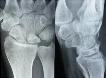
X-rayPreoperative wrist radiographs, anteroposterior and lateral views: stage II Kienböck disease with increased density of the lunate.
Source : Fouly et al., Journal of orthopaedic surgery and research, 2014, figure 1 · CC BY
The Lichtman classification dates from 1977. It remains the shared grammar of the whole literature and of every surgical discussion. It has two properties that must be held together: it governs treatment, and it is less reliable than is believed precisely where it governs most.
The principle is radiographic and progressive: normality, then increased density, then collapse, then osteoarthritis. The definitions below are those of the modified version, as published2.
| Stage | What the radiograph shows | What the surgeon does, in practice | What that changes for the physiotherapist |
|---|---|---|---|
| I | Nothing. Normal architecture and density of the lunate. The diagnosis is made on MRI, on a low T1 signal. | Non-operative treatment as first line: a splint or a below-elbow cast, for at least three months. In the ASSH survey, 74 % choose immobilisation first, then a radial shortening osteotomy if symptoms persist. | This is the stage where the trajectory can still be influenced. The physiotherapist has no treatment to offer here, but he is the one who can get the diagnosis made. After immobilisation, he picks up the consequences. |
| II | Increased density, diffuse sclerosis of the lunate. The architecture of the wrist is not compromised. | A shift towards surgery: 63 % of surgeons prefer to operate, mostly by radial shortening osteotomy. The aim: to unload the lunate. | Relative unloading, maintenance of range of motion on the operated side and of the proximal chain. Strength is not worked against a bone that is being unloaded. |
| IIIA | The lunate has collapsed, but carpal alignment and height remain preserved. | With negative ulnar variance, 69 % choose radial shortening. With neutral or positive variance, answers scatter: capitate shortening, vascularised bone graft. | Controlled loading only. The carpus is still holding: the aim is not to push it towards the next stage. |
| IIIB | Joint collapse, proximal migration of the capitate, palmar flexion of the scaphoid. The carpus is becoming disorganised. | Salvage procedures dominate: proximal row carpectomy (42 %), intercarpal arthrodesis (21 %), total wrist arthrodesis (10.7 %). | After surgery only. No loaded strengthening on a carpus that is collapsing can be justified. |
| IIIC | Complete coronal plane fracture of the lunate, whatever the morphology of the bone and of the wrist may otherwise be. | A stage added to the classification in its own right: the coronal line changes the strategy independently of everything else. It is seen on CT or MRI, rarely on the radiograph alone. | Not applicable outside the surgical context. Worth knowing in order to understand an operative report. |
| IV | Collapse of the lunate and radiocarpal or midcarpal osteoarthritis. | 87 % of surgeons choose proximal row carpectomy or arthrodesis. Motion-preserving options exist and give acceptable results30. | Rehabilitation of an osteoarthritic or operated wrist: functional gain, adaptation of movement, joint protection. No prospect of restoration. |
The tipping point is IIIA against IIIB, and it is the most fragile
This table reveals a boundary: on one side of IIIA, the aim is to save the lunate; on the other, it is abandoned in order to save function. It is the heaviest decision in this disease, and it is the one the radiograph discriminates least well. Goldfarb and his team measured this as early as 2003: overall reproducibility of the classification was good (kappa 0.63), but that of stage IIIA fell to 0.388.
The reproducibility of the classification has been measured three times, twenty years apart, with different protocols. The results do not agree, and the divergence is instructive.
Is the Lichtman classification reproducible?
Interobserver kappa coefficients of agreement, in three independent studies.
Jafarnia K et al., Journal of Hand Surgery American 2000;25(3):529-53414 : mean absolute agreement 74 %, mean weighted kappa 0.71, intraobserver reproducibility 79 % (kappa 0.77). Goldfarb CA et al., same journal, 2003;28(1):74-808 : adding a radioscaphoid angle of 60 degrees to subdivide stage III raises overall agreement to 0.81 and that of stage IIIA to 0.75. Aydemir AN et al., Joint Diseases and Related Surgery 2020;31(1):34-389 : 30 readers split between residents, orthopaedic surgeons and hand specialists. The bars are proportional to a scale from 0 to 1.
How should this gap from 0.71 to 0.203 be read? The protocols differ, and that is exactly the point. Jafarnia and Goldfarb had films read by four observers, all involved in the study and all specialists. Aydemir had forty films read by thirty professionals of three levels of experience, which is much closer to the real world where a film is read by whoever is there. His conclusion is explicit: “the Lichtman classification alone is insufficient and must be supplemented by other imaging and measurement techniques”.
A classification that governs treatment and whose agreement between readers is 0.203 under real conditions does not say what should be done. It says what should be considered.
David Lichtman has not defended his system as an absolute. With Bain and Pientka, he published in 2017 and then in 2022 two successive proposals that add two further classifications to his own : a classification based on contrast-enhanced MRI, which assesses perfusion and viability, and an arthroscopic classification based on the state of the articular cartilage, known as the Bain and Begg classification738.
The guiding idea of these two papers is that the radiograph is not enough because it answers only one question out of three. A lunate can be deformed and alive, or preserved in appearance and dead; it can carry intact cartilage on a collapsed bone. The authors therefore propose crossing three axes: bone, perfusion, cartilage. The title of the 2022 paper, Precision Medicine for Kienböck Disease, says clearly enough that radiographic staging on its own is regarded by its own authors as a crude tool. The review by the American Academy of Orthopaedic Surgeons published in 2020 goes so far as to describe the disease as “well known but poorly understood”, and the very abundance of treatment options it lists is the symptom of that uncertainty6.
The series published by MacLean and Bain in 2021 illustrates what a decision based on the state of the joint rather than on the radiograph alone produces: 27 patients, median follow-up of ten years, pain down from a median of 5 to 2 on the visual analogue scale, 18 of the 20 working patients back at the same level of work, and only one secondary recourse to a salvage procedure36. A case series, without a control group: the result is encouraging, it does not establish the superiority of the approach.
Key points from this chapter
It is the most cited hypothesis in the whole literature on this disease, and the one that governs the most frequent surgical choice. It is solidly associated with the disease. It has not been demonstrated to be its cause, and the distinction has practical consequences.
Ulnar variance is the difference in height, on a posteroanterior radiograph, between the distal articular surface of the ulna and that of the radius. It is said to be negative when the ulna is shorter than the radius. The mechanical reasoning is immediate: if the ulna does not carry its share, the load transmitted by the hand passes preferentially through the radius, therefore through the lunate, which receives it overhanging the radial edge.
This reasoning has a direct surgical consequence: shorten the radius to restore the distribution. That is the radial shortening osteotomy, the most commonly performed operation in this disease. In the survey of members of the American Society for Surgery of the Hand, 90 % of surgeons say they change their strategy according to ulnar variance28.
The association exists and it is robust. Bonzar and his team compared the ulnar variance of 44 affected patients with that of 99 controls from a general clinic, on standardised radiographs. They found, in both groups, a relationship between age and variance, and confirmed the association between negative variance and the disease after adjustment for age17.
The decisive work came fifteen years later, from a team in Tübingen. Stahl and his co-workers did three things in the same paper: a case-control study of 81 patients against 212 healthy controls, a meta-analysis of six case-control studies, and a systematic confrontation with the nine Bradford Hill criteria of causality16.
Strong association, causality not established
The three results of Stahl et al. 2013, which point in opposite directions.
After Stahl S, Stahl AS, Meisner C, Hentschel PJH, Valina S, Luz O, Schaller HE, Lotter O. Plastic and Reconstructive Surgery 2013;132(4):899-90916. The authors conclude that the observed association probably reflects a selection bias rather than causality, cross-sectional imaging having been performed only in affected patients in all the studies examined.
The second result deserves a pause. If negative ulnar variance caused the disease, an affected patient should have an affected wrist more negatively varied than his healthy wrist. The opposite is observed in three cases out of four. Yet the two wrists of the same subject are exposed to the same genome, the same hormones and much the same life. This result does not cancel the association, but it moves the question: negative variance could be a marker of a predisposition, not the mechanism of injury.
The third point is methodological and it is severe. In all the included studies, MRI was performed only in the affected group. In other words, the controls were never explored with the examination capable of detecting early forms. Some of them could therefore have carried a stage I Kienböck's disease that nobody looked for. The bias works exactly in the direction that inflates the association.
There is a more elementary problem, known since 1982 and regularly forgotten. Epner, Bowers and Guilford showed that the measured ulnar variance changes with the position of the forearm at the time of the film : supination increases the measurement of negative variance, pronation reduces it18. Wrist deviation and the orientation of the beam in the longitudinal plane also influence the result. The authors proposed a standardised radiographic protocol, with internal landmarks, and recommended applying it to all wrist films.
Forty years later, ulnar variance is still measured on films whose standardisation is not always documented. A parameter that decides on an osteotomy and that moves with forearm rotation deserves to be read with that in mind.
The systematic review by the same Tübingen team went through 220 references published since the original description of 1910. One hundred and fifty-two articles were retained, and 140 of them, that is 92 %, are level IV evidence, that is case series19.
The four most discussed aetiological hypotheses
Number of articles addressing them, out of the 152 retained by the systematic review.
After Stahl S, Stahl AS, Meisner C, Rahmanian-Schwarz A, Schaller HE, Lotter O. BMC Musculoskeletal Disorders 2012;13:22519. The review concludes that Kienböck's disease does not meet the International Labour Organization criteria defining an occupational disease related to vibration, while calling for studies of a better level of evidence. One article may discuss several hypotheses: the percentages do not add up.
This finding is worth knowing in France, where the question of occupational attribution comes up regularly for trades exposed to vibrating tools. The state of the literature allows neither confirmation nor refutation of the link: the authors say so in those terms, and that absence of a conclusion is itself the information.
Wassef's series of 57 cases, published in 2025, tested a set of radiographic parameters against the stage at the time of diagnosis. Two stand out: a reduced radial inclination is associated with stage IIIB or beyond at the first consultation, and a reduced ulnar variance is as well5. The mean values in the series were 21.9 degrees of radial inclination, 10.7 millimetres of radial height and an ulnar variance of minus 1.23 millimetres.
This is a retrospective single-centre level IV study, with an exploratory model. The authors accordingly phrase their conclusion conditionally: this correlation “suggests that it could be a risk factor for progression”. To be taken as a lead, not as an established fact.
Key points from this chapter
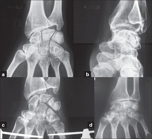
X-rayWrist radiographs at four time points: preoperative anteroposterior and lateral views showing stage II Kienböck disease, then postoperative films at 8 weeks and at 5-year follow-up showing preserved carpal height.
Source : Meena et al., Indian journal of orthopaedics, 2009, figure 2 · CC BY
We have to start with the conclusion, because it changes the reading of everything that follows: only one randomised trial has compared two treatments of Kienböck's disease with each other, on numbers of about ten patients per arm, and it finds no difference. Everything else is case series, pooled by systematic reviews. What is written below describes what is done and what has been observed, never what has been shown to be superior.
A Europe PMC query restricted to the publication type “randomised controlled trial”, searching every name of the disease, returns only two records for more than a century of literature.
The first is the only trial comparing two treatments with each other. Mazhar and his team randomly allocated patients at stage II or III between radial shortening and capitate shortening, with clinical and radiological assessment at 6 and 12 months. Fifty-two patients were assessed for eligibility; the paper reports 12 patients in the radial shortening group and 17 still present at the end of the study. None of the outcome measures differed significantly between the groups : range of motion, grip strength, pinch strength. The authors conclude that capitate shortening, performed through a smaller incision and in less time, may be suitable for stages II and III whatever the ulnar variance31.
This result must be read for what it is worth. About ten patients per arm does not allow equivalence to be concluded: the absence of a significant difference on such numbers is the expected result even if a real difference exists. And the abstract does not clearly report the number finally analysed. This is the best comparative level of evidence available for this disease.
The second randomised trial does not compare strategies: it tests low-intensity pulsed ultrasound on healing at the osteotomy site after forearm shortening, in 27 patients, with a 27 % reduction in the time to cortical union40. Useful, but that is a question of an adjunct, not of treatment choice.
The systematic review by Wang et al. devoted to stages IIIA and IIIB screened 1,489 titles to retain 30 articles. Their quality assessment gives 3 low-quality studies and 27 of moderate quality, none of high quality. The authors' conclusion: all modalities provide pain relief and functional improvement, and motion-preserving procedures give clinical results comparable with those of salvage procedures at both stages29.
What the evidence is worth, modality by modality
The nature of the data available for each option, and what they allow us to conclude.
Synthesis of the levels of evidence after Shin YH et al. 201826, Wang PQ et al. 202029 and 202330, Tsantes AG et al. 201935, Schuind F et al. 200825. The labels VERY LOW, NONE, LOW and MODERATE qualify the confidence in a comparison between treatments; they do not say that a treatment is ineffective, but that nothing allows it to be declared superior to another. The only randomised comparison available is that of Mazhar et al.31.
It is the first step, at stage I as often at stage II, and it is the one the physiotherapist meets downstream. The reported protocol is a splint or a below-elbow cast for at least three months2. In the ASSH survey, 74 % of surgeons choose it as first line at stage I28.
The review in the Journal of Bone and Joint Surgery British volume states its scope unambiguously: many patients are improved by temporary immobilisation of the wrist, which does not stop the progression of carpal collapse25. Both halves of that sentence must be taken. Immobilisation relieves. It does not cure.
The most worrying result of conservative treatment
In the comparison by Salmon, Stanley and Trail, 18 conservatively treated patients were followed for a mean of 3.6 years (from 1.5 to 9 years) and compared with 15 patients operated on with a radial shortening. The authors report that in some stage 3 patients treated conservatively, deterioration towards carpal collapse was rapid27.
This is not an argument against watchful waiting. It is an argument against unmonitored watchful waiting: a stage III wrist left to itself can tip quickly, and there is no clinical way of knowing which one will do so.
This is the best documented comparison, and its result is more nuanced than what is usually remembered. Shin and his team gathered 17 studies following patients for at least ten years: 5 studies of non-operative treatment, 12 of radial osteotomy, with comparable ages and stage distributions at the outset26.
Radial osteotomy against non-operative treatment, at ten years and beyond
Systematic review of 17 studies. The first two results separate the groups, the third does not.
After Shin YH, Kim JK, Han M, Lee TK, Yoon JO. Journal of Bone and Joint Surgery American 2018;100(14):1231-124026. The arc of motion values are given with their standard deviation in the publication: 107.4 ± 10.0 degrees against 88.8 ± 13.2 degrees. The bars are proportional to scales from 0 to 40 % and from 0 to 125 degrees.
This dissociation is the central message of the treatment of Kienböck's disease. Surgery improves what the patient experiences without changing the radiographic fate of his bone. We do not operate to stop the disease, we operate to live better with it.
Two series with very long follow-up confirm this order of magnitude. Watanabe et al. followed 19 patients operated on with a radial shortening, of whom 13 answered the questionnaire, with a mean follow-up of 21 years: mean DASH score of 8 out of 100, motion at 81 % in flexion and 82 % in extension of the opposite side, grip strength at 88 %; but radiographic progression in 6 of the 12 assessable patients32. Matsui et al., on 11 wrists followed for a mean of 14.3 years, report a modified Mayo score of 92 and a DASH of 5, with no progression of stage33. Two series, two opposite radiographic results, on numbers of about ten patients: that is exactly the level of certainty available.
| Procedure | Target stages | What the data report |
|---|---|---|
| Radial shortening | I to IIIA, negative variance | Moderate to severe pain in 5.7 % against 23.2 % without surgery at ten years and beyond; arc of motion nearly 19 degrees greater; radiographic progression unchanged26. |
| Capitate shortening | IIIA, neutral or positive variance | An alternative when radial shortening is ruled out by the variance. Surgeons' answers scatter in this situation28. |
| Metaphyseal core decompression | Early and advanced | Systematic review of 18 studies, 382 patients, mean age 34.5 years: return to work in 91 to 100 %, reoperation rate 4.2 to 8.3 %, variable radiographic progression with no consistent correlation with the clinical course37. |
| Vascularised bone graft | II to IIIB | Systematic review of 917 patients across all indications: in Kienböck's disease, progression despite the procedure in 13 % ; strength and pain clearly improved, motion not significantly improved35. |
| Proximal row carpectomy | IIIB and IV | Review of 6 series at more than ten years, 147 patients: grip strength at 68.4 % of the opposite side, 21 failures (14.3 %) requiring revision. Better results expected in early stages and outside heavy manual work34. |
| Intercarpal arthrodesis, total arthrodesis | IIIB and IV | Chosen by 21 % and 10.7 % of surgeons at stage IIIB, and predominant at stage IV along with proximal row carpectomy28. Motion is sacrificed for the sake of freedom from pain. |
| Motion-preserving procedures at stage IV | IV | 24 studies, 114 patients: flexion-extension arc of 95 degrees against 73 for salvage procedures (p = 0.0001); grip strength 86 % against 79 % (difference not significant, p = 0.28)30. |
The childhood form is not the same disease, and it is important not to transpose. Irisarri, Kalb and Ribak report 13 cases and a review of the literature: in children under 12, the prognosis is very good after immobilisation alone. Persistent symptoms may indicate surgery, probably a radial shortening, in some older adolescents, with a result that appears better than in adults39. The authors indeed propose to reserve the term lunatomalacia for this form, to distinguish it explicitly from the adult disease.
This contrast says something useful: in a growing bone, with a potential for revascularisation, the natural history is favourable. In the adult it is not, and no rehabilitation modality restores that potential.
Key points from this chapter
This chapter is the most important in the article, and the shortest to sum up: rehabilitation does not revascularise a lunate. What follows sets out why, what that forbids, and what remains, which is not nothing.
Nothing. It has to be put that way. A Europe PMC query crossing Kienböck's disease with physiotherapy, physical therapy or occupational therapy brings up articles where rehabilitation appears only as a passing mention within a surgical protocol : “proximal row carpectomy with rehabilitation”, “conservative treatment then surgery and rehabilitation”. No study has assessed a rehabilitation programme in this condition, neither as a treatment, nor as an adjunct, nor against a comparator.
This absence is not surprising for a disease whose incidence is estimated at 7 per 100,0002. It is simply the fact we have to work with. An article presenting a rehabilitation protocol for Kienböck's disease would be making it up.
The reasoning rests on three observations drawn from the previous chapters.
First, the problem is vascular and structural. The lunate receives on average 2.3 palmar vessels and 1.4 dorsal vessels, of the order of 118 to 136 micrometres in diameter, and two cadaveric specimens out of fourteen have no dorsal supply at all20. No rehabilitation modality creates a nutrient vessel. That is precisely what surgery attempts with vascularised grafts, and it fails to prevent progression in 13 % of cases despite a transplanted arterial supply35.
Second, even unloading surgery does not stop radiographic progression. That is the result of Shin et al. at ten years26 and of Salmon et al. at medium term27 : radial shortening relieves pain without reversing or preventing carpal collapse. If mechanically removing part of the load with an osteotomy is not enough, it would be unreasonable to expect more from a programme of exercises.
Third, symptoms do not follow the disease. A patient who is doing better under rehabilitation is not a patient whose lunate is doing better. Van Leeuwen et al. established this in the most direct way: symptoms are not a good indicator of severity or of prognosis3. Clinical improvement under treatment can perfectly well accompany structural worsening. It is the most dangerous trap of this condition for a rehabilitation professional, because it rewards the wrong decision.
The reflex to defuse: strengthening grip
The patient complains that his hand no longer grips. The dynamometer confirms it. The professional reflex is to work on grip strength.
But gripping means compressing the central column of the carpus, therefore loading the lunate, precisely what all the surgery of this disease seeks to avoid : joint-levelling osteotomies, capitate shortening and metaphyseal core decompression have no other aim than to unload this bone27.
The pathophysiological model of Bain and Lichtman points the same way: in the active subject, it is repeated loading that produces the initial subchondral stress fracture, and collapse occurs when this fracture becomes comminuted or crosses the bone in the coronal plane21. This model is presented by its authors as a postulate, not as a demonstration. But there is no competing model that would make loading harmless.
Practical conclusion: faced with an unexplained fall in grip strength, the first question is not “how do I strengthen it” but “why is it falling”. As long as the answer is unknown, loaded strengthening of the wrist has no justification.
| What is within his remit | What is not |
|---|---|
| Screening. Recognising a trajectory that is not going the right way, putting numbers on it, and referring on. It is the strongest lever available to him, given a mean diagnostic delay of 33.3 months5. | Making the diagnosis. It is made on imaging, and the imaging that counts is MRI. |
| Measuring and documenting. Range of motion, strength on a dynamometer, a validated functional scale, with their dates. That is what makes the doubt transmissible to the referring doctor49. | Staging. The Lichtman stage is read on imaging, and even among specialists there is no consensus on it9. |
| Recovering after immobilisation. Three months of cast or splint leave stiffness, muscle wasting and a loss of sensory input, and those do fall squarely within rehabilitation2. | Revascularising the lunate. No modality does that. The vascularised graft itself fails in 13 % of cases35. |
| Supporting the perioperative period. Preparing before, recovering after. Surgical series report high return-to-work rates, up to 91 to 100 % after metaphyseal core decompression37, and 18 working patients out of 20 back at the same level in the MacLean and Bain series36. | Stopping carpal collapse. The unloading osteotomy does not achieve it26. |
| Preserving function. Useful range of motion, adaptation of occupational and domestic movements, joint protection, management of load across the day. | Restoring previous function. After proximal row carpectomy, strength plateaus at around 68 % of the unaffected side34. |
| Informing. The quality of the information available online about this disease is poor and its reading level too high48. A patient well informed by his clinician is better served than by his own searching. | Reassuring about the course. Nobody knows which lunate will collapse. Promising stabilisation is not defensible. |
| Treating the comorbidities of the wrist. Shoulder or elbow stiffness set up by the immobilisation, an associated nerve entrapment, a disorganised proximal chain: all of that is the ordinary business of the physiotherapist. | Loaded strengthening of grip as long as the diagnosis has not been made and the stage is not known. |
The physiotherapist is not powerless in the face of Kienböck's disease. He is powerful at one precise moment, before the diagnosis, and modest at all the others.
Two results converge on the same piece of advice. The systematic review of proximal row carpectomy notes that long-term results are worse in patients doing heavy manual work34. And the analysis of the SPARCS database on 499 operated patients identifies workers' compensation status as a factor increasing the risk of reoperation24.
The practical implication is direct and it falls to the physiotherapist: analysis of the workstation, the search for movement adaptations, referral to the occupational physician. In a tiler, a mechanic or a hairdresser, that work is worth more than one extra mobilisation session.
Three months minimum of below-elbow immobilisation is the protocol reported at stage I2. The patient who arrives in the practice after that period presents a perfectly classical picture, and for once entirely within the rehabilitation professional's field of competence:
On this ground, rehabilitation does exactly what it knows how to do. The difference from a post-fracture immobilisation lies in one point, and it must be kept in mind: the progression of loading must remain under the surgeon's control, because the bone that was being spared is still necrotic, and because resuming weight-bearing through the palm, carrying a load or a screwing movement does not mean the same thing as on a healed radius.
Key points from this chapter
The cases below are all published, all identified by their PMID. They were chosen because they show what pooled series erase: the path by which a patient reaches the diagnosis, and what he went through beforehand.
A 32-year-old right-handed woman, a neonatal intensive care nurse, attends a sports medicine clinic for left wrist pain of two months' duration. The mechanism is unremarkable: hypersupination of the wrist while catching her daughter as she fell41.
What follows is the typical scenario. Radiographs had been taken three weeks after the injury: they showed no fracture, no dislocation, no malalignment. She had nevertheless been told she had a stress fracture, and she had worn a cast for six weeks. After it was removed the pain persisted; she was using an over-the-counter thumb splint, for comfort.
It was MRI that settled it: diffuse low signal of the lunate on T1 sequences, high T2 signal on fluid-sensitive sequences, that is avascular necrosis. Lichtman stage I, the stage at which the radiograph shows nothing by definition. Management, conducted with a hand surgeon, combined a short below-elbow cast then an orthosis, occupational therapy rehabilitation and a home exercise programme, with resolution of the pain.
What this case teaches
A 41-year-old woman, with no history of trauma, attends for chronic left wrist pain with restricted movement, which had worsened over some twenty days. Blood tests are of little help: erythrocyte sedimentation rate of 14 millimetres per hour, slightly prolonged clotting time, the rest normal. Imaging shows collapse of the lunate, joint space narrowing, subchondral cysts and secondary osteoarthritis. Treatment was a proximal row carpectomy, with clear relief of pain and functional improvement. The authors entitled their paper “Diagnostic Hurdles”42.
A 29-year-old woman, with no trauma and no classical risk factor, attends for progressive left wrist pain with restricted motion, of six months' duration. Radiographs, CT and MRI confirm avascular necrosis of the lunate at stage IIIB. After proximal row carpectomy, the DASH score falls from 62 preoperatively to 8 at one year, and the Mayo score rises from 45 to 85. At one year the patient is pain-free at rest, with satisfactory motion and a return to activities of daily living44.
What these two cases have in common is that they contradict two received ideas. The disease is not confined to men doing heavy manual work: neither of these two patients had any trauma or identified occupational exposure. And the delay before consulting is nothing out of the ordinary, six months and twenty days of worsening, which did not prevent an already disorganised carpus from being found.
A series of three cases published in 2025 illustrates well how the stage governs management43.
| Patient | Delay | Stage | Imaging and management |
|---|---|---|---|
| Man, 20 years, right-handed | 4 months | IIIa | Radiographs: collapse and fragmentation of the lunate, joint spaces preserved. MRI: heterogeneous low T1 signal with marrow oedema, reduced enhancement after injection. Conservative treatment: immobilisation and activity modification. |
| Man, 30 years, left-handed, manual worker | 6 months | IV | CT: fragmentation, sclerosis and radiolunate joint erosion. Proximal row carpectomy, followed by rehabilitation. |
| Woman, 28 years | 3 months | IV | MRI: flattened and fragmented lunate with low T1 signal, subchondral cysts, overall absence of enhancement, pancarpal degenerative change. Total wrist arthrodesis. |
Three months of symptoms for a stage IV in a 28-year-old woman: the delay declared by the patient does not tell us how long the disease has been developing. Pain that appears is often the moment when an already diseased bone decompensates, not the moment when the disease begins.
A 17-year-old adolescent, right-handed, presents with severe and progressive wrist pain, resistant to conservative treatment, with a stage IIIB comprising collapse of the lunate and fixed rotation of the scaphoid. A proximal row carpectomy was performed. At two years he is pain-free, with full motion and strength, and he has gone back to competitive basketball. The authors stress that this result questions the usual contraindications based on age45.
A 74-year-old man attends for numbness of the right hand of several years' standing, and an inability to extend the middle and ring fingers that appeared two months earlier. The diagnosis made is a subcutaneous rupture of the extensor tendons caused by stage IV Kienböck's disease complicated by carpal tunnel syndrome. Treatment combined endoscopic carpal tunnel release and tendon transfers, with improvement at two years46.
This second case deserves to be known by every physiotherapist who treats hands. Advanced Kienböck's disease is not only a painful wrist: bone fragments and remodelling can wear a tendon through to rupture, and the picture then presents as a loss of finger extension. See the site's synthesis on carpal tunnel syndrome for the neurological side of this association.
A 32-year-old woman attends for chronic right wrist pain and a fall in grip strength of two years' duration. Examination finds tenderness over the lunate and restricted wrist motion. Radiographs and MRI show sclerosis and avascular necrosis of the lunate, classified at stage II. Management was first conservative, combining non-steroidal anti-inflammatory drugs and rehabilitation, before a radial shortening osteotomy, with clear improvement in pain and motion after surgery and rehabilitation47.
Two years of falling grip strength before the diagnosis was made, in a 32-year-old patient. It is very likely that a health professional saw that wrist during those two years. That is exactly the window in which a physiotherapist can act.
What these seven patients have in common
What this article really changes in a day at the practice comes down to a few actions, none of them spectacular.
The information available online about this disease is of poor quality: in an analysis of 38 websites, the mean quality score was 13.3 out of 30 and the reading level corresponded to a tenth grade of schooling, far above the level recommended for a patient with limited literacy48. In other words, the patient who searches alone will search badly.
Three messages hold up and deserve to be said:
No. Stage I of the Lichtman classification is defined precisely by a normal radiograph: the lunate there retains normal architecture and density2. Only MRI then shows the disease, as a diffuse low T1 signal with enhancement after injection. Faced with mechanical wrist pain that persists beyond four to six weeks of management, a normal radiograph is no longer an argument for exclusion.
None. There is neither a specific manoeuvre nor a pathognomonic sign. The usual presentation combines unilateral reduction of wrist motion, pain and swelling2, which overlaps the picture of many other wrist conditions. The diagnosis is made on imaging. The clinician's role is to recognise an abnormal trajectory and to document it.
No, and there is no evidence suggesting otherwise. The problem is a bone necrosis linked to a defect of blood supply, and no rehabilitation modality restores a vascular supply. The vascularised bone graft, which literally transplants an artery, itself fails to prevent progression in 13 % of patients35. Rehabilitation has a real role, but it lies before the diagnosis (screening) and after treatment (functional recovery).
Not without knowing the stage and without instructions from the surgeon. Gripping loads the central column of the carpus, therefore the lunate, which is what all the surgery of this disease seeks to unload: joint-levelling osteotomies, capitate shortening, metaphyseal core decompression738. Grip strength remains an excellent indicator for follow-up, measured on a dynamometer at regular intervals. It is not a training goal as long as the diagnosis and the stage are not established.
No. The synthesis published in the Journal of Bone and Joint Surgery British volume is explicit: many patients are improved by temporary immobilisation of the wrist, which does not stop the progression of carpal collapse25. Immobilisation nevertheless remains the first step at stage I, for at least three months2, and 74 % of the surgeons surveyed choose it as first line at that stage28.
The question has no general answer. No randomised trial has compared surgery with no surgery, and only one has compared two techniques with each other: radial shortening against capitate shortening, with no difference found in range of motion or in strength31. What is known, at more than ten years of follow-up, comes down to a systematic review of 17 studies: moderate to severe pain persists in 5.7 % of patients operated on with a radial osteotomy against 23.2 % of patients not operated on, and the arc of motion is 107.4 degrees against 88.8, but radiographic worsening is comparable in both groups26. The decision is taken case by case, with a hand surgeon.
It is associated with it, with an odds ratio of 3.58 in meta-analysis, but causality is not established: six of the nine Bradford Hill criteria do not support it, and in 75 % of cases the negative variance is identical or less marked on the unaffected side of the same patient16. The measurement itself varies with the position of the forearm at the time of the film18. That does not stop 90 % of surgeons from basing their strategy on this parameter28.
The state of the literature allows neither confirmation nor refutation. The systematic review that examined this question across 220 references concludes that none of the nine Bradford Hill criteria was supported for the relationship between hand-transmitted vibration and Kienböck's disease, and that the disease therefore does not meet the International Labour Organization criteria defining an occupational disease. The authors explicitly call for studies of a better level of evidence19.
It is good, and in no way comparable with that in adults. Irisarri, Kalb and Ribak report that in children under 12 the prognosis is very good after immobilisation alone; in older adolescents surgery may be necessary but the result appears better than in adults39. The authors propose to reserve the term lunatomalacia for this form, precisely in order to avoid transposing one prognosis to the other.
Rarely. Bilateral forms account for about 4 % of cases2. Bilateral involvement should prompt a search for a systemic cause, and registry studies note associations with corticosteroid therapy, diabetes, alcohol, coagulation disorders and rheumatic diseases2223.
Figures, not a diagnostic hypothesis. Flexion and extension range measured with a goniometer on both sides, grip strength on a dynamometer with its comparison with the opposite side, the score on a validated functional scale49, each with its baseline date and its current date, and a sentence stating the absence of progress under well-conducted management. The referring doctor remains free to decide on referral; he has the fact that justifies it.
Topography points the way first: Kienböck sits centrally and dorsally, ulnocarpal impaction on the ulnar side, with pain reproduced on ulnar deviation and loaded pronation, and an ulnar variance that is readily positive13. The confusion is above all a trap on imaging: ulnocarpal impaction produces lunate signal abnormalities described as Kienböck pseudo-lesions12. So it is the reading of the sequences, and not the mere presence of a signal abnormality, that separates them: reviews of imaging of the painful wrist set out the criteria to weigh up15.
Because the quality of the information available is poor, and this has been measured. Across 38 websites analysed, the mean quality score was 13.3 out of 30, the accuracy score 10.4 out of 12, and the reading level corresponded to a tenth grade of schooling, far above the level recommended for a patient with limited literacy48. The websites that were hardest to read were also the least accurate. The authors recommend directing patients to specialty society websites and referring back to the doctor in case of doubt.
The 48 references below all carry a PubMed identifier, checked one by one against the MEDLINE database (full author list, journal, year, volume, issue, pagination), and cross-checked against CrossRef for the main ones. Levels of evidence are given when the publication declares them. A methodological reminder: the disease is rare, almost all of this literature consists of case series and reviews of case series. Only one randomised trial compares two treatments with each other31.
Behind this article
An author who explains, a reviewer who checks.
How we write and check our content

Physiotherapist · co-founder of Physio Learning
Marked for life by his first four-hour lecture without a single image, he took a master’s in instructional design so that it would never happen to anyone again. He hunts down publication bias and unreadable slides with the same intransigence.

Head of scientific content
Physiotherapist specialising in neuro-musculoskeletal practice and holder of a master’s in public health. He checks the methodological rigour of every article: primary sources, levels of evidence, no exceptions.
