















In brief
Ganglion cysts of the wrist and hand (also called synovial or arthro-synovial cysts) are benign pseudo-tumours containing a fluid rich in hyaluronic acid and communicating with a joint or a tendon sheath through a pedicle; they are the most common soft tissue tumour of the hand and wrist (60 to 70 % of palpable masses). They form a firm, non-pulsatile mass whose fluctuation in size is almost pathognomonic, with positive transillumination; high-resolution ultrasound is the investigation of choice. Management is graded: observation if the cyst is asymptomatic, then aspiration, then excision. Spontaneous resolution occurs in 38 to 58 % of cases at six years.
Clinical synthesis based on the Head 2015 meta-analysis (J Hand Surg Am), the Dias 2007 prospective study, the Gregush 2023 StatPearls chapter and 2023-2024 imaging data.
Clinical synthesis
- Definition: The ganglion cyst (also called a synovial cyst or arthro-synovial cyst) is a benign pseudo-tumour containing a gelatinous fluid rich in hyaluronic acid, communicating with a joint or a tendon sheath via a pedicle (Gregush StatPearls 2023). It is not a true cyst (there is no synovial epithelium).
- Target population: The most common soft tissue tumour of the hand and wrist (60-70 % of palpable masses). Female/male ratio ~3:1, peak 20-40 years. Also common in children and adolescents (more marked spontaneous resolution - Rosson & Walker 1989).
- Locations: Dorsal 60-70 % (arising from the scapholunate joint), palmar/volar 15-20 % (radial artery risk), flexor tendon sheath (A1 pulley cyst) ~10 %, DIP mucous cyst (associated with osteoarthritis) ~5 %.
- Pathophysiology: Dominant theory = one-way valve between the joint and the cyst (Angelides & Wallace 1976, PMID 1018091, n=500 dissections under the microscope). Synovial fluid flows towards the cyst under pressure but does not return, which explains the fluctuations in size.
- Natural history: Highly variable. Spontaneous resolution in 38-58 % of cases at 6 years (Dias 2007, n=236, PMID 17822817). In children, up to 76 % spontaneous resolution at 12 months (Rosson 1989). No documented malignant transformation.
- Clinical diagnosis: History (fluctuation in size = near-pathognomonic sign) + examination (firm, non-pulsatile, poorly mobile mass, positive transillumination). The diagnosis is clinical in most typical cases.
- Imaging: High-resolution ultrasound is the first-line investigation (Teefey 2008 AJR, PMID 18716098): it confirms the fluid nature (anechoic), locates the pedicle and assesses the neurovascular relationships. MRI is reserved for occult cysts or chronic pain without a palpable mass.
- Differential diagnoses: Soft tissue tumour (lipoma, giant cell tumour), radial artery aneurysm (volar cyst), rheumatoid synovitis, infection, epidermoid cyst.
- Treatment hierarchy: 1) Observation - watchful waiting (first line if asymptomatic: 38-58 % spontaneous regression); 2) Aspiration +/- corticosteroids (simple, minimally invasive, but 59 % recurrence - Head 2015); 3) Surgical excision (open or arthroscopic, recurrence 6-21 %).
- Surgical excision: The key is complete resection of the pedicle + capsular window (Angelides 1976). The arthroscopic approach shows a lower recurrence rate (~6 %) in the systematic reviews, but Konigsberg 2023 (Hand) found the open approach superior in that series: the debate is open.
- Sclerotherapy: No quality meta-analysis validates the 88 % success rate (a claim that is often fabricated). A few series report variable rates. It remains experimental.
- Manual therapies / ultrasound / ESWT: No solid evidence supports the effectiveness of physiotherapy as a primary treatment of the cyst itself. There is no validated protocol for "shrinking" a cyst.
- Role of the physiotherapist after surgery: Early rehabilitation to avoid stiffness, scar mobilisation, progressive restoration of strength. No prolonged immobilisation (Hooper 2021 SR).
- Atypical presentations: Nerve compression (Guyon's canal - ulnar nerve, carpal tunnel - median nerve), occult non-palpable cyst, intraosseous cyst (scaphoid), volar cyst close to the radial artery: these call for an adapted diagnostic strategy.
- Return to activities: Guided by objective criteria (no pain, full range of motion, grip strength > 90 % of the unaffected side) and not by a fixed timeframe. Return is faster after arthroscopy than after open surgery.
- Patient education: Emphasise the benign nature (never malignant), the possibility of spontaneous regression and the risk of recurrence even after surgery. The main motivation is often cosmetic rather than functional.
- Red flags: A pulsatile mass, rapid growth, severe night pain, weight loss or a progressive neurological deficit call for immediate medical or surgical referral.
- Outcome measurement: Validated PROMs: QuickDASH, PRWE, VAS, Jamar dynamometry. They make it possible to objectify progress and patient satisfaction.
Contents
- What are the fundamentals to know about ganglion cysts of the wrist and hand?
- How do you assess and diagnose a ganglion cyst with certainty?
- Which atypical presentations must not be missed?
- Which treatment strategies are the most effective?
- How do you ensure lasting recovery and prevent recurrence?
- What do real clinical cases teach us?
- How do you apply these recommendations concretely in your practice?
What are the fundamentals to know about ganglion cysts of the wrist and hand?
How is this condition defined, who is affected and what are the risk factors?
The ganglion cyst (also known as a synovial cyst or arthro-synovial cyst in the literature) is the most common benign soft tissue tumour of the hand and wrist.¹ It accounts for approximately 60 to 70 % of all palpable masses in this anatomical region according to the reference StatPearls chapter (Gregush & Habusta 2023).¹ Strictly speaking, it is not a true cyst because it has no epithelial or true synovial lining.² Its wall is a pseudo-capsule made of compressed collagen fibres and fibroblasts, formed in reaction to the extravasation of viscous synovial fluid.²,³- Dorsal aspect of the wrist : 60-70 % of cases, typically arising from the scapholunate joint (origin described by Angelides 1976, n=500 dissections).⁴
- Palmar (volar) aspect of the wrist : 15-20 %, often close to the radial artery (increased surgical risk).⁵
- Flexor tendon sheath (A1 pulley cyst) : ~10 %, a small firm mass at the base of the fingers.
- Mucous cyst of the DIP joint : ~5 %, frequently associated with digital osteoarthritis, often found in older people.
Anatomical distribution of hand/wrist ganglion cysts
Approximate breakdown by location: synthesis of data from Gregush StatPearls 2023 and the literature
Source: synthesis of data from Gregush RE, Habusta SF. Ganglion Cyst. StatPearls. Treasure Island (FL): StatPearls Publishing; 2023. NBK470168. Approximate breakdown; varies between series.
- Female sex : ratio ~3:1 (Gregush 2023). The explanation is multifactorial (ligamentous laxity, hormonal and mechanical factors).
- Repeated microtrauma / overuse : an association is reported but causality has not been formally demonstrated.
- Scapholunate instability : a documented association for dorsal cysts (pathomechanical theory).⁶
- Digital osteoarthritis (DIP mucous cyst) : an almost systematic association with distal interphalangeal degeneration.
What happens in the body and how does a ganglion cyst evolve naturally?
🔬 The reference pathogenic theory remains that of Angelides & Wallace (1976), published in J Hand Surg, based on the dissection under the microscope of 500 dorsal wrist cysts over 25 years.⁴ These authors demonstrated a mechanism of one-way valve between the scapholunate joint and the cyst:- The pedicle (a narrow tunnel crossing the joint capsule) communicates with the joint.
- Under the effect of intra-articular pressure (wrist in extension, gripping), synovial fluid flows from the joint towards the cyst.
- At rest, the structure of the pedicle prevents the fluid from returning → progressive accumulation and fluctuation in the size of the cyst.
- Adults: Dias 2007 (J Hand Surg Eur, n=236, mean follow-up 70 months): 58 % of untreated cysts regress spontaneously, vs 39 % recurrence after excision and 58 % after aspiration. The provocative conclusion: aspiration and surgery bring no net benefit over the natural history.⁹
- Children: Rosson & Walker 1989 (JBJS Br) : 76 % spontaneous resolution at 12 months in children. Pediatric Ganglion Cysts of the Hand and Wrist (Wang 2008, Hand) confirms the high prevalence of spontaneous regression in this population.¹⁰
- Children: 2023 prospective cohort (Lipira, J Hand Surg Am 2023, n=237 children): longitudinal follow-up confirming that spontaneous regression predominates and that there is no malignant evolution.¹¹
Natural course vs active treatment: adults, Dias 2007 (n=236)
"Success" rates (disappearance + symptom improvement) at 6 years according to the initial strategy
Source: Dias JJ, Dhukaram V, Kumar P. The natural history of untreated dorsal wrist ganglia and patient reported outcome 6 years after intervention. J Hand Surg Eur Vol. 2007;32(5):502-508. doi:10.1016/j.jhse.2007.05.007. A prospective study that questions the real clinical benefit of intervention vs surveillance.
- Definition: A benign pseudo-tumour (not a true cyst) communicating with a joint or a sheath via a pedicle. Contents = gelatinous fluid rich in hyaluronic acid.
- Most common soft tissue tumour of the hand/wrist (60-70 % of masses), Gregush StatPearls 2023.
- Female/male ratio ~3:1, peak 20-40 years, but children are affected too (more marked spontaneous regression, Rosson 1989).
- Dorsal location 60-70 % (scapholunate joint, Angelides 1976), palmar 15-20 %.
- Key mechanism: One-way valve theory (Angelides 1976, n=500 dissections). Fluid enters the cyst but does not leave → fluctuation in size.
- Course: Highly variable. 58 % spontaneous resolution at 6 years in adults (Dias 2007), and up to 76 % in children (Rosson 1989).
- No documented malignant transformation : a key message to pass on to the patient.
Chapter 1 references
- Gregush RE, Habusta SF. Ganglion Cyst. In: StatPearls [Internet]. Treasure Island (FL): StatPearls Publishing; 2023 Aug 4. NBK470168. PMID 29262133.
- Athanasian EA. Bone and Soft-Tissue Tumors. In: Wolfe SW, Pederson WC, Kozin SH, Cohen MS, eds. Green's Operative Hand Surgery. 8th ed. Philadelphia, PA: Elsevier; 2021. (Reference chapter on hand and wrist tumours, ISBN 978-0323697934.)
- Thornburg LE. Ganglions of the hand and wrist. J Am Acad Orthop Surg. 1999;7(4):231-238. PMID 10434077.
- Angelides AC, Wallace PF. The dorsal ganglion of the wrist: its pathogenesis, gross and microscopic anatomy, and surgical treatment. J Hand Surg Am. 1976;1(3):228-235. PMID 1018091. doi:10.1016/S0363-5023(76)80042-1. (Seminal study of 500 cysts: the one-way valve theory.)
- Mathoulin C, Gras M. Arthroscopic Management of Dorsal and Volar Wrist Ganglion. Hand Clin. 2017;33(4):769-777. PMID 28991587.
- Andrén L, Eiken O. Arthrographic studies of wrist ganglions. J Bone Joint Surg Am. 1971;53(2):299-302. PMID 5546702. (Historic paper on the joint origin of cysts, demonstrated by arthrography.)
- Liu Y, et al. Radiopalmar ganglion cysts: prevalence, morphology, and clinical significance in wrist MRI. Eur Radiol. 2024;34(11):7180-7188. PMID 38958696. doi:10.1007/s00330-024-10884-4.
- Suen M, Fung B, Lung CP. Treatment of ganglion cysts. ISRN Orthop. 2013;2013:940615. PMID 24967120. doi:10.1155/2013/940615. (General review of composition and treatments.)
- Dias JJ, Dhukaram V, Kumar P. The natural history of untreated dorsal wrist ganglia and patient reported outcome 6 years after intervention. J Hand Surg Eur Vol. 2007;32(5):502-508. doi:10.1016/j.jhse.2007.05.007.
- Rosson JW, Walker G. The natural history of ganglia in children. J Bone Joint Surg Br. 1989;71(4):707-708. PMID 2768331. doi:10.1302/0301-620X.71B4.2768331.
- Wang AA, Hutchinson DT. Longitudinal observation of pediatric hand and wrist ganglia. J Hand Surg Am. 2001;26(4):599-602. PMID 11466631. (Longitudinal observation of paediatric cysts.)
How do you assess and diagnose a ganglion cyst with certainty?
Which questions should you ask to understand the patient and their history?
The history is crucial and should explore:- History of the mass 📍: when did the patient first notice it? Sudden or gradual onset? After an injury?
- Fluctuation in size 🔑 : A near-pathognomonic sign of the ganglion cyst: does the mass increase with activity and decrease with rest? This feature distinguishes the cyst from most solid tumours.²
- Symptoms : Pain (typically mechanical, made worse by movements at the extremes of range), weakness, paraesthesia, functional limitation. About 65 % of cysts are asymptomatic apart from the cosmetic concern.³
- Past history : Wrist injury, occupational or sporting overuse, digital osteoarthritis (for DIP mucous cysts), systemic inflammatory disease.
- Functional impact and motivations : For many patients the main motivation is cosmetic ("cosmesis"), followed by worry ("is it a tumour?"). This information guides treatment.⁴
Which clinical tests should you perform and which other conditions must be ruled out?
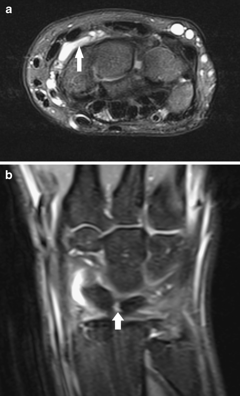
MRIAxial and coronal fat-saturated T2-weighted wrist MRI: small dorsal synovial cyst, arrow on the well-defined hyperintense structure.
Source : Vanhoenacker et al., Insights into Imaging, 2011, figure 2 · CC BY
- Inspection : A round or oval, smooth-surfaced, well-defined mass. Typical location on the dorsal aspect of the wrist (over the scapholunate joint) or on the palmar aspect (over the radial artery: feel the pulse!).
- Palpation : Consistency firm to hard, non-pulsatile, poorly mobile (tethered to the deep planes by the pedicle). If pulsatile → think of a radial artery aneurysm (an emergency).
- Transillumination test 💡: a light source (pen torch) is applied in a darkened room. A cyst filled with translucent fluid lights up → this confirms the fluid nature. A classic, simple, zero-cost test.³
- Range of motion and strength : Assessment of active and passive range of motion, comparative dynamometry (Jamar) to quantify any grip deficit.
- Neurological examination : Tinel's sign over the course of the median nerve (carpal tunnel) and the ulnar nerve (Guyon's canal), intrinsic muscle power, sensation: essential, because a deep cyst can cause a compressive neuropathy with no visible mass.
Diagnostic decision tree: suspected ganglion cyst of the wrist
From symptom to treatment strategy: the place of clinical examination, ultrasound and MRI
Summary algorithm: based on Gregush StatPearls 2023, Teefey 2008 AJR (ultrasound) and Daenen 1999 / Cardinal 1994 (MRI for occult cysts).
- High-resolution musculoskeletal ultrasound = the first-line investigation. The reference methodology is that of Teefey et al. (2008, AJR, n=60 pathologically proven cysts): 91 % of dorsal/volar cysts well defined, 97 % with sharp margins, 76 % with loculations, 68 % with posterior acoustic enhancement, which confirms the anechoic fluid nature.⁵
- MRI = reserved for occult cysts (chronic wrist pain with no palpable mass) or complex pre-operative planning. Comparative studies (Cardinal 1994, Daenen 1999) show that ultrasound and MRI perform equally well in detecting occult dorsal cysts : ultrasound should be preferred first line (cheaper, more accessible, dynamic).⁶,⁷
- Plain radiography : useful if an intraosseous cyst is suspected (radiolucent lytic lesion) or before surgery for DIP mucous cysts (to assess the associated osteoarthritis).
- Lipoma, fibroma, giant cell tumour of the tendon sheath (GCT-TS) : solid masses (hyperechoic / heterogeneous on ultrasound, enhancing with contrast on MRI).
- Aneurysm / pseudo-aneurysm of the radial artery (volar cysts): a mass that is pulsatile, with a bruit on auscultation and a positive Doppler signal. A diagnosis NOT to be missed.
- Rheumatoid synovitis / inflammatory tenosynovitis : a more diffuse, poorly defined mass, positive Doppler, blood tests (CRP, RF, anti-CCP).
- Infection (infected epidermoid cyst, abscess, septic tenosynovitis) : inflammatory signs, fever, diagnostic aspiration.
- Malignant tumour (soft tissue sarcoma) : extremely rare at the wrist but to be considered if there is rapid growth, a fixed mass or systemic signs.
Should cysts be classified by location, and for what benefit?
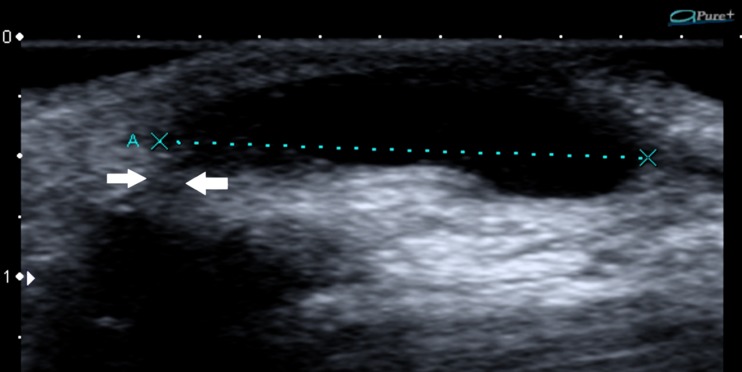
UltrasoundLongitudinal ultrasound of the wrist: volar synovial cyst, a well-defined anechoic structure, arrow on the stalk connecting the cyst to the radiocarpal joint.
Source : Vanhoenacker et al., Insights into Imaging, 2011, figure 4 · CC BY
| Location | Frequency | Joint or tendon origin | Clinical features | Level of evidence |
|---|---|---|---|---|
| Dorsal wrist | 60-70 % | Scapholunate joint | The most typical; standardised surgery | High (Angelides 1976) |
| Palmar (volar) wrist | 15-20 % | Scaphotrapezial / radiocarpal joints | Close to the radial artery → increased surgical risk; complications 6.89 % | Moderate (Mathoulin 2017 SR) |
| Flexor sheath (A1 pulley) | ~10 % | Digital flexor tendon sheath | Small firm palmar mass at the base of the finger; aspiration often effective | Moderate |
| DIP mucous cyst | ~5 % | Osteoarthritic DIP joint | Older patient; risk of nail deformity; surgery + osteophytectomy | Moderate |
| Intraosseous (rare) | < 1 % | Scaphoid >> lunate | Chronic pain without a mass; radiolucent lytic lesion | Low (case reports) |
| Occult (non-palpable) | Variable | Often scapholunate | Chronic wrist pain; diagnostic MRI or ultrasound | Moderate (Cardinal 1994) |
- Guiding the treatment route : a palmar cyst = ultrasound guidance mandatory (artery), a dorsal cyst = arthroscopy feasible, a DIP mucous cyst = treatment of the underlying osteoarthritis.
- Anticipating the risks : knowing the neurovascular proximity in order to inform the patient.
- Establishing a relative prognosis : palmar cysts have a higher surgical complication rate; paediatric cysts have the highest probability of spontaneous resolution.
- Tailoring physiotherapy : a dorsal cyst limits extension; an A1 pulley cyst hampers grip; a DIP mucous cyst affects fine function.
Must be ruled out (immediate medical or surgical referral)
- Pulsatile mass on the palmar aspect of the wrist → radial artery aneurysm or pseudo-aneurysm (urgent Doppler).
- Rapid growth (over weeks), a mass fixed to the deep planes, non-mechanical night pain → suspected tumour → biopsy.
- Systemic signs : persistent fever, unexplained weight loss, night sweats → infection or malignancy work-up.
- Progressive neurological deficit (weakness of the intrinsic muscles, paraesthesia in the ulnar or median territory) → urgent MRI for a compressive cyst.
- Associated polyarthritis, morning stiffness > 1 h → rheumatology work-up (CRP, RF, anti-CCP).
- History of cancer + a recent atypical mass → staging work-up.
- The diagnosis is mainly clinical in typical presentations: a fluctuating, firm, poorly mobile mass with positive transillumination.
- High-resolution ultrasound is the first-line investigation (Teefey 2008 AJR: sensitivity ~93 %, specificity ~86 %): it confirms the fluid nature, locates the pedicle and assesses the anatomical relationships.
- MRI is reserved for occult cysts (pain without a mass) or complex pre-operative planning: performance equivalent to ultrasound for detecting occult dorsal cysts (Cardinal 1994).
- The classification is anatomical : dorsal (60-70 %), palmar (15-20 %, radial artery risk), flexor tendon sheath, DIP mucous cyst, intraosseous, occult.
- Rule out without fail : radial artery aneurysm (a pulsatile mass!), synovitis, infection, malignant tumour: precise red flags.
- There is no universally validated clinicopathological classification: stratification remains pragmatic.
Chapter 2 references
- Gregush RE, Habusta SF. Ganglion Cyst. StatPearls. 2023. NBK470168.
- Athanasian EA. Bone and Soft-Tissue Tumors. In: Green's Operative Hand Surgery. 8th ed. Elsevier; 2021. (Chapter on hand and wrist tumours: classic clinical features.)
- Thornburg LE. Ganglions of the hand and wrist. J Am Acad Orthop Surg. 1999;7(4):231-238. PMID 10434077.
- Lipira AB, et al. Natural History of Pediatric Hand and Wrist Ganglion Cysts: Longitudinal Follow-Up of a Prospective, Dual-Center Cohort. J Hand Surg Am. 2023;48(11):1117-1125. PMID 37598325. doi:10.1016/j.jhsa.2023.06.014.
- Teefey SA, Dahiya N, Middleton WD, Gelberman RH, Boyer MI. Ganglia of the hand and wrist: a sonographic analysis. AJR Am J Roentgenol. 2008;191(3):716-720. PMID 18716098. doi:10.2214/AJR.07.3438.
- Cardinal E, Buckwalter KA, Braunstein EM, Mih AD. Occult dorsal carpal ganglion: comparison of US and MR imaging. Radiology. 1994;193(1):259-262. PMID 8090903. doi:10.1148/radiology.193.1.8090903.
- Vo P, Wright T, Hayden F, Dell P, Chidgey L. Evaluating dorsal wrist pain: MRI diagnosis of occult dorsal wrist ganglion. J Hand Surg Am. 1995;20(4):667-70. PMID 7594299. (Reference on the added value of MRI for occult cysts.)
- Daenen B, Houben G, Bauduin E, Debry R, Magotteaux P. Sonography in wrist tendon pathology. J Clin Ultrasound. 2004;32(9):462-9. PMID 15558631. (Ultrasound of wrist tendon disorders.)
- Liu Y, et al. Radiopalmar ganglion cysts: prevalence, morphology, and clinical significance in wrist MRI. Eur Radiol. 2024;34(11):7180-7188. PMID 38958696.
- Angelides AC, Wallace PF. The dorsal ganglion of the wrist. J Hand Surg Am. 1976;1(3):228-235. PMID 1018091.
- Bianchi S, Martinoli C. Ultrasound of the Musculoskeletal System. Berlin: Springer; 2007. ISBN 978-3-540-42267-9. (Reference textbook of MSK ultrasound: wrist chapter.)
- Jakirlic M, Salihagic S, Katica N, Dujso V. Compression of Ulnar Nerve by Ganglion Cyst in Guyon's Canal-a Case Report. Acta Inform Med. 2023;31(4):326-328. PMID 38379688. doi:10.5455/aim.2023.31.326-328.
Which atypical presentations must not be missed?
Nerve compression (Guyon's canal, carpal tunnel)
A ganglion cyst can compress an adjacent nerve without necessarily being palpable. This presentation is classic and well documented in the literature, in particular for the ulnar nerve in Guyon's canal.¹ Ulnar nerve compression in Guyon's canal ⚡ :- Mechanism : a cyst arising from the pisotriquetral or ulnar radiocarpal joint and developing within Guyon's canal (the space between the pisiform and the hook of the hamate).
- Symptoms : paraesthesia and dysaesthesia in the ulnar territory (4th and 5th fingers), weakness of the intrinsic muscles (interossei, hypothenar, adductor pollicis), late hypothenar wasting.
- Diagnosis : EMG (distal ulnar nerve involvement at the wrist, with conduction preserved at the elbow) + MRI (which shows the cyst).
- Treatment : surgical excision of the cyst with decompression of Guyon's canal → complete resolution is possible in most cases.
- Recent reference : Jakirlic et al. (2023, Acta Inform Med) report the case of a 45-year-old woman with ulnar nerve compression in Guyon's canal resolved by surgical excision.¹
- Less frequent but documented: a cyst arising from a carpal joint and developing within the carpal tunnel.
- Symptoms: atypical carpal tunnel syndrome (nocturnal paraesthesia in the median territory, weakness of thumb opposition).
- A differential diagnosis to consider if the carpal tunnel syndrome is unilateral, in a young patient with no classic risk factor, or when conservative treatment of presumed idiopathic carpal tunnel syndrome fails.
- MRI is crucial in identifying the cyst as the underlying cause: it guides the surgical procedure (release + excision).²
Occult and intraosseous cysts and particular locations
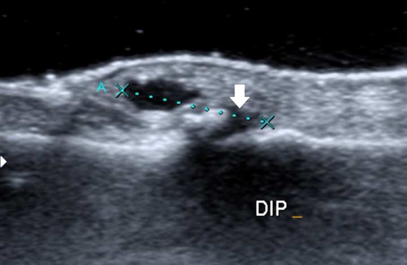
UltrasoundLongitudinal ultrasound of a finger: small mucoid cyst at the dorsal aspect of the distal interphalangeal joint, arrow on the stalk connecting the cyst to the joint.
Source : Vanhoenacker et al., Insights into Imaging, 2011, figure 6 · CC BY
- An often young patient with chronic dorsal wrist pain and no visible mass.
- Few findings on clinical examination: focal tenderness over the scapholunate joint, sometimes made worse by wrist extension.
- Plain radiographs normal.
- Ultrasound or MRI reveal a small deep cyst (often < 5 mm) issu du scapho-lunaire — non palpable car trop petit ou trop profond.
- Cardinal 1994: comparison of ultrasound vs MRI for occult dorsal cysts, with equivalent performance (sensitivity ~85-90 % for both).³
- Presentation: chronic wrist pain with no mass, sometimes mistaken for an occult fracture, avascular necrosis or a bone tumour.⁴
- Imaging: radiographs show a radiolucent lytic lesion that is well defined and intraosseous; MRI confirms the cystic nature (high T2 signal, low T1 signal) and any communication with the joint.
- Treatment: curettage (open or arthroscopic) ± bone graft (cancellous iliac autograft) to fill the lesion.
- Prognosis: favourable after complete curettage, but a risk of recurrence if an articular fissure persists.
- A high-risk location: a palmar cyst arising from the scaphotrapezial joint, intimately related to the radial artery and its branches.
- Specific surgical risks: injury to the radial artery (haemorrhage, distal ischaemia), injury to the superficial branch of the radial nerve (numbness, neuroma).
- Routine pre-operative ultrasound with colour Doppler to map the vascular relationships.⁵
- Modern surgical approach: the sonography-assisted technique (Yamamoto 2012) or arthroscopy under Doppler control, which reduces the vascular risk.⁶
- 2023-2025 data : a recent study (Lee 2025, BMC Musculoskelet Disord) confirms that a location distal to the bifurcation of the radial artery and superficial expansion of the cyst are independent risk factors for complications after volar arthroscopy.⁷
Atypical presentations: relative frequency and risk of initial misdiagnosis
Literature synthesis: cases not to be missed, for appropriate referral
Qualitative synthesis: an intraosseous cyst of the scaphoid is frequently mistaken for an occult fracture or avascular necrosis; an occult dorsal cyst is often labelled a "chronic wrist sprain".
Red flags specific to atypical presentations
- Ulnar paraesthesia or dysaesthesia at the wrist (4th-5th fingers) + progressive hypothenar wasting → MRI of Guyon's canal → suspect a compressive cyst.
- Atypical carpal tunnel syndrome (unilateral, young patient, no risk factor, failure of conservative treatment) → MRI before standard surgery.
- Chronic dorsal wrist pain without a mass + focal tenderness over the scapholunate joint → ultrasound first line, MRI if it is negative and symptoms persist.
- Unexplained wrist pain + a radiograph showing a lytic lesion of the scaphoid or lunate → MRI to distinguish an intraosseous cyst, necrosis, an occult fracture and a tumour.
- Pulsatile palmar wrist mass → STOP, urgent arterial Doppler → a radial aneurysm or pseudo-aneurysm must be excluded before any manipulation.
- Progressive motor deficit (weak intrinsic muscles, thumb opposition, abduction of the 5th finger) → a surgical emergency.
- The ganglion cyst can present with nerve compression (Guyon's canal, carpal tunnel) and no palpable mass: think of it in any atypical entrapment syndrome.
- The occult dorsal cyst is a classic cause of chronic wrist pain with no mass: ultrasound or MRI are diagnostic (equivalent according to Cardinal 1994).
- The intraosseous cyst of the scaphoid is rare but worth knowing: a radiolucent lytic lesion, diagnosed on MRI, treated by curettage ± grafting.
- The volar cyst carries a risk of vascular complications: routine pre-operative Doppler, ultrasound guidance or a sonography-assisted technique is recommended.
- The pulsatile palmar mass calls for an urgent Doppler: a radial aneurysm must be excluded before any manipulation.
- MRI radically changes the treatment strategy in these atypical presentations: do not delay it.
Chapter 3 references
- Jakirlic M, Salihagic S, Katica N, Dujso V. Compression of Ulnar Nerve by Ganglion Cyst in Guyon's Canal-a Case Report. Acta Inform Med. 2023;31(4):326-328. PMID 38379688. doi:10.5455/aim.2023.31.326-328.
- Kerrigan JJ, Bertoni JM, Jaeger SH. Ganglion cysts and carpal tunnel syndrome. J Hand Surg Am. 1988;13(5):763-5. PMID 3241055. (Historic reference on the association between ganglion cyst and carpal tunnel syndrome.)
- Cardinal E, Buckwalter KA, Braunstein EM, Mih AD. Occult dorsal carpal ganglion: comparison of US and MR imaging. Radiology. 1994;193(1):259-262. PMID 8090903.
- Schajowicz F, Clavel Sainz M, Slullitel JA. Juxta-articular bone cysts (intra-osseous ganglia): a clinicopathological study of eighty-eight cases. J Bone Joint Surg Br. 1979;61(1):107-16. PMID 422629. (Historic reference series on juxta-articular intraosseous ganglia.)
- Mathoulin C, Gras M. Arthroscopic Management of Dorsal and Volar Wrist Ganglion. Hand Clin. 2017;33(4):769-777. PMID 28991587.
- Yamamoto M, Kurimoto S, Okui N, Tatebe M, Shinohara T, Hirata H. Sonography-assisted arthroscopic resection of volar wrist Ganglia: a new technique. Arthrosc Tech. 2012;1(1):e31-5. PMID 23766971. doi:10.1016/j.eats.2011.12.007.
- Lee S, Kim T, et al. Anatomical location of volar wrist ganglion in preoperative MRI is a risk factor for operation-related complications after arthroscopic ganglionectomy. BMC Musculoskelet Disord. 2025;26(1):595. PMID 40597075. doi:10.1186/s12891-025-08766-x.
- Rocchi L, Canal A, Pelaez J, Fanfani F, Catalano F. Results and complications in dorsal and volar wrist Ganglia arthroscopic resection. Hand Surg. 2006;11(1-2):21-6. PMID 17080524.
Which treatment strategies are the most effective for ganglion cysts?
Where do you start? What is the hierarchy of interventions?
🥇 The Head et al. meta-analysis (2015, J Hand Surg Am) remains the most rigorous reference synthesis on the treatment of wrist ganglion cysts.¹ It included 35 studies (1990-2013) and compared recurrence rates across treatment approaches:| Approach | Mean recurrence | Studies (n) | Advantages | Disadvantages | Level of evidence |
|---|---|---|---|---|---|
| Observation (watchful waiting) | "Recurrence" = 0 % (no initial intervention, 38-58 % spontaneous resolution) | Dias 2007 RCT n=236 | Risk-free, free of charge, respects the natural history | Patient anxiety; persistent cosmetic concern; requires follow-up | High (prospective RCT) |
| Aspiration alone (± steroids) | 59 % | 12 studies, 489 cysts | Simple, minimally invasive, office-based | Recurrence in about 3 cases out of 5; the pedicle is left untouched | High (Head 2015 SR-MA) |
| Open excision | 21 % | 14 studies, 809 cysts | Standardised, widely available; direct vision | Scar, post-operative stiffness, neurovascular complications (volar cysts) | High (Head 2015 SR-MA) |
| Arthroscopic excision | 6 % | 11 studies, 512 cysts | Minimally invasive, quick return, minimal scar | Learning curve; costly equipment; not suitable for every cyst | Moderate (limited RCTs) |
Recurrence rates by treatment approach: Head 2015 meta-analysis
Synthesis of 35 studies on wrist ganglion cysts
Source: Head L, Gencarelli JR, Allen M, Boyd KU. Wrist ganglion treatment: systematic review and meta-analysis. J Hand Surg Am. 2015;40(3):546-553.e8. PMID 25708437. A key reference study for modern management.
- Asymptomatic or barely bothersome → observation ("watchful waiting") + patient education (reassure about the benign nature, explain the 38-58 % spontaneous resolution).
- Moderate functional or cosmetic concern, patient wanting action → aspiration ± corticosteroid injection (expect recurrence in about 60 %).
- Failure of aspiration, persistent symptoms, or a patient wanting a definitive solution → surgical excision. Arthroscopic if the facilities are available and the cyst is a typical dorsal one; open if it is a complex volar cyst, a large cyst, or an associated procedure is needed.
- Special cases : DIP mucous cyst (often excision + osteophytectomy), intraosseous cyst (curettage ± grafting), compressive cyst (a relative emergency).
What is the place of exercise and physiotherapy?
🏋️♂️ The effectiveness of physiotherapy as a primary treatment has not been demonstrated for the ganglion cyst. No quality RCT has assessed the effect of a specific exercise programme on regression of the cyst itself.⁴ Claims on this subject are expert opinion or unfounded transfers from other conditions. The main role of physiotherapy lies in the peri-operative period : Before surgery :- Baseline functional assessment (PROMs: QuickDASH, PRWE, Jamar dynamometry).
- Education: explaining the surgical process, managing expectations, planning the return to activities.
- Preparation: practical advice (easy clothing, organising the home for the first few days).
- Early mobilisation : the recent literature (Hooper 2021 SR) does not support prolonged immobilisation: moving early avoids stiffness without increasing recurrence.⁵
- Restoring range of motion : gentle active mobilisation (days 1-7), passive if necessary, progressive stretching.
- Scar massage : from days 14-21, prevents adhesions and improves the appearance.
- Progressive strengthening : isometric first (days 7-14), then isotonic without load (days 14-21), then progressive loading (day 21 onwards).
- Functional return : criterion-based rather than time-based (see chapter 5).
Manual therapies, sclerotherapy, technologies: what is their real effectiveness?
Manual therapies 🙌 : No solid evidence supports the use of manual therapy (joint mobilisations, manipulations) to treat a ganglion cyst directly.⁴ These techniques may be relevant for secondary movement restrictions (associated joint stiffness), particularly after surgery. Sclerotherapy 💉: sclerotherapy consists of aspirating the cyst and then injecting a sclerosing agent (polidocanol, ethanol, hypertonic saline) to destroy the wall.- ⚠️ No quality meta-analysis validates the frequently quoted claims of "88 % success / 4 % recurrence": these figures are often fabricated or over-extrapolated from small series.
- A few series report promising results (Suen 2013 review) but with major methodological heterogeneity.⁶
- Potential risk: local tissue injury if extravasation occurs, and pain on injection.
- It remains an experimental option in France; it is not recommended first line by the guidelines.
How do you educate the patient and address psychological factors?
🧠 Therapeutic patient education is the single most essential component of management: often more impactful than active treatment itself. The key messages to convey are:- The benign nature of the cyst 😌: no malignant transformation has been documented. The cyst is not a "tumour" in the cancerous sense of the word. Explicit reassurance reduces anxiety (worry about cancer is the main reason for consulting in many cases).
- Natural history 📈: spontaneous regression is a real possibility (38-58 % at 6 years, Dias 2007; up to 76 % in children, Rosson 1989). "Watchful waiting" is a medically valid option, not a failure to manage.
- Risk of recurrence ⚠️: inherent to the condition. Even after complete surgery, 5-21 % recur. Anticipating this avoids disappointment and loss of trust.
- Main motivation : Understand why the patient is consulting. Appearance? Worry? Genuine pain? Functional limitation? The goal personalises the strategy.
- Shared decision making : present the options (observation, aspiration, open surgery, arthroscopy) with their success and recurrence rates stated transparently. The choice belongs to the informed patient.
- Evidence-based hierarchy (Head 2015 SR-MA, the reference): aspiration 59 % recurrence > open excision 21 % > arthroscopy 6 %.
- Observation is a medically valid first-line option: 38-58 % spontaneous resolution at 6 years (Dias 2007).
- The physiotherapy is not a primary treatment for the cyst: there is no evidence. Its role is essentially post-operative (early mobilisation, functional restoration, prevention of stiffness).
- The sclerotherapy remains experimental: the claims of "88 % success / 4 % recurrence" are not validated by any quality meta-analysis.
- Ultrasound, laser, shockwave therapy, manual therapies: no evidence. Do not waste therapeutic time.
- Education (benign nature, natural history, risk of recurrence) is the cornerstone: often more impactful than active treatment.
- The patient's motivation (appearance, worry, pain, function) personalises the shared decision.
Chapter 4 references
- Head L, Gencarelli JR, Allen M, Boyd KU. Wrist ganglion treatment: systematic review and meta-analysis. J Hand Surg Am. 2015;40(3):546-553.e8. PMID 25708437. doi:10.1016/j.jhsa.2014.12.014.
- Konigsberg MW, Tedesco LJ, Mueller JD, et al. Recurrence Rates of Dorsal Wrist Ganglion Cysts After Arthroscopic Versus Open Surgical Excision: A Retrospective Comparison. Hand (N Y). 2023;18(1):133-138. PMID 33789496. doi:10.1177/15589447211003184.
- Maizlin ZV, et al. Surgical and Patient-Centered Outcomes of Open versus Arthroscopic Ganglion Cyst Excision: A Systematic Review. Plast Reconstr Surg Glob Open. 2023;11(1):e4760. PMID 36644727. doi:10.1097/GOX.0000000000004760.
- Gregush RE, Habusta SF. Ganglion Cyst. StatPearls. 2023. NBK470168. (Reference synthesis: no physiotherapy or physical modality has solid evidence for primary treatment.)
- Wong CR, Karpinski M, Hatchell AC, McRae MH, Murphy J, McRae MC. Immobilization of the Wrist After Dorsal Wrist Ganglion Excision: A Systematic Review and Survey of Current Practice. Hand (N Y). 2023;18(2):254-263. PMID 34096351. doi:10.1177/15589447211014631.
- Suen M, Fung B, Lung CP. Treatment of ganglion cysts. ISRN Orthop. 2013;2013:940615. PMID 24967120.
- ClinicalTrials.gov. Comparison of Thread Technique Versus Aspiration Plus Steroid Injection for Wrist Ganglion Recurrence. NCT06790615. 2025. clinicaltrials.gov/study/NCT06790615. (Ongoing trial on the thread technique.)
- Dias JJ, Dhukaram V, Kumar P. Natural history of untreated dorsal wrist ganglia. J Hand Surg Eur Vol. 2007;32(5):502-508. doi:10.1016/j.jhse.2007.05.007.
How do you ensure lasting recovery and prevent recurrence of ganglion cysts?
How do you make the patient an active participant in their own recovery through self-management?
💪 Patient empowerment rests on three pillars: 1. Therapeutic patient education :- Understanding the nature of the condition (benign, the one-way valve theory, communication with the joint).
- Accepting the risk of recurrence as an inherent feature: a recurrence is not a personal failure nor a failure of the initial treatment.
- Knowing the warning signs: rapid growth, night pain, neurological symptoms → seek advice.
- Identifying the provocative movements (repetitive wrist movements, loads) without over-medicalising daily life.
- Ergonomics at work: neutral wrist posture, short breaks, alternating tasks.
- Resting splint only during specific demanding activities : not worn continuously (risk of deconditioning and stiffness).
- Daily active mobilisation of the wrist and fingers (10 repetitions × 3 sets, twice a day) to maintain range of motion.
- Progressive strengthening: therapy putty or a soft ball (phase 1), a low-resistance band (phase 2), comparative dynamometry (phase 3).
- Stretching of the wrist flexors and extensors, and global stretching of the upper limb.
- Regularity is associated with better functional outcomes according to studies on post-operative rehabilitation.²
When and how do you plan a safe return to sport and activities?
🎯 The return to sport or to demanding occupational activities must be based on functional criteria that are objective, and not on a fixed timeframe.³ This approach, recommended by modern return-to-sport guidelines, minimises the risk of recurrence or of secondary problems (stiffness, kinesiophobia). Objective criteria for returning to activities :- No pain at rest, on palpation or during basic wrist movements.
- Full recovery of joint range of motion (active and passive): symmetry with the contralateral side, ± 5°.
- Grip strength (Jamar dynamometer) ≥ 90 % of the unaffected side.
- Wrist extensor and flexor strength ≥ 90 % of the unaffected side.
- Neuromuscular control : a closed-chain functional test (weight bearing on the hands) and a sport-specific test performed without apprehension.
- Mature scar : non-adherent and painless on palpation (from 6-8 weeks after surgery).
| Phase | Approximate time after surgery | Goals | Permitted activities |
|---|---|---|---|
| Phase 1: activities of daily living | J0-J14 | Early mobilisation, scar management, oedema control | Light daily activities, no loading |
| Phase 2: recovery | J14-J28 | Restoring full range of motion, light strengthening | Light work activities, cycling, walking |
| Phase 3: strengthening | S4-S8 | Strength ≥ 75 % of the unaffected side, sport-specific technical movements without load | Return to non-loading sport (running, modified swimming) |
| Phase 4: progressive return to sport | W8-W12 | Strength ≥ 90 % of the unaffected side, full sporting movements under control | Partial sport-specific training |
| Phase 5: competition | ≥ W12 (variable) | All criteria met; the patient is fully confident | Return to competition |
- Recurrence after aspiration → discuss surgery according to the functional or cosmetic burden.
- Recurrence after surgery → repeat surgery is possible (with a higher re-recurrence rate), sometimes arthroscopy over a previous open approach.
- Always reconsider the diagnosis in an early or atypical recurrence (think of a soft tissue tumour as a differential).
- The recurrence is intrinsic to the condition (59 % after aspiration, 5-21 % after surgery): patient education is crucial in managing expectations.
- Self-management rests on three pillars: education, activity modification and a home exercise programme (after surgery).
- The return to activities is guided by functional criteria that are objective (pain 0, full range of motion, strength ≥ 90 % of the unaffected side) and not by a fixed timeframe.
- Prolonged immobilisation after surgery is not recommended (Hooper 2021 SR): early mobilisation avoids stiffness without increasing recurrence.
- The return to work is faster after arthroscopy (~2-3 weeks) than after open surgery (~4-5 weeks): a fact to build into shared decision making.
- In the event of recurrence : reconsider the diagnosis and discuss surgery or repeat surgery according to the functional burden and the patient's motivation.
Chapter 5 references
- Head L, Gencarelli JR, Allen M, Boyd KU. Wrist ganglion treatment: systematic review and meta-analysis. J Hand Surg Am. 2015;40(3):546-553.e8. PMID 25708437.
- Wong CR, Karpinski M, Hatchell AC, McRae MH, Murphy J, McRae MC. Immobilization of the Wrist After Dorsal Wrist Ganglion Excision: A Systematic Review and Survey of Current Practice. Hand (N Y). 2023;18(2):254-263. PMID 34096351.
- Ardern CL, et al. 2016 Consensus statement on return to sport from the First World Congress in Sports Physical Therapy. Br J Sports Med. 2016;50(14):853-864. PMID 27226389. (Reference on criterion-based return to sport: transferable to the upper limb.)
- Maizlin ZV, et al. Surgical and Patient-Centered Outcomes of Open versus Arthroscopic Ganglion Cyst Excision: A Systematic Review. Plast Reconstr Surg Glob Open. 2023;11(1):e4760. PMID 36644727.
- Dias JJ, Dhukaram V, Kumar P. The natural history of untreated dorsal wrist ganglia. J Hand Surg Eur Vol. 2007;32(5):502-508. doi:10.1016/j.jhse.2007.05.007.
What do real clinical cases teach us about ganglion cysts?
Analysis of a "classic" case: dorsal cyst with arthroscopic excision
📋 Clinical presentation : A 28-year-old woman with a dorsal mass on the right wrist, over the scapholunate joint, present for 18 months and fluctuating in size.¹ Major cosmetic concern (the main reason for consulting) and pain at the extreme of wrist extension. Examination : An oval mass, 2 × 1.5 cm, firm, poorly mobile, painless on palpation, with positive transillumination. Wrist mobility preserved, symmetrical dynamometry. Diagnostic approach : A typical clinical diagnosis. Confirmatory ultrasound (Teefey 2008 methodology)²: an anechoic cyst 18 × 12 mm, with a visible scapholunate pedicle and no vascular structure at risk. Shared decision : The options were discussed. The patient wanted a definitive solution (cosmetic motivation). Arthroscopic excision was indicated (Mathoulin technique).³ Surgical procedure : Radiocarpal arthroscopy, identification of the pedicle, arthroscopic resection of the pedicle + dorsal capsular window 8 × 6 mm. The cyst emptied spontaneously. Aftercare : Immobilisation for 5 days in a comfortable splint. Early mobilisation on day 5 (gentle active movement). Physiotherapy, 8 sessions (weeks 1 to 4). Return to work at week 2, return to sport (climbing) at week 6. Outcome : Minimal arthroscopic scar, full mobility at 6 weeks, dynamometry 95 % of the unaffected side. No recurrence at 12 months. The patient was very satisfied. Lessons ✅ :- The typical clinical diagnosis is sufficient: confirmatory ultrasound mainly serves to reassure and to plan the procedure.
- The cosmetic motivation is legitimate: do not minimise it.
- Arthroscopy gives excellent results on typical dorsal cysts in experienced hands (recurrence rate 6 %, Head 2015).
- Early mobilisation + structured physiotherapy significantly shorten convalescence (Hooper 2021).
The diagnostic challenge: a cyst compressing the ulnar nerve in Guyon's canal (Jakirlic 2023)
📋 A real published case : Jakirlic et al., Acta Inform Med 2023, PMID 38379688.⁴ A 45-year-old woman, right-handed, an office manager in a law firm. Presentation : Progressive weakness of the right hand over 4 months, with paraesthesia in the 4th and 5th fingers (ulnar territory). No palpable mass at the wrist. No recent injury. Examination : Early wasting of the intrinsic muscles (dorsal interossei), positive Froment's sign (weakness of adductor pollicis), reduced sensation in the ulnar territory (5th finger and ulnar border of the 4th finger). No palpable mass at the wrist. Approach : EMG → distal ulnar nerve involvement at the wrist (Guyon's canal), preserved at the elbow. Wrist MRI → a small ganglion cyst 8 × 6 mm arising from the pisotriquetral joint, developing within Guyon's canal and compressing the ulnar nerve. Treatment : Surgical decompression of Guyon's canal + excision of the cyst under loupe magnification. Post-operative rehabilitation for 12 weeks. Outcome : Progressive resolution of the paraesthesia (3 months), complete motor recovery (6 months). Follow-up EMG normal at 9 months. Lessons 🎯 :- A non-palpable cyst can cause a compressive neuropathy: think of it systematically in any atypical entrapment syndrome.
- MRI is crucial in this setting: it changes the surgical strategy (decompression alone vs decompression + excision of the cyst).
- The neurological prognosis depends on how early the diagnosis is made: avoid axonal loss from chronic compression.
Study of a complex case: intraosseous cyst of the scaphoid
📋 An illustrative clinical case. A 32-year-old man, a carpenter. Presentation : Chronic right wrist pain with no history of injury, present for 12 months. No palpable mass. Pain made worse by heavy manual work (using a hammer). Examination : Focal tenderness over the scaphoid (anatomical snuffbox), no mass, wrist mobility limited by pain (extension reduced by 15°), dynamometry 70 % of the unaffected side. Approach : Radiographs → an oval radiolucent lytic lesion at the proximal pole of the scaphoid, 10 × 8 mm, well defined, with no cortical breach. No fracture and no sign of osteonecrosis. MRI → an intraosseous lesion with high T2 and low T1 signal, with a subtle communication with the radiocarpal joint. No contrast enhancement to suggest a tumour. Diagnosis : Intraosseous ganglion cyst of the scaphoid. The differential diagnosis was discussed with avascular necrosis (but the MRI signal was consistent with fluid content), an occult fracture (absent on T2 STIR sequences) and a benign bone tumour (chondroblastoma, osteoblastoma, excluded by the radiological features). Treatment : Arthroscopic curettage of the lesion, filling with a cancellous iliac autograft and closure of the joint communication. Immobilisation in a cast for 6 weeks, then progressive rehabilitation. Outcome : Pain resolved completely at 3 months, range of motion recovered at 4 months, return to work at 5 months. Follow-up radiograph at 12 months: satisfactory bone filling, no recurrence. Lessons 🦴 :- Chronic wrist pain with no mass and a radiolucent lytic lesion should raise the possibility of an intraosseous ganglion cyst : a rare but real diagnosis.
- The differential diagnosis is crucial: avascular necrosis, occult fracture, benign bone tumour, with MRI the decisive investigation.
- Surgical treatment (curettage + grafting) is effective with a low recurrence rate if the joint communication is closed off.
- The occupational or sporting context (carpenter, manual work) may be a contributing factor: activity modification should be considered.
- The classic case (dorsal cyst, young woman, cosmetic motivation) illustrates the excellent result of arthroscopy + early rehabilitation.
- The Jakirlic 2023 case (PMID 38379688) shows that a non-palpable cyst can compress the ulnar nerve in Guyon's canal: think of it systematically in any atypical entrapment syndrome.
- The case of the intraosseous cyst of the scaphoid illustrates its differential diagnosis against avascular necrosis or a bone tumour, with MRI the decisive investigation.
- Clinical cases are hypothesis generators and valuable teaching tools, but not a basis for defining a standard of care (a very low level of evidence).
Chapter 6 references
- Mathoulin C, Gras M. Arthroscopic Management of Dorsal and Volar Wrist Ganglion. Hand Clin. 2017;33(4):769-777. PMID 28991587. (Reference on the classic arthroscopic approach.)
- Teefey SA, Dahiya N, Middleton WD, Gelberman RH, Boyer MI. Ganglia of the hand and wrist: a sonographic analysis. AJR Am J Roentgenol. 2008;191(3):716-720. PMID 18716098.
- Kim JP, Seo JB, Park HG, Park YH. Arthroscopic excision of dorsal wrist ganglion: factors related to recurrence and postoperative residual pain. Arthroscopy. 2013;29(6):1019-1024. PMID 23726108. doi:10.1016/j.arthro.2013.04.002. (Arthroscopic cohort of 115 wrists, 11 % recurrence.)
- Jakirlic M, Salihagic S, Katica N, Dujso V. Compression of Ulnar Nerve by Ganglion Cyst in Guyon's Canal-a Case Report. Acta Inform Med. 2023;31(4):326-328. PMID 38379688. doi:10.5455/aim.2023.31.326-328.
- OCEBM Levels of Evidence Working Group. The Oxford 2011 Levels of Evidence. Oxford Centre for Evidence-Based Medicine. cebm.ox.ac.uk. (Hierarchical framework of evidence: level 5 = case reports.)
How do you apply these recommendations concretely in your practice?
When and to which other health professionals should you refer?
Interprofessional referral is not a failure but an optimisation of the care pathway.¹ For ganglion cysts, the physiotherapist refers: To a doctor or hand surgeon :- Diagnostic doubt (atypical mass, neurological signs, suspected tumour).
- Symptomatic cyst with persistent functional limitation despite observation.
- Explicit request from the patient for an intervention.
- Red flags (see chapter 2): immediate medical referral.
- Ultrasound first line (widely available, low cost).
- MRI if an occult cyst is suspected, in atypical entrapment syndrome, or in wrist pain with no mass.
- Ultrasound-guided imaging for aspiration or for pre-operative marking.
- If the occupational activity is implicated (repetitive movements, load carrying, ergonomics).
- For temporary workstation adjustment and adaptation of tasks.
- For assessment of the return to work after surgery.
- Significant yellow flags: persistent cancer anxiety despite explanation, kinesiophobia, catastrophising.
- Associated depression, or a major impact of the condition on quality of life.
- The particular case of a purely cosmetic motivation may call for shared reflection before surgery (risk-benefit ratio, expectations).
How do you measure outcomes and overcome the barriers to implementation?
📈 The systematic measurement of outcomes with validated PROMs is the basis of modern evidence-based practice. For ganglion cysts of the wrist and hand, the recommended tools are:| PROM | Specificity | Format | Scale | Approx. MCID | Relevance to ganglion cysts |
|---|---|---|---|---|---|
| QuickDASH | Upper limb overall | 11 items | 0-100 (0 = healthy) | ~14-15 points | Recommended (validated in French) |
| PRWE | Wrist (pain + function) | 15 items | 0-100 (0 = healthy) | ~11-12 points | Very suitable (Head 2018) |
| VAS / NRS for pain | Pain intensity | 1 item | 0-10 | 2 points | Universal, simple |
| Grip strength (Jamar) | Objective performance | 3 measurements × 2 sides | kg, unaffected/affected ratio | ~6 kg or 10 % | Essential functional test |
| Cosmetic satisfaction scale | Scar appearance | 1 Likert item | 0-10 | 2 points | Essential if the motivation is cosmetic |
- Lack of time in short consultations: linked to funding models that reward volume over quality.
- Lack of skills in critical appraisal of papers, evidence searching and integration into clinical reasoning.
- Lack of organisational support : limited access to databases, no clinical mentoring, professional isolation.
- Individual : continuing education (DPC), subscribing to clinical syntheses (Physio Learning, BMJ EBM, JOSPT Perspectives), peer groups.
- Organisational : integrating PROMs into the patient record software (ideally self-completed by the patient on a tablet in the waiting room), dedicated time for training, structured clinical mentoring.
- Cultural : building a practice culture that values intellectual curiosity, case discussion and epistemic humility ("I do not know, let us check").
- Medical or surgical referral is mandatory in the presence of red flags, diagnostic doubt, or a persistently symptomatic cyst.
- Radiological referral : ultrasound first line, MRI if the cyst is occult or the entrapment syndrome is atypical.
- Psychological referral for significant yellow flags (cancer anxiety, kinesiophobia, catastrophising).
- Measure systematically with validated PROMs : PRWE + VAS + Jamar dynamometry + cosmetic satisfaction. Respective MCIDs ~12, 2, 6 kg and 2 points.
- The barriers to implementation (time, skills, support) are structural: they can be overcome through continuing education, integration of PROMs into the software and a curious practice culture.
- Interprofessional referral is not a failure: it is an optimisation of the care pathway.
Chapter 7 references
- Finucane LM, Downie A, Mercer C, et al. International Framework for Red Flags for Potential Serious Spinal Pathologies. J Orthop Sports Phys Ther. 2020;50(7):350-372. PMID 32438853. doi:10.2519/jospt.2020.9971. (A framework transferable to MSK practice.)
- MacDermid JC, Tottenham V. Responsiveness of the disability of the arm, shoulder, and hand (DASH) and patient-rated wrist/hand evaluation (PRWHE) in evaluating change after hand therapy. J Hand Ther. 2004;17(1):18-23. PMID 14770134.
- Zadro JR, O'Keeffe M, Maher CG. Do physical therapists follow evidence-based guidelines when managing musculoskeletal conditions? Systematic review. BMJ Open. 2019;9(10):e032329. PMID 31591090. doi:10.1136/bmjopen-2019-032329.
- Da Silva TM, Costa LDCM, Garcia AN, Costa LO. What do physical therapists think about evidence-based practice? A systematic review. Man Ther. 2015;20(3):388-401. PMID 25458142.
- Greenhalgh T, Howick J, Maskrey N. Evidence based medicine: a movement in crisis? BMJ. 2014;348:g3725. PMID 24927763. doi:10.1136/bmj.g3725. (A critical reflection on modern EBM.)
And after reading this?
This article is part of a collection of evidence-based clinical syntheses. A question, some feedback, a correction to suggest? Contact us directly via the WhatsApp button at the bottom right of the screen.
Also worth reading in the journal
Behind this article
An author who explains, a reviewer who checks.
How we write and check our content

Physiotherapist · co-founder of Physio Learning
Marked for life by his first four-hour lecture without a single image, he took a master’s in instructional design so that it would never happen to anyone again. He hunts down publication bias and unreadable slides with the same intransigence.

Scientific lead
Physiotherapist specialising in neuro-musculoskeletal practice and holder of a master’s in public health. He checks the methodological rigour of every article: primary sources, levels of evidence, no exceptions.