














On-siteDPCFIFPL

Upper limb
Advance your hand and wrist rehabilitation: assess and empower your patients
Tessadit Aissaoui
Go further
A fist that hits a wall, a knuckle that disappears under the skin, a joint that flattens out: the boxer's fracture looks serious and behaves well.
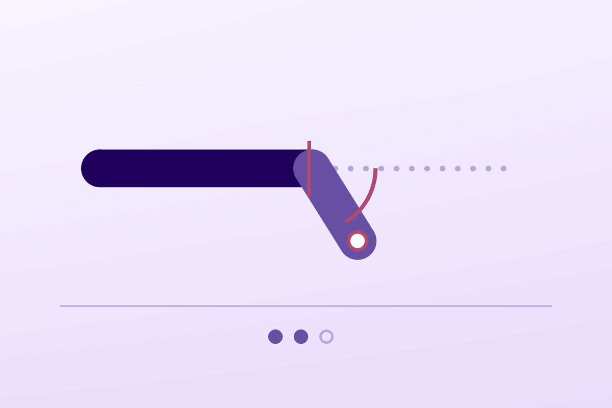
Volar angulation is well tolerated: trials have tested it up to 70° with no measurable loss of function, well beyond the 30-40° thresholds usually cited.
39.7%of metacarpal fractures affect the fifth metacarpal
What follows covers each of these points in detail, with sources. It is there if you need it.
This topic is taught in a course: Advance your hand and wrist rehabilitation: assess and empower your patients, on-site with Tessadit Aissaoui.
1 / 12
11 article chapters · 40 min in total
Understanding◔ 4 min
The fracture line runs through the metacarpal neck, and the angulation comes from the interosseous muscles, not the impact.
Population◔ 3 min
A young man who punches a wall, not a boxer: the fracture is not primarily a sporting injury.
Examination◔ 5 min
Making the patient close the fist detects the rotational deformity that no X-ray shows.
Angulation thresholds◔ 4 min
The thresholds usually cited rest on no controlled trial.
Treatment◔ 6 min
Early buddy taping is as good as a cast, without stiffness or loss of strength.
Bites◔ 3 min
Any wound over an MCP after a fight is a fight bite until proven otherwise.
Surgery◔ 2 min
Surgery is required for a rotational deformity or a joint wound, not for the angle.
Rehabilitation◔ 4 min
Rehabilitation produces significant gains in function, strength and range.
Clinical cases◔ 3 min
Three published cases each document a possible error not to repeat.
In practice◔ 3 min
The decision tree follows a precise order: wound, then rotational deformity, before the angle.
Questions◔ 3 min
The knuckle does not come back, but going without a cast is not substandard care.
Train on this
Upper limb
Tessadit Aissaoui
Next comes practice: the course that teaches this topic, with Tessadit Aissaoui.
Our courses on this topic may be covered by DPC or FIFPL, the French continuing-education funding schemes, depending on your profession and situation.
A fist that hits a wall, a knuckle that disappears under the skin, a joint that flattens out: the boxer's fracture looks serious and behaves well. It also has the rare feature of tolerating a deformity better than a cast; up to 70 degrees of angulation, no study has measured the loss of function that common sense promised. The real risk lies elsewhere, in a defect that does not show on the AP radiograph and that the examination takes ten seconds to flush out.
A synthesis written from primary sources verified one by one on PubMed: identifier resolved, journal, year and author list checked, abstract read before citation. Full bibliography at the end of the article.
The boxer's fracture in three figures
Three independent pieces of work that shift the question from “how do we straighten this bone?” to “what here really deserves treating?”
Sources: Statius Muller MG et al., Arch Orthop Trauma Surg 2003, randomised trial, 40 patients, mean angulation 39° (PMID 14639483); Pellatt R et al., Ann Emerg Med 2019, randomised trial, 97 patients (PMID 30853124); Medeiros I, Saconato H, Cochrane Database Syst Rev 2001, OR 0.10 (95 % CI 0.01-0.86), NNT 4 (CI 2-50) (PMID 11406003).
What to have in mind
Three red flags in a fractured fist
The boxer's fracture is one of the rare fractures where the radiographic appearance is more worrying than the functional result, and where immobilisation costs more than it brings in.
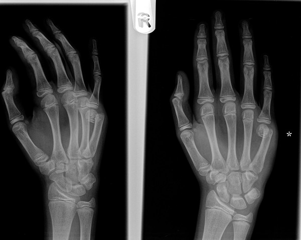
X-rayOblique and anteroposterior X-rays of the hand: displaced fracture of the fifth metacarpal neck, angulation visible at the fracture site.
Source : Mohammed et al., Journal of orthopaedic surgery and research, 2011, figure 1 · CC BY
Understanding volar angulation means understanding why it cannot be held reduced, and why that does not matter.
The boxer's fracture means a subcapital fracture of the fifth metacarpal: the fracture line runs at the neck, the narrowed portion immediately proximal to the metacarpal head, the one that forms the “knuckle” of the joint when the fist is closed. The mechanism is almost always the same: a closed fist that strikes a hard object, with the force transmitted along the axis of the fifth ray1.
That location is not incidental. The neck is a transition zone between a strong cortical shaft and a cancellous epiphyseal head: mechanically it is the weak link of the ray. And the fifth ray is, of all the metacarpals, the most exposed: first because it is on the ulnar side, at the border of the fist, so the first in contact; and second because its carpometacarpal joint is the most mobile of the long rays, which shifts a share of the loading towards it.
The European series by Dominguez-Prado, which classified 1,341 hand fractures in a population of 470,000 inhabitants, places the fifth metacarpal at the top of the metacarpals involved, with 39.7 % of the fractures in that group, and the distal end as the commonest location in the young man2.
Why the head tilts towards the palm
The displacement is not due to the impact: it is produced, and then maintained, by the intrinsic muscles that cross the fracture
An explanatory diagram built on the commonly accepted mechanical description of the subcapital fracture. The clinical consequence, loss of the reduction obtained, is itself measured: in the Martínez-Catalán trial, the reduction was systematically lost by 3 weeks, with equivalent residual angulation in both arms at 3 and 9 weeks (PMID 32718787).
The distal fragment, the head, tilts towards the palm. That angulation is not the direct result of the impact, but of the intrinsic muscles: the interossei pass in front of the longitudinal axis of the metacarpal, and their pull flexes the distal fragment as soon as cortical continuity is broken. Those muscles do not relax because the hand has been put in plaster.
That is the whole logic of what follows. A reduction is easy to obtain under anaesthesia, by pushing on the head through the flexed phalanx. It is almost impossible to hold by external means. The randomised trial by Martínez-Catalán measured it directly: in the reduced and casted group, the reduction was lost by three weeks of follow-up, and the residual radiographic angulation observed at 3 and 9 weeks was equivalent to that of the group simply buddy taped with no attempt at reduction at all6.
Key points
The cosmetic complaint deserves to be heard rather than brushed aside, because it is often the real reason for the follow-up appointment. The angulation makes the prominence of the metacarpal head disappear from the back of the hand: the closed fist shows a hollow where there should be a knuckle. It is visible, permanent if the fracture unites in angulation, and with no demonstrated functional consequence.
The series by France, which specifically looked for fractures united with more than 70° of angulation, provides here the argument most useful in the clinic: of 364 fractures, 40 (11 %) had united beyond 70°, and among the 15 patients reviewed (mean angulation 73°, up to 77°), all scored at the highest level of the hand function scale, and 80 % reported a QuickDASH of zero, that is no trouble at all. Nearly half of them worked in a manual trade, and the dominant hand was affected in 87 % of cases8.
That result does not say that the deformity is desirable. It says that between a deformed hand and an immobilised hand, the measured functional gap tilts towards mobility.
A male, young and rarely sporting fracture: the name misleads about the real mechanism.
Four figures that place the population concerned
Data from the American national NEISS registry (160,790 metacarpal fractures estimated) and from two population series
Sources: Nakashian MN et al., Hand (N Y) 2012, NEISS database 2002-2006 (PMID 24294164); Poolman RW et al., Cochrane Database Syst Rev 2005 (PMID 16034891) and Retrouvey H et al., Plast Surg 2022 (PMID 35096686) for the 20 % figure; Aitken S, Court-Brown CM, Injury 2008, 1,430 hand fractures (PMID 18656191).
Nakashian's study on the American NEISS database, which extrapolates to 160,790 metacarpal fractures over five years, gives the clearest portrait available. Overall incidence is 13.6 per 100,000 person-years. It peaks in 10 to 19-year-olds (38.8) then in 20 to 29-year-olds (28.4), and collapses afterwards. The male-to-female incidence ratio is 5.08 : 23.0 against 4.5 per 100,000. The two dominant mechanisms are contact with a wall or a door and falling; the commonest place is the home, ahead of leisure venues1.
In other words: the name “boxer's fracture” refers to a fracture that, in the great majority of cases, does not happen in boxing. The trained boxer strikes with the second and third rays, protected by a wrap and a glove; it is the untrained fist, striking a wall with its ulnar border, that produces this fracture. The paediatric literature has even devoted a famous title to that scene: “another angry adolescent, another boxer's fracture?”.
Incidence of metacarpal fractures by age band
Per 100,000 person-years, NEISS database 2002-2006: the fracture is a marker of age as much as of mechanism
Source: Nakashian MN, Pointer L, Owens BD, Wolf JM. Incidence of metacarpal fractures in the US population. Hand (N Y) 2012;7(4):426-30 (PMID 24294164). The values by age band cover all metacarpal fractures, of which the fifth metacarpal neck fracture is the commonest form.
Aitken and Court-Brown analysed 1,430 hand fractures occurring in one year in Edinburgh: 22.4 % were sport-related, with 86 % men and a mean age of 24. Metacarpals accounted for 33.8 % of those sporting fractures, and the first and fifth rays were the most affected. A notable fact for practice: 87.2 % of those fractures were treated as outpatients, and open fractures were only 2.2 %3.
That proportion is useful to bear in mind so as not to over-read the context: the boxer's fracture is not primarily a sports injury, and the typical patient is not a return-to-competition problem but a return-to-work and everyday-activities one.
The psychosocial context is not a clinical detail
The dominant mechanism, punching a wall, often signals distress, anger or intoxication, sometimes domestic violence or a brawl. The literature on metacarpal fractures in prison settings exists precisely because that population concentrates the mechanism. Without turning the physiotherapist into a social worker, it is legitimate, faced with a recurrence or a suggestive context, to refer to the general practitioner. It is also, very concretely, the factor that decides adherence to functional treatment.
Key points
The whole point of the examination comes down to one sentence: the radiograph shows what does not count, and misses what does.
Volar angulation is measured on a lateral or oblique radiograph. Rotational deformity, on the other hand, is visible on no standard view. It shows only in flexion, because it is in flexion that the axes of the fingers converge: the four long fingers, flexed together, must all point towards the scaphoid tubercle. A ray whose metacarpal has united in rotation makes its finger deviate out of that bundle, and the little finger comes to overlap the ring finger.
It is a ten-second examination, and it is the only one that changes what you do. It is done before any treatment and is rechecked at every follow-up appointment, because secondary displacement can appear. Royle, in a prospective study of 98 metacarpal fractures, points out a practical difficulty met at the bedside: pain often limits metacarpophalangeal flexion just after the fracture, which makes assessment awkward. For that reason he recommends adding examination of the nail seen end-on, whose plane must stay parallel to that of the neighbouring nails10.
The same study gives the order of magnitude of the problem: of 91 consecutive patients, a quarter had minor rotation of less than 10°, only five exceeded that threshold, and two required surgery for rotational instability10. Significant rotational deformity is therefore rare: that is precisely why it gets left out, and precisely why it must be looked for systematically.
The rotation test, in three steps
What you look at, in what order, and what counts as an abnormal result
Built after Royle SG. Rotational deformity following metacarpal fracture. J Hand Surg Br 1990;15(1):124-5 (PMID 2307874), which documents the frequency of rotation (a quarter of fractures under 10°, 2 cases out of 91 operated on) and explicitly recommends adding the end-on view of the nail, metacarpophalangeal mobility often being limited after fracture.
Examination of a hand injured after a punch follows a sequence whose order matters: what is urgent first, what is deforming next, what is functional last.
| Order | What you look for | How | What changes if it is positive |
|---|---|---|---|
| 1 | A wound over a metacarpophalangeal joint | Dorsal inspection, fist closed and open: the wound moves with the skin and can hide itself in extension | Presumed fight bite: surgical opinion the same day, washout, antibiotics16 |
| 2 | Rotational deformity | Simultaneous flexion of the four long fingers; failing that, nails seen end-on | A surgical indication to discuss, whatever the angulation10 |
| 3 | Deficit of active extension of the 5th finger | Active metacarpophalangeal extension against gravity, compared with the healthy side | Look for tendon rupture21 or sagittal band injury23 |
| 4 | Volar angulation | AP, lateral and oblique radiographs; the oblique shows the neck best | Discussion of the threshold (next chapter), rarely decisive on its own |
| 5 | Associated involvement of the carpus or of the bases | Palpation of the metacarpal bases and of the carpometacarpal joint line | Fracture of the 4th and 5th bases: a rare entity, reduction and fixation more demanding22 |
| 6 | Flattening of the metacarpal head | Fist closed, bilateral comparison | Informing the patient: a real cosmetic loss, with no demonstrated functional consequence8 |
Three views are enough in the vast majority of cases: AP, lateral and oblique. The oblique at 30-45° clears the neck of the fifth metacarpal from the overlap of the neighbouring rays and gives the most reliable measure of angulation: it was in fact on the oblique view that the France series measured its angulations above 70°8. The New England Journal of Medicine published a reference clinical and radiographic image of it, useful for calibrating on the typical appearance25.
Ultrasound has been the subject of one description: Thom et al. report the use of point-of-care ultrasound to guide a closed reduction and check the improvement in angulation before radiographic confirmation24. It is a case report, not a validated practice, and it is chiefly worth noting as a reminder that ultrasound sees the metacarpal neck. For the physiotherapist, the interest is indirect: it explains why some patients arrive with an ultrasound report and no recent radiograph.
The trap of the follow-up radiograph
A check at three weeks showing angulation identical to that on the day of the fracture is not a treatment failure: it is the expected behaviour, measured in the casted arm of the Martínez-Catalán trial, where the reduction was systematically lost6. What should be looked at on that check is the absence of rotation and the presence of signs of union, not the restoration of an angle.
Key points
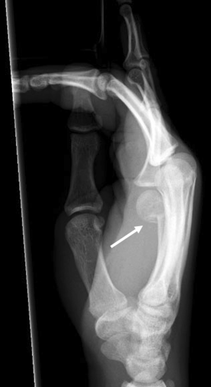
X-rayLateral X-ray of the hand: fracture of the fifth metacarpal neck, known as a boxer's fracture, with volar angulation, arrow on the fracture site.
Source : Oetgen et al., Current reviews in musculoskeletal medicine, 2008, figure 5 · CC BY-NC
This is the question most often asked about this fracture, and the one where the gap between the textbooks and the trials is widest.
You regularly read tolerance thresholds of 30°, 40°, sometimes 50° for the fifth metacarpal, presented as limit values beyond which reduction is required. Those figures have an apparent logic: the more the head tilts, the more the metacarpal functionally shortens, the more the mechanics of the extensor change.
The literature search conducted for this article found no controlled trial establishing those thresholds. What it does find are trials that tested tolerance to far greater angulations and found no functional loss. It is a reversal that has to be stated clearly, because it changes what you do.
The reference work is that of Statius Muller et al. Forty patients were randomised between an ulnar gutter cast for three weeks and a compression bandage for one week then immediate mobilisation within the limits of pain. The inclusion criterion was an angulation accepted up to 70° ; the actual mean angulation was 39°, with values ranging from 15 to 70°. At 6 and 12 weeks, no statistical difference was found in metacarpophalangeal range, satisfaction, pain, return to work and leisure, or the need for physiotherapy. The authors conclude, in terms worth quoting precisely, that reducing a fracture angulated by less than 70° “does not seem to be of use” as regards metacarpophalangeal mobility5.
The relevant threshold is not the one at which the deformity becomes visible, but the one at which function deteriorates. No trial has managed to place it below 70°.
That is the next question, and the France series answers it, with the limitations of its level of evidence. It is a retrospective review of adult patients with an isolated closed fracture of the neck of the fifth metacarpal, united with more than 70° of angulation, treated without surgery, with a minimum follow-up of six months.
Of 364 fractures identified, 40 (11 %) had angulation above 70°, between 71 and 82°. Fifteen patients could be reviewed, with a mean angulation of 73° and a mean follow-up of 32 months. The results: all obtained the highest rating on the hand function scale, 80 % reported a QuickDASH of zero, and 87 % had SF-12 quality of life scores at or above average. Seven of the fifteen patients worked in a manual trade, and thirteen out of fifteen had the dominant hand affected8.
This result has to be read for what it is: a retrospective series, with an inclusion rate of 38 % among eligible patients, therefore exposed to selection bias, since dissatisfied patients may have been harder to reach, or the reverse. It does not demonstrate that 80° is equivalent to 20°. What it does demonstrate is that the opposite claim, very widespread, has no support : you cannot tell a patient that an angulation of 75° condemns them to a deficient hand.
What the trials actually tested, in degrees of angulation
Each bar represents the range of angulation actually included in the study, not a recommended threshold
Sources: Statius Muller MG et al., Arch Orthop Trauma Surg 2003 (PMID 14639483); Martínez-Catalán N et al., J Hand Surg Am 2020 (PMID 32718787); Zawam SH et al., Eur J Trauma Emerg Surg 2024 (PMID 38151577); France TJ et al., Hand (N Y) 2023 (PMID 34991365). The thresholds of 30° and 40° are shown for the record: the search conducted for this article did not find a controlled trial establishing them.
Saying that angulation is tolerated does not mean that it has no effect. Three consequences are documented or mechanically expected, and they must be distinguished by their level of evidence:
Key points
Five randomised trials, a Cochrane review and a network meta-analysis converge on the same answer, and it is counter-intuitive.
The Cochrane review by Poolman et al. compared functional treatment with immobilisation, and the different immobilisation methods with each other, for closed fractures of the neck of the fifth metacarpal in adults. Five trials, 252 participants. The verdict is cautious: no non-operative treatment regimen can be recommended as superior to another, the studies being of limited quality and heterogeneous. The authors note a major methodological point: none of the studies included used a validated measure of hand function as its primary outcome4.
That review is from 2005. It would be a mistake to stop there, because the trials that were missing then, the ones that measure the DASH and quickDASH, have since been published. It is a case where the benchmark systematic review is older than the decisive data, and where reading only Cochrane would lead you to conclude “we do not know” when the later trials do know.
Pellatt 2019, Annals of Emergency Medicine. A randomised trial in two Queensland hospitals, 97 patients aged 18 to 70 with an uncomplicated boxer's fracture. Comparison: buddy taping of the ring and little fingers against a cast. Primary outcome: quickDASH at 12 weeks. Result: a median of 0 in both groups (interquartile range 0 to 2.3 for buddy taping, 0 to 4 for the cast; difference 0, 95 % CI from 0 to 0). The buddy taping group lost a median of 0 days of work against 2 days for the cast. The authors explicitly recommend the minimal intervention7.
Martínez-Catalán 2020, Journal of Hand Surgery (American). This is the highest-level trial on the question: therapeutic level I. Seventy-two patients with angulation below 70° and without rotational deformity were randomised between buddy taping with no reduction and closed reduction then a cast. At three weeks, the DASH was significantly better in the buddy taping group: 19.7 ± 19.7 against 44.6 ± 15.0. At nine weeks, the advantage persisted in range and in the DASH, with a mean difference of 6.3 points that did not exceed the threshold of clinical relevance. There were more complications in the cast group. And above all: time off work was 28 days shorter with buddy taping6.
Retrouvey 2022, Plastic Surgery. A Canadian multicentre trial, 37 participants, comparing an elastic bandage with early protected movement against immobilisation in a splint. The hand score (bMHQ) did not differ. But at eight weeks, grip strength reached 93 % of Canadian reference values in the early movement group, against 64 % in the splint group : a significant difference11.
A 2022 meta-analysis brought together seven trials and 454 patients on that precise question: it concludes that buddy taping improves pain, range and strength, with no difference in the DASH or in satisfaction, and recommends buddy taping rather than a cast for uncomplicated fractures12.
Buddy taping against cast: what the trials measure
A low DASH score is better than a high one; a high grip strength is better than a low one
Sources: Martínez-Catalán N et al., J Hand Surg Am 2020;45(12):1134-1140 (PMID 32718787); Retrouvey H et al., Plast Surg (Oakv) 2022;30(1):6-15 (PMID 35096686); Pellatt R et al., Ann Emerg Med 2019;74(1):88-97 (PMID 30853124). The three trials have slightly different populations and comparators; the values are not interchangeable between studies.
The convergence of these trials is easily explained. You immobilise to hold a reduction; but the reduction does not hold, whatever the means: that is measured6. So you end up with the same radiographic result, but paying the price of immobilisation: metacarpophalangeal stiffness, loss of strength, prolonged time off work.
The systematic review by Feehan and Bassett in the Journal of Hand Therapy, covering six studies and 459 patients, had already framed the cautious conclusion that was warranted then: early mobilisation after an extra-articular hand fracture has the potential to restore mobility and strength faster, to allow an earlier return to work, and does not alter fracture alignment, with a consistent benefit and no significant risk identified, but on evidence of insufficient quality14. The trials of 2019, 2020 and 2022 filled exactly that gap.
And what about pinning?
The network meta-analysis by Zong et al. compared four strategies (conservative treatment, antegrade intramedullary nailing, transverse pinning and plating), on the outcome of total complications, from six randomised trials and 288 patients. The ranking is clear-cut: conservative treatment comes first (SUCRA probability of 94.1 %), followed by plating (52.9 %), nailing (37.3 %), and transverse pinning comes last, with the highest complication rate (15.7 %)9.
An Egyptian randomised trial of 2024 in 84 patients with angulations of 30 to 70° compared an ulnar gutter cast with transverse pinning: time to union of 7.76 against 7.38 weeks, with no significant difference in union or in functional results, all the patients returning to their previous work13. When two treatments give the same result, the one that does not expose the patient to a pin-track infection wins.
Treatment modalities and level of evidence
An appraisal of the level of evidence following the GRADE principles, from the sources cited in this chapter
Functional treatment (buddy taping or bandage) with early mobilisation, at least as effective as immobilisation on every functional outcome measured, with faster return to work and better early strength. Three concordant randomised trials, one of them level I6711.
Not reducing an angulation below 70° : a dedicated randomised trial finds no benefit of reduction on mobility5, corroborated by the consistent loss of reduction observed in the 2020 trial6. Modest sample sizes.
A structured rehabilitation programme : a gain in function, strength and range in meta-analysis, but across only five studies, two of them cohorts15.
Tolerance of angulations above 70° : a single retrospective series of 15 patients, an inclusion rate of 38 %, with no control group8. Enough to reassure, not enough to make a rule of.
Reduction thresholds at 30° or 40° : no trial found establishing them. Those values circulate with no identified support in the sources consulted here.
Transverse pinning as first-line treatment : last of the four treatments compared in network meta-analysis, with the highest total complication rate9.
An appraisal built following the GRADE principles from the sources cited, and not taken from a published GRADE assessment: none of the reviews consulted produced a formal GRADE table on this question. The 2005 Cochrane review explicitly stopped at a finding of insufficient evidence4.
Key points
This is the only chapter of this article where an error is paid for with a destroyed joint. It alone justifies looking at the back of the hand before the radiograph.
The fight bite (fight bite, or clenched fist injury) occurs when the thrown fist meets the opponent's teeth. The tooth perforates the skin over a metacarpophalangeal joint, crosses the extensor tendon, and inoculates oral flora into the joint.
The trap is anatomical, and it is twofold. First, the wound is made with the fist closed, the joint in flexion; when the hand opens for the examination, the skin, tendon and capsular planes move relative to each other, and the three openings are no longer aligned. A wound of three millimetres, apparently clean, can therefore cover a septic arthrotomy. Second, the closing of those planes shuts the organism inside a closed, poorly vascularised space, where it thrives before the patient becomes concerned.
De Smet and Stoffelen put it bluntly in their series: these injuries result from the impact of the fist with the opponent's teeth, with perforation of the skin and of the joint, and severe septic arthritis of the metacarpophalangeal joint of the dominant hand is a frequent consequence19.
The retrospective series by Goon et al., covering 34 patients with a confirmed human bite to the hand over seven years, gives the figures most telling for practice:
The four-day delay is the central item. The patient does not consult for the wound: they consult when the hand swells, that is, when the infection is established.
Immediate management of a wound over a metacarpophalangeal joint after a fight
Human and animal bites most often lead to polymicrobial infections, mixing aerobic and anaerobic organisms. Pasteurella is characteristic of dog and cat bites; Eikenella is characteristic of human wounds ; staphylococci, streptococci and anaerobes are common to all mammals16. That distinction is not merely academic: it explains why the antibiotic spectrum chosen must cover Eikenella, an organism that escapes certain penicillinase-resistant penicillins.
The review by Malahias et al. is also a reminder of the scale of the problem: mammalian bites account for up to 1 % of emergency department attendances in the United Kingdom, and clenched fist injuries are reputed to be the worst of the human bites20.
What that means for the physiotherapist
A patient referred for rehabilitation of a “boxer's fracture” who has a dorsal scar over a metacarpophalangeal joint, persistent thickening, disproportionate stiffness or night pain should raise the suspicion of a joint infection that went unnoticed or was insufficiently treated. That is not stiffness to be mobilised harder: it is a reason to refer back. Timing counts: a hand that gets worse after a few days of rehabilitation is not a hand that needs more sessions.
Key points
The indications come down to three lines, and none of them rests on an angle.
The trials cited above all share a methodological feature that must be read carefully: they exclude certain fractures. Martínez-Catalán excludes fractures with rotational deformity6 ; Zawam excludes old, open or malrotated fractures13 ; Pellatt recruits only “uncomplicated” fractures7. Those exclusions draw the surgical indications in outline, better than a guideline would:
| Situation | Why it falls outside the scope | Source |
|---|---|---|
| Rotational deformity | Digital overlap in flexion, not corrected spontaneously, with a direct impact on grip. Excluded from every functional treatment trial | Royle 199010 ; exclusion criteria of Martínez-Catalán6 and Zawam13 |
| Open fracture, including fight bite | An infectious problem before being a bone problem: debridement, washout, antibiotics | Kennedy 201516 ; Goon 200818 |
| Multiple or unstable fractures | The relative stability provided by the neighbouring ray, the very principle of buddy taping, disappears when several rays are involved | Trial exclusions613 |
| Involvement of the metacarpal bases | A different entity, technically demanding reduction and fixation, risk of carpometacarpal subluxation | Amirthalingam 202222 |
| Old fracture, united in a malposition | A matter for corrective osteotomy, a specialist decision | Zawam exclusion13 |
You will notice what is not in that list: an angle. None of the sources consulted makes angulation alone an indication for surgery within the ranges studied.
When it is indicated, surgery delivers on its promise of stability. The interesting question is that of the choice of technique, and that is where Zong's network meta-analysis is useful: on the outcome of total complications, the order is conservative treatment (94.1 %), plating (52.9 %), antegrade intramedullary nailing (37.3 %), transverse pinning (15.7 %)9.
That ranking deserves cautious reading: the outcome is total complications, not effectiveness of reduction. A treatment that does nothing obviously cannot cause a hardware infection: the ranking mechanically favours doing nothing. It remains that, among the surgical techniques themselves, the hierarchy is informative, and that transverse pinning appears there as the greatest source of complications.
Zawam's trial confirms that point from another angle: a plaster gutter against transverse pinning in 84 active adults, angulations of 30 to 70°. Time to radiological union of 7.76 weeks against 7.38 weeks, with no significant difference in union or in functional results, all the patients returning to their previous work and activity level13. Two-thirds of a week gained on union does not justify a percutaneous wire.
With this fracture, the debate is no longer “operate or not” but “why would you operate”, and the answer lies in rotation, in skin breach and in instability, never in an isolated angle.
The physiotherapist rarely intervenes in the union. They intervene in what immobilisation would have cost, and in what remains when it has taken place.
The most recent meta-analysis on this precise point, non-thumb metacarpal fractures, was published in 2026 by Hakami et al., preregistered on PROSPERO. Five studies, three randomised trials and two cohorts. Rehabilitation programmes significantly improve:
The authors note that early mobilisation and adherence strategies, including playful ones, are associated with the best functional results, and that minimal immobilisation approaches such as buddy taping suit stable fractures15. The number of studies included (five) means those intervals must be treated with caution: they are narrow for such a small corpus.
Timetable of care after an uncomplicated fracture of the neck of the 5th metacarpal
Markers built from the protocols of the trials cited; the times to union are measured, the session contents belong to everyday practice
Times to union: Zawam SH et al., Eur J Trauma Emerg Surg 2024, 7.76 weeks conservatively and 7.38 with pinning (PMID 38151577). Strength target at 8 weeks: Retrouvey H et al., Plast Surg 2022 (PMID 35096686). Time off work: Pellatt R et al., Ann Emerg Med 2019 (PMID 30853124). The detail of the session contents belongs to everyday practice and does not come from a published protocol.
That is the deficit that gets in the way, and the one immobilisation creates. The metacarpophalangeal joint stiffens in extension: its collateral ligaments are slack in extension and taut in flexion, so that immobilisation in extension lets them shorten in a position from which flexion becomes hard to win back. That is why, when immobilisation is necessary, the so-called intrinsic-plus position (metacarpophalangeal joints flexed, interphalangeal joints extended) is preferred.
In practice, the session covers isolated active metacarpophalangeal flexion, the composite fist, and tendon glides. Frequency matters more than duration: several short sets spread through the day are better than one long session, and home work does most of the work.
That is the most measurable deficit, and Retrouvey's trial gives the target: 93 % of reference values at eight weeks in the group treated with early movement, against 64 % in the immobilised group11. A patient who, at eight weeks, plateaus at two-thirds of their expected strength is on the trajectory of the immobilised group, which justifies intensifying, not becoming alarmed.
The fifth ray has a particular function that should be targeted explicitly: it carries the power grip, that of the hammer, the suitcase, the handlebar. That is where the deficit is noticed in manual workers, and it is the work-specific movement that should finish the rehabilitation.
Persistent dorsal oedema mechanically stiffens the metacarpophalangeal joints and makes flexion painful: it precedes stiffness rather than accompanying it. Elevation, repeated active mobilisation, gentle compression and muscle-pump work are its core treatment.
What should stop rehabilitation and prompt re-referral
Key points
Three published reports, chosen because each documents a possible error: believing the fracture is isolated, believing the closed fist only fractures the neck, believing the wound is superficial.
Closed rupture of both extensor tendons of the little finger after a neck fracture
Lardenoye S, Hannemann PFW, Ten Bosch JA. Case Reports in Plastic Surgery and Hand Surgery 2020;7(1):30-33 (PMID 32128350).
The authors report a rupture of both extensor tendons of the fifth finger occurring after a closed fracture of the neck of the fifth metacarpal. They stress that this complication had never been reported before in that context, and conclude on the importance of a complete clinical examination so as not to miss associated injuries.
What the case teaches. The boxer's fracture is almost always an isolated and benign injury, and that is precisely what makes people stop examining. A deficit of active extension of the little finger must never be put down to pain or oedema without being retested. For the physiotherapist receiving the patient some time after the emergency visit, it is the first test to repeat: the fracture may be known, the tendon may not be.
A bad punch, a rare fracture: the bases of the fourth and fifth metacarpals
Amirthalingam S, Sameer M, Harshavardhan JKG. Journal of Orthopaedic Case Reports 2022;12(11):110-113 (PMID 37013244).
A 37-year-old right-handed man presents with pain and swelling of the right hand after punching a wall. It is not a neck fracture: the authors describe an isolated and displaced fracture of the bases of the fourth and fifth metacarpals, with no carpometacarpal subluxation or carpal fracture, an entity they describe as extremely rare. Treatment consisted of reduction and fixation with Kirschner wires through a mini-open approach, with ten months of follow-up.
The authors state the lesson in their conclusion: a closed fist injury does not always mean a boxer's fracture. They specify that the site of the fracture depends on the type and direction of the blow, and that these fractures are easily misread by an inexperienced practitioner.
What the case teaches. The name of the injury directs the gaze, and the gaze directs the radiograph. Faced with an injured fist, palpation must go down to the metacarpal bases and the carpometacarpal joint line, whose involvement completely changes the management and the prognosis.
The clenched fist injury, a pitfall for the patient as for the surgeon
De Smet L, Stoffelen D. Clenched fist injury: a pitfall for patients and surgeons. Acta Orthopaedica Belgica 1997;63(2):113-7 (PMID 9265797), a case series.
The authors describe the typical sequence: the impact of the fist with the opponent's teeth perforates the skin and the joint; severe septic arthritis of the metacarpophalangeal joint of the dominant hand frequently follows. They stress the long-term medical and medicolegal consequences of failing to recognise this injury.
What the case teaches. The title says the essential: the pitfall applies to the patient, who plays down a three-millimetre wound and consults four days later18, and to the clinician, who sees a fracture on the radiograph and does not look at the skin. The question to ask is not “do you have a wound?” but “exactly how did you do this?”.
Why these cases and not others
Clinical cases are the classic weak point of review articles: the too-neat story, the 28-year-old runner cured in six sessions. The three reports above are published, indexed case reports, cited with their PubMed identifier, and chosen because each documents an error of reasoning rather than a therapeutic success. No case in this chapter was composed for the purposes of the demonstration.
What reading the trials changes in practice, for the patient who arrives at the clinic with a swollen hand and a prescription.
What to do with a fracture of the neck of the fifth metacarpal
The order of the questions matters: the first rules out an emergency, the second a surgical indication
A tree built from the inclusion and exclusion criteria of the randomised trials cited: Martínez-Catalán 2020 (PMID 32718787), Pellatt 2019 (PMID 30853124), Zawam 2024 (PMID 38151577), and from the bite series of Goon 2008 (PMID 26815618) and De Smet 1997 (PMID 9265797). It reproduces no published guideline: none of the learned societies consulted has produced a formal recommendation on this fracture.
Three messages come up at every consultation, and they are better formulated in advance:
| Error | Consequence | What avoids it |
|---|---|---|
| Not making them close the fist | A missed rotational deformity, permanent trouble with grip | Ten seconds of examination, at every consultation10 |
| Not looking for the wound | Septic arthritis of the metacarpophalangeal joint, joint sequelae | Dorsal inspection with the fist closed and open19 |
| Immobilising “to be safe” | Stiffness, loss of strength at 8 weeks, time off work extended by nearly a month | Buddy taping and early mobilisation611 |
| Reacting to persistent angulation at the follow-up | Useless repeat reductions, sometimes a surgical indication made in error | Knowing that the reduction is always lost again6 |
| Mobilising harder a hand that is getting worse | Diagnostic delay in an infection or a tendon injury | A hand that worsens after a few sessions is referred back1821 |
With this fracture, the most useful action costs nothing: ask the patient to close their fist and look at their fingers.
On the same ground
No, if the fracture unites in angulation. The prominence of the metacarpal head on the back of the hand is flattened by the displacement of the fragment, and union fixes that position. It is a real cosmetic loss, which should be announced rather than played down. The France series, which reviewed patients united with more than 70° of angulation, finds a QuickDASH of zero in 80 % of them and the best functional rating in all of them8 : the hand works, but it no longer has the same shape.
Because the trials that compared the two strategies find no advantage in the cast. In Pellatt's trial, the functional score at 12 weeks was 0 in both groups7. In Martínez-Catalán's, buddy taping did better at three weeks and allowed a return to work 28 days earlier6. The cast does not hold the reduction, which is lost whatever happens, and it costs stiffness and strength.
The search conducted for this article found no controlled trial establishing a 40° threshold. The available trials tested angulations up to 70° without showing any loss of function56, and the only series above 70° finds good functional results8. The surgical indications that emerge from the sources are rotational deformity, skin breach and instability, not an isolated angle.
By closing the fist slowly: the four long fingers must converge on the same area of the wrist, with none overlapping its neighbour. If pain prevents full flexion, look at the nails end-on: their planes must stay parallel10. Frank overlap justifies a surgical opinion, because it does not correct itself.
If it happened while hitting someone, yes, potentially very. A wound of a few millimetres over a metacarpophalangeal joint may correspond to a fight bite, with inoculation of oral flora into the joint. In a series of 34 patients, 76 % arrived already infected at a mean delay of four days, and 59 % had an open joint18. That is a same-day appointment, not tomorrow.
After union, which occurs on average around 7.4 to 7.8 weeks according to Zawam's trial13. A distinction has to be made between return to work and everyday activities, which is very early with functional treatment, and return to activities that expose the ray to a new direct impact, which requires union to be achieved and strength restored. No study found gives a validated return-to-impact-sport criterion for this particular fracture: the reasoning rests on radiological union, freedom from pain and strength compared with the healthy side.
In Statius Muller's trial, the need for physiotherapy did not differ between the two groups, and many patients recovered without it5. The 2026 meta-analysis nevertheless shows a clear benefit of structured programmes on function, strength and range15. In practice, a hand that moves early and well often recovers on its own; rehabilitation comes into its own when there has been immobilisation, persistent oedema, established stiffness, or when the job demands a high grip strength.
A deficit of active extension is to be investigated, never attributed straight away to oedema. Two documented causes: tendon rupture, described even after a closed fracture of the neck21, and sagittal band injury, which lets the extensor tendon sublux ulnarwards on flexion and which often responds to an extension splint in the acute phase23. The distinction is clinical and is made on active extension against gravity and on the behaviour of the tendon in flexion. That last injury, classically described on the middle finger, has also been reported on the little finger, which forbids ruling it out on that ray26.
Twenty-six references, all verified individually on PubMed: identifier resolved, title, journal, year and author list checked, abstract read before citation. The links point to the PubMed record.
A note on method
The bibliographic base was built before writing, subject by subject, by searching PubMed through the E-utilities interface: every identifier was resolved, every record checked for its authors, journal and year, and every abstract read before being cited. The figures put forward in this article carry their source at the exact place where they are written. The appraisals of the level of evidence follow the GRADE principles without taking over a published GRADE assessment, none existing on this question. Two claims often read elsewhere, the reduction thresholds of 30° and 40°, are explicitly flagged as not found in the sources consulted, rather than repeated without a source. Article written on 15 August 2026.
Behind this article
An author who explains, a reviewer who checks.
How we write and check our content

Physiotherapist · co-founder of Physio Learning
Marked for life by his first four-hour lecture without a single image, he took a master’s in instructional design so that it would never happen to anyone again. He hunts down publication bias and unreadable slides with the same intransigence.

Head of scientific content
Physiotherapist specialising in neuro-musculoskeletal practice and holder of a master’s in public health. He checks the methodological rigour of every article: primary sources, levels of evidence, no exceptions.
