
















On-siteDPCFIFPL

Upper limb
Advance your hand and wrist rehabilitation: assess and empower your patients
Tessadit Aissaoui
Go further
Miss the Stener lesion and no orthosis heals it. Ultrasound rules out rupture (96% sensitivity), MRI settles displacement (92% specificity against 72%).
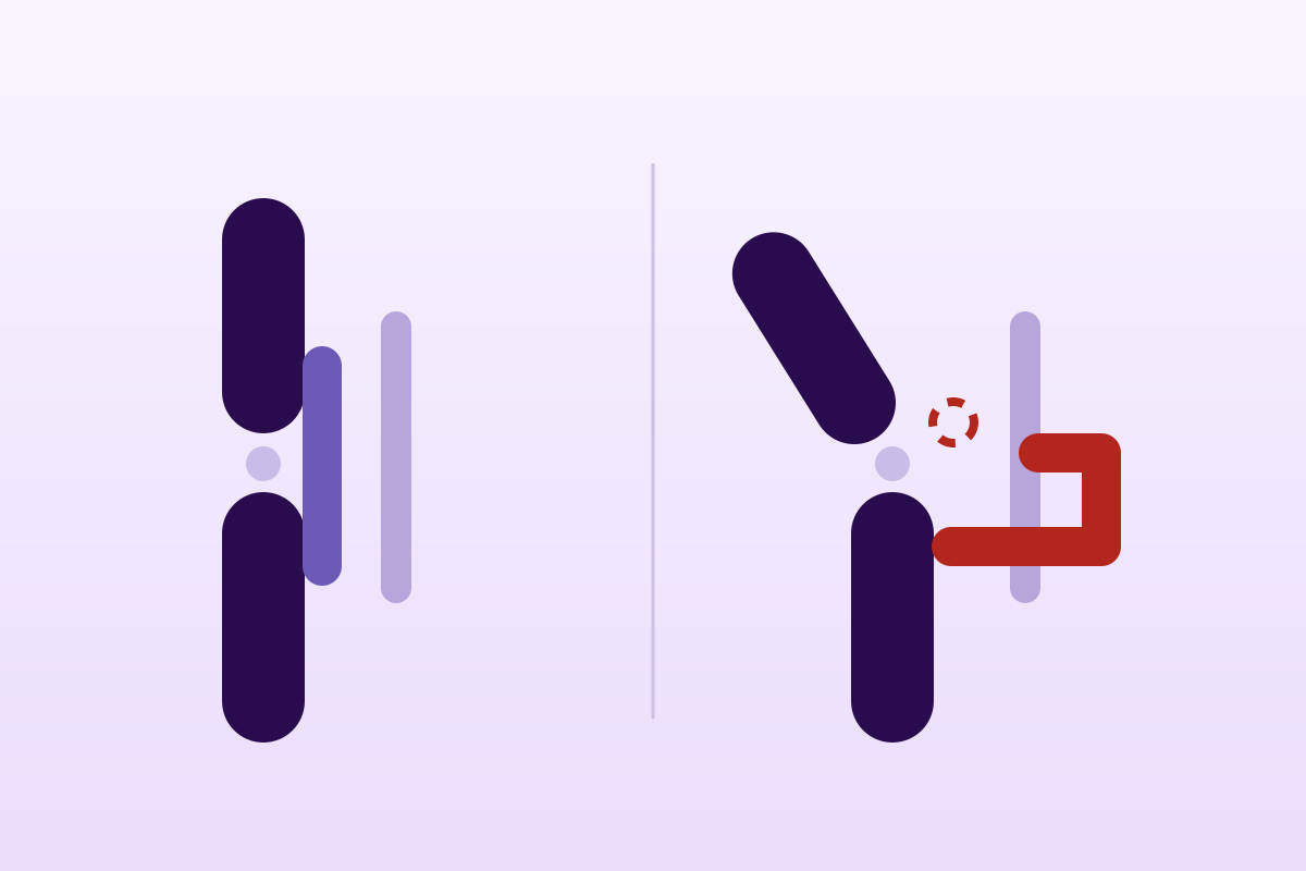
A thumb sprain raises a single question: is the rupture complete, and is the ligament stump displaced, which defines the Stener lesion.
87%of thumbs with more than 35° of laxity in extension had a Stener lesion
What follows covers each of these points in detail, with sources. It is there if you need it.
This topic is taught in a course: Advance your hand and wrist rehabilitation: assess and empower your patients, on-site with Tessadit Aissaoui.
1 / 12
11 article chapters · 61 min in total
Understand◔ 8 min
Untreated, this lesion can lead to reduced pinch strength, instability and osteoarthritis.
Stener◔ 5 min
In the Stener lesion, the ligament can no longer reach its insertion bed, whatever the immobilisation.
Test◔ 9 min
A normal, well conducted and comparative test is a good argument for going no further.
Image◔ 6 min
Three 2021 meta-analyses give different figures: the target and the reference differ from one study to another.
Distinguish◔ 4 min
It would be excessive to conclude that a thumb not repaired quickly is lost.
Rehabilitate◔ 10 min
On this subject, no recommendation for detailed week-by-week rehabilitation is supported by trials.
Return◔ 4 min
Return to sport reaches 98.1 % in aggregate, with no drop in performance after surgery.
Children◔ 3 min
In children, the expected lesion is bony: pure ligamentous injury remains the exception.
Case studies◔ 3 min
In one case, ultrasound reached an exact diagnosis, avoiding any further imaging.
In practice◔ 4 min
Without a radiograph, do not stress test: ask for medical advice first.
FAQ◔ 5 min
A controlled and gentle exam, thumb held without rotation, should not create an iatrogenic Stener lesion.
Train on this
Upper limb
Tessadit Aissaoui
Next comes practice: the course that teaches this topic, with Tessadit Aissaoui.
Our courses on this topic may be covered by DPC or FIFPL, the French continuing-education funding schemes, depending on your profession and situation.
A thumb sprain turns on one question, and one only: has the ruptured ligament stayed in contact with its insertion, or has the adductor aponeurosis slipped between the two? In the second case, the Stener lesion, no orthosis will make it heal, and six weeks of immobilisation will only delay surgery. Here is how not to miss it.
Review written from three diagnostic accuracy meta-analyses (2021), two systematic reviews of interventions, two randomised trials and the reference anatomical and clinical series. Every figure carries its source at the point where it is written; the 40 references are listed at the end of the article with their PMID or DOI.
What the literature says, in one page, before the detail of the chapters.
Ulnar collateral ligament (UCL) sprain of the metacarpophalangeal (MCP) joint of the thumb is an injury that is commonplace in its frequency and formidable in its capacity to be misclassified. The mechanism is stereotyped: forced abduction-extension of the thumb, most often during a fall in which an object held in the hand, a ski pole, a handlebar, a ball, acts as a fulcrum and forces the thumb column to open. The popular name, "skier's thumb", comes from that.
The difficulty is not recognising the sprain: it is separating the injuries that will heal in an orthosis from those that will never heal. That separation rests on an anatomical fact described by Bertil Stener in 1962: when the UCL ruptures completely at its distal insertion, its free end can flip outwards and above the adductor pollicis aponeurosis, which then interposes itself between the ligament and the bony bed where it should reattach (DOI 10.1302/0301-620X.44B4.869). The ligament is there, it is alive, it is simply on the wrong side of a fibrous sheet. No immobilisation crosses that sheet.
Key points
The course of action that follows from this reasoning is simple to state. A sprain with no laxity, or with laxity comparable to the uninjured side, calls for immobilisation in an orthosis, then graded rehabilitation. Marked laxity with a Stener effect calls for surgery: reattaching the ligament has no conservative alternative. Between the two, complete rupture without displacement remains, in the words of a recent French-language review, a "controversial" treatment (PMID 28636299).
"Interposition of the adductor aponeurosis distinguishes the Stener lesion from other UCL injuries and prevents healing, thereby making surgery necessary."
Beutel, Melamed & Rettig, Bull Hosp Jt Dis 2019, review of the Stener lesion (PMID 30865860).What follows sets out this reasoning in the order in which it unfolds in the consulting room: what the injury is, why displacement changes everything, how to test it without error, what each investigation really adds, what becomes of a thumb left unstable, and what rehabilitation can and cannot do.
Red flags: when not to rehabilitate and to refer
A two-tier anatomy, a mechanism with a single movement, and an epidemiology that depends entirely on where the counting is done.
The thumb MCP joint is a condylar joint whose main function is stability, not mobility. It transmits to the pulp most of the loads of the thumb-index pinch: every time we turn a key, close a jar or hold a pen, the thumb column resists a force that tends to open it outwards. It is the ulnar structures that absorb that opening.
This ulnar layer is made of two bundles, and that duality is not an anatomist's subtlety: it explains the whole of the diagnostic reasoning set out below.
This distribution has been measured directly. Palmer and Louis examined 750 normal thumbs to establish the physiological radioulnar mobility of the MCP joint in full extension, at 15° of flexion and in full flexion: it is in full flexion that the joint is most stable. In 25 cadaveric thumbs, sectioning the adductor aponeurosis and the capsule produced only minimal instability; it was the additional sectioning of the ulnar collateral ligament that produced marked instability, and it showed up best in full flexion. Sectioning the whole set (aponeurosis, dorsal capsule, proper ligament, accessory ligament and volar plate) produced complete instability in every position tested (PMID 722028).
Running over this ligamentous layer is the adductor pollicis aponeurosis, a tendinous expansion that covers the ulnar side of the joint before joining the extensor apparatus. Its arrangement as a superficial sheet, while the ligament lies deep, is exactly what makes the Stener lesion possible. A recent anatomical study has spelled out the detail: in 37 cadaveric thumbs, the intramuscular tendon of adductor pollicis has three components, dorsal, volar and distal, arranged in a lambda; the dorsal component inserts not only on the aponeurosis but also on the joint capsule, dorsal to the ulnar sesamoid, and the thickened portion of the capsule usually called the proper ulnar collateral ligament has a distinct bony insertion, on the proximal aspect of the lateral tubercle of the proximal phalanx, separate from the adductor insertion (PMID 29395587). This anatomical separation is what allows the ligament to detach and retract under then above a structure that itself stays in place.
The vocabulary, and what it hides
Skier's thumb denotes the acute traumatic form. Gamekeeper's thumb: pouce du garde-chasse in French, is the historical term, described in 1955 by C. S. Campbell in Scottish gamekeepers in whom the repeated movement of killing game progressively stretched the ulnar layer (DOI 10.1302/0301-620X.37B1.148). The original description is therefore that of a chronic attrition injury, and not of an acute sprain, but usage has shifted the term towards UCL injuries as a whole. One also reads player's thumb for the repetitive forms seen in athletes. These three names cover two conditions whose management differs (see the chapter on chronic forms).
The mechanism is a forced abduction and/or hyperextension of the MCP joint. In the surgical series of Chuter and colleagues, covering 127 patients operated on over ten years, at least 66 % of the injuries arose from a hyperextension-abduction mechanism (PMID 19389670).
The role of the ski pole has been studied with rare precision. Engkvist and colleagues compared injured skiers with a control population of 1,619 uninjured skiers: no type of grip in use eliminated the risk of thumb injury, and the frequency was even higher with a grip carrying a large upper platform. The way the skier held the pole in relation to the strap, by contrast, made no difference at all. The explanation the authors propose is mechanical: during the fall, the skier keeps hold of the pole until the last moment before the hand hits the ground; the grip then stays in the palm and acts as a hypomochlion, that is, as a fulcrum around which the thumb is forced into abduction and extension (PMID 7068299).
This observation has a practical consequence that comes as a surprise: prevention does not run through equipment. A review devoted to the subject puts it bluntly: poles without straps do not reduce the incidence of skier's thumb, but teaching the skier to let go of the pole during the fall might reduce the risk (PMID 7740248). Thirty years later, a two-season study in the Dolomites observed that none of the patients with distal upper limb injuries was wearing wrist or thumb protection (PMID 41155774).
This is where general figures deserve mistrust, and the literature itself supplies the counter-example that prevents generalisation.
Seen from a ski resort, skier's thumb is a skiing condition: in Engkvist's series, thumb injuries came just after knee injuries among alpine skiing injuries, at 17 % of all injuries, and three quarters of them were UCL injuries of the MCP joint (PMID 7068299). A prospective study run over two consecutive seasons in the Dolomites, between December 2023 and March 2025, finds a comparable profile: of 195 patients attending the emergency department for injury to the forearm, wrist, hand or fingers, 96 (49.2 %) had a fracture and 33 (16.9 %) an isolated UCL injury. Fractures occurred more often on blue runs (56.2 % against 33.3 %, p < 0.001), while UCL injuries were concentrated on red runs (54.5 %) and were frequently linked to the pole being trapped during the fall (PMID 41155774).
Seen from a city trauma department, the picture is reversed. In the British series of Chuter and colleagues, 127 patients operated on for acute UCL rupture over ten years, mean age 40 years (12 to 81 years), male to female ratio 3 to 2, the commonest cause was a simple fall (49 %), followed by sports accidents; skiing accounted for only 2.4 % of cases. The authors say so explicitly in their conclusion: in their population, UCL injuries of the thumb are rarely caused by skiing (PMID 19389670).
The practical lesson is twofold. First, do not require a history of skiing to consider the diagnosis : most of the thumbs operated on in the British series had never seen a ski slope. Second, do not carry rates from one population to another: a practice in a mountain area and an urban practice do not see the same proportions, and there is no single incidence figure valid everywhere.
The functional consequence of UCL insufficiency is well identified. A systematic review devoted to outcomes after thumb UCL injury states it in its aim: untreated, this injury can lead to reduced pinch strength, pain, instability and osteoarthritis (PMID 23615487). An orthopaedic review confirms this mechanism: UCL injuries are painful and unstable especially during lateral pinch and grip (PMID 36548149).
In other words, the patient does not complain about the thumb when opening the hand: the complaint comes when the thumb is used as a pillar. This is an important practical point for the history, and for the choice of functional follow-up tests.
Key points
Because it changes neither the pain, nor the appearance, nor the mechanism: only the fact that healing has become impossible.
In 1962, Bertil Stener published in the Journal of Bone and Joint Surgery an observation that has organised the management of this sprain for sixty years: when the ulnar collateral ligament ruptures completely at its distal insertion on the proximal phalanx, its free end can retract proximally and flip above the proximal border of the adductor aponeurosis. The aponeurosis, left in place, is then found interposed between the ligament and the bony surface on which it should reattach (DOI 10.1302/0301-620X.44B4.869).
This configuration bears his name. It has one unique consequence, and therein lies all its clinical importance: the ligament can no longer reach its insertion bed, whatever the position of immobilisation and however long it lasts. A review devoted to this lesion puts it thus: interposition of the aponeurosis distinguishes the Stener lesion from other UCL injuries and prevents healing, which makes surgery necessary (PMID 30865860).
The stump flips only under two conditions. First a complete distal rupture : as long as one bundle stays in continuity, the ligament cannot retract enough. Then an abduction wide enough for the free end to cross the proximal border of the aponeurosis, which behaves like a ledge: easy to cross one way, hard to cross back the other.
The anatomical study by Sato and colleagues sheds light on this mechanism by showing that the ligament and the adductor do have distinct bony insertions on the lateral tubercle of the proximal phalanx: the ligament can therefore detach independently, while the adductor stays anchored and keeps its sheet stretched across the field (PMID 29395587). The same study notes that the dorsal component of the intramuscular tendon of the adductor also inserts on the joint capsule, which gives this sheet a stiffness that is underestimated when it is described as a mere "aponeurosis".
"The results of the present study suggest the anatomical basis of a possible pathophysiological mechanism of the Stener lesion."
Sato et al., J Hand Surg Am 2018;43(7):682.e1-682.e8, in 37 cadaveric thumbs (PMID 29395587).The question "what percentage of complete ruptures are Stener lesions?" comes up constantly, and it has no single answer, for a methodological reason that must be named: a Stener lesion is only known to be present by operating, and only thumbs judged unstable are operated on. Every available series is therefore a series of patients already selected by a clinical threshold, and the rate they report is that of their threshold, not that of the condition.
What can honestly be written are conditional rates, with their condition:
These two figures, 87 % and 94.1 %, are positive predictive values of given clinical or radiographic signs, and not the prevalence of the Stener lesion among thumb sprains. Confusing them leads to a common error of reasoning: concluding that "about 9 complete ruptures in 10 are Stener lesions", which none of these data establishes.
The trap of the orphan figure
One often reads a single prevalence of the Stener lesion among complete ruptures, quoted with no reference population. Before repeating it, ask three questions: who was operated on (and therefore what threshold selected the series), what is called a complete rupture in that series, and who judged displacement, the surgeon intraoperatively or preoperative imaging? The three 2021 meta-analyses available on the subject also stress that all the included studies were at high risk of bias (PMID 33596684).
The difference in prognosis is not played out in the weeks that follow, but in those that will have been lost. A thumb whose ligament is displaced can improve perfectly well in an orthosis: pain decreases, swelling settles, the patient resumes activity. This is not healing, it is a lull. The instability persists and shows itself later, at the pinch, and that is when the patient comes back, several months on, with what will now be called chronic instability.
The French series of Agout and colleagues on chronic instability of the thumb MCP joint opens on exactly this point: the functional result of a severe sprain is good when repair is carried out at the acute stage, but the diagnosis is often missed, leading to chronic instability (PMID 28576699).
Red flags in this chapter
Key points
The valgus stress test is easy to describe and hard to make say anything. Here is the order of operations, the positions, the published thresholds, and why they are not the same from one article to another.
This is the point on which the French-language literature is most explicit. In the review by Poujade and Chick, bilateral comparative clinical examination is the central element of the diagnosis, but it must be preceded by a radiograph (PMID 28636299). The reason is twofold: a displaced avulsion fracture changes management straight away without any test being needed, and forcing a thumb carrying an unstable bone fragment brings nothing but risk.
The radiograph is not a confirmatory investigation, it is a triage investigation. It looks for:
The thumb MCP joint is not tested in one position but in two, and the two do not inform about the same structure. This follows directly from the two-bundle anatomy described above.
| Test position | Structure mainly placed under tension | What laxity there means |
|---|---|---|
| MCP in flexion (30° to full flexion) | The proper collateral ligament, isolated from the accessory layer | Injury to the proper bundle. Position of greatest physiological stability: laxity there is all the more significant. |
| MCP in extension | The accessory collateral ligament and volar plate, in addition to the proper one | Marked laxity there indicates injury to both bundles, and therefore an extensive lesion. |
This division is not a convention: it has been measured. On cadaveric specimens, sectioning the proper collateral ligament alone significantly increased valgus instability of the flexed MCP joint, with much less laxity when the joint was tested in extension. When the accessory ligament/volar plate complex was sectioned in turn, instability in extension increased to the point of no longer differing significantly from the values obtained at 30° of flexion (PMID 8519106). Palmer and Louis had established the same hierarchy by a different route, showing in 750 normal thumbs that full flexion is the position of greatest stability, and on cadaveric specimens that sectioning the UCL reveals instability there most clearly (PMID 722028).
The fear of "creating" a Stener lesion by testing a thumb is an old one. It has been tested directly on six fresh cadaveric specimens, with sequential sectioning of the accessory ligament, the proper ligament and then the ulnar sagittal band, and measurement of radial deviation in neutral rotation, in pronation and in supination, at 0° and at 30° of flexion. The result: no Stener lesion was created in any position as long as the fascial origin of the ulnar sagittal band remained intact. After a defect had been created in that band, a Stener lesion was produced in two specimens, but only with the thumb flexed and in supination. Pronation added stability, supination took it away (PMID 28774254).
"Performing a physical examination to assess the degree of instability of an ulnar collateral ligament injury did not create a Stener lesion if the examination was carried out in a controlled and gentle manner, with the thumb held without rotation."
Lankachandra et al., J Hand Surg Asian Pac Vol 2017;22(3):350-354, a study on six cadaveric specimens (PMID 28774254).The practical conclusion applies directly: test in neutral rotation, without supinating the thumb, with a progressive and controlled stress. This is not a precaution of principle, it is the condition under which the test was shown not to be harmful.
The manoeuvre, step by step
This is where the literature calls for most caution. Three values circulate; they do not come from the same study, they are not measured in the same position and they do not answer the same question.
Three clarifications are worth putting in black and white, because they are what separates quoting a threshold from understanding it.
Heyman's 35° threshold is measured in extension. This is not a detail: in the same study, testing in flexion gives different values for the same injuries. Applying 35° to a test performed in flexion means using a figure outside its measurement setting.
Fricker's two thresholds are cumulative. Their text couples radial deviation under stress greater than 30° and a difference greater than 20° from the uninjured side, not one or the other (PMID 7740248). A thumb that is naturally lax on both sides can exceed 30° without exceeding a 20° difference.
Bilateral comparison is not optional. Palmer and Louis examined 750 normal thumbs precisely because physiological radioulnar mobility varies a great deal between individuals (PMID 722028). This is what the French-language review recalls when it makes "bilateral and comparative" examination the central element of the diagnosis (PMID 28636299).
Measuring an angle in the consulting room is approximate, and admitting it is more useful than pretending otherwise. The quality of the end of range provides distinct information: in the study of the displaced fleck sign, all 17 patients with this sign had frank instability with no endpoint in the consulting room, and all had a grade III rupture confirmed intraoperatively (PMID 39831878).
A wide opening that stops firmly and a moderate opening with no stop do not tell the same story. The report sent to the doctor therefore gains from describing both: the estimated range, in flexion and in extension, compared with the opposite side, and the quality of the endpoint.
The meta-analysis by Rashidi and colleagues, covering 17 studies and 519 subjects, assessed clinical examination, ultrasound and MRI separately. To rule out a UCL rupture, clinical examination reaches a pooled sensitivity of 97 % (95 % CI: 93-99), statistically comparable to that of ultrasound (96 %) and of MRI (99 %), p = 0.3. But to confirm a rupture, its specificity falls to 85 % (95 % CI: 78-91), significantly lower than that of MRI (100 %, p = 0.04) (PMID 33459856).
Translated into practice: a normal test, well conducted and comparative, is a good argument for going no further. An abnormal test, on the other hand, is not enough to decide between the situations that call for an orthosis and those that call for theatre, which is exactly what imaging is there for.
Red flags in this chapter
Key points
Three meta-analyses were published in 2021 on exactly this question. They do not give the same figures, and that is instructive.
The subject has the rare distinction of having been synthesised three times in the same year, by three independent teams, with slightly different questions. Putting them side by side is more honest than quoting only one.
| Meta-analysis | Volume | Diagnostic target | Main results |
|---|---|---|---|
| Rashidi et al. Eur Radiol 2021 PMID 33459856 |
17 studies, 519 subjects (clinical 8, ultrasound 12, MRI 5) |
UCL rupture, then distinction displaced / non-displaced | Pooled sensitivities: clinical 97 % (93-99), ultrasound 96 % (94-98), MRI 99 % (92-100), p = 0.3. Specificities: MRI 100 % (87-100), ultrasound 91 % (86-95), clinical 85 % (78-91). For displaced ruptures: MRI specificity 92 % (73-99) against ultrasound 72 % (63-80). |
| Raheman et al. J Plast Surg Hand Surg 2021 PMID 33156740 |
17 studies, 593 injuries | Ultrasound alone, three distinct targets | Stener lesion: Se 0.96 (0.89-0.99), Sp 0.90 (0.81-0.94), AUC 0.98. Complete non-displaced ruptures: Se 0.81 (0.66-0.93), Sp 0.87 (0.67-0.96). Complete ruptures without a Stener lesion: Se 0.82 (0.66-0.92), Sp 0.94 (0.85-0.98). |
| Qamhawi et al. J Hand Surg Eur Vol 2021 PMID 33596684 |
ultrasound 9 studies / 315 thumbs MRI 6 studies / 107 thumbs |
Stener lesion only (reference: surgical exploration or clinical stability) |
Ultrasound: Se 95 %, Sp 94 %. MRI: Se 93 %, Sp 98 %. Conclusion: ultrasound is an appropriate first-line modality. All the included studies were at high risk of bias. |
An attentive reader will notice that the specificity of ultrasound for the Stener lesion is 90 % in Raheman, 94 % in Qamhawi, and that the ultrasound specificity for displaced rupture falls to 72 % in Rashidi. These three figures describe three different things, and it should be said rather than choosing the most flattering one:
What this implies for a report
A "normal" ultrasound in a clinically unstable thumb does not close the file. This is exactly the situation in which MRI adds something ultrasound does not: settling displacement, with a specificity of 92 % against 72 % (PMID 33459856). Conversely, in a clinically stable thumb, normal imaging simply confirms what the examination already said with a sensitivity of 97 %.
Ultrasound has two advantages of its own that neither radiography nor MRI possesses: it is dynamic and it is comparative in real time. A reported case illustrates exactly this use: in a patient attending for conservative management of thumb pain after a fall on the outstretched hand, the radiograph showed an avulsion fracture at the ulnar base of the proximal phalanx, and ultrasound revealed a UCL that was ruptured and displaced proximal to the adductor aponeurosis, the dynamic imaging confirming the displacement. The diagnosis led directly to surgical repair, with no further imaging (PMID 27298646).
Its ultrasound criteria have been formalised. Melville and colleagues reviewed 26 thumbs with surgically proven rupture, 17 complete displaced ruptures, 7 complete non-displaced and 2 partial, and identified two criteria present in all the displaced cases: the non-visualisation of the ligament in its normal position, and the presence of a heterogeneous mass proximal to the MCP joint. Applied at a second reading, these two criteria reached 100 % sensitivity, specificity and accuracy (PMID 23001117).
This 100 % deserves to be read with the caveats the authors themselves give: it is a retrospective re-reading, by the same readers, of criteria derived from the same series. It is a demonstration of internal consistency, not a prospective validation, a difference of the same order as the one separating the fitting of a model from its verification on new data. The closest prospective series included only 10 patients, of whom 2 displaced ruptures were correctly identified (PMID 32930402).
The main ultrasound pitfall is named in a review devoted to interpretation errors in hand and wrist ultrasound: the confusion between the adductor aponeurosis and a displaced rupture (PMID 25148155). That is, precisely, the error that would produce a false positive Stener lesion, and it explains part of the imperfect specificities in the table above.
"Both ultrasound and MRI demonstrate high diagnostic accuracy for detecting Stener lesions. Ultrasound is an appropriate first-line imaging modality."
Qamhawi et al., J Hand Surg Eur Vol 2021;46(9):946-953 (PMID 33596684). The authors state in the same article that the fifteen included studies were all at high risk of bias.Stress radiographs have long been proposed, with two limitations. The first is that they require a stress applied during the exposure, which raises exactly the questions of safety and reproducibility dealt with in the previous chapter. The second is that the indirect signs proposed have not lived up to their promise: a French anatomical study set out to verify the Rotella and Urpi criterion, the loss of parallelism between the sesamoids and the metacarpal head as a sign of complete rupture, and to establish its relationship with the Stener lesion (PMID 17897862).
The radiographic sign that does have a documented value is static and requires no stress: it is the bone fleck displaced proximal to the joint line on a plain film, with a positive predictive value of 100 % for grade III rupture and of 94.1 % for the Stener lesion, and excellent interobserver reproducibility (κ = 0.94), at the cost of a low frequency, since it was present in only 17 of the 228 patients in the series, i.e. 7.5 % (PMID 39831878). A rare but near-certain sign: when it is there, it saves going further; when it is absent, it says nothing.
Key points
Two pictures that everyday vocabulary confuses, and whose treatment has almost nothing in common.
The history of the vocabulary is worth recalling, because it explains the confusion. In 1955, C. S. Campbell described in the Journal of Bone and Joint Surgery an injury of the ulnar layer of the thumb MCP joint in Scottish gamekeepers, in whom the repeated occupational movement progressively stretched the ligament: the gamekeeper's thumb, in French the pouce du garde-chasse (DOI 10.1302/0301-620X.37B1.148). It is therefore a chronic attrition injury that gave the condition its name, and a recent orthopaedic review recalls this 1955 precedence (PMID 36548149).
The term "skier's thumb" came later to denote the acute traumatic form, and usage has ended up applying the two interchangeably. One also comes across "player's thumb" for the forms seen in athletes exposed to repeated valgus loading. This imprecision is not merely lexical: it leads to applying to a chronically lax thumb the reasoning built for a freshly ruptured one, and to immobilising for six weeks an injury that is months old.
| Feature | Acute form ("skier's thumb") | Chronic form ("gamekeeper's thumb", "player's thumb") |
|---|---|---|
| Onset | Single injury, dated to the hour | Insidious, or an old injury that went unnoticed |
| Pain | Sharp, immediate, with ulnar swelling and bruising | Moderate, mechanical, on pinch and grip |
| Dominant complaint | Pain | Weakness and give-way of the pinch |
| Testing | Often limited by pain; frank laxity possible | Painless or barely painful laxity, well tolerated on testing |
| Radiograph | May show a fleck, displaced or not (7.5 % of displaced flecks in a series of 228 patients) | May show osteoarthritic change at the MCP joint |
| Diagnostic issue | Spot a Stener lesion before it becomes chronic | Decide between repair, reconstruction and arthrodesis |
| Role of the physiotherapist | Screen, refer, then run the immobilisation and the return to activity | Document the functional impact, support the decision, rehabilitate after surgery |
The French series of Agout and colleagues is the most useful reference on this point, because it follows the three surgical options for chronic instability over seven years. Of 67 patients operated on between 2000 and 2012 for chronic post-traumatic instability of the thumb MCP joint, 55 were reassessed after a mean follow-up of 84 months (24 to 164 months). Of these, 48 (87.3 %) described themselves as satisfied or very satisfied (PMID 28576699).
The detailed results draw a hierarchy that was not necessarily expected:
The authors conclude that primary repair should be preferred whenever it is possible and that, contrary to earlier publications, the results of ligament reconstruction were no better than those of arthrodesis (PMID 28576699). In other words: the chronic thumb has no solution as clean as the acute thumb, and the option that best restores strength is also the one that abolishes joint motion.
"Severe sprain of the thumb metacarpophalangeal joint is a common injury whose functional result is good when repair is carried out at the acute stage. The diagnosis is, however, often missed, leading to chronic instability."
Agout et al., Orthop Traumatol Surg Res 2017;103(6):923-926, a series of 67 patients (PMID 28576699).It would be excessive to conclude that a thumb not repaired within days is lost, and two sets of data forbid it.
The systematic review by Samora and colleagues, covering 14 studies and 293 thumbs (32 treated without surgery, 261 operated on, including 200 acute and 93 chronic injuries, with a mean follow-up of 42.8 months), concludes that there are excellent clinical results after surgical treatment, of acute as well as chronic injuries, with no significant difference between repair and reconstruction respectively. It even specifies that after a long delay or after a failed non-operative treatment, excellent results remain achievable (PMID 23615487).
In professional athletes, delay is sometimes a deliberate strategy. In the NFL series of Werner and colleagues, covering 36 thumbs in 32 players followed from 1991 to 2014, repair, when it was needed, was deferred to the end of the season, and all the players, including those with combined ulnar and radial injuries, played again the following season (PMID 27566241).
These two results do not contradict the urgency of the diagnosis: they shift it. The diagnosis is urgent; surgery is not always. What costs is not operating late, it is never knowing that an operation was needed.
Red flags in this chapter
Key points
A short literature, two useful randomised trials, and a table of evidence levels where the empty space takes up more room than the full. Saying so is more useful than filling it in.
The reference systematic review on therapeutic interventions in acute complete rupture of the thumb UCL searched MEDLINE, EMBASE, CINAHL and SPORTDiscus from inception to January 2018. It retained six studies, and describes them all as at high risk of bias. Three were retrospective comparative series of surgical techniques; three were randomised trials, of which two compared rehabilitation regimens in operated patients, cast against early mobilisation, and a new splint against a standard splint (PMID 30057756).
This is little, and the methodological consequence must be drawn: on this subject, a week-by-week rehabilitation recommendation is not supported by trials. What follows therefore distinguishes explicitly between what rests on data and what rests on biomechanical reasoning.
Sollerman and colleagues randomised 63 consecutive thumbs with a UCL injury of the MCP joint, operated or not, between plaster immobilisation and functional treatment with a splint. The splint allowed flexion-extension of the MCP joint while preventing ulnar and radial deviation of the thumb. Of the 40 thumbs treated without surgery, 21 received a cast and 19 a splint; of the 23 operated on, 10 received a cast and 13 a splint. At a mean follow-up of 15 months (11 to 41 months), there was no difference in stability, range of motion, thumb strength or time off work between the groups. Patients did, on the other hand, judge the splint markedly more comfortable than the cast (PMID 1767639).
This result is old, 1991, but it remains the only randomised trial directly comparing the two forms of immobilisation, and its conclusion is consistent with what is seen elsewhere in upper limb trauma: at equal effectiveness, the removable orthosis wins over rigid immobilisation on comfort and adherence.
The second support is biomechanical. Gil and colleagues tested, on 10 fresh cadaveric hands, a radial-based orthosis stabilising the MCP joint, made by a certified hand therapist, under increasing valgus loads of 20, 40, 60, 80 and 100 N applied 15 mm distal to the joint. The orthosis significantly reduced the mean abduction angles at every load applied, even though it leaves the interphalangeal and trapeziometacarpal joints free (PMID 28711411).
What these two studies allow one to say, and no more
Two sources give an explicit timetable, and it is useful to quote them as they stand rather than produce an average of them.
Fricker and Hintermann propose: controlled active range-of-motion exercises from the 3rd-4th week (after the injury for conservative treatment, after the operation for those operated on), the protective splint continued until the 6th week, and unrestricted use from the 12th week (PMID 7740248).
Poujade and Chick indicate, whatever treatment is chosen, a rehabilitation started as early as the 4th week to limit stiffness, with return to sport depending on the stage of the sprain and on the possibility of wearing a rigid splint (PMID 28636299).
These two timetables are authors' proposals, consistent with each other, published in peer-reviewed journals but not derived from a comparative trial. Presenting them as a validated protocol would credit them with more than they carry.
This is one of the rare points where a randomised trial, small though it is, exists. Crowley and colleagues compared, after UCL repair with a Mitek bone anchor, an early active mobilisation regimen with standard immobilisation in a thumb spica orthosis for 4 to 6 weeks. The results of this pilot trial: return to full hand function on average at 6 weeks against 8, return to work at 7 weeks against 11, and no difference in final range of motion (PMID 23970193).
The reach of this result is real but bounded: a pilot trial, a small sample, and conditional on repair with a bone anchor, the authors relying explicitly on biomechanical work suggesting that this type of construct is strong enough to tolerate early controlled active mobilisation. It extrapolates neither to other constructs nor to thumbs treated without surgery.
The caveat deserves to be stated in both directions. An orthopaedic review recalls that no repair or reconstruction technique using native tissue restores strength equivalent to that of the ligament before injury and that augmentation with suture tape gives excellent short-term results with an earlier return to function, though the literature is lacking in the long term (PMID 36548149). The physiotherapist who receives an operated thumb therefore has every interest in knowing the construct used before choosing a progression.
This is the point on which the literature is most uncomfortable, and where honesty means not deciding in its place.
On one side, the systematic review by Samora and colleagues writes that non-operative treatment often failed, requiring surgery, a finding based on 32 thumbs treated without surgery against 261 operated on, an imbalance that itself reflects prevailing practice (PMID 23615487). On the other, the French-language review explicitly describes as "controversial" the therapeutic choice in cases of laxity without a Stener effect, and reserves the mandatory surgical indication for laxity with a Stener effect (PMID 28636299).
What the physiotherapist can do with this, in concrete terms: do not promise a patient with a complete non-displaced rupture that the orthosis will be enough, and set a dated reassessment criterion from the outset. Laxity that persists on comparative testing at the end of the immobilisation period is not a failure of rehabilitation, it is information that belongs in the record and that justifies going back for a surgical opinion.
For want of dedicated trials, the progression below is built from the published time frames above and from the anatomical reasoning of the earlier chapters. It is presented as such, and not as a validated protocol.
| Phase | Main objective | Content | Criterion for moving on |
|---|---|---|---|
| Protection S0 – S3-S4 |
Give the ligament the position for healing, without stiffening the rest of the hand | Orthosis blocking MCP deviation; free active mobilisation of the thumb IP joint and the long fingers; swelling control; maintenance of shoulder, elbow and wrist | Time frame reached and pain at rest resolved |
| Controlled mobility S3-S4 – S6 |
Restore MCP flexion-extension without loading the ulnar layer | Controlled active range in pure flexion-extension, orthosis kept between sessions and for activities; no abduction stress | Flexion-extension range recovered, comparative testing stable |
| Progressive loading W6–W12 |
Rebuild pinch and grip | Progressive strengthening of the thumb-index pinch then of global grasp; reintegration of everyday and workplace tasks; protective orthosis during at-risk activities | Pinch strength compared with the uninjured side, no pain on loading |
| Return without restriction from S12 |
Resume maximal loading, sport included | Activity-specific movements, progressive and controlled valgus loading | See the next chapter |
The table below grades each modality using GRADE logic, not by how reasonable it seems, but by what the retrieved literature actually supports.
| Modality | Level | What supports it | What limits it |
|---|---|---|---|
| Functional orthosis blocking deviation, rather than a cast | Moderate | Randomised trial, 63 thumbs, no difference in stability, mobility, strength or time off work; greater comfort (PMID 1767639) | Single study, 1991, modest sample, non-standardised outcome measures |
| Radial-based orthosis leaving the IP and CMC joints free | Low | Significant reduction in abduction up to 100 N in 10 cadaveric hands (PMID 28711411) | Cadaveric study: no clinical data on healing or recurrence |
| Early active mobilisation after repair with a bone anchor | Low | Randomised pilot trial: function at 6 weeks against 8, work at 7 against 11, identical final range (PMID 23970193) | Pilot, small sample, conditional on the anchor construct; not transferable to conservative treatment |
| Surgical treatment of a complete displaced rupture (Stener) | Moderate | Consistency of the anatomical reasoning and of the series: interposition prevents healing (PMID 30865860) with excellent surgical results and rare complications (PMID 23615487) | No randomised trial against conservative treatment, and it would be hard to conduct one ethically |
| Conservative treatment of a complete non-displaced rupture | Very low | Practised and reported; 32 non-operated thumbs in the available systematic review | "Often fails" according to that review (PMID 23615487) and called "controversial" elsewhere (PMID 28636299) |
| Progressive pinch strengthening | Very low | Consistent with the dominant functional complaint and with the outcome criteria of the surgical series, which measure pinch strength (PMID 28576699) | No dedicated intervention trial found for this condition |
| Proprioceptive work and motor control of the thumb | Very low | Extrapolated from other ligamentous joints | No study found on the thumb MCP joint after UCL injury |
| Immobilisation rather than surgery in children, avulsion fracture | Low | Retrospective comparison of 47 children; union achieved even in fractures meeting the surgical criteria (PMID 30857433) | Retrospective, single centre; longer and more variable recovery time |
| Prevention through equipment (poles, straps) | Very low | — | No type of grip eliminates the risk; the large-platform grip increases it (PMID 7068299) and poles without straps do not reduce the incidence (PMID 7740248) |
Red flags in this chapter
Key points
A very high return to sport rate, a spread of time frames that is just as high, and a decisive criterion that is not elapsed time.
The most complete systematic review on the subject collected 23 articles, 11 reporting patients and 12 giving expert opinion, for a total of 311 patients operated on for an injury of the ulnar ligament complex of the thumb. Return to sport reached 98.1 % in aggregate, no drop in performance was found after surgery, and 32 patients (10.3 %) had a postoperative complication. The methodological quality of the included studies was modest, with a mean MINORS score of 9.4. Above all, the authors note that the recommended return times vary by sport and by author, while all recommend initial immobilisation of the thumb (PMID 37323971).
This spread is found again in the professional series. Of 23 American football players operated on for a thumb UCL injury, 22 (95.7 %) played again in the NFL, after a mean delay of 132.2 days, with a standard deviation of 126.1 days. One-year career survival was 87.0 %, with no significant difference in games per season, career length or performance compared with matched control players (PMID 29480741).
This standard deviation, almost as large as the mean, deserves emphasis: it means that the time to return is essentially individual, and that no mean value amounts to a prescription.
Three elements condition the return, and their hierarchy is not the one people expect.
Stability, first. A thumb that stays lax on comparative testing is not ready, whatever the number of weeks that have passed. It is also the criterion that decides whether to go back for a surgical opinion rather than continue rehabilitation.
The possibility of protecting, next. The French-language review is explicit: return to sport depends on the stage of the sprain and on the possibility of wearing a rigid splint (PMID 28636299). That is why a rugby or American football player sometimes returns before a skier or a climber: in some sports a rigid orthosis is tolerated by the rules and by the movement; in others it is not.
Pinch strength, finally, as the criterion for resuming maximal loading. It is the parameter that the surgical series measure when they assess their results: 89 %, 84 % and 94 % of the uninjured side depending on the technique in Agout's series (PMID 28576699). Measuring it with a dynamometer, comparatively, gives the record an objective criterion where the patient's own sense discriminates poorly.
"Return-to-play time recommendations vary by sport and by author, but all recommend initial immobilisation of the thumb."
Allahabadi et al., J Hand Surg Glob Online 2023;5(3):349-357, systematic review of 23 articles and 311 patients (PMID 37323971).Return to work is the most concrete criterion for most patients, and it was measured in two of the available trials. Sollerman and colleagues found no difference in time off work between cast and functional splint (PMID 1767639). Crowley and colleagues, after repair with a bone anchor, observed a return to work at 7 weeks with early active mobilisation against 11 weeks with standard immobilisation (PMID 23970193).
These figures describe populations, not jobs. A thumb does not recover at the same pace depending on whether it turns a screwdriver all day, types on a keyboard or holds a motorbike handlebar. The occupational history, which movements, how often, what scope for adjustment, is more informative here than the calendar.
Key points
In children, load is directed preferentially towards bone rather than ligament, and the management that follows is more permissive than one might imagine.
In adults, forced abduction ruptures the ligament or avulses a small bone fragment at its insertion. In children, the same load finds a different weak point, and a purely ligamentous injury is unusual enough to have been the subject of a case report entitled, precisely, " True skier's thumb in childhood ", a true skier's thumb in a child (PMID 11890924). The title says the essential: in this age group, the expected injury is bony, and the ligamentous injury is the exception worth reporting.
This difference in site has an immediate practical consequence: in children the radiograph has a diagnostic value that it does not have in adults, where it mainly serves to rule out. The avulsion fracture of the base of the proximal phalanx is the usual presentation there.
Huynh, Tang and Cheung retrospectively reviewed all patients under 18 managed at their centre for an avulsion fracture of the thumb UCL, that is, 47 children, grouping them by initial treatment and measuring the time to return to full activity (PMID 30857433). The results are counter-intuitive and deserve to be read in full:
The authors conclude that, while surgery provides definitive treatment without delaying recovery, immobilisation is not an unreasonable choice even in the presence of a large or displaced fracture, and that in the event of failure, second-line surgery gives good results (PMID 30857433).
What this study does not say
This is a retrospective, single-centre series, not randomised, in which the children operated on at the outset had larger, more displaced and more rotated fractures than those immobilised, so the groups are not comparable. The usable result is not "one should not operate", but " immobilisation keeps a real place in children, including for fractures that adult criteria would send to theatre ", and that failure of immobilisation does not compromise the result of secondary surgery. The decision remains a surgical one.
Red flags in this chapter
Key points
Four published observations, each chosen because it defeats a different certainty. All are genuine indexed case reports, cited with their identifier.
The situation. A patient attends for conservative management of thumb pain that arose after a fall on the outstretched hand. The plain radiograph shows an avulsion fracture on the ulnar side of the base of the proximal phalanx (PMID 27298646, full text open access: PMC4879008).
What was done. Diagnostic ultrasound shows an ulnar collateral ligament ruptured and displaced proximal to the adductor aponeurosis, that is, a Stener lesion. The dynamic imaging confirms the displacement of the completely ruptured ligament. Surgical repair follows the diagnosis.
What this case defeats. The idea that a patient referred for conservative treatment has already been triaged. The authors note that ultrasound made it possible, in this case, to reach an exact diagnosis while avoiding any further imaging, and that early intervention allowed an optimal result. A thumb arriving in rehabilitation has not necessarily been tested, or imaged.
The situation. A woman of 63 presents with thumb instability, pain, and a small palpable mass on the ulnar side of the metacarpophalangeal joint, the classic clinical expression of the ligament stump folded above the aponeurosis (PMID 37116280, full text open access: PMC10163635).
What was found. Intraoperatively, the mass proved to be a collection of granulation tissue, and not a displaced ligament. The rupture was indeed present but lay at the proximal insertion of the ligament and not at its usual distal insertion. The authors call this presentation a pseudo-Stener lesion. The ligament was repaired and the patient resumed her daily activities without restriction after six weeks.
What this case defeats. The absolute value given to the palpable mass. It remains a major warning signal, but it is not pathognomonic: what is felt under the finger is not always the ligament. Management does not change, this thumb had to be referred, but what is said to the patient must remain cautious.
The situation. Three rare cases of skier's thumb with a two-level injury, reported by a Swiss hand surgery team (PMID 39697532, full text open access: PMC11650722).
What the authors draw from it. They set out the diagnostic difficulties, the surgical treatments and the anatomical explanations of these forms, and conclude explicitly on the importance of a systematic diagnostic approach in any skier's thumb type injury, comprising clinical examination, radiography and ultrasound.
What this case defeats. The idea that a commonplace mechanism produces an unambiguous injury. The authors' conclusion is also the practical conclusion of this article: it is not the rarity of a form that justifies a systematic approach, it is the impossibility of knowing in advance which one you are dealing with.
The situation. An imaging observation published in a physical and rehabilitation medicine journal, under the title "Stener lesion: an uncommon variant of skier's thumb" (PMID 31464751).
What this case adds. It is a reminder that this injury belongs to the catalogue of situations encountered by rehabilitation practitioners, and not only by hand surgeons. The presence of this image in a PRM journal is in itself an indication of the real patient pathway: many of these thumbs pass through rehabilitation before the full diagnosis is made.
What these four observations have in common
"It is imperative to restore joint stability to prevent a decrease in grip strength and the early onset of metacarpophalangeal osteoarthritis."
Lerman, Bullock & Trzeciak, Int J Surg Case Rep 2023;106:108141 (PMID 37116280).Key points
What is checked at the first appointment, what is written to the doctor, and what to think of when it is not a UCL sprain.
Seven checks, in this order
A useful report fits in five lines and avoids wording that commits to nothing. Concretely:
The sentence not to write
"Thumb stable, rehabilitation started." It is true on the day it is written and it will be held against everyone six months later, if the thumb turns out to be unstable. The wording that protects the patient and the record is the one that dates and conditions: "comparative testing today: laxity in extension estimated at X°, uninjured side Y°, firm endpoint, test carried out without limitation by pain. Reassessment scheduled at the end of the immobilisation period."
A painful thumb or hand after injury, or with no clear injury, may be something else entirely. The corpus of this site covers several of these diagnoses in detail.
| What points elsewhere | Diagnosis to consider | What distinguishes it from UCL sprain |
|---|---|---|
| Pain at the base of the thumb, coming on without injury, in a patient over 50, with loss of pinch strength | Thumb base osteoarthritis (trapeziometacarpal osteoarthritis) | The pain sits at the trapeziometacarpal joint, one level below the MCP joint; gradual onset, no dated traumatic event |
| Dorsal or volar swelling of the wrist, painless or barely painful, of gradual onset | Ganglion cysts of the wrist and hand and the question of the surgical indication in "is it serious, should it be operated on?" | The mass is fluctuant and mobile, with no associated joint instability; ultrasound settles it easily |
| Palmar nodule or cord, a finger that no longer fully extends, no pain | Dupuytren's disease | Involvement of the palmar aponeurosis, progressive and painless extension deficit, with no history of injury |
| Paraesthesia of the first three fingers, waking at night, clumsiness | Carpal tunnel syndrome | The picture is neurological rather than mechanical; no laxity on testing the thumb |
| Triggering or locking of a finger in flexion, pain over the A1 pulley | Trigger finger (stenosing tenosynovitis) | Triggering that is reproducible, tendinous rather than ligamentous in site |
| Pain on the dorsal or ulnar side of the wrist made worse by repeated pronation-supination movements | Wrist extensor tendinopathies | Pain at the wrist and not at the thumb MCP joint; no instability |
Two further situations are worth keeping in mind, because they accompany UCL sprain rather than replacing it. The first is the combined injury of the ulnar and the radial collateral ligament : it accounted for 25 % of the thumb MCP ligament injuries in an American football series followed over 23 seasons, and all required surgery; the authors stress that it is probably frequently missed, and recommend testing both sides (PMID 27566241). The second is the associated dorsal capsular injury, found in 57 % of the operated thumbs in a series of 127, with dorsal capsular invagination in 29 % of cases (PMID 19389670).
Key points
Yes, under precise conditions. A study on six fresh cadaveric specimens showed that no Stener lesion was created by an examination reproducing clinical testing, as long as the fascial origin of the ulnar sagittal band remained intact. A lesion was produced only after a defect had been created in that band, and only with the thumb flexed and in supination. The authors conclude that an examination carried out in a controlled and gentle manner, with the thumb held without rotation, should not create an iatrogenic Stener lesion (PMID 28774254). The radiograph still comes first (PMID 28636299).
Both, because they do not probe the same structure. Flexion tensions the proper collateral ligament and extension also tensions the accessory ligament and the volar plate. On cadaveric specimens, sectioning the proper ligament alone significantly increased instability of the flexed joint, with far less laxity in extension; instability in extension reached the level seen in flexion only after the accessory complex had also been sectioned (PMID 8519106). Marked laxity in extension therefore indicates a more extensive injury.
There is no single threshold, and it is important to know that before quoting one. Two values are documented, in two different settings. Heyman and colleagues observed that valgus laxity greater than 35°, tested in extension, consistently indicated rupture of the proper and accessory ligaments, with a Stener lesion in 15 of the 17 cases concerned (PMID 8519106). Fricker and Hintermann jointly require radial deviation under stress greater than 30° and a difference greater than 20° from the uninjured side (PMID 7740248). These thresholds are not interchangeable: they are not measured in the same position and they do not answer the same question.
To rule out a rupture, ultrasound is amply sufficient: its pooled sensitivity is 96 % (95 % CI: 94-98), statistically comparable to that of MRI (PMID 33459856). Two meta-analyses also find it excellent for detecting the Stener lesion itself, with a sensitivity of 95 to 96 % (PMID 33596684), (PMID 33156740). The gap widens when it comes to confirming displacement : specificity of 92 % for MRI against 72 % for ultrasound (PMID 33459856). In practice: ultrasound first line, MRI if displacement remains the open question.
It depends entirely on displacement. When a Stener lesion is present, the interposed aponeurosis prevents healing, which makes surgery necessary (PMID 30865860). In the absence of displacement, the question is explicitly described as controversial in the literature (PMID 28636299), and a systematic review notes that non-operative treatment often failed, requiring surgery (PMID 23615487). A conservative trial remains possible, provided it is dated and reassessed.
A randomised trial on 63 thumbs, operated and non-operated, found no difference in stability, range of motion, strength or time off work between a cast and a functional splint allowing flexion-extension while blocking deviation. Patients found the splint markedly more comfortable (PMID 1767639). For equal results, comfort and adherence decide.
Nothing requires it. A radial-based orthosis stabilising the MCP joint, leaving the interphalangeal and trapeziometacarpal joints free, significantly reduced abduction angles under valgus loads of up to 100 N in ten cadaveric hands (PMID 28711411). Immobilising more than necessary means causing stiffness for nothing.
Two published timetables agree. Fricker and Hintermann propose controlled active exercises from the 3rd-4th week, with the protective splint continued until the 6th week and unrestricted use permitted at the 12th (PMID 7740248). Poujade and Chick indicate rehabilitation started as early as the 4th week to limit stiffness (PMID 28636299). These are authors' proposals, not protocols validated by trial.
After repair with a bone anchor, a pilot randomised trial compared early active mobilisation with standard immobilisation for 4 to 6 weeks: return to full function at 6 weeks against 8, return to work at 7 weeks against 11, with no difference in final range (PMID 23970193). This result is conditional on the type of construct: it does not extrapolate to other techniques, nor to conservative treatment. Ask for the operative report before deciding.
There is no single answer, and the data show it rather than merely suggest it: in 23 professional players who underwent surgery, return to play occurred on average at 132.2 days with a standard deviation of 126.1 (PMID 29480741). A systematic review of 311 patients reports an overall return to sport of 98.1 % with 10.3 % of complications, and notes that the recommended delays vary by sport and by author (PMID 37323971). The factor that genuinely brings the return forward is the possibility of wearing a rigid splint while playing (PMID 28636299).
The available data are disappointing and deserve to be stated as such. Compared with 1,619 uninjured skiers, no type of grip in use eliminated the risk, and the frequency was even higher with a large-platform grip; the way the pole was held in relation to the strap made no difference at all (PMID 7068299). Poles without straps do not reduce the incidence; learning, on the other hand, to let go of the pole during the fall might reduce the risk (PMID 7740248).
This is not a hopeless situation. A systematic review of 293 thumbs concludes that clinical outcomes after surgical treatment are excellent, for acute as well as chronic injuries, with no significant difference between repair and reconstruction respectively, and notes that excellent results remain achievable after a long delay or after failed conservative treatment (PMID 23615487). A French series with seven years of follow-up adds a caveat, however: after ligament reconstruction, 6 patients out of 10 remained unstable, and primary repair should be preferred whenever it is still possible (PMID 28576699).
No. The usual presentation is the avulsion fracture rather than ligament rupture, and immobilisation keeps a wider place: in a series of 47 children, 19 fractures that met the surgical criteria united under immobilisation in 7.1 ± 2.9 weeks. The conservative time frame nonetheless remains longer and far more variable (9.2 ± 8.3 weeks against 6.2 ± 1.8 after surgery), with 11 % of surgical conversions that initial characteristics did not predict (PMID 30857433).
All the references below have been verified at source: an existing PubMed PMID or CrossRef DOI, and the content checked to make sure it does support the statement it is attached to in the text.
These four references do not support any figure in the body of the article: they are offered as review reading, and flagged as such so as not to inflate the bibliography artificially.
Method note
This article was built by first laying down the bibliographic base, then writing only what it supports. Every PMID was checked against PubMed and every DOI against CrossRef; the content of each source was checked to make sure it does establish what is attributed to it. Where a frequently quoted figure could not be verified at its primary source, notably the sample sizes of Stener's original 1962 publication, which is not indexed with an abstract, it was not used. Statements that arise from clinical reasoning rather than from a source are flagged as such in the text.
To continue on the hand and wrist
Behind this article
An author who explains, a reviewer who checks.
How we write and check our content

Physiotherapist · co-founder of Physio Learning
Marked for life by his first four-hour lecture without a single image, he took a master’s in instructional design so that it would never happen to anyone again. He hunts down publication bias and unreadable slides with the same intransigence.

Head of scientific content
Physiotherapist specialising in neuro-musculoskeletal practice and holder of a master’s in public health. He checks the methodological rigour of every article: primary sources, levels of evidence, no exceptions.
