















In brief
Wrist extensor tendinopathies comprise extensor carpi ulnaris (ECU) tendinopathy, the leading non-traumatic cause of ulnar-sided wrist pain in athletes, and lateral epicondylalgia (tennis elbow), which mainly affects the extensor carpi radialis brevis (ECRB). ECU tendinopathy combines dorso-ulnar pain, tenderness on palpation of the groove, a positive Ruland-Hogan test and possible dynamic instability; dynamic ultrasound is the investigation of choice and a triangular fibrocartilage complex (TFCC) tear is the main differential diagnosis. Conservative management comes first: activity modification, a wrist splint for 4 to 6 weeks, then progressively loaded exercise (isometric, isotonic/HSR, functional), with no contraction mode proving superior. The incidence of lateral epicondylalgia is around 9.7 per 1,000 per year.
Clinical synthesis based on Thirumavalavan 2024 (Hand), Zarro 2024 (Hand), the JOSPT 2022 CPG (Lucado et al.), Karanasios 2021 (BJSM) and prospective data from 2024-2026.
Clinical synthesis
- Wrist extensor tendinopathies mainly comprise extensor carpi ulnaris (ECU) tendinopathy, the leading non-traumatic cause of ulnar-sided wrist pain in athletes, and lateral epicondylalgia (proximal extensors, mainly the ECRB). The management principles are largely transferable.
- The ECU is subjected to peak loads in supination + flexion + ulnar deviation (the tennis backhand, the golf swing). Its stability depends on the subsheath, a structure independent of the dorsal retinaculum, whose rupture leads to recurrent tendon instability.1,2
- The epidemiology of ECU tendinopathy is poorly documented in the general population; by contrast, up to 75% of asymptomatic recreational tennis players show sonographic abnormalities and 42% dynamic subluxation (Sole 2015).
- Lateral epicondylalgia is better studied: an annual incidence of about 9.7 per 1,000 people (Korean nationwide study 2025), female predominance, peak at 35-54 years, rising prevalence.
- The pathophysiology follows the tendon continuum of Cook & Purdam: reversible reactive phase → dysrepair → chronic degenerative phase with neovascularisation/hyperinnervation.3
- The diagnosis of ECU tendinopathy rests on a triad: tenderness on palpation of the groove, Ruland-Hogan test (ECU synergy test, J Hand Surg Am 2008, Se 74% Sp 86%), and assessment for dynamic instability. Dynamic ultrasound is the investigation of choice.
- The main differential diagnosis is a tear of the triangular fibrocartilage complex (TFCC), which may coexist in throwing athletes. MRI and arthroscopy remain the reference standards.
- Conservative management is first line: activity modification, a wrist splint in slight pronation (protecting the subsheath) for 4 to 6 weeks, then progressive rehabilitation (Zarro 2024).
- Progressively loaded exercise is the intervention with the highest level of evidence: isometric first (analgesia), then isotonic (HSR/eccentric), then sport-specific functional work.4,5
- No contraction type is superior: the Karanasios 2021 meta-analysis (2,123 participants, 30 RCTs) shows no difference between concentric-eccentric and eccentric alone. The progression of load matters more than the mode.
- The HSR (Heavy Slow Resistance) is recommended for lateral epicondylalgia (JOSPT CPG 2022 Lucado, grade B). In practice, adherence is limited (32% in the 2024 Norwegian RCT).
- Extracorporeal shockwave therapy (ESWT) has documented efficacy in chronic lateral epicondylalgia (Yao 2020, meta-analysis of 13 studies, 1,035 patients).
- The corticosteroid injections relieve symptoms at 6 weeks but are deleterious at 12 months (Smidt 2002 Lancet, Bisset 2006 BMJ): to be avoided as a first-line option.
- Return to sport should be based on functional criteria (grip strength ≥ 80-90% of the uninjured side, no pain on sport-specific movements) and not on a timetable.
- Symptomatic recurrent ECU instability despite 3-6 months of conservative treatment may require surgical reconstruction of the subsheath (Mastroianni 2024: 14 elite athletes, all returning to their previous level).
- Red flags: recent trauma + deformity, persistent mass, fever, history of malignancy → prompt medical referral.
Contents
- What are the fundamentals to know about wrist extensor tendinopathies and the ECU?
- How can ECU tendinopathy be assessed and diagnosed with confidence?
- Which treatment strategies are the most effective?
- How do you secure lasting recovery and prevent flares?
- What specific management does the athlete, the most exposed subgroup, require?
- What do real case studies teach us about ECU tendinopathies?
- How do you apply these recommendations concretely in your practice?
What are the fundamentals to know about wrist extensor tendinopathies and the ECU?
How are ECU tendinopathy and lateral epicondylalgia defined, who is affected and what are the risk factors?
According to the contemporary review by Thirumavalavan et al. (Hand 2024), ECU tendinopathy is defined as dorso-ulnar wrist pain with tenderness on palpation of the ulnar groove, made worse by supination and ulnar deviation.² ECU instability refers to a palpable or audible displacement of the tendon out of its groove, caused by rupture or stretching of the subsheath.² The two entities may coexist. The epidemiology of ECU tendinopathy in the general population remains poorly documented, most of the data coming from case series and sporting cohorts. By contrast, lateral epicondylalgia has an annual incidence of about 9.7 cases per 1,000 people according to the Korean nationwide study of 2013-2017 (Lee et al. 2024), with a steadily rising prevalence (from 969 to 1,248 cases per 100,000 between 2011 and 2020) and a moderate female predominance.⁴,⁵ Prevalence in the general population is more conventionally estimated at 1-3 % according to the landmark Finnish study by Shiri et al. (Am J Epidemiol 2006, n = 4,783).⁶📊 Prevalence of ECU abnormalities and of lateral epicondylalgia across different populations
The gap between general-population epidemiology (1-3%) and that of asymptomatic athletes (75%) illustrates the imaging-symptom dissociation typical of tendinopathies
Sources: Sole G et al. PM&R. 2015;7(3):255-263; Lee SH et al. Medicine. 2025 (Korea nationwide); Shiri R et al. Am J Epidemiol. 2006;164(11):1065-1074.
What happens inside the tendon and how do these conditions evolve naturally?
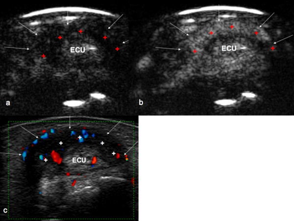
UltrasoundTransverse ultrasound of the wrist over the extensor carpi ulnaris (ECU) tendon, showing peritendinous effusion and vascularity associated with tenosynovitis.
Source : Klauser et al., Arthritis Research & Therapy, 2010, figure 1 · CC BY
- Reactive tendinopathy : a non-inflammatory adaptive response to sudden overload, with transient thickening and an increase in ground substance. This phase is reversible with load modulation.⁸
- Dysrepair (tendon dysrepair) : partial failure of healing, focal disorganisation of collagen, cell and matrix proliferation. Partial reversibility.⁸
- Degenerative tendinopathy : advanced structural changes, acellular areas, major collagen disorganisation, neovascularisation and hyperinnervation correlated with persistent pain. Limited reversibility.⁹
📈 The tendon continuum model (Cook & Purdam, BJSM 2009)
The three phases with the main therapeutic strategy and estimated reversibility
Adapted from Cook JL, Purdam CR. Br J Sports Med. 2009;43(6):409-416 (PMID 18812414) and Cook 2016 (PMID 27127294).
- The ECU is stabilised by a subsheath independent of the dorsal retinaculum: its rupture causes tendon instability, to be distinguished from pure tendinopathy.
- The epidemiology of lateral epicondylalgia is better documented than that of the ECU: incidence ~9.7 / 1,000 / year (Korea 2013-2017), prevalence 1-3 % in the general population.
- In asymptomatic tennis players, 75% show sonographic ECU abnormalities and 42% dynamic subluxation: imaging-symptom dissociation.
- Best-documented risk factor: a high Strain Index (Bretschneider 2022, GRADE high).
- The Cook & Purdam continuum (reactive → dysrepair → degenerative) guides the therapeutic strategy: analgesia first, then progressive loading.
- The natural history of lateral epicondylalgia is favourable but slow (~80% at 52 weeks). Corticosteroids help in the short term but make matters worse at 12 months.
References
- Ahmad Z, Siddiqui N, Malik SS, Abdus-Samee M, Tytherleigh-Strong G, Rushton N. Lateral epicondylitis: a review of pathology and management. Bone Joint J. 2013;95-B(9):1158-1164. PMID 23997125.
- Thirumavalavan J, Ibrahim Z, Byrne RA, Arant KR, Gil JA. Extensor Carpi Ulnaris Instability: A Comprehensive Review of Pathology and Operative Techniques. Hand (NY). 2024;19(7):1090-1096. PMID 37226412.
- Campbell D, Campbell R, O'Connor P, Hawkes R. Sports-related extensor carpi ulnaris pathology: a review of functional anatomy, sports injury and management. Br J Sports Med. 2013;47(17):1105-1111. PMID 24096897.
- Lee SH, Park HJ, Park JH, et al. Epidemiology of lateral and medial epicondylitis in South Korea: A nationwide population-based study. Medicine (Baltimore). 2025;104(8):e41597. PMC 11875595.
- Han SH, Yoon JH, et al. Epidemiology and Etiology of Elbow Pain Based on the Healthcare Bigdata Hub in Korea: A Longitudinal Observational Study. Ewha Med J. 2024. PMC 12093584.
- Shiri R, Viikari-Juntura E, Varonen H, Heliövaara M. Prevalence and determinants of lateral and medial epicondylitis: a population study. Am J Epidemiol. 2006;164(11):1065-1074. PMID 16968862.
- Bretschneider SF, Los FS, Eygendaal D, Kuijer PPFM, van der Molen HF. Work-relatedness of lateral epicondylitis: Systematic review including meta-analysis and GRADE. Am J Ind Med. 2022;65(1):41-50. PMID 34674287.
- Cook JL, Purdam CR. Is tendon pathology a continuum? A pathology model to explain the clinical presentation of load-induced tendinopathy. Br J Sports Med. 2009;43(6):409-416. PMID 18812414.
- Cook JL, Rio E, Purdam CR, Docking SI. Revisiting the continuum model of tendon pathology: what is its merit in clinical practice and research? Br J Sports Med. 2016;50(19):1187-1191. PMID 27127294.
- Bisset LM, Beller E, Jull G, Brooks P, Darnell R, Vicenzino B. Mobilisation with movement and exercise, corticosteroid injection, or wait and see for tennis elbow: randomised trial. BMJ. 2006;333(7575):939. PMID 17012266.
- Smidt N, van der Windt DA, Assendelft WJ, Devillé WL, Korthals-de Bos IB, Bouter LM. Corticosteroid injections, physiotherapy, or a wait-and-see policy for lateral epicondylitis: a randomised controlled trial. Lancet. 2002;359(9307):657-662. PMID 11879861.
- Zarro M, Goel R, Bickhart N, May CC, Abzug JM. Extensor Carpi Ulnaris Tendinopathy in Athletes: A Review of the Conservative and Rehabilitative Options. Hand (NY). 2024;19(3):407-413. PMID 36250572.
- Vaquero-Picado A, Barco R, Antuña SA. Lateral epicondylitis of the elbow. EFORT Open Rev. 2017;1(11):391-397. PMID 28461918.
How can ECU tendinopathy be assessed and diagnosed with confidence?
Which questions should you ask to understand the patient and their history?
The history should systematically explore:- Location : dorso-ulnar wrist pain, over the ulnar head or along the course of the ECU tendon (patients often point to it precisely).¹,²
- Mechanisms : an acute episode (hyperflexion-supination trauma, a tennis backhand, a golf swing) points to rupture of the subsheath; a gradual onset with slow worsening points to an overuse tendinopathy.¹,²,³
- Provoking activities : resisted supination, active loaded ulnar deviation, gripping tasks (pouring, opening a jar, turning a steering wheel). In sport: the two-handed tennis backhand, the golf swing, throwing and striking in baseball or hockey.²,³
- Mechanical phenomenon : the audible or palpable "snap" with a sense of giving way indicates dynamic instability of the tendon (subluxation/dislocation), more readily referred for surgery.²,⁴
- Response to load : pain that "warms up" then eases with effort, or conversely worsens with activity, locates the stage within the continuum.⁵
- Systemic context : a history of rheumatoid arthritis (subsheath vulnerability), gout, lupus, scleroderma.²
- Functional impact : use validated PROMs, namely the Patient-Rated Wrist Evaluation (PRWE) for the wrist, Patient-Rated Tennis Elbow Evaluation (PRTEE) for the elbow.
Which clinical tests should be performed and which other conditions must be ruled out?
Provocation tests
- Palpation of the ECU groove : elective tenderness on direct palpation along the course of the tendon, from the ulnar head to the base of the 5th metacarpal. Performed in pronation to expose the tendon, then in supination to look for displacement.²
- Ruland-Hogan test (ECU synergy test) : the patient holds the elbow flexed to 90°, forearm in full supination on the table. The examiner grasps the patient's thumb and middle finger and asks for resisted radial abduction of the thumb, which synergistically recruits the ECU. Reproduction of dorso-ulnar pain makes the test positive.6 First described by Ruland & Hogan (J Hand Surg Am 2008, PMID 19084177). The study by Sato et al. (J Ultrasound Med 2016) reports a sensitivity of 74% and a specificity of 86% against ultrasound as the reference standard.
- Testing for instability : forearm in active resisted supination with ulnar deviation; a visible or palpable snap of the tendon out of its groove indicates instability. To be confirmed on dynamic ultrasound.²,⁴
- For lateral epicondylalgia (elbow) : Cozen's test (resisted wrist extension), Maudsley's test (resisted middle finger extension), Mill's test (passive stretch).⁷
Differential diagnosis 🗺️
Ulnar-sided wrist pain is a genuine diagnostic melting pot . Mirza & Mirza (J Hand Surg Am 2024, PMID 38435942) propose a systematic guide based on 30 years of clinical experience.⁸ The main competing hypotheses to rule out:| Differential diagnosis | Key clue | Confirmatory test |
|---|---|---|
| TFCC tear (1st DDx) | Painful "press test", pain on ulnar-sided pivoting | MRI, arthroscopy (reference standard) |
| Ulnar impaction syndrome | Positive ulnar variance, pain on loaded ulnar deviation | Loaded pronation radiograph |
| Pisotriquetral osteoarthritis | Crepitus, pisiform tenderness | Oblique-view radiograph |
| DRUJ instability | Ulnar ballottement test + | Dynamic ultrasound, MRI |
| Neuropathy of the dorsal cutaneous branch of the ulnar nerve | Dorso-ulnar hypoaesthesia, Tinel + | EMG if in doubt |
| Ulnar styloid fracture | Recent trauma, exquisite tenderness | Radiograph, CT |
🚩 Red flags in ulnar-sided wrist pain
- Recent trauma + visible deformity or major loss of function → suspected fracture-dislocation of the distal ulna or distal radius → urgent imaging.
- A pulsatile or firm painless mass in the dorso-ulnar region → suspected neoplasm (synovial sarcoma, giant cell tumour) or aneurysm.
- Fever + oedema + redness → suspected infective tenosynovitis (a surgical emergency).
- Known history of malignancy + deep nocturnal bone pain → work-up for metastases.
- Active rheumatoid arthritis with progressive polyarticular involvement → rheumatology referral for systemic management.
- Neurological symptoms (paraesthesia, motor deficit) not following the territory of the dorsal cutaneous branch of the ulnar nerve → neurological work-up.
⚠️ Any red flag calls for prompt medical referral before physiotherapy management.
What is the role of imaging and of ECU lesion classification?
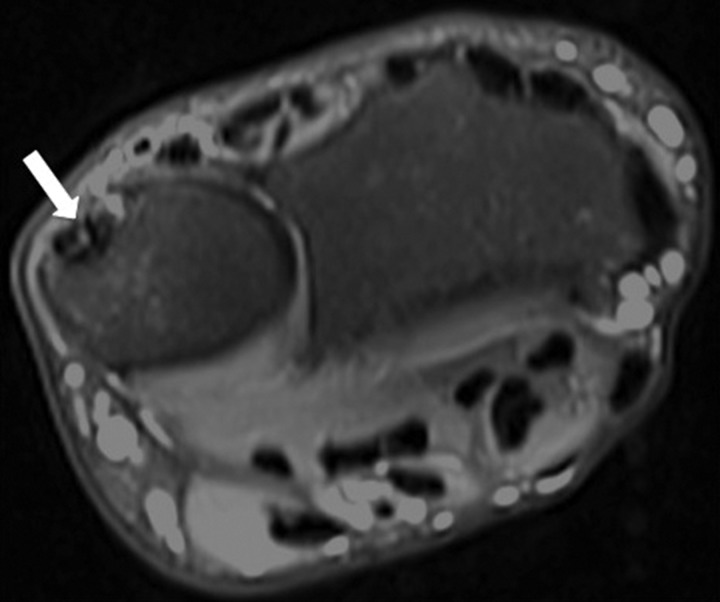
MRIAxial fat-saturated T2-weighted wrist MRI: longitudinal tear of the extensor carpi ulnaris tendon, arrow on the area of hyperintensity.
Source : Campbell et al., British Journal of Sports Medicine, 2013, figure 7 · CC BY-NC
- Morphological assessment of the tendon (thickening, hypoechogenicity, neovascularisation on Doppler).
- The search for dynamic subluxation of the tendon out of its groove during active supination.
- Detection of tenosynovitis (peritendinous effusion).
📋 Diagnostic algorithm for ulnar-sided wrist pain with suspected ECU involvement
A pragmatic hierarchy combining clinical findings + dynamic ultrasound + MRI if in doubt
Sources: Thirumavalavan 2024 (PMID 37226412), Mirza & Mirza 2024 (PMID 38435942), Iwamoto 2022 (PMID 36037819).
- The history looks for dorso-ulnar pain aggravated by supination + ulnar deviation, and for any snapping suggesting instability.
- The Ruland-Hogan test (PMID 19084177) is the reference clinical test: Se 74%, Sp 86% vs ultrasound.
- Dynamic ultrasound is the first-line investigation: it detects subluxation in real time, which a static MRI misses.
- The main differential diagnosis is a TFCC tear, which may coexist with ECU tendinopathy in throwing athletes.
- Beware the imaging-symptom dissociation (75% sonographic abnormalities in asymptomatic tennis players, Sole 2015).
- The Iwamoto 2022 classification distinguishes tendinopathy, tenosynovitis, acute/chronic instability and rupture in order to stratify treatment.
References
- Zarro M, Goel R, Bickhart N, May CC, Abzug JM. Extensor Carpi Ulnaris Tendinopathy in Athletes: A Review of the Conservative and Rehabilitative Options. Hand (NY). 2024;19(3):407-413. PMID 36250572.
- Thirumavalavan J, Ibrahim Z, Byrne RA, Arant KR, Gil JA. Extensor Carpi Ulnaris Instability: A Comprehensive Review of Pathology and Operative Techniques. Hand (NY). 2024;19(7):1090-1096. PMID 37226412.
- Campbell D, Campbell R, O'Connor P, Hawkes R. Sports-related extensor carpi ulnaris pathology: a review of functional anatomy, sports injury and management. Br J Sports Med. 2013;47(17):1105-1111. PMID 24096897.
- Inoue G, Tamura Y. Recurrent dislocation of the extensor carpi ulnaris tendon. Br J Sports Med. 1998;32(2):172-174. PMC 1756093.
- Cook JL, Purdam CR. Is tendon pathology a continuum? A pathology model to explain the clinical presentation of load-induced tendinopathy. Br J Sports Med. 2009;43(6):409-416. PMID 18812414.
- Ruland RT, Hogan CJ. The ECU synergy test: an aid to diagnose ECU tendonitis. J Hand Surg Am. 2008;33(10):1777-1782. PMID 19084177.
- Lucado AM, Day JM, Vincent JI, et al. Lateral Elbow Pain and Muscle Function Impairments: Clinical Practice Guidelines (APTA). J Orthop Sports Phys Ther. 2022;52(12):CPG1-CPG111. PMID 36453071.
- Mirza A, Mirza JB. Ulnar-Sided Wrist Pain: A Diagnostic Evaluation Guide From 30-Plus Years of Experience. Plast Reconstr Surg Glob Open. 2024;12(2):e5602. PMID 38435942.
- Sole G, Strauss J, Cassidy M, et al. Sonographic Evaluation of the Extensor Carpi Ulnaris in Asymptomatic Tennis Players. PM&R. 2015;7(3):255-263. PMID 25217825.
- Burda R, Morochovič R, Rudnay M, Špaková T. A new comprehensive Classification Scheme for ECU Tendon Problems at the Wrist. Handchir Mikrochir Plast Chir. 2022;54(5):427-433. PMID 36037819.
Which treatment strategies are the most effective?
Where do you start? What is the recommended hierarchy of interventions?
The therapeutic hierarchy, structured as a cascade:- First line: education + load modulation : explaining the condition (tendon continuum, pain ≠ damage), identifying and adapting the provoking movements, modifying activity without complete rest (which is counterproductive).¹
- First line: wrist splint in slight pronation for 4-6 weeks for acute ECU instability or florid tenosynovitis, to protect the subsheath and allow healing.²,³
- Cornerstone: progressively loaded exercise therapy : isometric first (analgesia), then isotonic (HSR or eccentric), then functional.¹,⁴,⁵
- Adjuncts: manual therapy (mobilisation with movement, Mulligan) for the elbow (grade B in the JOSPT CPG 2022); carpal joint mobilisations for the ECU if there is associated stiffness.¹,⁶
- Adjuncts: extracorporeal shockwave therapy (ESWT) in chronic lateral epicondylalgia (efficacy documented by Yao 2020 and later meta-analyses).⁷
- Last conservative resort: injections (PRP, prolotherapy) if treatment fails; the corticosteroids should be avoided because of their deleterious effect at 12 months.⁸,⁹
- Surgery for symptomatic recurrent ECU instability after 3-6 months of well-conducted conservative care (Yan 2024, Thirumavalavan 2024).¹⁰,²
What is the role of exercise and is any one approach superior?
Progressively loaded exercise is the intervention with the highest level of evidence for upper limb tendinopathies.⁴,¹¹ The meta-analysis by Karanasios et al. (Br J Sports Med 2021, 30 RCTs, 2,123 participants) on lateral epicondylalgia concludes that exercise interventions produce better outcomes than passive interventions, but with a modest effect size (a clinically small but statistically significant difference).¹¹ Above all, the meta-analysis finds no difference between concentric-eccentric and eccentric alone, confirming that load progression matters more than the type of contraction.¹¹ This conclusion is consistent with the earlier review by Cullinane et al. (Clin Rehabil 2014, PMID 23881334), which found benefits of eccentric work within a multimodal programme but did not demonstrate its superiority in isolation.⁵ Likewise, the Stasinopoulos 2022 editorial "Stop Using Eccentric Exercises as the Gold Standard" formalises this conceptual shift.¹² The pragmatic programme in three phases that is recommended:📈 The loading continuum: a pragmatic 3-phase protocol
No contraction type has been shown to be superior: load progression is what matters
Sources: Rio E et al. Br J Sports Med. 2015;49(19):1277-1283 (PMID 25979840); Karanasios S et al. Br J Sports Med. 2021; Zarro M et al. Hand. 2024 (PMID 36250572); Kongsgaard M et al. Scand J Med Sci Sports. 2009 (PMID 19793213).
Manual therapy, shockwave therapy, injections: how effective are they really?
| Intervention | Level of evidence (GRADE) | Expected effect | Position in the algorithm |
|---|---|---|---|
| Education + load modulation | High | Therapeutic foundation, prevention of recurrence | 1st line, systematic |
| Progressive exercise (any form) | High | Δ pain and function small to moderate vs passive care (Karanasios 2021) | Cornerstone, from phase 1 |
| High-intensity isometrics | Mod. | Immediate transient analgesia (Rio 2015) | Phase 1, severe pain |
| HSR | Mod. | Grade B in the JOSPT CPG 2022; limited adherence (32-50%) | Phase 2, after analgesia |
| Manual therapy (Mulligan MWM) | Mod. | Short-term benefit when combined with exercise (Lucado 2019) | Facilitating adjunct |
| Shockwave therapy (ESWT) | Mod. | Yao 2020 meta-analysis: significant Δ pain vs placebo (13 RCTs) | Refractory chronic cases |
| Counterforce brace | Low | Short-term relief, to be combined with exercise | Occasional adjunct |
| Dry needling | Low | Heterogeneous data, short term only | Optional |
| Corticosteroid injection | Low | ↑ short term (6 weeks) but ↓ at 52 weeks (Smidt 2002, Bisset 2006) | To be avoided; last line if short-term relief is genuinely needed |
| PRP / prolotherapy | Low | Equivocal data; not for routine use | Refractory cases only |
| Ultrasound, low-level laser | Low | No clinically significant benefit | Not recommended |
⚠ GRADE levels of evidence: the strength of recommendation in the JOSPT CPG 2022 uses grades A (strong evidence) to F (expert consensus). The GRADE levels above are a pragmatic synthesis combining the CPG and later meta-analyses.
The meta-analysis by Yao et al. on ESWT (BioMed Res Int 2020, PMC7106907) included 13 RCTs (1,035 patients) with chronic lateral epicondylalgia, concluding that it is superior to placebo for pain and function, with an excellent safety profile.⁷ This efficacy has been confirmed again by later meta-analyses (Du 2024, Zhang 2024), although the effect size remains modest and cost and access are practical limitations. The manual therapy, in particular Mulligan's mobilisation with movement (MWM), is documented by the meta-analysis of Lucado-Day et al. (J Hand Ther 2019, PMID 29705077): 20 RCTs showing a short-term benefit for pain and function, especially when combined with exercise; its long-term effect is limited, which positions it as a facilitator of the exercise programme rather than a stand-alone curative treatment.⁶ The debate over corticosteroid injections is settled: while they give dramatic short-term relief (6 weeks), the Smidt 2002 (Lancet, PMID 11879861) and Bisset 2006 (BMJ, PMID 17012266) RCTs demonstrated a significantly poorer outcome at 12 months than watchful waiting or exercise, with higher recurrence rates.⁸,⁹ The JOSPT CPG 2022 advises against their routine use.¹Beyond the physical: how do you educate the patient and address psychological factors?
Chronic lateral epicondylalgia is associated with adverse psychosocial factors (catastrophising, kinesiophobia, anxiety) that predict persistence and disability.¹,¹⁷ Therapeutic education must include:- An accessible explanation of the tendon continuum and of the fact that pain does not mean ongoing damage.⁸
- Introducing the concept of acceptable pain (≤ 3-4/10 during exercise, no worsening at 24 h) to normalise mild discomfort during rehabilitation.¹⁸
- Setting realistic collaborative goals (recovery typically 8-12 weeks at minimum, sometimes longer).
- Early identification of yellow flags (catastrophising, fear of movement) and referral for a cognitive-behavioural approach where needed.
Critique and controversy
Three areas of uncertainty persist. First, extrapolating lateral epicondylalgia data to the ECU is necessary but imperfect: the biomechanics of the ECU (bony groove, subsheath, oblique course) differ from those of a pure tension tendon such as the ECRB. ECU-specific recommendations remain largely based on case series and expert opinion (CEBM level 4-5). Secondly, the controversy over exercise protocols persists: HSR has its place but suffers from poor adherence in practice (32% in the 2024 Norwegian RCT)¹⁶; lighter but more engaging loading exercises may be preferable for some patients. The individualisation of the programme is probably more decisive than the theoretical selection of the "best" protocol. Thirdly, the role of adjunctive technologies (ESWT, dry needling, PRP) is statistically significant but clinically modest, with variable costs and uneven access. Their place in the routine treatment algorithm is not definitively established.- 1st line : therapeutic education + load modulation + a splint if there is acute instability.
- Cornerstone : progressively loaded exercise. No superiority has been shown for any one contraction type (Karanasios 2021, meta-analysis of 30 RCTs).
- Pragmatic protocol: isometric → HSR → sport-specific functional.
- Manual therapy (MWM) and ESWT are useful adjuncts but not stand-alone treatments.
- Avoid corticosteroids : improvement at 6 weeks but worse outcomes at 12 months (Smidt 2002, Bisset 2006).
- Address the psychosocial factors (catastrophising, kinesiophobia) in chronic cases.
References
- Lucado AM, Day JM, Vincent JI, et al. Lateral Elbow Pain and Muscle Function Impairments: Clinical Practice Guidelines Linked to the ICF. J Orthop Sports Phys Ther. 2022;52(12):CPG1-CPG111. PMID 36453071.
- Thirumavalavan J, Ibrahim Z, Byrne RA, Arant KR, Gil JA. Extensor Carpi Ulnaris Instability: A Comprehensive Review. Hand (NY). 2024;19(7):1090-1096. PMID 37226412.
- Zarro M, Goel R, Bickhart N, May CC, Abzug JM. Extensor Carpi Ulnaris Tendinopathy in Athletes. Hand (NY). 2024;19(3):407-413. PMID 36250572.
- Rio E, Kidgell D, Purdam C, et al. Isometric exercise induces analgesia and reduces inhibition in patellar tendinopathy. Br J Sports Med. 2015;49(19):1277-1283. PMID 25979840.
- Cullinane FL, Boocock MG, Trevelyan FC. Is eccentric exercise an effective treatment for lateral epicondylitis? A systematic review. Clin Rehabil. 2014;28(1):3-19. PMID 23881334.
- Lucado AM, Dale RB, Vincent JI, Day JM. Do joint mobilizations assist in the recovery of lateral elbow tendinopathy? A systematic review and meta-analysis. J Hand Ther. 2019;32(2):262-276.e1. PMID 29705077.
- Yao G, Chen J, Duan Y, Chen X. Efficacy of Extracorporeal Shock Wave Therapy for Lateral Epicondylitis: A Systematic Review and Meta-Analysis. Biomed Res Int. 2020;2020:2064781. PMC 7106907.
- Smidt N, van der Windt DA, Assendelft WJ, et al. Corticosteroid injections, physiotherapy, or a wait-and-see policy for lateral epicondylitis. Lancet. 2002;359(9307):657-662. PMID 11879861.
- Bisset L, Beller E, Jull G, Brooks P, Darnell R, Vicenzino B. Mobilisation with movement and exercise, corticosteroid injection, or wait and see for tennis elbow. BMJ. 2006;333(7575):939. PMID 17012266.
- Lari A, Burhamah W, Alherz M, et al. Operative Management of Extensor Carpi Ulnaris Instability: A Systematic Review. J Wrist Surg. 2024;13(3):272-281. PMID 38808191.
- Karanasios S, Korakakis V, Whiteley R, Vasilogeorgis I, Woodbridge S, Gioftsos G. Exercise interventions in lateral elbow tendinopathy have better outcomes than passive interventions, but the effects are small: a systematic review and meta-analysis of 2123 subjects in 30 trials. Br J Sports Med. 2021;55(9):477-485. doi:10.1136/bjsports-2020-102525.
- Stasinopoulos D. Stop Using the Eccentric Exercises as the Gold Standard Treatment for the Management of Lateral Elbow Tendinopathy. Healthcare. 2022. PMC 8911334.
- Coombes BK, Wiebusch M, Heales L, Stephenson A, Vicenzino B. Isometric Exercise Above but Not Below an Individual's Pain Threshold Influences Pain Perception in People With Lateral Epicondylalgia. Clin J Pain. 2019;35(2):162-168. PMID 31425384.
- Kongsgaard M, Kovanen V, Aagaard P, et al. Corticosteroid injections, eccentric decline squat training and heavy slow resistance training in patellar tendinopathy. Scand J Med Sci Sports. 2009;19(6):790-802. PMID 19793213.
- Beyer R, Kongsgaard M, Hougs Kjær B, et al. Heavy Slow Resistance Versus Eccentric Training as Treatment for Achilles Tendinopathy: A Randomized Controlled Trial. Am J Sports Med. 2015;43(7):1704-1711. doi:10.1177/0363546515584760.
- Sveinall H, Brox JI, Engebretsen KB, et al. Heavy slow resistance training, radial extracorporeal shock wave therapy or advice for patients with tennis elbow in the Norwegian secondary care: a randomised controlled feasibility trial. BMJ Open. 2024;14(12):e085916. PMID 39806585.
- Coombes BK, Bisset L, Vicenzino B. A new integrative model of lateral epicondylalgia. Br J Sports Med. 2009;43(4):252-258. PMID 19050004.
- Cook JL, Rio E, Purdam CR, Docking SI. Revisiting the continuum model of tendon pathology. Br J Sports Med. 2016;50(19):1187-1191. PMID 27127294.
How do you secure lasting recovery and prevent flares?
How can the patient be made an active participant in recovery through self-management?
The physiotherapist's role is changing: from "technician" to "coach" in long-term management. Self-management rests on three pillars:- Therapeutic education : understanding the tendon continuum, dispelling the automatic pain = damage link, explaining that the capacity of the tendon is built by progressive stimulation and not by avoidance.²,³
- Load management (load management): identifying provoking activities, modulation (volume × intensity × frequency), possibly keeping a symptom-activity diary.²
- Home exercise programme that is simple, short (10-15 min) and built into a daily routine: long-term adherence is the main challenge.
⚖️ The acceptable pain model (Silbernagel 2007)
A clinical tool to guide progression, applied to extensor tendinopathies
Adapted from Silbernagel KG et al. Am J Sports Med. 2007;35(6):897-906 (PMID 17307888). Rule: pain may reach 5/10 during activity provided that it does not worsen over the following 24 hours.
When and how should a safe return to sport and activity be planned?
Return to sport (RTS) after ECU tendinopathy or lateral epicondylalgia must be based on measurable functional criteria, never on an arbitrary time frame.²,⁵ The principles of van der Vlist et al. (Sports Med 2021), originally developed for the Achilles, are largely applicable to the wrist and elbow extensors.⁵ Typical functional RTS criteria (Zarro 2024 for the ECU) :- Pain ≤ 2/10 at rest and during activities of daily living.
- Range of movement symmetrical with the uninjured side (ulnar deviation, supination, wrist flexion-extension).
- Grip strength ≥ 80-90% of the uninjured side on the Jamar dynamometer.²
- Ability to perform sport-specific movements (tennis strokes, the golf swing) without significant pain or worsening at 24 h.
- A sport-specific functional test passed at full intensity.
🚦 Return-to-sport algorithm after ECU tendinopathy (criterion-based)
Three successive lights: baseline criteria → sport-specific reintroduction → full competition
Adapted from Zarro M et al. Hand. 2024 (PMID 36250572); van der Vlist AC et al. Sports Med. 2021; Silbernagel KG. AJSM. 2007 (PMID 17307888).
- The empowerment of the patient is the key to preventing recurrence: education, load management, a long-term exercise programme.
- The acceptable pain model (Silbernagel 2007) allows pain of ≤ 5/10 provided it does not worsen at 24 h.
- Return to sport rests on measurable functional criteria (grip strength ≥ 80-90%), not on elapsed time.
- Typical progression: the 10% per week rule in volume or intensity, to avoid re-overloading.
- Long-term adherence to the strengthening programme is the main clinical challenge: favour simplicity and patient motivation.
References
- Lucado AM, Day JM, Vincent JI, et al. Lateral Elbow Pain and Muscle Function Impairments: CPG. J Orthop Sports Phys Ther. 2022;52(12):CPG1-CPG111. PMID 36453071.
- Zarro M, Goel R, Bickhart N, May CC, Abzug JM. Extensor Carpi Ulnaris Tendinopathy in Athletes. Hand (NY). 2024;19(3):407-413. PMID 36250572.
- Cook JL, Rio E, Purdam CR, Docking SI. Revisiting the continuum model of tendon pathology. Br J Sports Med. 2016;50(19):1187-1191. PMID 27127294.
- Silbernagel KG, Thomeé R, Eriksson BI, Karlsson J. Continued Sports Activity, Using a Pain-monitoring Model, During Rehabilitation in Patients With Achilles Tendinopathy. Am J Sports Med. 2007;35(6):897-906. PMID 17307888.
- van der Vlist AC, Winters M, Weir A, et al. Which treatment is most effective for patients with Achilles tendinopathy? A living systematic review with network meta-analysis. Br J Sports Med. 2021;55(5):249-256. doi:10.1136/bjsports-2019-101872.
- Karanasios S, Korakakis V, Whiteley R, et al. Exercise interventions in lateral elbow tendinopathy. Br J Sports Med. 2021;55(9):477-485. doi:10.1136/bjsports-2020-102525.
- Thirumavalavan J, Ibrahim Z, Byrne RA, Arant KR, Gil JA. Extensor Carpi Ulnaris Instability: A Comprehensive Review. Hand (NY). 2024;19(7):1090-1096. PMID 37226412.
- Lucado AM, Vincent JI, Day JM. Physical Therapy for People with Lateral Elbow Tendinopathy: Using the Evidence to Guide Musculoskeletal Rehabilitation Clinical Practice. J Orthop Sports Phys Ther. 2023;53(1):1-2. doi:10.2519/jospt.2023.0501.
What specific management does the athlete, the most exposed subgroup, require?
Which sports and movements place the ECU tendon most at risk?
The reference study is that of Montalvan et al. (Br J Sports Med 2006, PMID 16632573), which followed 28 cases of ECU pathology in tennis players at elite level and identified three distinct clinical patterns :¹- Acute instability : direct trauma or a forced movement (two-handed backhand), rupture of the subsheath.
- Overuse tendinopathy : gradual pain, overuse, preserved structure.
- Tendon rupture : rare, on a chronically degenerative tendon or in an inflammatory context.
- 75 % structural sonographic abnormalities (most often a partial split distal to the groove).
- 42 % dynamic instability (subluxation on active supination).
- 91 % of the instabilities were subluxations, not complete dislocations.
- No complete rupture, effusion or tenosynovitis in this asymptomatic population.
🎾 Sports at greatest risk of ECU pathology
Main biomechanical mechanisms and reported incidence
Sources: Montalvan B et al. Br J Sports Med. 2006;40(5):424-429 (PMID 16632573); Sole G et al. PM&R. 2015;7(3):255-263 (PMID 25217825); Campbell D et al. Br J Sports Med. 2013 (PMID 24096897); Zarro M et al. Hand. 2024 (PMID 36250572).
Return-to-competition strategies in the elite athlete
The prospective study by Mastroianni et al. (Hand 2024) followed 14 elite athletes who underwent radially based subsheath reconstruction between 2011 and 2021. All returned to their previous level of competition, showing that well-indicated targeted surgery and rigorous rehabilitation allow a full return to performance, including at the highest level.⁴ Principles specific to the elite athlete:- Early diagnostic imaging (dynamic ultrasound + MRI if in doubt), because the timescale of a season leaves no room for watchful waiting.
- A conservative vs surgical decision made quickly when instability is established: prolonged watchful waiting can cost the whole season with no benefit.
- A supervised rehabilitation programme (a dedicated sports physiotherapist), not home self-rehabilitation alone.
- Equipment adjustment : racket grip size, string tension, protective gloves (no direct ECU evidence, but transposed from lateral epicondylalgia).
- Technique adjustment with the coach: the two-handed backhand, the golf swing, managing repeated loading.
- Monitoring of external load (training hours, number of strokes, intense sessions per week) and of internal load (discomfort, fatigue) during the return.
🚩 Red and yellow flags specific to the sporting context
- An audible symptomatic subluxation recurring under load → strong suspicion of subsheath rupture → semi-urgent dynamic ultrasound + referral to a hand surgeon.
- Failure of 3-6 months of conservative care in a competitive athlete → early re-discussion of surgery, without prolonging the wait.
- Pressure from those around the athlete (coach, sponsor, federation) for a premature return → a firm professional stance, a criterion-based return, not a calendar-based one.
- Post-injury kinesiophobia in the athlete (fear of re-injury) → psychological work + a gradual return that reassures and demonstrates capability.
- Suspected TFCC comorbidity in a thrower or a golfer → systematic MRI before discharge.
⭐ Key points
- The sports at greatest risk are tennis, golf, hockey/lacrosse, baseball, rowing and combat sports: the common thread is supination + ulnar deviation under load.
- Sole 2015 (PMID 25217825): 75% sonographic abnormalities and 42% subluxation in tennis players who are asymptomatic : imaging alone does not make the decision.
- Montalvan 2006 (PMID 16632573) described three ECU patterns in 28 tennis players: acute instability, overuse tendinopathy, rupture.
- Mastroianni 2024 (Hand): 14 elite athletes reconstructed, all returning to their previous level of competition.
- In the athlete, the decision to operate is sometimes taken sooner than in the general population, to save the season.
- The return rests on an interdisciplinary team (physiotherapist, sports physician, coach) and objective functional criteria.
References
- Montalvan B, Parier J, Brasseur JL, Le Viet D, Drape JL. Extensor carpi ulnaris injuries in tennis players: a study of 28 cases. Br J Sports Med. 2006;40(5):424-429. PMID 16632573.
- Campbell D, Campbell R, O'Connor P, Hawkes R. Sports-related extensor carpi ulnaris pathology: a review of functional anatomy, sports injury and management. Br J Sports Med. 2013;47(17):1105-1111. PMID 24096897.
- Sole G, Strauss J, Cassidy M, et al. Sonographic Evaluation of the Extensor Carpi Ulnaris in Asymptomatic Tennis Players. PM&R. 2015;7(3):255-263. PMID 25217825.
- Mastroianni MA, Leibman M, Belsky M, Vitale MA, Ruchelsman DE. Radially Based Extensor Retinacular Sling Reconstruction for Extensor Carpi Ulnaris Subsheath Injuries in Elite Athletes. Hand (NY). 2024. doi:10.1177/15589447231151433.
- Zarro M, Goel R, Bickhart N, May CC, Abzug JM. Extensor Carpi Ulnaris Tendinopathy in Athletes. Hand (NY). 2024;19(3):407-413. PMID 36250572.
- Thirumavalavan J, Ibrahim Z, Byrne RA, Arant KR, Gil JA. Extensor Carpi Ulnaris Instability: A Comprehensive Review. Hand (NY). 2024;19(7):1090-1096. PMID 37226412.
- Pang EQ, Yao J. Ulnar-sided wrist pain in the athlete (TFCC/DRUJ/ECU). Curr Rev Musculoskelet Med. 2017;10(1):53-61. PMID 28185125.
What do real case studies teach us about ECU tendinopathies?
Analysis of a "typical" case: from assessment to conservative resolution
The typical case is that of a racket or stick sport athlete with dorso-ulnar pain of gradual onset over several weeks, made worse by sport-specific movements. The series by Fisch et al. (J Hand Surg Eur Vol 2020, PMID 31533512) is instructive: 4 patients with ECU tendinopathy refractory to initial conservative treatment underwent minimally invasive surgical tendon lengthening , with complete recovery in all of them.¹ For most cases, however, structured conservative treatment is enough. The typical 12-week pathway (synthesised from Zarro 2024 and Thirumavalavan 2024):²,³- Weeks 0-2 : relative rest, modulation of the provoking movements, a wrist splint in slight pronation, analgesics as needed, patient education.
- Weeks 2-6 : analgesic isometrics (resisted wrist extension, radial thumb abduction 5×30 sec at 70% MVIC), forearm and carpal mobilisation, manual therapy (distal radioulnar joint mobilisation).
- Weeks 6-10 : introduction of HSR for the wrist extensors (3 sets × 10-15 repetitions, slow 3-1-3 tempo), progression criterion: pain ≤ 4/10 with no worsening at 24 h.
- Weeks 10-12 : functional exercises including resisted supination, progressive speed, first sport-specific loading.
- Weeks 12+ : gradual return to sport according to the functional criteria (grip strength ≥ 80-90% of the uninjured side).
The diagnostic challenge: when ECU pathology mimics or coexists with another condition
The main diagnostic pitfall is confusion with, or coexistence of, a triangular fibrocartilage complex (TFCC) tear.⁴,⁵ The symptoms can be almost identical: dorso-ulnar pain, a clicking sensation, worsening with ulnar deviation and forearm rotation. Both throwers (baseball, javelin) and golfers are particularly exposed to coexisting ECU + TFCC pathology, because of the combined axial (compression) and torsional (shear) loads applied to the wrist.⁴ Mirza & Mirza (2024, PMID 38435942), a synthesis of 30+ years of clinical experience in ulnar-sided wrist pain, propose the following differentiating clues:⁴- In favour of the ECU : elective tenderness along the tendon, a positive Ruland-Hogan test, a click or snap that is palpable on the ulnar border of the wrist, abnormal dynamic ultrasound.
- In favour of the TFCC : press test positive (pain on axial compression in ulnar deviation), foveal sign, tenderness of the ulnar fovea, abnormal MRI or arthroscopy.
- Coexistence likely : an audible snap + foveal sign + residual pain after a diagnostic DRUJ injection.
Study of a complex case: recurrent instability and surgery
The complex case par excellence is symptomatic recurrent ECU instability, which results from rupture of the subsheath. The tendon audibly "jumps" out of its groove with every active supination or loaded ulnar deviation, creating pain and apprehension in the athlete.⁶ The most instructive series is that of Mastroianni et al. (Hand 2024) : 14 elite athletes operated on by radially based subsheath reconstruction between 2011 and 2021. Indications: dynamic instability confirmed on ultrasound + disabling symptoms + failure of 3-6 months of conservative treatment. Result: all the athletes returned to their previous level of competition.⁶ The systematic review by Yan et al. (J Wrist Surg 2024, PMID 38808191) pooled 8 retrospective studies totalling 97 operated wrists :⁷- 40% primary repairs of the subsheath (with anchors and sutures) when tissue quality allows.
- 60% reconstructions using an extensor retinaculum flap or an allograft / palmaris longus graft.
- Satisfactory rates of return to previous activity in most series (generally > 80-90%), but with considerable methodological heterogeneity.
- Overall level of evidence low (retrospective series, CEBM level 4), with no RCT available.
🏛️ GRADE pyramid of the evidence available in ECU pathology
Horizontal card format, from the strongest to the weakest level of evidence
Practical implication: clinical decisions favour levels 1-3 (meta-analyses, RCTs, CPGs) over levels 4-5 (case series, expert opinion). For the ECU specifically, most of the literature is level 4-5, hence the importance of extrapolating intelligently from the principles of better-studied tendinopathies (Achilles, patellar) and of lateral epicondylalgia.
Critique and controversy
Several reservations deserve to be highlighted about the case-based literature on the ECU. First, the overall level of evidence remains low : most ECU-specific recommendations rest on retrospective series (CEBM level 4) or expert opinion (level 5). No RCT of quality comparable to those available for lateral epicondylalgia exists for the ECU specifically.²,⁷ Secondly, extrapolating the principles of lower limb tendinopathies (Achilles, patellar) and of lateral epicondylalgia to the wrist is necessary but imperfect: the biomechanics of the subsheath, the oblique course of the tendon in its groove, and the particular sensitivity to supination are not found in the tendons usually studied. Thirdly, the timing of surgery remains controversial: should the elite athlete be operated on early (3 months) to save the season, or should 6-12 months of well-conducted conservative care be tried first? Practice varies significantly between centres, with no solid comparative data.²,⁷ Fourthly, the operator-dependent reliability of dynamic ultrasound is an often-overlooked factor. The diagnosis of dynamic instability depends on the quality of the examination, the operator's experience and the patient's cooperation. Specific training of the radiologist or of the clinician performing the scan is essential.- The typical case of ECU tendinopathy responds to structured conservative treatment over 12-16 weeks.
- The diagnostic challenge that matters most is the TFCC tear, which may mimic ECU pathology or coexist with it: MRI or arthroscopy settles it.
- The complex case of recurrent instability (subsheath rupture) responds to surgery: Mastroianni 2024 reports 14 elite athletes returning to their previous level.
- The systematic review by Yan 2024 (PMID 38808191) consolidates 97 operated wrists: 40% primary repairs, 60% reconstructions, broadly good results but a low level of evidence.
- The GRADE pyramid illustrates how level 4-5 literature predominates for the ECU, hence the importance of drawing on the principles of better-studied tendinopathies.
References
- Fisch R, Meals C, Meals R. Effectiveness of extensor carpi ulnaris tendon lengthening in treating four patients with tendinopathy. J Hand Surg Eur Vol. 2020;45(1):88-90. PMID 31533512.
- Thirumavalavan J, Ibrahim Z, Byrne RA, Arant KR, Gil JA. Extensor Carpi Ulnaris Instability: A Comprehensive Review of Pathology and Operative Techniques. Hand (NY). 2024;19(7):1090-1096. PMID 37226412.
- Zarro M, Goel R, Bickhart N, May CC, Abzug JM. Extensor Carpi Ulnaris Tendinopathy in Athletes. Hand (NY). 2024;19(3):407-413. PMID 36250572.
- Mirza A, Mirza JB. Ulnar-Sided Wrist Pain: A Diagnostic Evaluation Guide From 30-Plus Years of Experience. Plast Reconstr Surg Glob Open. 2024;12(2):e5602. PMID 38435942.
- Pang EQ, Yao J. Ulnar-sided wrist pain in the athlete (TFCC/DRUJ/ECU). Curr Rev Musculoskelet Med. 2017;10(1):53-61. PMID 28185125.
- Mastroianni MA, Leibman M, Belsky M, Vitale MA, Ruchelsman DE. Radially Based Extensor Retinacular Sling Reconstruction for Extensor Carpi Ulnaris Subsheath Injuries in Elite Athletes. Hand (NY). 2024. doi:10.1177/15589447231151433.
- Lari A, Burhamah W, Alherz M, et al. Operative Management of Extensor Carpi Ulnaris Instability: A Systematic Review. J Wrist Surg. 2024;13(3):272-281. PMID 38808191.
- Inoue G, Tamura Y. Recurrent dislocation of the extensor carpi ulnaris tendon. Br J Sports Med. 1998;32(2):172-174. PMC 1756093.
- Campbell D, Campbell R, O'Connor P, Hawkes R. Sports-related extensor carpi ulnaris pathology. Br J Sports Med. 2013;47(17):1105-1111. PMID 24096897.
- Burda R, Morochovič R, Rudnay M, Špaková T. A new comprehensive Classification Scheme for ECU Tendon Problems at the Wrist. Handchir Mikrochir Plast Chir. 2022;54(5):427-433. PMID 36037819.
How do you apply these recommendations concretely in your practice?
When and to which other health professionals should you refer?
The detection of red flags is the first skill required in direct-access physiotherapy. The international framework of Finucane et al. (JOSPT 2020, PMID 32438853) , originally developed for spinal pathology, proposes a reasoned approach combining isolated signs and clusters of symptoms rather than mechanical box-ticking.¹ For ulnar-sided wrist pain and ECU pathology, the specific red flags were detailed in chapter 2 (trauma + deformity, mass, fever, history of malignancy, systemic polyarticular involvement). The absence of a red flag allows physiotherapy to continue; the presence of one or more calls for prompt referral. Referral to a hand surgeon in the following situations:²,³- Confirmed ECU instability (dynamic ultrasound) with disabling symptoms despite 3-6 months of well-conducted conservative care.
- Tendon rupture that is acute (clinical findings + imaging).
- An associated TFCC tear requiring surgical assessment (diagnostic +/- therapeutic arthroscopy).
- A lesion on inflammatory arthropathy (rheumatoid arthritis, gout) with structural involvement.
- In the elite athlete, surgery is discussed earlier in order to save the season.
- Atypical presentation (disproportionate pain, systemic signs).
- Non-musculoskeletal comorbidity (suspected inflammatory arthropathy, neuropathy).
- A need for further investigations (advanced imaging, blood tests).
- Failure of conservative treatment without a clear surgical diagnosis.
How do you measure outcomes and overcome the barriers to implementation?
Systematic outcome measurement is a marker of evidence-based practice. For wrist extensor tendinopathies, the validated PROMs (Patient-Reported Outcome Measures) to use are:| Tool | Target condition | Score | Approx. MCID |
|---|---|---|---|
| PRTEE (Patient-Rated Tennis Elbow Eval.) | Lateral epicondylalgia | 0-100 (100 = worst) | ~ 11 points |
| PRWE (Patient-Rated Wrist Eval.) | Wrist pain and function (ECU) | 0-100 (100 = worst) | ~ 11-14 points |
| DASH / Quick DASH | Upper limb overall | 0-100 (100 = worst) | ~ 10-15 points |
| Pain VAS | Pain intensity | 0-10 | ~ 2 points |
| Jamar dynamometer (grip strength) | RTS criterion | kg, % ratio to the uninjured side | ≥ 80-90% for RTS |
- Lack of time : the main barrier cited (short consultations, administrative burden).
- Lack of critical appraisal skills for the literature.
- Insufficient organisational support (no dedicated time to keep up with the science).
- Limited access to databases and to full-text articles.
- Professional habits that are entrenched (belief in historical passive modalities).
- Continuing education targeted at the most recent CPGs (the JOSPT CPG 2022 for lateral epicondylalgia is freely available).
- Use of structured clinical summaries (clinical practice guidelines, infographics, specialist podcasts).
- Peer mentoring and case discussion within a network.
- Integration of PROMs into the electronic patient record to reduce the administrative burden.
- Shared decision-making with the patient, to encourage adherence to treatment.
Critique and controversy: between the evidential ideal and clinical reality
Three tensions persist. First, the tyranny of red flags : searching for them systematically is necessary, but their individual predictive value is low, which can lead to excessive imaging and iatrogenic anxiety. Finucane 2020 calls for a contextualised probabilistic interpretation, not mechanical box-ticking.¹ Secondly, the burden of measurement : PROMs add consultation time and can decontextualise the patient-clinician encounter. The balance between methodological rigour and the therapeutic alliance still has to be found locally. Thirdly, the persistent gap between research and practice (know-do gap): the barriers identified for decades remain stable (time, training, resources), suggesting that the solutions must be systemic (health policy, funding quality rather than volume) and not merely individual. It is a collective fight as much as a personal one.- The Finucane 2020 framework (PMID 32438853) guides referral: combined red flags + contextual reasoning, not mechanical box-ticking.
- Surgical referral in cases of established ECU instability + disabling symptoms after 3-6 months of conservative care, or earlier in the elite athlete.
- Recommended PROMs: PRTEE for lateral epicondylalgia, PRWE for the wrist, DASH for the upper limb overall.
- The Jamar grip strength (ratio to the uninjured side ≥ 80-90%) is a key return-to-sport criterion.
- EBP comes up against persistent barriers (time, training, resources): the solution is multi-level (individual + organisational + systemic).
References
- Finucane LM, Downie A, Mercer C, et al. International Framework for Red Flags for Potential Serious Spinal Pathologies. J Orthop Sports Phys Ther. 2020;50(7):350-372. PMID 32438853.
- Thirumavalavan J, Ibrahim Z, Byrne RA, Arant KR, Gil JA. Extensor Carpi Ulnaris Instability: A Comprehensive Review. Hand (NY). 2024;19(7):1090-1096. PMID 37226412.
- Lari A, Burhamah W, Alherz M, et al. Operative Management of Extensor Carpi Ulnaris Instability: A Systematic Review. J Wrist Surg. 2024;13(3):272-281. PMID 38808191.
- Coombes BK, Bisset L, Vicenzino B. A new integrative model of lateral epicondylalgia. Br J Sports Med. 2009;43(4):252-258. PMID 19050004.
- Lucado AM, Day JM, Vincent JI, et al. Lateral Elbow Pain and Muscle Function Impairments: CPG. J Orthop Sports Phys Ther. 2022;52(12):CPG1-CPG111. PMID 36453071.
- Zarro M, Goel R, Bickhart N, May CC, Abzug JM. Extensor Carpi Ulnaris Tendinopathy in Athletes. Hand (NY). 2024;19(3):407-413. PMID 36250572.
- Lucado AM, Vincent JI, Day JM. Physical Therapy for People with Lateral Elbow Tendinopathy. J Orthop Sports Phys Ther. 2023;53(1):1-2. doi:10.2519/jospt.2023.0501.
- Karanasios S, Korakakis V, Whiteley R, Vasilogeorgis I, Woodbridge S, Gioftsos G. Exercise interventions in lateral elbow tendinopathy. Br J Sports Med. 2021;55(9):477-485. doi:10.1136/bjsports-2020-102525.
What next, after this article?
This article is part of a collection of evidence-based clinical syntheses. A question, some feedback, a correction to suggest? Contact us directly via the WhatsApp button at the bottom right of the screen.

