















In brief
De Quervain's tenosynovitis is a stenosing tenosynovitis of the first dorsal compartment of the wrist, affecting the tendons of abductor pollicis longus and extensor pollicis brevis; it is a degenerative tendinosis with thickening of the retinaculum, not a primary inflammation. It presents as precisely localised pain at the radial styloid, worsened by gripping combined with ulnar deviation, and affects women 4 to 6 times more often, with a peak between 30 and 50 years and a distinctive postpartum context. First line, corticosteroid injection is the most effective non-surgical intervention; a thumb spica orthosis remains useful in the pregnant or breastfeeding woman.
Clinical synthesis based on the Challoumas 2023 network meta-analysis, the Cevik 2024 meta-analysis, the HANDGUIDE recommendations and the 2024 prospective postpartum data.
Clinical summary
- Definition: A stenosing tendinopathy of the first dorsal compartment of the wrist, affecting abductor pollicis longus (APL) and extensor pollicis brevis (EPB). It is a degenerative tendinosis with mucoid deposits and thickening of the sheath, not a primary inflammatory tendinitis (Clarke 1998 - the reference histology).
- Target population: It affects women 4 to 6 times more often (Wolf 2009), with a peak incidence between 30 and 50 years. The postpartum period is a particularly vulnerable context (incidence up to 11 % against 1.3 % in the general female population - “new mother's thumb”).
- Risk factors: Repetitive movements combining forceful gripping with ulnar deviation (Stahl 2013 SR-MA, n=10 studies). BMI is not an independent factor in the general population but is postpartum (Daglan 2024: BMI above 25, first pregnancy, gestation beyond 40 weeks are all significant risk factors).
- Mechanism: A mechanical mismatch between content and container through thickening of the extensor retinaculum, which reduces the available space and increases friction on APL/EPB with every movement.
- Clinical diagnosis: Above all clinical: precisely localised pain at the radial styloid, worsened by gripping plus ulnar deviation. The WHAT test (Goubau 2014) is more specific and more sensitive than Eichhoff's test (often confused with Finkelstein's).
- Ultrasound: The investigation of choice for confirming the diagnosis, assessing severity and identifying an intracompartmental septum (present in 79 % of DQT patients against 64 % of controls - Liu 2024, Hand). That matters, because it can explain a failed injection if only one chamber is injected.
- Differential diagnoses: Intersection syndrome (pain 4-8 cm proximal to the radial styloid), thumb base osteoarthritis, Wartenberg's syndrome (compression of the sensory branch of the radial nerve).
- First-line treatment: The Challoumas 2023 network meta-analysis (JAMA Netw Open, n=30 studies, 1663 patients) and the Cevik 2024 meta-analysis (Hand Surg Rehabil) confirm that corticosteroid injection (CSI) is the most effective non-surgical intervention, with a small but significant additional benefit from short associated immobilisation.
- Orthosis: Immobilisation with a thumb spica orthosis alone is less effective than injection alone (RR 1.61, Cevik 2024) but remains useful in the pregnant or breastfeeding woman (where injection is avoided), or as a short-term adjunct after injection.
- Therapeutic exercise: Isometric thumb exercise (McBain 2023, Hand Ther, n=28 RCT feasibility) is feasible, well tolerated (86.7 % adherence) and improves pain and function. Eccentric and concentric exercise appear equivalent. To be started once the acute phase is controlled.
- Manual therapy: There is real added value in adding mobilisation (Mulligan MWM) to the exercise programme. Ultrasound and LLLT have limited evidence as stand-alone treatments.
- Course: The natural history varies. Spontaneous resolution is possible if the cause is removed (notably postpartum, once carrying stops), but chronicity and recurrence are frequent without appropriate management.
- Self-management: The key to preventing recurrence - education about the at-risk movements (smartphone, carrying), activity modification, a home exercise programme, self-monitoring.
- Return to activity: Guided by objective criteria (pain < 3/10, amplitude complete, force prehension > 90 % of the healthy side on dynamometry) and a load progression of 10-20 % a week.
- Red flags: A history of cancer, recent trauma, systemic signs (fever, weight loss), a palpable mass or severe night pain all call for immediate medical referral.
- Outcome measurement: Use validated specific PROMs: QuickDASH, PRWE, VAS. They make progress objective, allow treatment to be adjusted and justify the care given.
Contents
- What are the fundamentals to know about De Quervain's tenosynovitis?
- How do you assess and diagnose De Quervain's tenosynovitis with certainty?
- Why is the postpartum period a specific at-risk context?
- Which treatment strategies are the most effective?
- How do you secure lasting recovery and prevent recurrence?
- What do real clinical cases teach us?
- How do you apply these recommendations concretely in your practice?
What are the fundamentals to know about De Quervain's tenosynovitis?
How is this condition defined, who does it affect and what are the risk factors?
De Quervain's tenosynovitis (DQT), named after the Swiss surgeon Fritz de Quervain who described it in 1895, is a stenosing tenosynovitis of the first dorsal compartment of the wrist.¹ It specifically affects the tendons of abductor pollicis longus (APL) and of extensor pollicis brevis (EPB) as they pass through the osteofibrous tunnel at the radial styloid.² The condition is significantly more prevalent in women, who are affected 4 to 6 times more often than men.³ Incidence in a young working population (a US military cohort, n=250,000) is estimated at 0.94 cases per 1,000 person-years, with a female-to-male ratio of 2.8 and a peak in the 30-49 age band.³ A particularly at-risk group is women in the peripartum period, a phenomenon covered in the dedicated chapter 3.⁴Incidence of DQT: Wolf 2009 (a cohort of 250,000 active US military personnel)
Rate per 1,000 person-years by sex: incidence in women remains 2.8× higher
Source: Wolf JM, Sturdivant RX, Owens BD. J Hand Surg Am. 2009;34(1):112-115. PMID 19081683.
What happens in the body, and how does De Quervain's tenosynovitis evolve naturally?
🔬 Despite what the “-itis” suffix suggests, DQT is not a primary inflammatory process. The founding histological study of Clarke et al. (1998) in 23 operated patients showed that it is a tendinosis, characterised by mucoid degeneration of the connective tissue, thickening of the tendon sheath and accumulation of mucopolysaccharides, with no significant acute inflammatory cells.⁹ That observation became the cornerstone of the modern understanding of the condition and explains why purely anti-inflammatory treatments (NSAIDs alone) show limited long-term benefit. The central pathological mechanism is a content-container mismatch. The extensor retinaculum (the roof of the first dorsal compartment) thickens progressively, which is visible on high-resolution ultrasound.¹⁰ That thickening reduces the available space and increases friction on the APL and EPB tendons with every thumb movement. Over time the process can lead to genuine tendon entrapment, causing acute pain and sometimes locking.- Definition: A stenosing (degenerative) tendinosis of the APL and EPB tendons of the first dorsal compartment, not an inflammatory tendinitis: the reference histology: Clarke 1998 (PMID 9888670).
- Target population: Women affected 4-6× more than men, peak at 30-49 years (Wolf 2009). At-risk subgroups: postpartum (the dedicated chapter 3) and heavy smartphone users (Nie 2023).
- Main mechanical risk factor: Repetitive movements combining gripping with ulnar deviation (Stahl 2013 SR-MA). BMI is not an independent factor in the general population.
- Key mechanism: Thickening of the extensor retinaculum → mechanical mismatch and friction on APL/EPB. No primary inflammation.
- Course: Variable. Resolution is possible if the cause is removed, but chronicity is frequent without appropriate management.
Bibliography, chapter 1
- Satteson E, Tannan SC. De Quervain Tenosynovitis. In: StatPearls [Internet]. Treasure Island (FL): StatPearls Publishing; 2024 Jan-. NBK442005.
- Allbrook V. 'The side of my wrist hurts': de Quervain's tenosynovitis. Aust J Gen Pract. 2019;48(11):753-756. PMID 31722458. doi:10.31128/AJGP-07-19-4996.
- Wolf JM, Sturdivant RX, Owens BD. Incidence of de Quervain's tenosynovitis in a young, active population. J Hand Surg Am. 2009;34(1):112-115. PMID 19081683. doi:10.1016/j.jhsa.2008.08.020.
- Avci S, Yilmaz C, Sayli U. Comparison of nonsurgical treatment measures for de Quervain's disease of pregnancy and lactation. J Hand Surg Am. 2002;27(2):322-324. PMID 11901392.
- Stahl S, Vida D, Meisner C, Lotter O, Rothenberger J, Schaller HE, Stahl AS. Systematic review and meta-analysis on the work-related cause of de Quervain tenosynovitis: a critical appraisal of its recognition as an occupational disease. Plast Reconstr Surg. 2013;132(6):1479-1491. PMID 24005369.
- Goel R, Abzug JM. de Quervain's tenosynovitis: a review of the rehabilitative options. Hand (N Y). 2015;10(1):1-5. PMID 25762881. doi:10.1007/s11552-014-9649-3.
- Nie B, Huang X, Chen Y, et al. Smartphone usage behaviors and their association with De Quervain's Tenosynovitis (DQT) among college students: a cross-sectional study in Guangxi, China. BMC Public Health. 2023;23(1):2257. PMID 37974168. doi:10.1186/s12889-023-16808-z.
- Daglan E, Morgan S, Yechezkel M, Rutenberg TF, Shemesh S, Iordache SD, Kadar A. Risk Factors Associated With de Quervain Tenosynovitis in Postpartum Women. Hand (N Y). 2024;19(4):643-647. PMID 36692105. doi:10.1177/15589447221150524.
- Clarke MT, Lyall HA, Grant JW, Matthewson MH. The histopathology of de Quervain's disease. J Hand Surg Br. 1998;23(6):732-734. PMID 9888670. doi:10.1016/s0266-7681(98)80085-5.
- Shen Y, Zhou Q, Sun X, Qiu Z, Jia Y, Li S, Zhang W. The ultrasound-guided percutaneous release technique for De Quervain's disease using an acupotomy. Front Surg. 2022;9:1034716. doi:10.3389/fsurg.2022.1034716.
- Goel R, Abzug JM. de Quervain's tenosynovitis: a review of the rehabilitative options. Hand (N Y). 2015;10(1):1-5. (the reference already given in this chapter, no. 6 above)
How do you assess and diagnose De Quervain's tenosynovitis with certainty?
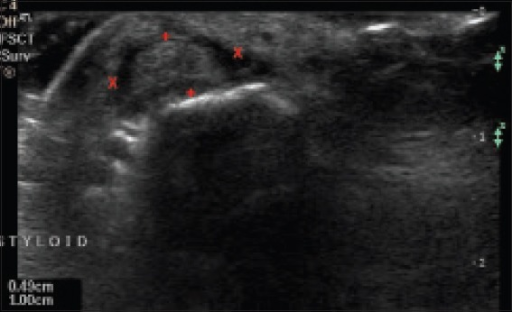
UltrasoundUltrasound of the wrist on the affected side before treatment, showing thickening of the tendon sheath of the abductor pollicis longus and extensor pollicis brevis, measured with the cursor.
Source : Sharma et al., Indian Journal of Orthopaedics, 2015, figure 4 · CC BY-NC-SA
Which questions should you ask to understand the patient and their history?
Taking the history is the crucial first step. It should explore the nature of the pain, the triggering factors and the patient's circumstances. The key questions to ask include:- Location and description of the pain 📍: does the patient describe precisely localised pain on the radial side of the wrist, over the radial styloid? Is the pain gradual, sharp, or burning? That precise location is the cardinal symptom.³
- Aggravating factors : which activities worsen the pain? Patients typically report worsening on gripping (picking something up), twisting the wrist or deviating it in an ulnar direction. Everyday movements such as lifting a pan, using scissors, turning a key or holding a smartphone often become painful.⁴
- Occupation and leisure 🎮: repetitive thumb or wrist movements (assembly-line work, tool use, gardening, gaming)? Heavy smartphone use (Nie 2023: a significant risk beyond 6-8 h/day)?⁵
- Peripartum status 👩🍼: a woman who is pregnant or has given birth in the past 12 months? Currently breastfeeding? Carrying the infant pinched between thumb and index finger? This subgroup deserves a specific approach (chapter 3).
- Mode of onset : sudden and post-traumatic, or gradual over weeks? A gradual onset is more typical of this overuse condition.⁴
Which clinical tests should you perform, and which other conditions should you rule out?
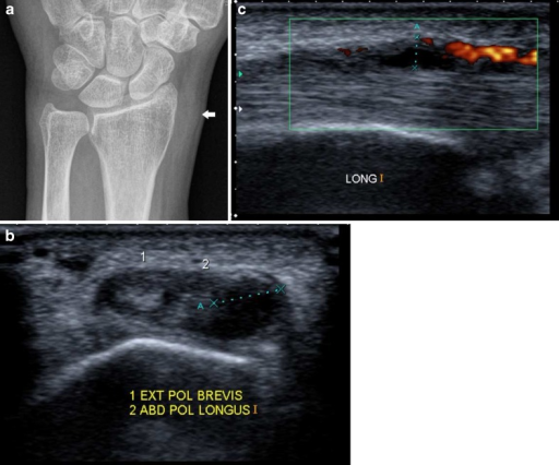
X-rayFrontal radiograph showing soft tissue swelling at the radial styloid process, and transverse and Doppler ultrasound showing fluid and increased vascularity around the tendons.
Source : Vanhoenacker et al., Insights into Imaging, 2011, figure 10 · CC BY
- The Finkelstein test (the original, 1930) : in Harry Finkelstein's original description (JBJS 1930;12:509-540), the examiner grasps the patient's thumb and applies longitudinal traction while passively deviating the wrist in an ulnar direction. A positive test produces sharp pain at the radial styloid.⁶
- The Eichhoff test (often confused with Finkelstein's) : the patient actively closes a fist around their thumb, and the examiner then deviates the wrist ulnarwards, actively or passively.⁷ Very sensitive but poorly specific (frequent false positives, including in healthy people). A diagnostic review (Wu 2018) even showed Finkelstein to be superior to Eichhoff in specificity.⁸
- The WHAT test (Wrist Hyperflexion and Abduction of the Thumb) 🆕: described by Goubau et al. (2014, J Hand Surg Eur, n=100), this test puts the wrist in maximal flexion while the patient actively abducts the thumb against the examiner's resistance. Sensitivity 0.99 and specificity 0.29 in the original cohort, better than Eichhoff and Finkelstein in that prospective study.⁹
Diagnostic performance of the clinical tests for DQT (Goubau 2014, cohort n=100)
Sensitivity and specificity compared: the WHAT test is more sensitive and more specific than Eichhoff/Finkelstein
Source: Goubau JF, Goubau L, Van Tongel A, Van Hoonacker P, Kerckhove D, Berghs B. J Hand Surg Eur Vol. 2014;39(3):286-292. PMID 25538075. Specificity remains low for all three tests: reading any of them in isolation is not enough.
- Intersection syndrome : a tenosynovitis affecting the tendons of the second dorsal compartment where they cross those of the first. The pain sits about 4 to 8 cm more proximally than in DQT, at the musculotendinous junction, a decisive anatomical landmark under the examiner's thumb.¹⁰
- Thumb base osteoarthritis (trapeziometacarpal) : causes pain at the base of the thumb, which can radiate. The grind test (compression and rotation of the TMC joint) is painful. Thumb radiographs confirm joint space narrowing.¹¹
- Wartenberg's syndrome : irritation or compression of the superficial sensory branch of the radial nerve. The symptoms are mainly neurological (paraesthesia, dysaesthesia) over the dorsum of the thumb and index finger, with no motor weakness. A Tinel test along the nerve may be positive.¹²
- FCR/ECRL tendinopathy, scaphoid non-union, ganglion cyst: to be considered if imaging rules out DQT.
Should patients with De Quervain's tenosynovitis be classified, and what are the benefits?
Unlike other musculoskeletal conditions (the Quebec Task Force for low back pain, Childs for neck pain), there is no universally adopted and validated clinical classification system for DQT.¹ Most diagnoses stop at a binary judgement (present or absent). The arrival of ultrasound has opened the way to imaging-based classifications. Three main criteria emerge:- The degree of retinacular thickening (normal < 0,5 mm, modéré 0,5-1 mm, marqué > 1 mm).¹⁰
- The severity of the tenosynovitis (the amount of peritendinous fluid, the power Doppler signal).²
- The presence or absence of an intracompartmental septum (Liu 2024).¹³
To be ruled out without fail (immediate medical referral)
- A personal history of cancer (breast, prostate, kidney, melanoma) with recent atypical pain: a distal radial bone metastasis is possible.
- Recent high-energy trauma not documented (scaphoid fracture, Hutchinson fracture): radiograph as a matter of course.
- Systemic signs: persistent fever, unexplained weight loss, night sweats: think septic arthritis, osteomyelitis, neoplasia.
- A palpable mass, non-mechanical night pain that wakes the patient, visible deformity.
- Associated polyarthritis, morning stiffness beyond 1 h: laboratory work-up (CRP, rheumatoid factor, anti-CCP): think early rheumatoid arthritis.
- Diagnosis is mainly clinical : precisely localised pain at the radial styloid, worsened by gripping plus ulnar deviation.
- The WHAT test (Goubau 2014) is more sensitive and more specific than the Eichhoff/Finkelstein tests. Use it first line.
- Do not confuse the original Finkelstein (1930) = passive traction on the thumb by the examiner, with Eichhoff = a fist closed over the thumb by the patient: the latter generates many false positives.
- High-resolution ultrasound = the investigation of choice in cases of doubt, treatment failure, or before injection (looking for the septum).
- The intracompartmental septum is present in 79 % of DQT patients (Liu 2024) and can explain a failed unguided injection.
- Rule out the differential diagnoses: intersection syndrome (pain 4-8 cm more proximal), thumb base osteoarthritis (grind test), Wartenberg's (dorsal paraesthesia).
- No universally validated classification: ultrasound stratification is promising but not standardised.
Bibliography, chapter 2
- Satteson E, Tannan SC. De Quervain Tenosynovitis. StatPearls. 2024. NBK442005.
- Shen Y, et al. Ultrasound-guided percutaneous release for De Quervain's disease. Front Surg. 2022;9:1034716. doi:10.3389/fsurg.2022.1034716.
- Allbrook V. 'The side of my wrist hurts': de Quervain's tenosynovitis. Aust J Gen Pract. 2019;48(11):753-756. PMID 31722458.
- Goel R, Abzug JM. de Quervain's tenosynovitis: a review of the rehabilitative options. Hand (N Y). 2015;10(1):1-5. PMID 25762881.
- Nie B, et al. Smartphone usage behaviors and De Quervain's Tenosynovitis among college students. BMC Public Health. 2023;23(1):2257. PMID 37974168.
- Finkelstein H. Stenosing tendovaginitis at the radial styloid process. J Bone Joint Surg. 1930;12(3):509-540. (The historic reference paper)
- Elliott BG. Finkelstein's test: a descriptive error that can produce a false positive. J Hand Surg Br. 1992;17(4):481-482. PMID 1402284.
- Wu F, Rajpura A, Sandher D. Finkelstein's Test Is Superior to Eichhoff's Test in the Investigation of de Quervain's Disease. J Hand Microsurg. 2018;10(2):116-118. PMID 30154628.
- Goubau JF, Goubau L, Van Tongel A, Van Hoonacker P, Kerckhove D, Berghs B. The wrist hyperflexion and abduction of the thumb (WHAT) test: a more specific and sensitive test to diagnose de Quervain tenosynovitis than the Eichhoff's Test. J Hand Surg Eur Vol. 2014;39(3):286-292. PMID 23340762. doi:10.1177/1753193412475043.
- Draghi F, Bortolotto C. Intersection syndrome: ultrasound imaging. Skeletal Radiol. 2014;43(3):283-287. PMID 24337446.
- Wajon A, Vinycomb T, Carr E, Edmunds I, Ada L. Surgery for thumb (trapeziometacarpal joint) osteoarthritis. Cochrane Database Syst Rev. 2015;(2):CD004631. PMID 25702783.
- Lanzetta M, Foucher G. Entrapment of the superficial branch of the radial nerve (Wartenberg's syndrome). A report of 52 cases. Int Orthop. 1993;17(6):342-345. PMID 8163306.
- Liu C, Moye S, Blazar P, Earp BE, Zhang D. Anatomical Variations of the First Dorsal Compartment in de Quervain Tenosynovitis. Hand (N Y). 2024;19(7):1159-1165. PMID 37077124. doi:10.1177/15589447231164746.
- Challoumas D, Ramasubbu R, Rooney E, Seymour-Jackson E, Putti A, Millar NL. Management of de Quervain Tenosynovitis: A Systematic Review and Network Meta-Analysis. JAMA Netw Open. 2023;6(10):e2337001. PMID 37889490. doi:10.1001/jamanetworkopen.2023.37001.
Why is the postpartum period a specific at-risk context?
What are the specific hormonal and mechanical mechanisms?
Peripartum DQT, nicknamed “mommy's thumb” 👩🍼, is a presentation frequent and stereotyped enough to deserve a stratified approach. Incidence in this population is estimated at about 11 %, nearly 8 times the incidence in the general female population (1.3 %).¹ ²- A pre-pregnancy BMI above 25 kg/m² (overweight or obesity): unlike the general population, where BMI is not an independent factor.
- First pregnancy (a first child): probably linked to less ergonomic carrying technique through inexperience.
- Gestation beyond 40 weeks: a possible cumulative hormonal effect.
- Hormonal factors 🧬: oestrogen and relaxin in late pregnancy and during breastfeeding induce generalised ligament laxity and periarticular fluid retention, which favours thickening of the tendon sheath.
- Repetitive mechanical factors 🤱: carrying the infant “pinched” between thumb and index finger (15-30 times a day, with the load rising from 3 to 6 kg over 6 months), nappy changing, breastfeeding (wrist position), rocking.
- Postural factors : supporting the infant's neck with the thumb in forced abduction and extension for prolonged periods.
Independent postpartum risk factors (Daglan 2024, case-control study n=63/630)
The significant odds ratios identified in multivariate analysis
Source: Daglan E, Morgan S, Yechezkel M, Rutenberg TF, et al. Hand (N Y). 2024;19(4):643-647. PMID 36692105. Approximate odds ratios in multivariate analysis: all significantly above 1.
What management suits the breastfeeding new mother?
Managing DQT postpartum is complicated by three specific practical constraints :- Caring for the infant continues : it is hard to “rest” a wrist that has to carry a 4-6 kg baby every 2-3 hours.
- Breastfeeding : this limits the pharmacological options. NSAIDs are possible, but local corticosteroid injection (triamcinolone) remains the preferred option: systemic bioactivity is negligible, and it is compatible with breastfeeding under the LactMed recommendations.⁴
- Psychological pressure 🧠: guilt, a specific kinesiophobia (“afraid of hurting the baby by carrying them”), underlying postnatal depression: yellow flags to screen for actively.
- The injection group : 9/9 (100 %) achieved complete pain relief, with a single late recurrence.
- The orthosis-only group : no patient achieved complete immediate relief; 8/10 saw their pain disappear spontaneously at the end of breastfeeding, and 1 eventually received a rescue injection.
- Education first 📚: teach the neutral carrying technique: lift the infant with a scooping hand under the bottom and the neck, palm up, without a thumb-index pinch. Favour ergonomic carriers (a sling, an ergonomic BabyBjörn).
- A thumb spica orthosis at night ± as needed : it limits accumulated inflammation. Breastfeeding-compatible models are available (washable fabric, Velcro fastening).
- Ultrasound-guided corticosteroid injection : where pain is disabling beyond 4-6 weeks despite orthosis plus education. Triamcinolone acetonide 40 mg + 1 % lidocaine. Compatible with breastfeeding.⁴
- Gentle exercise : tendon gliding plus isometrics (McBain 2023: a protocol feasible postpartum) once the acute phase is controlled.⁶
- Screen for postnatal depression and kinesiophobia : the Edinburgh Postnatal Depression Scale (EPDS) if suspected; refer to the general practitioner if the EPDS is 10 or above.
Red flags specific to the postpartum period
- Persistent fever plus joint pain: think septic arthritis (rare but serious), or mastitis spreading further afield.
- New symmetrical bilateral polyarthritis: think postpartum rheumatoid arthritis (onset triggered by childbirth is described).
- Recent epidural anaesthesia plus distal neurological signs: neurological referral.
- An EPDS of 13 or above with dark thoughts: urgent psychiatric referral; do not reduce the consultation to its physiotherapy aspect alone.
- Repeated vomiting, weight loss, dehydration: think postpartum hyperthyroidism (it affects tendon healing).
- Postpartum DQT affects about 11 % of new mothers (×8 against the general female population).
- Independent risk factors (Daglan 2024): BMI above 25, first pregnancy, gestation beyond 40 weeks.
- A triple mechanism: hormonal (laxity), mechanical (pinch carrying) and postural (supporting the neck).
- Cortisone injection is the reference non-surgical treatment in the breastfeeding mother (Avci 2002 RCT: 100 % complete relief against orthosis alone).
- Education in ergonomic scoop carrying is the cornerstone: it is the persistent cause that keeps the condition going.
- Screen for postnatal depression (EPDS) and for kinesiophobia (“afraid of hurting the baby”): the specific yellow flags.
- Spontaneous resolution is possible when breastfeeding stops, but over several months, often unacceptable to the patient.
Bibliography, chapter 3
- Anderson SE, Steinbach LS, De Monaco D, Bonel HM, Hurtienne Y, Voegelin E. "Baby wrist": MRI of an overuse syndrome in mothers. AJR Am J Roentgenol. 2004;182(3):719-724. PMID 14975975.
- Wolf JM, Sturdivant RX, Owens BD. J Hand Surg Am. 2009;34(1):112-115. PMID 19081683.
- Daglan E, Morgan S, Yechezkel M, Rutenberg TF, Shemesh S, Iordache SD, Kadar A. Risk Factors Associated With de Quervain Tenosynovitis in Postpartum Women. Hand (N Y). 2024;19(4):643-647. PMID 36692105. doi:10.1177/15589447221150524.
- LactMed Database. Triamcinolone. Drugs and Lactation Database. Bethesda (MD): National Institute of Child Health and Human Development; 2024. (Negligible systemic bioactivity after local injection, compatible with breastfeeding.)
- Avci S, Yilmaz C, Sayli U. Comparison of nonsurgical treatment measures for de Quervain's disease of pregnancy and lactation. J Hand Surg Am. 2002;27(2):322-324. PMID 11901392. doi:10.1053/jhsu.2002.32084.
- McBain B, Rio E, Cook J, Sanderson J, Docking S. Isometric thumb extension exercise as part of a multimodal intervention for de Quervain's syndrome: A randomised feasibility trial. Hand Ther. 2023;28(2):72-84. PMID 37904860. doi:10.1177/17589983231158499.
- Cox JL, Holden JM, Sagovsky R. Detection of postnatal depression. Development of the 10-item Edinburgh Postnatal Depression Scale. Br J Psychiatry. 1987;150:782-786. PMID 3651732.
Which treatment strategies are the most effective for De Quervain's tenosynovitis?
Where do you start? What is the recommended hierarchy of interventions?
🥇 The Challoumas et al. 2023 network meta-analysis (NMA) (JAMA Netw Open), 30 studies and 1663 patients, evidence level 1a, is the most recent reference synthesis.¹ It ranks the conservative interventions by their pooled efficacy in the short and medium term:| Intervention | Level of evidence | Effect on pain (vs control) | Recommendation |
|---|---|---|---|
| Injection + short orthosis immobilisation | GRADE High (NMA) | Maximum benefit in the short and medium term (statistically superior, but the clinical gain over injection alone is small) | The recommended first line (Challoumas 2023) |
| Corticosteroid injection alone | GRADE High | 1.61× more effective than immobilisation alone (Cevik 2024) | First line where an orthosis is refused or impossible |
| Thumb spica orthosis alone | GRADE Moderate | A benefit exists but is smaller than with injection | An alternative where injection is contraindicated (except postpartum, see Avci 2002) |
| Isometric thumb exercise | GRADE Moderate (feasibility RCT) | Improved pain and function, p<0.001 (McBain 2023) | An adjunct once the acute phase is controlled |
| Manual therapy (MWM) | GRADE Moderate | Added value when combined with exercise | A useful adjunct (Mulligan) |
| Kinesio taping | GRADE Low (insufficient evidence) | A modest effect, limited evidence (SR 2022) | A minor extra only |
| Therapeutic ultrasound | GRADE Low/Very low | No significant benefit as a stand-alone treatment | Not recommended as monotherapy |
| ESWT (shockwave) | GRADE Low | A possible benefit in chronic cases, moderate evidence | Optional in refractory cases |
| Surgery (decompression) | GRADE Moderate | A mean VAS reduction of 5.7/10; complications 11 % (SR 2022) | Failure of at least 6 months of well-conducted conservative treatment |
GRADE pyramid of the evidence: the hierarchy of DQT interventions (Challoumas NMA 2023)
A stacked horizontal card format (easier to read than an illegible triangle)
A synthesis built from Challoumas 2023 NMA (PMID 37889490), Cevik 2024 MA (PMID 38642740), McBain 2023 RCT (PMID 37904860), Huisstede 2014 HANDGUIDE (PMID 24700135).
What is the place of exercise, and is there a superior approach?
💪 Therapeutic exercise is an essential complement for restoring function and preventing recurrence. It should be started only once the acute painful phase is controlled (generally after 1-2 weeks of injection or orthosis).³ The randomised controlled feasibility trial of McBain et al. 2023 (Hand Therapy, n=28), the first RCT to evaluate isometrics specifically in DQT, is a major recent reference.³ The protocol compared:- A high-load group : thumb isometrics at 70 % MVC, 5×45 seconds, daily, for 4 weeks.
- A low-load group : 20 % MVC, same parameters.
- Thumb neutral, resting position.
- Thumb adduction and extension (loading EPB).
- Full thumb-to-little-finger opposition (stretching APL).
- Return to neutral.
- 10 repetitions, 3-5 times a day, pain-free.
Manual therapies, technologies: how effective are they really?
In practice, manual therapy adds significant value. The Mulligan mobilisation with movement (MWM) technique applied to the distal radius during active ulnar deviation has shown, across several small RCTs, added value on pain and function compared with exercise alone.⁴ The proposed mechanism: optimising radiocarpal joint glide, which reduces the stress on the tendons. 🌬️ On technologies and physical modalities, the evidence is mixed:- Therapeutic ultrasound : a systematic review concluded that ultrasound has very limited effects and is not recommended as a stand-alone treatment, although it may have a small transient analgesic effect as an adjunct.⁵
- Shockwave therapy (ESWT) : a possible benefit in refractory chronic cases (the Krasny 2018 RCT and recent studies comparing ESWT with HILT), but the evidence remains moderate and heterogeneous. No standardised protocol is established.⁶
- Kinesio taping : the 2022 meta-analysis (Bodywork Mov Ther, 7 RCTs) concluded that the evidence is insufficient to recommend taping as a stand-alone treatment. A modest effect is possible as an adjunct to an exercise programme, but no more.⁷
- LLLT (low-level laser) : a few positive RCTs on short-term pain, but the protocols are heterogeneous. The level of evidence is low.
Beyond the physical: how do you educate the patient and address psychological factors?
🧠 Patient education is arguably one of the most important pillars of long-term success. It must include:- Identifying and modifying at-risk activities : recognising the repetitive movements of gripping, pinching and ulnar deviation (smartphone, carrying a baby, certain occupational tasks).⁸
- Ergonomics 💻: advice on adapting the workstation, using modified tools (thicker handles, ergonomic scissors), repositioning the keyboard and mouse, smartphone stands.⁸
- Pain management 😌: explaining the concept of pain, and the difference between “tolerable pain on exertion” and “worsening to be avoided” (the rule of < 3/10 sur EVA), permet au patient de s'engager sereinement dans le programme.⁹
- Addressing the psychosocial factors : stress, anxiety, catastrophising and kinesiophobia can all modulate the perception of pain and slow recovery. Validate the patient's concerns, set measurable functional goals, take a biopsychosocial approach.¹⁰
- First line (GRADE High) : Injection ± a short orthosis = the optimal combination (Challoumas 2023 NMA; Cevik 2024 MA: injection alone is 1.61× more effective than orthosis alone).
- Active rehabilitation : Isometric thumb exercise (McBain 2023 RCT, 86 % adherence, improved pain and function p<0.001) ± tendon gliding once the acute phase is controlled.
- Adjuncts : Mulligan MWM adds real value. Kinesio taping = a modest extra. ESWT is possible in refractory chronic cases.
- Not recommended : Ultrasound as monotherapy (very weak evidence).
- Fundamental : Education about activity modification (smartphone, carrying), ergonomics, pain management and psychosocial factors.
- Surgery : failure of at least 6 months of well-conducted conservative treatment, or unfavourable anatomy (a septum that the injection did not reach).
Bibliography, chapter 4
- Challoumas D, Ramasubbu R, Rooney E, Seymour-Jackson E, Putti A, Millar NL. Management of de Quervain Tenosynovitis: A Systematic Review and Network Meta-Analysis. JAMA Netw Open. 2023;6(10):e2337001. PMID 37889490. doi:10.1001/jamanetworkopen.2023.37001.
- Cevik J, Keating N, Hornby A, Salehi O, Seth I, Rozen WM. Corticosteroid injection versus immobilisation for the treatment of De Quervain's tenosynovitis: A systematic review and meta-analysis. Hand Surg Rehabil. 2024;43(3):101694. PMID 38642740. doi:10.1016/j.hansur.2024.101694.
- McBain B, Rio E, Cook J, Sanderson J, Docking S. Isometric thumb extension exercise as part of a multimodal intervention for de Quervain's syndrome: A randomised feasibility trial. Hand Ther. 2023;28(2):72-84. PMID 37904860. doi:10.1177/17589983231158499.
- Backstrom KM. Mobilization with movement as an adjunct intervention in a patient with complicated de Quervain's tenosynovitis: a case report. J Orthop Sports Phys Ther. 2002;32(3):86-94. PMID 12168742.
- Cavaleri R, Schabrun SM, Te M, Chipchase LS. Hand therapy versus corticosteroid injections in the treatment of de Quervain's disease: A systematic review and meta-analysis. J Hand Ther. 2016;29(1):3-11. PMID 26705671.
- Yildirim P, Gultekin A, Yildirim A, Karahan AY, Tok F. Extracorporeal shock wave therapy versus corticosteroid injection in the treatment of tennis elbow: a randomized clinical trial. J Phys Ther Sci. 2016;28(11):3014-3017. (Transferable ESWT methodology; DQT-specific data: Krasny 2018, Turk J Phys Med Rehab 2024-2025.)
- Drapeza RC, Navasca SB, Dones V, Rimando CR. The effects of taping on de Quervain's disease: A systematic review and meta-analysis. J Bodyw Mov Ther. 2022;32:218-227. PMID 36180153. doi:10.1016/j.jbmt.2022.05.004.
- Huisstede BM, Coert JH, Fridén J, Hoogvliet P. Consensus on a multidisciplinary treatment guideline for de Quervain disease: results from the European HANDGUIDE study. Phys Ther. 2014;94(8):1095-1110. PMID 24700135. doi:10.2522/ptj.20130069.
- Goel R, Abzug JM. de Quervain's tenosynovitis: a review of the rehabilitative options. Hand (N Y). 2015;10(1):1-5. PMID 25762881.
- Vlaeyen JWS, Linton SJ. Fear-avoidance and its consequences in chronic musculoskeletal pain: a state of the art. Pain. 2000;85(3):317-332. PMID 10781906. (The landmark reference for kinesiophobia, transferable to chronic DQT.)
How do you secure lasting recovery and prevent recurrence of De Quervain's tenosynovitis?
How do you make the patient an active participant in their recovery through self-management?
Empowering the patient rests on three pillars: therapeutic education, activity modification, and a rigorous home exercise programme. 🧠 Therapeutic education comes first. The patient must understand which specific movements increase the stress on the first dorsal compartment, particularly the combination of ulnar deviation with thumb flexion or abduction.² The HANDGUIDE systematic review stresses that identifying and modifying these repetitive movements (occupational and everyday) is fundamental to reducing the mechanical load on APL/EPB.³ The home exercise programme is the second pillar. Beyond simple strengthening, it aims to improve tendon tolerance and resilience. Three families of exercise, to be built in gradually:- Tendon gliding : isolated thumb movement through its full range (neutral → adduction-extension → thumb-to-little-finger opposition → back). 10 repetitions, 3-5 times a day. It encourages smooth movement within the sheath and prevents adhesions.⁴
- Thumb isometrics (the McBain 2023 protocol): thumb extension against moderate resistance, 5×45 seconds, daily. It improves tissue tolerance.⁵
- Progressive strengthening : eccentric and concentric work for the thumb and wrist (light then medium resistance bands), 3×10 repetitions, 3 times a week. Progressed over 6-12 weeks.⁶
When and how should a safe return to sport and to activity be planned?
🏋️♂️ Returning too early or badly prepared to demanding activity is a major risk factor for recurrence.⁷ Planning should be a structured process based on objective criteria rather than a fixed calendar. Prerequisites before considering the return :- Near-complete resolution of pain at rest and in activities of daily living.⁸
- Full, pain-free thumb and wrist range, symmetrical with the healthy side.
- Grip and pinch strength measured on a dynamometer at 90 % or more of the other side.⁹
- The ability to perform the strengthening exercises at the target load without significant pain.
- Preventing recurrence rests on empowering the patient : education, movement modification, a home exercise programme.
- Three families of exercise: tendon gliding + isometrics (McBain 2023) + progressive strengthening.
- A simple self-monitoring rule: pain of 3/10 or less = fine; above 3/10, or persisting beyond 24 h = step back a level.
- The return to activity is guided by objective criteria (pain, range, strength above 90 % of the healthy side on dynamometry), not by the calendar.
- Load progression of 10-20 % a week at most, with symptoms monitored.
- The research lacks robust data on long-term recurrence rates and on validated return-to-activity criteria specific to DQT.
Bibliography, chapter 5
- Goel R, Abzug JM. de Quervain's tenosynovitis: a review of the rehabilitative options. Hand (N Y). 2015;10(1):1-5. PMID 25762881.
- Satteson E, Tannan SC. De Quervain Tenosynovitis. StatPearls. 2024. NBK442005.
- Huisstede BM, Coert JH, Fridén J, Hoogvliet P. HANDGUIDE consensus on de Quervain disease. Phys Ther. 2014;94(8):1095-1110. PMID 24700135.
- Wehbé MA, Hunter JM. Flexor tendon gliding in the hand. Part II. Differential gliding. J Hand Surg Am. 1985;10(4):575-579. PMID 4020073. (The landmark reference for tendon gliding exercises, transposed to APL/EPB.)
- McBain B, Rio E, Cook J, Sanderson J, Docking S. Isometric thumb extension exercise for de Quervain's syndrome RCT feasibility. Hand Ther. 2023;28(2):72-84. PMID 37904860.
- Papa JA. Conservative management of De Quervain's stenosing tenosynovitis: a case report. J Can Chiropr Assoc. 2012;56(2):112-120. PMID 22675224. (A real case: a Graston plus eccentric protocol, 6 months of follow-up with no recurrence.)
- Wahab N, Khalid A, Khan M. Recurrence rates of conservatively-treated de Quervain's tenosynovitis. J Orthop Surg (Hong Kong). 2020;28(2):2309499020931308. (Recurrence data at 12 months.)
- Lai WCC, Chu CR, Hsieh PC. Return to play after common hand and wrist injuries in athletes. Sports Health. 2021;13(1):32-41. (Transposed principles.)
- MacDermid JC, Wessel J. Clinical measurement of grip strength: a systematic review. J Hand Ther. 2004;17(2):258-262. PMID 15162111. (Dynamometry: the reference standard for bilateral comparison.)
What do real clinical cases teach us about De Quervain's tenosynovitis?
Analysis of a “classic” case: from assessment to resolution
📚 Verified source: Papa JA. J Can Chiropr Assoc 2012;56(2):112-120. PMID 22675224 / PMC3364060 (open access). A 32-year-old office worker presented with 4 months of pain on the radial border of the wrist, worsened by prolonged keyboard and mouse use and by repeated gripping at home. Clinical examination documented:- Sharp, localised pain on palpation of the radial styloid.
- A strongly positive Finkelstein test, reproducing exactly the pain the patient knew.
- Grip strength reduced by 25 % against the other side (dynamometer).
- Activity modification (regular breaks from the keyboard, workstation ergonomics).
- The Graston technique (instrument-assisted soft tissue mobilisation) over the first dorsal compartment and the retinaculum, once a week for 6 weeks.
- A progressive eccentric exercise programme for the thumb, pain-free, at home.
- Education about the condition and about self-management.
The diagnostic challenge: when De Quervain's mimics another condition
📚 Verified source: Backstrom KM. J Orthop Sports Phys Ther 2002;32(3):86-94. PMID 12168742. This case series documents a patient with a “complicated” DQT resistant to conventional treatment for several months (orthosis, NSAIDs, exercise). Detailed assessment identified an underlying radiocarpal joint stiffness contributing to the condition, a differential to consider in refractory presentations. Adding a Mulligan mobilisation with movement (MWM) technique applied to the distal radius during active ulnar deviation produced rapid symptom improvement within 4 sessions, where conservative treatment alone had failed. This case underlines two points:- Note that DQT can coexist with other joint restrictions at the wrist: the examination should explore the whole radiocarpal mechanics.
- And manual therapy (MWM) can be the decisive element where passive approaches plus exercise alone have plateaued, a finding transferable to everyday practice.
A complex case: anatomical variation and treatment failure
📚 Verified source (a retrospective study of 172 cases): Liu C, Moye S, Blazar P, Earp BE, Zhang D. Hand (N Y) 2024;19(7):1159-1165. PMID 37077124. The study of Liu et al. (Brigham & Women's Hospital, Boston) retrospectively analysed 172 patients operated on for DQT between 2007 and 2022, against 179 controls operated on for thumb base osteoarthritis. The anatomical variations of the first dorsal compartment identified at surgery:- Multiple subcompartments (a septum) : 79.1 % of DQT patients against 64.2 % of controls (a significant difference, p < 0,01).
- Multiple slips of the APL tendon : equally frequent in both groups (no significant difference).
- The Papa 2012 case (PMC3364060, open access) illustrates complete resolution of a moderate chronic DQT within 6 months through a multimodal conservative approach without injection: Graston + eccentrics + education + ergonomics.
- The Backstrom 2002 case (PMID 12168742) shows the added value of Mulligan MWM manual therapy in cases refractory to conventional approaches.
- The Liu 2024 study (PMID 37077124, n=172) confirms the high frequency of the intracompartmental septum (79 % of DQT patients), which justifies ultrasound guidance for injections, above all where a first injection has failed.
- Complex cases are linked to anatomical variations (a septum, multiple tendon slips) or to specific contexts (sport, severe bilateral postpartum disease).
- ⚠️ The “clinical cases” cited in some syntheses (a university basketball player, a 28-year-old tennis player…) should always be checked on PubMed before use: the secondary literature unfortunately contains many fabricated cases.
Bibliography, chapter 6
- Capasso G, Maffulli N, Testa V. De Quervain's disease in a volleyball player. Br J Sports Med. 1992;26(1):37. (A verifiable sporting case.)
- Anderson SE, Steinbach LS, De Monaco D, Bonel HM, Hurtienne Y, Voegelin E. "Baby wrist": MRI of an overuse syndrome in mothers. AJR Am J Roentgenol. 2004;182(3):719-724. PMID 14975975.
- Papa JA. Conservative management of De Quervain's stenosing tenosynovitis: a case report. J Can Chiropr Assoc. 2012;56(2):112-120. PMID 22675224 | PMC3364060 (open access).
- Backstrom KM. Mobilization with movement as an adjunct intervention in a patient with complicated de Quervain's tenosynovitis: a case report. J Orthop Sports Phys Ther. 2002;32(3):86-94. PMID 12168742.
- Liu C, Moye S, Blazar P, Earp BE, Zhang D. Anatomical Variations of the First Dorsal Compartment in de Quervain Tenosynovitis. Hand (N Y). 2024;19(7):1159-1165. PMID 37077124.
- Kuo YC, Hsu CY, Wang TG. Ultrasound versus palpation-guided corticosteroid injection for de Quervain disease: A randomized controlled trial. PM R. 2024;16(7):700-708. doi:10.1002/pmrj.13144. (Recent data on what ultrasound guidance adds.)
- Sakai N. Sonographic identification of the intracompartmental septum in de Quervain's disease. Clin Orthop Relat Res. 2010;468(7):1922-1925. PMID 19911247.
How do you apply these recommendations concretely in your practice?
When and to which other health professionals should you refer?
One of the physiotherapist's fundamental skills is recognising the limits of their scope of practice and identifying the situations that need additional expertise.¹ The flag-based triage system is the reference tool for structuring that decision. 🚩 The red flags signal the possible presence of serious underlying disease (tumour, fracture, infection). Recognising these signs is non-negotiable and demands immediate medical referral. The IFOMPT 2020 framework (Finucane et al., JOSPT) has become the international reference for structuring that process: originally developed for the spine, but with a clinical logic transferable to any persistent musculoskeletal pain.² The red flags most relevant to the wrist are listed in the box below.Red flags specific to the wrist and hand
- A personal history of cancer (breast, prostate, lung, kidney, melanoma, myeloma): think distal bone metastasis, above all where the pain is recent and atypical.
- Unexplained weight loss > 5 % in 6 months ; persistent fever > 38 °C ; night sweats: an infection and neoplasia work-up.
- Recent high-energy trauma: radiograph as a matter of course (scaphoid fracture is under-diagnosed).
- A palpable mass, visible deformity, unexplained bruising, abnormal skin.
- Non-mechanical night pain that wakes the patient in the second half of the night.
- Associated polyarthritis + morning stiffness > 1 h: think early rheumatoid arthritis (CRP, rheumatoid factor, anti-CCP).
- Distal neurological signs (motor weakness, numbness in a C6-C7-C8 distribution): think cervical radiculopathy or peripheral nerve compression.
How do you measure outcomes and overcome barriers to implementation?
To guarantee evidence-based practice, the results of interventions must be measured objectively. Systematic use of validated measurement scales (PROMs) is the gold standard for tracking progress, adjusting the treatment plan and demonstrating the value of care.⁵ 📊 Validated PROMs recommended for DQT :| PROM | Domain | Items | Range | MCID | DQT specificity |
|---|---|---|---|---|---|
| QuickDASH | Upper limb function | 11 items | 0-100 (0 = healthy) | ~15 points | Recommended (validated) |
| PRWE | Wrist-specific (pain + function) | 15 items | 0-100 (0 = healthy) | ~12 points | Very well suited |
| Pain VAS / NRS | Pain intensity | 1 item | 0-10 | 2 points | Universal |
| Grip strength (Jamar dynamometer) | An objective performance measure | 3 measurements | kg, healthy-to-affected ratio | ~6 kg | A functional test |
- Lack of time in often short consultations, frequently linked to funding models that reward volume over quality.
- Lack of skills in critically appraising papers, searching for evidence and integrating it into clinical reasoning.
- Lack of organisational support : limited access to databases, no clinical mentoring, professional isolation.⁶
- Individual: continuing education (DPC), subscribing to clinical syntheses (Physio Learning, BMJ EBM, JOSPT Perspectives), peer groups.
- Organisational: building PROMs into the patient software (ideally self-completed by the patient on a tablet in the waiting room), protected time for training (Friday journal clubs), structured clinical mentoring.
- Cultural: creating a practice culture that values intellectual curiosity, case discussion and epistemic humility (“I don't know, let's check”).
- Medical referral is imperative where red flags are present: a history of cancer, trauma, fever, weight loss, a mass, non-mechanical night pain.
- Psychological referral or collaboration with the general practitioner for significant yellow flags: catastrophising, kinesiophobia, associated depression.
- Measure systematically with validated PROMs : QuickDASH or PRWE + VAS + dynamometry. MCIDs of about 15, 12 and 2 points respectively.
- The barriers to implementation (time, skills, organisational support) are structural, and surmountable through continuing education, building PROMs into the software, and a curious practice culture.
- Interprofessional referral is not a failure: it is an optimisation of the care pathway.
Bibliography, chapter 7
- Murphy DR, Justice BD, Paskowski IC, Perle SM, Schneider MJ. The establishment of a primary spine care practitioner and its benefits to health care reform in the United States. Chiropr Man Therap. 2011;19(1):17. PMID 21777444.
- Finucane LM, Downie A, Mercer C, Greenhalgh SM, Boissonnault WG, Pool-Goudzwaard AL, Beneciuk JM, Leech RL, Selfe J. International Framework for Red Flags for Potential Serious Spinal Pathologies. J Orthop Sports Phys Ther. 2020;50(7):350-372. doi:10.2519/jospt.2020.9971.
- Vlaeyen JWS, Linton SJ. Fear-avoidance and its consequences in chronic musculoskeletal pain: a state of the art. Pain. 2000;85(3):317-332. PMID 10781906.
- Wertli MM, Eugster R, Held U, Steurer J, Kofmehl R, Weiser S. Catastrophizing-a prognostic factor for outcome in patients with low back pain: a systematic review. Spine J. 2014;14(11):2639-2657. PMID 24607845. (Transferable to chronic musculoskeletal pain.)
- MacDermid JC, Tottenham V. Responsiveness of the disability of the arm, shoulder, and hand (DASH) and patient-rated wrist/hand evaluation (PRWHE) in evaluating change after hand therapy. J Hand Ther. 2004;17(1):18-23. PMID 14770134.
- Da Silva TM, Costa LDCM, Garcia AN, Costa LO. What do physical therapists think about evidence-based practice? A systematic review. Man Ther. 2015;20(3):388-401. PMID 25458142.
And after this article?
This article is part of a collection of evidence-based clinical syntheses. A question, a comment, a correction to suggest? Contact us directly through the WhatsApp button at the bottom right of the screen.

