

















In brief
Trigger finger, or stenosing flexor tenosynovitis, is a mechanical conflict between the flexor tendons and a thickened A1 pulley rather than pure inflammation. The diagnosis is essentially clinical: a tender nodule over the A1 pulley at the distal palmar crease, audible or palpable triggering during flexion and extension of the finger, morning stiffness; the four-grade Green classification guides the treatment decision. Conservative management follows a hierarchy of education and activity modification, orthosis, tendon gliding exercises and corticosteroid injection, with surgery remaining the last resort. Lifetime prevalence is 2 to 3%, rising to 10-20% in people with diabetes.
Evidence-based clinical synthesis 2023-2026: A1 pulley / flexor tendon conflict, the Green classification, the place of ultrasound, the orthosis / injection / ESWT / surgery hierarchy, at-risk subpopulations (diabetes, paediatrics, rheumatoid arthritis, aromatase inhibitors) and prevention of recurrence.
Clinical summary
- Trigger finger is a mechanical conflict between the flexor tendons (FDP/FDS) and the thickened A1 pulley (ICD-11 FB40.4). Not a pure "tendon disease": it is a tendinosis with fibrocartilaginous metaplasia rather than a purely inflammatory tenosynovitis (Brozovich 2019).
- Lifetime prevalence 2-3% in the general population, but 10-20% in people with diabetes (Hellgren 2021), 5-10% in rheumatoid arthritis, and up to 5.3% in women taking aromatase inhibitors (Regent-Smith 2023).
- The diagnosis is essentially clinical : a tender nodule over the A1 pulley at the distal palmar crease, audible or palpable triggering during active flexion and extension, morning stiffness. High-resolution ultrasound is reserved for atypical cases (sensitivity > 90%, Bianchi 2019).
- The Green classification in four grades (I pre-triggering → IV fixed contracture) guides the treatment decision and the prognosis.
- The natural history is variable: spontaneous resolution is unusual in adults beyond a few weeks, but common in children (Tang 2024 paediatric MA).
- The conservative treatment hierarchy is: education + activity modification → static MCP extension orthosis (Lin 2025) → corticosteroid injection (Peters-Veluthamaningal Cochrane 2025) → ESWT as an alternative (Yildirim 2016) → surgery (Fiorini Cochrane 2018) as a last resort.
- Corticosteroid injection remains the most effective non-surgical option (70% success at 6 months) but with 30 to 50% recurrence at 1 year (Sato 2012).
- The tendon gliding exercises (Wehbe 1987) combined with an orthosis improve hand function compared with the orthosis alone (Yanko 2025).
- The diabetic patients respond less well to conservative treatment and need a prolonged approach + strict glycaemic control (Stirling 2020).
- The congenital trigger thumb in children (Notta's node) has a high spontaneous resolution rate (up to 64% at 4 years, Tang 2024): conservative treatment first line, before surgery.
- The red flags to screen for are: local signs of infection, an atypical palpable mass, a history of penetrating trauma, multiple bilateral trigger digits (drug-induced / RA), failure of well-conducted conservative treatment > 6 months.
- The prevention of recurrence rests on self-management: education, ergonomics (wide-handled tools, breaks), night orthosis, differential tendon gliding exercises and a progressive return to gripping.
Contents
- What are the fundamentals to know about trigger finger?
- How do you assess and diagnose trigger finger with certainty?
- Which treatment strategies are the most effective?
- Where do you start? What is the hierarchy of interventions?
- What is the place of exercise, and is there a superior approach?
- Adjuvant therapies, ESWT, technologies: what is the real effectiveness?
- When should you operate and what results can be expected?
- Beyond the physical: education and psychosocial factors
- How do you secure lasting recovery and prevent flares?
- How do you adapt management to at-risk subpopulations?
- What do real clinical cases teach us?
- How do you apply these recommendations concretely in your practice?
1. What are the fundamentals to know about trigger finger?
How is this condition defined, who does it affect and what are the risk factors?
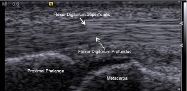
UltrasoundLongitudinal finger ultrasound, anatomical landmarks labelled at the level of the A1 pulley: superficial and deep flexor tendons, proximal phalanx, metacarpal. Normal anatomy, no visible lesion.
Source : Chuang et al., BioMedical Engineering OnLine, 2017, figure 1 · CC BY
Trigger finger, or stenosing flexor tenosynovitis (ICD-11 FB40.4; ICD-10 M65.3), is a common cause of hand pain and disability characterised by painful locking or audible triggering during flexion and extension of a finger¹. The historical name "tenosynovitis" is misleading: contemporary histological studies have shown that it is a fibrocartilaginous metaplasia of the A1 pulley with a degenerative process (tendinosis) rather than pure synovial inflammation².
Lifetime prevalence is estimated at 2 to 3% in the general adult population³. It rises considerably in certain populations: up to 10 to 20% in patients with diabetes (type 2 more than type 1)⁴,⁵, and around 5 to 10% in rheumatoid arthritis⁶. Female predominance is marked (F:M ratio 2:1 to 6:1) with a peak incidence between 50 and 60 years of age¹,⁵.
Any digit can be affected. In order of frequency: ring finger > thumb > middle finger > index finger > little finger³. Multiple involvement (several digits at once) should prompt a search for a systemic cause (diabetes, rheumatoid arthritis, amyloidosis, hypothyroidism, aromatase inhibitors)⁶.
Distribution of affected digits in adult trigger finger
Relative frequency observed in clinical series (Wong 2024, Lunsford 2019)
The ring finger and the thumb account for 60% of cases. Multiple involvement should prompt a search for a systemic cause. Source: synthesis of Wong 2024 BMC Musculoskelet Disord; Lunsford 2019 J Hand Ther.
The systemic risk factors that are best documented are:
- The role of diabetes mellitus (type 1 and 2): the strongest risk factor. A Swedish longitudinal cohort study spanning more than 20 years confirmed a significant excess risk in people with diabetes⁴.
- The role of rheumatoid arthritis⁶.
- Hypothyroidism¹.
- Amyloidosis (rare, but to be considered when involvement is multiple and bilateral)¹.
- The link with Dupuytren's disease : a common comorbidity, and trigger finger can precipitate the onset of post-surgical Dupuytren's disease⁷.
- The coexisting carpal tunnel syndrome : a frequent association, explained by shared risk factors (diabetes, manual loading).
- Chronic haemodialysis and renal failure¹.
- The aromatase inhibitors (anastrozole, letrozole, exemestane) used in hormone-dependent breast cancer induce a musculoskeletal syndrome with a trigger finger incidence of 5.3% vs 1.5% in non-users⁸.
The mechanical and occupational risk factors include powerful repetitive gripping, the use of vibrating tools, and certain sports (climbing, golf, tennis, string instruments)⁹. However, the relative weight of systemic factors outweighs that of mechanical factors in recent epidemiological series.
Risk factors for trigger finger: approximate odds ratios (OR)
Synthesis of recent cohorts (Hellgren 2021, Stirling 2020, Kim 2024, Regent-Smith 2023)
The ORs are approximate pooled estimates from recent cohorts. Diabetes remains the strongest risk factor, which justifies systematic screening. Sources: Hellgren 2021 (PMC10012113), Stirling 2020 (PMID 32443949), Kim 2024 Clin Orthop Surg, Regent-Smith 2023 Hand (NY).
What happens in the body and how does trigger finger progress?
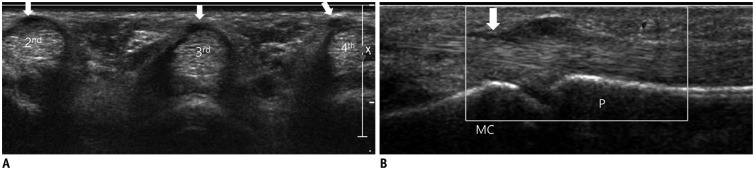
UltrasoundFinger ultrasound, transverse and longitudinal sections: hypoechoic nodular thickening of the A1 pulley at the level of the metacarpophalangeal joint.
Source : Park et al., Korean journal of radiology, 2016, figure 10 · CC BY-NC
The pathophysiology is above all mechanical. It results from a conflict between the flexor tendons (flexor digitorum superficialis FDS and flexor digitorum profundus FDP) and their retinacular pulley system, in particular the A1 pulley located at the base of the finger, at the distal palmar crease (over the metacarpal head)¹,².
For multifactorial reasons (repeated microtrauma, systemic metabolic factors, collagen ageing), the A1 pulley thickens and narrows (stenosis), while the tendon develops a nodule or local thickening opposite it. This size mismatch creates a conflict: the tendon glides less and less freely, and the audible or palpable triggering occurs when the tendon nodule forces its way through the stenosed pulley⁹.
Modern histological studies have called the term "stenosing tenosynovitis" into question: this is more a tendinosis / fibrocartilaginous metaplasia than pure inflammation². That nuance has treatment implications: non-steroidal anti-inflammatory drugs are of little use, whereas mechanical approaches (orthosis, gliding exercises, surgical release) target the cause directly.
The natural history varies with severity and duration. Mild, recent forms may resolve on their own, but most cases established for several weeks become chronic without intervention. A prospective observational study showed that spontaneous resolution is rare beyond 3 months in adults, in contrast with the frequent spontaneous resolution seen in children (congenital trigger thumb of Notta)¹⁰. Without intervention, the condition can progress to chronic pain, loss of range of motion, permanent locking in flexion and a fixed contracture of the proximal interphalangeal joint (Green grade IV).
Key points
- Trigger finger is a mechanical condition (conflict between a nodular tendon and a thickened A1 pulley), not simple tendon inflammation.
- Prevalence of 2-3% in the general population, but 10-20% in people with diabetes and higher in rheumatoid arthritis and on aromatase inhibitors.
- The female-to-male ratio is 2:1–6:1, with a peak between 50 and 60 years of age. The ring finger and the thumb are the most affected.
- The spontaneous resolution rate is low in adults beyond 3 months, in contrast with paediatric cases (Notta's thumb).
- Involvement that is multiple or bilateral calls for a search for a systemic cause (diabetes, RA, amyloidosis, aromatase inhibitors).
Chapter 1 bibliography
- Jeanmonod R, Harberger S, Waseem M. Trigger Finger. StatPearls [Internet]. Treasure Island (FL): StatPearls Publishing; 2024 (updated 8 Feb 2024). NBK459310.
- Brozovich N, Agrawal D, Reddy G. A Critical Appraisal of Adult Trigger Finger: Pathophysiology, Treatment, and Future Outlook. Plast Reconstr Surg Glob Open. 2019;7(8):e2360. PMID 31592381.
- Wong J, Wee SK, et al. From diagnosis to rehabilitation of trigger finger: a narrative review. BMC Musculoskelet Disord. 2024;25:55. doi:10.1186/s12891-024-08192-5.
- Hellgren MI, et al. Diabetes Mellitus as a Risk Factor for Trigger Finger - a Longitudinal Cohort Study Over More Than 20 Years. Front Clin Diabetes Healthc. 2021. PMC10012113.
- Stirling PHC, et al. Functional outcomes of trigger finger release in non-diabetic and diabetic patients. J Hand Surg Eur Vol. 2020;45(8):867-871. PMID 32443949.
- Adams JE, Habbu R. Tendinopathies of the Hand and Wrist. J Am Acad Orthop Surg. 2015;23(12):741-750. PMID 26510626.
- Maasarani S, Wee CE, Lee CD, Khalid SI, Layon S, Noland SS. Surgical Trigger Finger Release Is Associated With New-Onset Dupuytren Contracture in the Short-Term Postoperative Period: A Matched Analysis. Hand (NY). 2023. PMID 35253506.
- Regent-Smith AJ, Childers EJ, Dzwierzynski WW, Morgan AL. Incidence and Treatment Efficacy of Trigger Finger in the Breast Cancer Population on Aromatase Inhibitors. Hand (NY). 2023;18(2):250-253. PMC10035093.
- Lunsford D, Valdes K, Hengy S. Conservative management of trigger finger: A systematic review. J Hand Ther. 2019;32(2):212-221. PMID 29290504.
- Makkouk AH, Oetgen ME, Swigart CR, Dodds SD. Trigger finger: etiology, evaluation, and treatment. Curr Rev Musculoskelet Med. 2008;1(2):92-96. PMID 19468879.
2. How do you assess and diagnose trigger finger with certainty?
Which questions should you ask to understand the patient and their history?
A structured history makes it possible to reconstruct the course of the condition and to identify the associated systemic factors¹. Three lines of enquiry to explore systematically:
- Description of the symptoms : palmar pain at the base of the finger (over the A1 pulley), a sensation of clicking or audible triggering during flexion and extension, locking in flexion sometimes requiring passive help from the other hand to extend the finger. Morning stiffness is typical and reflects the mechanical stenosis, made worse by immobility overnight².
- Systemic risk factors to screen for :
- Diabetes mellitus (type 1 and 2): the most important factor, multiplying the risk by 4³.
- Rheumatoid arthritis or other inflammatory rheumatic diseases.
- Hypothyroidism, amyloidosis, renal failure / haemodialysis.
- Aromatase inhibitors in women treated for breast cancer⁴.
- Associated carpal tunnel syndrome or Dupuytren's disease.
- Occupational and leisure activities : repetitive, powerful gripping, vibrating tools, string instruments, climbing, golf, tennis. Quantify the daily duration and the weekly frequency⁵.
Involvement that is multiple or bilateral should raise the suspicion of an underlying systemic cause. Onset that is post-traumatic with a wound calls for a search for a foreign body or infection (suppurative tenosynovitis).
Which clinical tests should you perform and which other conditions must be ruled out?
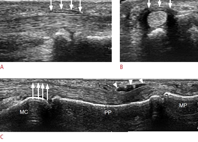
UltrasoundFinger ultrasound, longitudinal and transverse sections: hypoechoic thickening of the A1 pulley, a sign of stenosing tenosynovitis, arrows on the pulley.
Source : Lee et al., Ultrasonography (Seoul, Korea), 2016, figure 16 · CC BY-NC
The physical examination is simple, quick and highly reliable. Two key manoeuvres:
- Palpation of the A1 pulley at the level of the distal palmar crease (over the metacarpal head). You are looking for localised tenderness and a palpable nodule corresponding to thickening of the flexor tendon¹,⁶.
- Active flexion-extension manoeuvre : the patient repeatedly closes and opens the fist. You watch for and palpate the characteristic triggering. In severe cases (grade III) the finger stays locked in flexion and needs passive help to be extended.
Differential diagnoses to rule out
Differential diagnoses for locking or palmar pain in a finger
| Diagnosis | Distinguishing features | Useful imaging |
|---|---|---|
| Trigger finger | A1 nodule, audible triggering on flexion-extension, morning stiffness | Ultrasound (doubtful cases) |
| Dupuytren's disease | Progressive irreducible contracture, palmar cords, no triggering | Clinical examination is enough |
| De Quervain's tenosynovitis | Pain over the radial styloid (thumb), positive Finkelstein test | Ultrasound of the first compartment |
| Giant cell tumour (TGCT) of the sheath | Firm painless mass, slowly progressive, sometimes pseudo-locking | MRI, biopsy |
| Infectious tenosynovitis (Kanavel) | Finger held in flexion, pain on passive extension, fusiform swelling, fever | Emergency: ultrasound + blood tests |
| Osteoarthritis / arthritis of the PIP or MCP joint | Diffuse joint stiffness, no tendon triggering | Plain radiography |
| Ganglion cyst | Fluctuant mass, sometimes mechanical pseudo-locking | Ultrasound / aspiration |
| Post-traumatic foreign body | History of a wound, atypical localised triggering, persistent inflammation | High-resolution ultrasound |
The place of imaging
Imaging is not routine. In the vast majority of cases the clinical diagnosis is enough¹. However, high-resolution dynamic ultrasound (15-22 MHz linear probe) has become a reference tool for atypical cases⁷,⁸.
The typical ultrasound findings are⁷:
- Thickening of the A1 pulley (> 0.5 mm is often considered pathological).
- Thickening of the flexor tendon opposite the pulley, sometimes with focal hypoechogenicity.
- Hyperaemia on power Doppler indicating an active inflammatory component.
- Dynamic study of the conflict during flexion and extension: direct visualisation of the tendon catching.
The broader indications for ultrasound include: a suspected intratendinous mass (TGCT), a history of penetrating trauma (foreign body), failure of well-conducted conservative treatment, injection guidance (to avoid intratendinous injection), and pre-surgical assessment⁷. A recent narrative review confirms the growing place of personalised dynamic ultrasound in treatment planning⁹.
Should patients be classified, and what are the benefits?
Yes. Grading severity guides the treatment decision, allows standardised communication between professionals and makes progress easier to follow. The Green classification (modified from Quinnell 1980⁶) is the most widely used⁶.
Green classification of adult trigger finger
Four grades of increasing severity, guiding the treatment strategy
Source: Green classification (Wessel 2008), modified from Quinnell RC. Practitioner. 1980;224(1340):187-90. PMID 7367373. Grades III-IV are the preferred indications for injection and then surgery.
Critique and controversies
Several debates persist in the recent literature:
- Routine or targeted ultrasound? Its diagnostic accuracy is excellent (sensitivity and specificity > 90% in the best series⁷,⁸), but its cost and limited availability argue for targeted use: atypical cases, diagnostic doubt, injection guidance, suspected mass or foreign body.
- "Tenosynovitis" or "tendinosis"? The historical term stenosing tenosynovitis is inadequate histologically. Contemporary studies argue for a degenerative fibrocartilaginous metaplasia². That nuance explains the limited effectiveness of systemic NSAIDs and the value of mechanical approaches (orthosis, exercises, ESWT, surgery).
- Is the Green classification a continuum? Some patients stay at grade I-II for years, while others progress quickly to locking. The predictors of progression (diabetes, duration, multiple involvement) remain imperfectly characterised.
Key points
- The diagnosis is eminently clinical : a tender nodule over the A1 pulley, audible or palpable triggering during active flexion and extension, morning stiffness.
- The history must systematically look for the systemic comorbidities : diabetes, rheumatoid arthritis, hypothyroidism, aromatase inhibitors.
- Ultrasound is not routine but is becoming the standard for atypical cases, palpable masses, a history of trauma or injection guidance.
- The four-grade Green classification (I pre-triggering → IV fixed contracture) guides the treatment decision.
- Differential diagnoses to know: Dupuytren, TGCT, infectious tenosynovitis (Kanavel, an emergency), ganglion cyst, osteoarthritis, foreign body.
Chapter 2 bibliography
- Jeanmonod R, Harberger S, Waseem M. Trigger Finger. StatPearls [Internet]. 2024. NBK459310.
- Brozovich N, Agrawal D, Reddy G. A Critical Appraisal of Adult Trigger Finger: Pathophysiology, Treatment, and Future Outlook. Plast Reconstr Surg Glob Open. 2019;7(8):e2360. PMID 31592381.
- Hellgren MI, et al. Diabetes Mellitus as a Risk Factor for Trigger Finger. Front Clin Diabetes Healthc. 2021. PMC10012113.
- Regent-Smith AJ, et al. Trigger Finger in Breast Cancer Population on Aromatase Inhibitors. Hand (NY). 2023;18(2):250-253. PMC10035093.
- Akhtar S, Bradley MJ, Quinton DN, Burke FD. Management and referral for trigger finger/thumb. BMJ. 2005;331(7507):30-33. PMID 15994689.
- Huisstede BM, Hoogvliet P, Coert JH, Fridén J, European HANDGUIDE Group. Multidisciplinary consensus guideline for managing trigger finger: results from the European HANDGUIDE Study. Phys Ther. 2014;94(10):1421-1433. PMID 24810861. doi:10.2522/ptj.20130135.
- Quinnell RC. Conservative management of trigger finger. Practitioner. 1980;224(1340):187-190. PMID 7367373.
- Wong J, Wee SK, et al. From diagnosis to rehabilitation of trigger finger: a narrative review. BMC Musculoskelet Disord. 2024;25:55. doi:10.1186/s12891-024-08192-5.
- Makkouk AH, Oetgen ME, Swigart CR, Dodds SD. Trigger finger: etiology, evaluation, and treatment. Curr Rev Musculoskelet Med. 2008;1(2):92-96. PMID 19468879.
- Lunsford D, Valdes K, Hengy S. Conservative management of trigger finger: A systematic review. J Hand Ther. 2019;32(2):212-221. PMID 29290504.
- Bianchi S, Gitto S, Draghi F. Ultrasound Features of Trigger Finger: Review of the Literature. J Ultrasound Med. 2019;38(12):3141-3154. PMID 31106876.
- Sconfienza LM, Adriaensen M, Albano D, et al. Clinical indications for musculoskeletal ultrasound updated in 2017 by European Society of Musculoskeletal Radiology (ESSR) consensus. Eur Radiol. 2018;28(12):5338-5351. PMID 29876703.
- Marsico S, et al. Trigger Finger: A Narrative Review of Dynamic Ultrasound and Personalized Therapies. J Clin Ultrasound. 2025. PMID 40119539.
3. Which treatment strategies are the most effective?
Where do you start? What is the recommended hierarchy of interventions?
International recommendations converge on a stepwise approach that favours the least invasive option first¹,². The European HANDGUIDE consensus (Huisstede 2014) remains the multidisciplinary reference³.
- Patient education and modification of repetitive or forceful gripping activities.
- Static orthosis holding the MCP joint in extension (10-15 degrees of flexion), worn for 6 to 10 weeks on a prolonged basis⁴,⁵.
- Corticosteroid injection (1 to 2 spaced injections): the non-surgical gold standard⁶,⁷.
- Shockwave therapy (ESWT) : a non-invasive alternative⁸,⁹.
- Surgery (open or percutaneous): for resistant grades III-IV or repeated recurrence⁹.
Corticosteroid injection: the non-surgical gold standard
The landmark Cochrane review by Peters-Veluthamaningal (2009)⁶, an update of which is under way⁶, established the effectiveness of local corticosteroid injection against placebo. Success rates at 6 months range between 50 and 90% depending on initial severity and the presence of comorbidities. The pivotal randomised trial by Sato 2012⁷ compared injection with percutaneous release and open surgery, confirming injection as first line.
Three important caveats⁷,⁶:
- High recurrence rate : 30 to 50% at 1 year, higher in people with diabetes.
- A moderate risk of infection after injection: if surgery follows within 90 days of the injection, the infection risk is multiplied (Matzon 2020)⁸.
- Local side effects : depigmentation, subcutaneous atrophy, a rare risk of tendon rupture, and transient hyperglycaemia in people with diabetes.
The orthosis: the main option in grade I-II and in patients who decline injection
The orthosis aims to immobilise the MCP joint in slight extension (10-15 degrees of flexion) in order to limit tendon excursion through the A1 pulley. A recent meta-analysis (Lin 2025)⁴ confirms short-term effectiveness, particularly in patients at grade I-II and with symptoms < 6 months. The randomised trial by Teo 2019⁹ showed that an orthosis blocking the PIP can be as effective as one blocking the MCP joint, with better functional tolerance.
The optimal parameters according to recent trials⁴,⁹:
- Material: low-temperature thermoplastic (custom-made by an occupational therapist) or a commercial prefabricated device.
- Position: MCP in slight extension (10-15 degrees of flexion), PIP and DIP free.
- Duration: 6 to 10 weeks continuously (day and night as far as possible), then a night-time regimen.
- Adherence: the key factor for success, since an orthosis that is not worn is ineffective.
Comparison table of treatment modalities for trigger finger
GRADE level of evidence according to the 2018-2025 meta-analyses
| Modality | Effectiveness | Evidence | Target population | Source |
|---|---|---|---|---|
| Corticosteroid injection | High (60-90%) | Moderate GRADE | Grades II-III | Peters-Veluthamaningal Cochrane 2009/2025 |
| Open surgery | High (> 90%) | Moderate GRADE | Grades III-IV / recurrence | Fiorini Cochrane 2018 |
| Percutaneous surgery | High (85-95%) | Moderate-to-low GRADE | Same; operator experience | Sato 2012; NMA 2025 |
| MCP/PIP orthosis | Moderate (50-80%) | Low-to-moderate GRADE | Grades I-II, < 6 months | Lin 2025; Teo 2019 |
| ESWT | Moderate | Low GRADE | Alternative to injection | Yildirim 2016; Yao 2024 |
| Tendon gliding exercises | A useful adjunct | Low GRADE | All grades, post-injection / post-operative | Wehbe 1987; Yanko 2025 |
| Education + activity modification | A systematic adjunct | Very low GRADE | All | HANDGUIDE 2014 |
| Oral NSAIDs | Low | Very low GRADE | Short-term symptomatic relief | HANDGUIDE 2014 |
| Low-level laser | Insufficient | Very low GRADE | Not recommended routinely | Lunsford 2019 |
| PRP / stem cells | Emerging, limited data | Very low GRADE | Research | Limited literature |
GRADE pyramid: levels of evidence for each modality
Adapted from Oxford CEBM / GRADE Working Group, readable horizontal cards
Pyramid adapted from the OCEBM Levels of Evidence (Oxford 2011) and the GRADE system. Source: author synthesis based on the references above.
What is the place of exercise, and is there a superior approach?
Exercise has long been undervalued. Recent research positions it as a key adjunct, particularly when combined with an orthosis or after injection or surgery⁷.
Tendon gliding exercises
The reference protocol remains the one described by Wehbe and Hunter 1985-1987⁷. It aims to maximise the differential excursion between FDS and FDP through their synovial sheath and to prevent adhesions. The sequence comprises five positions: flat hand, hook, full fist, straight fist and composite fist. Perform 5 to 10 repetitions of each, 3 to 5 times a day.
A recent prospective study (Yanko 2025)¹⁰ tested an interdisciplinary protocol redirecting patients to hand therapy before surgery: 22% complete resolution without injection or surgery, showing the value of a structured conservative approach with exercises.
Stretching and mobilisations
Gentle stretching of the flexors (with the wrist in passive extension and the fingers extended) maintains overall suppleness. Gentle passive mobilisations of the MCP and PIP joints can be useful after surgery or injection to restore range of motion.
Adjuvant therapies, ESWT, technologies: what is the real effectiveness?
Extracorporeal shockwave therapy (ESWT)
ESWT has become the most studied non-invasive alternative. A pivotal randomised trial (Yildirim 2016)¹² compared ESWT with corticosteroid injection: comparable results at 3 and 6 months for pain and function, without the risks specific to injection (tendon rupture, infection, skin atrophy). A 2024 meta-analysis (Yao)¹³ reinforced these conclusions across eight RCTs.
Advantages: non-invasive, repeatable, no infection risk and no systemic side effects. Drawbacks: limited availability (specialised equipment), cost, 3 to 5 sessions needed, transient pain during treatment.
Low-level laser therapy (LLLT) and therapeutic ultrasound
The evidence is insufficient for a routine recommendation in trigger finger. Recent systematic reviews (Lunsford 2019¹⁴) conclude that there is a low level of evidence, mainly because rigorous RCTs are lacking.
Manual therapies
Mobilisation of the palmar soft tissues, deep transverse friction, passive mobilisation of the A1 pulley: the evidence rests mainly on case reports and small series, which does not allow a strong recommendation. Use them as an adjunct after injection or surgery to encourage gliding.
When should you operate and what results can be expected?
Surgery is indicated⁹ in the case of:
- Failure of two corticosteroid injections that were well conducted.
- Green grade III-B or IV (irreducible locking or fixed contracture).
- Rapid recurrence after injection in a patient with diabetes.
- An atypical presentation requiring investigation (mass, foreign body).
Two main techniques:
- Open surgical release : division of the A1 pulley through a small palmar incision. The historical reference, success rate > 90%, complications < 5%⁹.
- Percutaneous needle release : division of the pulley with a hypodermic needle introduced percutaneously. Success rate 85-95% depending on operator experience, with a risk of incomplete division (~5-15%) and of injury to the collateral nerve of the thumb (rare but described). A 2025 NMA confirms short-term non-inferiority⁷.
The post-operative rehabilitation is generally short (2 to 4 weeks) with early tendon gliding exercises from day 1 or day 2 to prevent scar adhesions, and a gradual return to activity at 4-6 weeks.
Beyond the physical: how do you educate the patient and address psychological factors?
Therapeutic education is a pillar that is often neglected. The key points to cover³,⁷:
- Explain the mechanism : a conflict between the A1 pulley and the tendon, not a "disease" of the tendon itself. This understanding changes how the patient relates to the condition.
- Identify and modify aggravating activities : forceful repetitive gripping (shears, screwdriver, tennis), vibrating tools, musical instruments. Offer alternatives: wide padded handles, anti-vibration gloves, regular breaks.
- A realistic prognosis : most cases resolve with conservative treatment, but recurrence is possible. Kinesiophobia can lead to underuse of the hand and secondary stiffness.
- Concrete functional goals that are measurable (holding a cup without pain, buttoning a shirt, making a full fist) make adherence easier.
Critique and controversies: the grey areas of management
1. The "gold standard" status of injection is increasingly qualified. While its short-term effectiveness is established, the high recurrence rates and the risks (depigmentation, atrophy, post-operative infection if surgery follows) argue for a personalised approach. ESWT and orthosis + exercises appear as credible alternatives, particularly in patients with diabetes and in grades I-II.
2. The role of physiotherapy remains under-evaluated in the high-level literature. The major Cochrane reviews focus on medical and surgical interventions. Research on standardised physiotherapy protocols and their comparative effectiveness is still lacking.
3. The "phenotyping" of patients remains a challenge: a patient with diabetes and symptoms > 6 months has a very different prognosis from a healthy patient with recent symptoms. Current recommendations do not stratify management enough according to these profiles.
Key points
- A clear hierarchy : education and activity modification + MCP orthosis → corticosteroid injection (the gold standard, 60-90% success at 6 months) → ESWT as an alternative → surgery (open or percutaneous) for refractory cases.
- The MCP extension orthosis worn for 6-10 weeks has moderate effectiveness (50-80%) in grades I-II and symptoms < 6 months.
- The tendon gliding exercises (Wehbe 1987) combined with an orthosis improve functional outcomes.
- ESWT has effectiveness comparable to injection in the short term (Yildirim 2016), without the infection risk.
- Patient education and adherence to the protocol are the major determinants of lasting success.
Chapter 3 bibliography
- Lunsford D, Valdes K, Hengy S. Conservative management of trigger finger: A systematic review. J Hand Ther. 2019;32(2):212-221. PMID 29290504.
- Wong J, Wee SK, et al. From diagnosis to rehabilitation of trigger finger: a narrative review. BMC Musculoskelet Disord. 2024;25:55. doi:10.1186/s12891-024-08192-5.
- Huisstede BM, Hoogvliet P, Coert JH, Fridén J, European HANDGUIDE Group. Multidisciplinary consensus guideline for managing trigger finger: results from the European HANDGUIDE Study. Phys Ther. 2014;94(10):1421-1433. PMID 24810861.
- Lin C, et al. Efficacy of Splinting in Managing Adult Trigger Finger: SR of Short-Term Outcomes. J Hand Surg Glob Online. 2025. PMC12681533.
- Valdes K. A retrospective review of the long-term efficacy of orthotic devices for trigger finger. J Hand Ther. 2012;25(1):89-95. PMID 22265444.
- Peters-Veluthamaningal C, van der Windt DAWM, Winters JC, Meyboom-de Jong B. Corticosteroid injection for trigger finger in adults. Cochrane Database Syst Rev. 2009;(1):CD005617. PMID 19160256.
- Sato ES, Gomes Dos Santos JB, Belloti JC, Albertoni WM, Faloppa F. Treatment of trigger finger: RCT comparing corticosteroid injection, percutaneous release and open surgery. Rheumatology (Oxford). 2012;51(1):93-99. PMID 22039269.
- Matzon JL, Lebowitz C, Graham JG, Lucenti L, Lutsky KF, Beredjiklian PK. Risk of Infection in Trigger Finger Release Surgery Following Corticosteroid Injection. J Hand Surg Am. 2020;45(4):310-316. PMID 32113702.
- Teo SH, Ng DCI, Wong YKY. PIP-blocking vs MCP-blocking orthosis in trigger digit: RCT. J Hand Ther. 2019;32(4):444-451. PMID 30030005.
- Yanko E, et al. A collaborative interdisciplinary approach for trigger finger management. J Hand Ther. 2025;38(3):508-521. PMID 39818526.
- Wehbe MA. Tendon gliding exercises. Am J Occup Ther. 1987;41(3):164-167. PMID 3318478.
- Yildirim P, Gultekin A, Yildirim A, Karahan AY, Tok F. ESWT vs corticosteroid injection in trigger finger: RCT. J Hand Surg Eur Vol. 2016;41(9):977-983. PMID 26763271.
- Yao H, Wu Y, Li C, Mu X. Efficacy of Extracorporeal Shockwave Therapy for Trigger Finger: SR / MA. 2024.
- Fiorini HJ, Tamaoki MJ, Lenza M, Gomes Dos Santos JB, Faloppa F, Belloti JC. Surgery for trigger finger. Cochrane Database Syst Rev. 2018;2:CD009860. PMID 29460276.
- Shen PC, Chou SH, Lu CC, Fu YC, Lu CK, Liu WC, Huang PJ, Tien YC, Shih CL. Comparative effectiveness of treatment strategies for trigger finger by pairwise meta-analysis. Clin Rehabil. 2020;34(9):1217-1229. PMID 32539454.
- Amirfeyz R, et al. Evidence-based management of adult trigger digits. J Hand Surg Eur Vol. 2017;42(5):473-480. PMID 28488453.
4. How do you secure lasting recovery and prevent recurrence?
How do you make the patient an active partner in recovery through self-management?
Patient empowerment is the fundamental pillar of recurrence prevention¹. A passive approach (receiving an injection without understanding the causes) is often doomed to fail in the medium and long term. Four pillars of self-management:
- Therapeutic education : understanding the mechanics of the A1 pulley / flexor tendon conflict. Identifying the aggravating activities, movements or postures (forceful repetitive gripping, vibrating tools, string instruments).
- Ergonomic modification of activities :
- Wider, padded handles (foam grips) to reduce palmar pressure.
- Alternating tasks to avoid repeating the same movement pattern.
- Regular breaks (5 min every 30-60 min of sustained gripping).
- Anti-vibration gloves for tool users.
- Early recognition of the signs of overload.
- Night orthosis as a follow-on: holding the MCP joint in slight extension during sleep to avoid locking and morning stiffness²,³.
- Home self-rehabilitation exercises :
- Differential tendon gliding (Wehbe 1987)⁴: a sequence of five positions (flat hand, hook, full fist, straight fist, composite fist), 5-10 repetitions, 3-5 times a day.
- Gentle stretching of the flexors (wrist in extension, fingers extended, hold for 20-30 s).
- Progressive strengthening (after the acute phase): pinch grip with therapy putty, hand exerciser balls, in endurance mode and pain-free.
When and how do you plan a safe return to sport and to daily activities?
The return to demanding occupational or sporting activities (climbing, golf, tennis, DIY, music) must be planned in order to avoid recurrence. The guiding principle is a progressive, controlled return guided by the absence of symptoms⁵.
Entry criterion: complete clinical resolution
The return should only be considered when the patient shows:
- No locking and no mechanical triggering.
- No pain on palpation of the A1 pulley.
- Full, pain-free active and passive range of motion (TAM > 250 degrees as a minimum).
- The ability to hold a light object without pain for 1-2 minutes.
Stages of progression
- Phase 1 - Reintroducing specific movements without load : the sporting or occupational movement mimed without a tool, light gripping (a light object). 5-10 minutes, 1 to 2 times a day. Validation: no pain the next day.
- Phase 2 - Progressive increase in load : apply the "10% rule" (increase only one parameter per week, max 10-20%)⁶. Never increase intensity, volume and frequency at the same time.
- Phase 3 - Return to performance : volume and intensity close to the previous level. Continuous monitoring over 4 to 8 weeks. Resume a preventive night orthosis during periods of overload.
The no-pain rule
The patient must watch for any reappearance, however small, of pain or stiffness. The "pain monitoring" model validated in Achilles tendinopathy (Silbernagel 2007)⁷ is transferable: pain during the activity is acceptable if it settles within 24 h and does not accumulate. Otherwise, step back one stage.
Algorithm for a progressive return to gripping activities
Inspiration: Silbernagel pain monitoring 2007, Gabbett training paradox 2016
Sources: adapted from the principles of Silbernagel KG. Am J Sports Med. 2007;35(6):897-906 (pain monitoring in Achilles tendinopathy, PMID 17307888) and Gabbett TJ. BJSM. 2016;50(5):273-280 (training-injury paradox, PMID 26758673).
Critique and controversies
1. Heterogeneity of prevention protocols : there is no standardised protocol for wearing a preventive night orthosis, nor for the optimal duration of exercises after resolution. Most trials cover the first 6 to 12 months, with little data beyond that.
2. The place of injection in secondary prevention : should a second injection be offered at the first sign of recurrence, or should the patient go straight to surgery? Current guidelines favour 1 to 2 injections spaced 3-4 months apart before surgery⁶, but the cost-benefit ratio in patients with diabetes who keep relapsing remains debated.
3. Long-term adherence : the major challenge. Observational studies show adherence to the exercise programme and to wearing a night orthosis falling away beyond 3 months, which partly explains the recurrences.
Key points
- The prevention of recurrence is crucial: 30 to 50% recurrence at 1 year after injection, particularly in people with diabetes.
- Self-management rests on four pillars: education, ergonomic modification of activities, a night orthosis, and tendon gliding exercises.
- The return to manual or sporting activities must be strictly progressive, guided by the entry criteria (complete clinical resolution) and by the no-pain rule.
- Apply the 10% rule : only one parameter increased per week, max +10-20%.
- The "pain monitoring" model from Silbernagel (pain acceptable if it settles in < 24 h and is not cumulative) is transferable to trigger finger.
Chapter 4 bibliography
- Yanko E, et al. A collaborative interdisciplinary approach for trigger finger management. J Hand Ther. 2025;38(3):508-521. PMID 39818526.
- Valdes K. A retrospective review of the long-term efficacy of orthotic devices for trigger finger. J Hand Ther. 2012;25(1):89-95. PMID 22265444.
- Teo SH, Ng DCI, Wong YKY. PIP-blocking vs MCP-blocking orthosis in trigger digit: RCT. J Hand Ther. 2019;32(4):444-451. PMID 30030005.
- Wehbe MA. Tendon gliding exercises. Am J Occup Ther. 1987;41(3):164-167. PMID 3318478.
- Lin C, et al. Efficacy of Splinting in Managing Adult Trigger Finger: SR of Short-Term Outcomes. J Hand Surg Glob Online. 2025. PMC12681533.
- Huisstede BM, Hoogvliet P, Coert JH, Fridén J, European HANDGUIDE Group. Multidisciplinary consensus guideline for managing trigger finger: results from the European HANDGUIDE Study. Phys Ther. 2014;94(10):1421-1433. PMID 24810861.
- Peters-Veluthamaningal C, van der Windt DAWM. Corticosteroid injection for trigger finger in adults. Cochrane Database Syst Rev. 2009;(1):CD005617. PMID 19160256.
- Sato ES, Gomes Dos Santos JB, Belloti JC, Albertoni WM, Faloppa F. RCT corticosteroid vs percutaneous vs open surgery in trigger finger. Rheumatology (Oxford). 2012;51(1):93-99. PMID 22039269.
- Cook JL, Rio E, Purdam CR, Docking SI. Revisiting the continuum model of tendon pathology. BJSM. 2016;50(19):1187-1191. doi:10.1136/bjsports-2015-095422.
- Gabbett TJ. The training-injury prevention paradox: should athletes be training smarter and harder?. BJSM. 2016;50(5):273-280. PMID 26758673.
- Silbernagel KG, Thomeé R, Eriksson BI, Karlsson J. Continued sports activity using pain-monitoring model in Achilles tendinopathy: RCT. Am J Sports Med. 2007;35(6):897-906. PMID 17307888.
- Grindem H, Snyder-Mackler L, Moksnes H, Engebretsen L, Risberg MA. Decision rules to reduce ACL reinjury risk. BJSM. 2016;50(13):804-808. PMID 27162233.
- Atthakomol P, et al. MCID of Michigan Hand Outcomes Questionnaire and pain VAS in conservative trigger finger. J Hand Surg Eur Vol. 2023;48(9):863-871. PMID 37288517.
5. How do you adapt management to at-risk subpopulations?
The patient with diabetes: a phenotype in its own right
Diabetes mellitus is the most powerful risk factor for trigger finger. Prevalence reaches 10–20% in people with diabetes (vs 2-3% in the general population), with a relative excess risk of about RR x4¹. Involvement is more often multiple and bilateral, and the response to conservative treatment is less favourable².
Specific pathophysiology
Non-enzymatic glycation of proteins (AGEs, advanced glycation end-products) alters the collagen structure of the A1 pulley and of the flexor tendon: thickening, loss of elasticity, accelerated fibrosis. These changes explain the poorer treatment response and the increased recurrence rate³.
Treatment adaptations
Adapting management in the patient with diabetes
Synthesis of recent data (Stirling 2020, Hellgren 2021)
| Modality | Patient with diabetes | Adaptations |
|---|---|---|
| Orthosis | Less effective than in the general population | Longer duration (10-12 weeks), close follow-up |
| Corticosteroid injection | Success 50-65% vs 80-90% in non-diabetic patients | Risk of transient hyperglycaemia (day 0 to day 7). Monitor blood glucose. Space the injections (max 1-2 per year). |
| ESWT | Limited data, but no contraindication | A useful alternative if injection is declined or contraindicated |
| Surgery | Good effectiveness (95%+) | Infection risk x2 if HbA1c > 7%. Optimise glycaemic control before surgery. |
| Glycaemic control | Essential | Target HbA1c < 7%. Coordinate with the endocrinologist. |
A prospective study of A1 release surgery (Stirling 2020)² showed that patients with diabetes have functional outcomes (QuickDASH, VAS) comparable in the long term to those of non-diabetic patients after surgery, suggesting that when conservative treatment fails, surgery can be offered earlier in the patient with diabetes, to avoid prolonged painful chronicity.
Congenital trigger thumb in children (Notta's node)
Paediatric trigger thumb is a distinct entity from the adult picture. It presents in infants or young children (often noticed at around 1-3 years), with a thumb locked in flexion permanently at the PIP joint. Palpation reveals a firm nodule at the base of the thumb (Notta's node) corresponding to thickening of the flexor pollicis longus (FPL).
Spontaneous resolution: the rule rather than the exception
A recent meta-analysis (Tang 2024)⁴ covering 19 paediatric studies documented a spontaneous resolution rate of 49 to 64% at 4 years of follow-up with simple observation and no active treatment. That finding overturns the historical approach of routine early surgery.
Paediatric treatment strategy
- Simple watchful waiting first line in children < 3 years, with gentle passive thumb extension exercises done by the parents (educate, reassure)⁴,⁵.
- Static extension orthosis worn at night: moderate evidence, but not routine. Adherence is a problem in the very young.
- A1 pulley release surgery of the thumb : reserved for contractures that persist beyond 12 months of follow-up in children > 3 years, or in severe cases (passive extension impossible). Success rate > 90%⁵.
Juvenile idiopathic arthritis (JIA) is a differential diagnosis to consider when involvement is multiple or symmetrical in a child: look for associated synovitis, inflammatory blood tests (CRP, ESR), and a paediatric rheumatology opinion.
Rheumatoid arthritis, haemodialysis, amyloidosis: the rare systemic causes worth knowing
Rheumatoid arthritis
In the patient with RA, trigger finger may be the presenting symptom or a complication of established disease. You need to distinguish :
- Either pure mechanical locking at the A1 pulley (a true trigger finger).
- Or a tendon rupture (loss of active extension without triggering, common in long-standing RA).
- Or a joint subluxation at the MCP joint related to the underlying disease.
- Or a diffuse rheumatoid tenosynovitis requiring systemic treatment (biologic therapies).
Corticosteroid injections must be used with caution because of the increased risk of tendon weakening in this setting. Coordination with the rheumatologist is essential; optimising the underlying treatment (anti-TNF, anti-IL-6, JAK inhibitors) reduces the frequency of tenosynovitis episodes.
Chronic haemodialysis and amyloidosis
Amyloidosis secondary to chronic haemodialysis (beta-2-microglobulin deposits) can present as trigger digits that are multiple and bilateral, sometimes together with carpal tunnel syndrome. Synovial biopsy can confirm the diagnosis. Treatment is essentially surgical when function is affected, but recurrence is common.
Aromatase inhibitors and iatrogenic trigger finger
The aromatase inhibitors (anastrozole, letrozole, exemestane) are prescribed to millions of women treated for hormone-dependent breast cancer. They induce a musculoskeletal syndrome with arthralgia, myalgia and multiple bilateral trigger digits.
Recent epidemiological data
Regent-Smith 2023 (Hand NY)⁶ documented, in a matched cohort, an incidence of 5.3% of trigger finger in women taking aromatase inhibitors vs 1.5% in non-users (OR ~3.5). Involvement is typically multiple, bilateral and symmetrical, and appears within 6 to 24 months of starting treatment.
Management strategy
- Informing the patient and the oncologist about this common complication.
- Conservative treatment first line : orthosis, hand therapy, exercises.
- Corticosteroid injection : moderate effectiveness, high recurrence rate for as long as the treatment continues.
- Possible adaptation of the oncological treatment (a multidisciplinary decision): switching AI, temporary suspension, or a move to tamoxifen depending on the oncological context.
- The use of specialised hand therapy in these patients has shown a significant functional improvement (Murphy 2025)¹¹.
Red flags and systemic involvement
- Multiple and bilateral involvement : consider undiagnosed diabetes, RA, amyloidosis, aromatase inhibitors, hypothyroidism.
- A red, hot finger with severe pain on passive extension and fever : suppurative tenosynovitis (a surgical emergency, Kanavel's signs).
- An atypical palpable mass with progressive growth : giant cell tumour of the tendon sheath, MRI + biopsy.
- A history of penetrating trauma even long ago: foreign body, high-resolution ultrasound.
- Abrupt onset with systemic signs (rash, fever, polyarthralgia): reactive arthritis, viral infection, aetiological work-up.
- Failure of well-conducted conservative treatment > 6 months : reassess the diagnosis, consider ultrasound and a surgical opinion.
- A child with permanent locking > 12 months after the age of 3 : refer for surgery.
Key points
- Patients with diabetes : prevalence 10-20%, often multiple and bilateral involvement, poorer response to conservative treatment. HbA1c control < 7% is essential. Surgery can be considered earlier.
- Congenital trigger thumb in children (Notta) : 49-64% spontaneous resolution at 4 years (Tang 2024). Watchful waiting + passive exercises first line; surgery reserved for contractures persisting beyond 12 months.
- Rheumatoid arthritis : distinguish mechanical locking, tendon rupture, subluxation and rheumatoid tenosynovitis. Coordinate with the rheumatologist; be cautious with injections.
- Aromatase inhibitors : incidence 5.3% vs 1.5% (Regent-Smith 2023). Typically multiple and bilateral involvement. Specialised hand therapy is effective (Murphy 2025).
- Dialysis amyloidosis : multiple trigger digits, beta-2-microglobulin deposits. Biopsy for diagnosis, surgery often needed.
Chapter 5 bibliography
- Hellgren MI, et al. Diabetes Mellitus as a Risk Factor for Trigger Finger - Longitudinal Cohort Study Over More Than 20 Years. Front Clin Diabetes Healthc. 2021. PMC10012113.
- Stirling PHC, et al. Functional outcomes of trigger finger release in non-diabetic and diabetic patients. J Hand Surg Eur Vol. 2020;45(8):867-871. PMID 32443949.
- Brozovich N, Agrawal D, Reddy G. A Critical Appraisal of Adult Trigger Finger: Pathophysiology, Treatment, and Future Outlook. Plast Reconstr Surg Glob Open. 2019;7(8):e2360. PMID 31592381.
- Tang Y, et al. Prevalence of spontaneous resolution among pediatric trigger thumb: SR / MA. J Orthop Surg Res. 2024;19:524. PMID 39095911.
- Cardon LJ, Ezaki M, Carter PR. Trigger finger in children. J Hand Surg Am. 1999;24(6):1156-1161. PMID 10584935.
- Villarreal Acha D, et al. Clinical Review of Trigger Finger in Pediatric and Adult Patients: Evaluation and Management Strategies. Cureus. 2025;17(8). PMC12476290.
- Adams JE, Habbu R. Tendinopathies of the Hand and Wrist. J Am Acad Orthop Surg. 2015;23(12):741-750. PMID 26510626.
- Maasarani S, Wee CE, Lee CD, Khalid SI, Layon S, Noland SS. Surgical Trigger Finger Release Associated With New-Onset Dupuytren Contracture. Hand (NY). 2023. PMID 35253506.
- Akhtar S, Bradley MJ, Quinton DN, Burke FD. Management and referral for trigger finger/thumb. BMJ. 2005;331(7507):30-33. PMID 15994689.
- Regent-Smith AJ, Childers EJ, Dzwierzynski WW, Morgan AL. Incidence and Treatment Efficacy of Trigger Finger in the Breast Cancer Population on Aromatase Inhibitors. Hand (NY). 2023;18(2):250-253. PMC10035093.
- Murphy MR, et al. Effectiveness of hand therapy for breast cancer survivors with AI-induced MSK syndrome. 2025. PMID 40087098.
6. What do real clinical cases teach us?
Analysis of a "classic" case: from assessment to resolution
The archetypal case of adult trigger finger described in the clinical series (Yanko 2025¹, Wong 2024²) is typically that of a woman aged 50-60, sometimes with diabetes, presenting with:
- Palmar pain at the base of the ring finger or the thumb, present for several weeks.
- Morning stiffness with audible and palpable triggering during flexion and extension.
- Made worse by repetitive gripping activities (knitting, gardening, using the phone).
- Examination: a tender nodule over the A1 pulley, reproducible Green grade II triggering.
Step-by-step management (typical case, Yanko 2025 synthesis¹):
- Weeks 1-2 : education + modification of gripping activities. Static MCP extension orthosis at night and for part of the day. Tendon gliding exercises 5x/day.
- Weeks 4-6 : reassessment. Reduced pain and triggering frequency in 40-50% of cases.
- If it fails at 8 weeks : guided corticosteroid injection (ultrasound recommended to avoid intratendinous injection).
- Week 12 : reassessment. Complete resolution in 60-90% of cases.
- If there is recurrence at 6 months, or failure : a second injection or referral for surgery.
The diagnostic challenge: when trigger finger mimics another condition
The main pitfall is to make a standard clinical diagnosis without investigating an atypical presentation. Several published cases serve as a warning:
Tumour mimicry
The giant cell tumours of the tendon sheath (TGCT) can present as a tender nodule with functional impairment at the base of a finger. Clinical distinction from a trigger finger is sometimes impossible. Ultrasound or MRI are then needed: the TGCT appears as a well-defined nodular soft-tissue mass, often hypoechoic, distinct from the tendon and from the pulley. Biopsy confirms the diagnosis and allows surgical excision³.
Trigger finger secondary to Dupuytren's disease
A recent matched study (Maasarani 2023)⁴ showed that surgical release of a trigger finger can precipitate the onset of post-operative Dupuytren's disease in predisposed patients. Conversely, an early digital contracture attributed to Dupuytren can mask a genuine concomitant trigger finger. The examination must systematically look for dynamic triggering as well as palpating the palmar cords.
Suppurative tenosynovitis: a surgical emergency
The Kanavel signs (finger held in flexion, fusiform swelling, pain on passive extension, tenderness along the tendon sheath) must be sought systematically in any acute, painful and inflammatory presentation. Suppurative tenosynovitis is a surgical emergency (drainage, antibiotics). A recent puncture, even a minor wound, or an invasive procedure (injection) are warning signs.
The post-traumatic foreign body
Faced with a picture of trigger finger that is post-traumatic even after a symptom-free interval, high-resolution ultrasound can reveal a foreign body (a plant thorn, a metal fragment, a shard of glass) surrounded by a chronic inflammatory reaction. Treatment is surgical excision.
Complex PMC-verified case studies
Case 1: multiple bilateral trigger fingers on letrozole (Regent-Smith 2023)
A matched cohort⁵ documenting 5.3% of trigger digits in women taking aromatase inhibitors (vs 1.5% in non-users). Typical patients: 50-65 years, multiple bilateral involvement developing within 6-24 months of starting treatment. The response to conservative treatment is generally good, but recurrence is common for as long as the oncological treatment continues.
Case 2: congenital trigger thumb, spontaneous resolution (Tang 2024 MA)
The paediatric meta-analysis of 19 studies⁶ documented a spontaneous resolution of 49 to 64% at 4 years in infants and young children with a thumb locked in flexion (Notta's node). That high rate justifies a strategy of prolonged watchful waiting before surgery, unlike the historical approach of early release. Surgery remains indicated when a contracture persists beyond 12 months after the age of 3.
Case 3: new Dupuytren after trigger finger surgery (Maasarani 2023)
A matched analysis⁴ in a large cohort showing that patients who have undergone surgical trigger finger release have a significantly higher incidence of new Dupuytren contracture in the short term after surgery. Implications: inform the patient before surgery, monitor clinically, and do not label every new post-operative digital contracture a "complication" too quickly.
Case 4: a critical pathophysiological synthesis (Brozovich 2019 PRS GO)
A critical review⁶ positioning trigger finger as a fibrocartilaginous tendinosis rather than a purely inflammatory tenosynovitis. Treatment implications: it explains the limited effectiveness of systemic NSAIDs and the preference for mechanical approaches (orthosis, exercises, surgical release).
Critique and controversy: the limits of clinical cases
Individual clinical cases are by definition of a low level of evidence (Oxford CEBM level 4-5). Publication bias favours exceptional presentations and treatment failures at the expense of typical cases that resolve simply. This distortion can make the condition look more complex than it is in everyday practice. Cohort meta-analyses (Tang 2024 paediatric, Maasarani 2023 Dupuytren) provide a more solid quantitative framework for clinical decisions.
Key points
- The typical adult case responds in 60-90% of cases to structured conservative treatment (education + orthosis + exercises + injection if needed) within 8-12 weeks.
- Major diagnostic pitfalls: TGCT (giant cell tumour), coexisting Dupuytren, suppurative tenosynovitis (Kanavel, an emergency), foreign body after trauma.
- The aromatase inhibitors induce the typical multiple bilateral involvement in women treated for breast cancer.
- The paediatric trigger thumb resolves spontaneously in 50 to 64% of cases at 4 years, so watchful waiting is preferred.
- Surgical release of a trigger finger can be associated with a new Dupuytren contracture in predisposed patients.
Chapter 6 bibliography
- Yanko E, et al. A collaborative interdisciplinary approach for trigger finger management. J Hand Ther. 2025;38(3):508-521. PMID 39818526.
- Wong J, Wee SK, et al. From diagnosis to rehabilitation of trigger finger: a narrative review. BMC Musculoskelet Disord. 2024;25:55. doi:10.1186/s12891-024-08192-5.
- Bianchi S, Gitto S, Draghi F. Ultrasound Features of Trigger Finger: Review of the Literature. J Ultrasound Med. 2019;38(12):3141-3154. PMID 31106876.
- Maasarani S, Wee CE, Lee CD, Khalid SI, Layon S, Noland SS. Surgical Trigger Finger Release Associated With New-Onset Dupuytren Contracture: A Matched Analysis. Hand (NY). 2023. PMID 35253506.
- Regent-Smith AJ, et al. Trigger Finger in Breast Cancer Population on Aromatase Inhibitors. Hand (NY). 2023;18(2):250-253. PMC10035093.
- Brozovich N, Agrawal D, Reddy G. A Critical Appraisal of Adult Trigger Finger: Pathophysiology, Treatment, and Future Outlook. Plast Reconstr Surg Glob Open. 2019;7(8):e2360. PMID 31592381.
- Tang Y, et al. Prevalence of spontaneous resolution among pediatric trigger thumb: SR / MA. J Orthop Surg Res. 2024;19:524. PMID 39095911.
- Cardon LJ, Ezaki M, Carter PR. Trigger finger in children. J Hand Surg Am. 1999;24(6):1156-1161. PMID 10584935.
- Villarreal Acha D, et al. Clinical Review of Trigger Finger in Pediatric and Adult Patients. Cureus. 2025;17(8). PMC12476290.
- Lunsford D, Valdes K, Hengy S. Conservative management of trigger finger: A systematic review. J Hand Ther. 2019;32(2):212-221. PMID 29290504.
7. How do you apply these recommendations concretely in your practice?
When and to which other health professionals should you refer?
The physiotherapist is often a first-line professional, which brings a responsibility for screening and appropriate referral. Red flag screening (Finucane 2020¹) is a non-negotiable component of the initial assessment.
Urgent referral (24-48 h) to the doctor / emergency department
- Kanavel signs (suppurative tenosynovitis): finger held in flexion, pain on passive extension, fusiform swelling, fever.
- A recent penetrating wound with inflammation, suspected foreign body.
- Systemic signs of infection (chills, fever > 38.5 degrees).
Semi-urgent referral (1-2 weeks) to the hand surgeon
- Green grade IV (fixed contracture).
- Grade III-B (irreducible locking) with a failed injection.
- An atypical palpable mass suspected of being a tumour.
- Recent multiple bilateral involvement with no identified cause.
Planned referral to other professionals
- General practitioner / endocrinologist : screening for undiagnosed diabetes (HbA1c, fasting glucose) when there are multiple or recurrent trigger digits with no other explanation.
- Rheumatologist : suspected rheumatoid arthritis, symmetrical involvement, prolonged morning stiffness, systemic signs.
- Oncologist : women on aromatase inhibitors who develop multiple trigger digits - a benefit/risk discussion about the oncological treatment.
- Occupational therapist : making a custom orthosis, assessing occupational and daily-living activities, ergonomic advice.
- Psychologist specialising in pain : kinesiophobia, catastrophising (yellow flags), significant psychosocial impact.
- Nutritionist : if metabolic comorbidities (diabetes, obesity) are influencing the course.
Structured, concise communication with the other professionals (a summary of the assessment, the Green grade, the treatments tried, the reason for referral) is what makes the care pathway work well.
How do you measure outcomes and overcome the barriers to implementation?
Validated PROMs for trigger finger
Using standardised scales is the standard of evidence-based practice. For trigger finger, the most widely used tools are:
PROMs and functional tests for following up trigger finger
| Tool | Type / items | Approximate MCID | Indication |
|---|---|---|---|
| Pain VAS | 1 item, 0-10 cm | 1.4-3 cm | Overall pain, quick follow-up |
| QuickDASH | Hand PROMs, 11 items | 10-15 points | Upper limb function, short |
| MHQ (Michigan) | Hand PROMs, 6 domains | 7-23 points depending on the domain | Detailed hand function (Atthakomol 2023) |
| PRWE | Wrist/hand PROMs, 15 items | 11-17 points | Wrist pain + function |
| Green scale | Grade I-IV | Change in grade | Clinical severity, treatment decision |
| TAM (total active motion) | Sum of the active ranges MCP+PIP+DIP | 10-15 degrees | Functional goal, post-operative follow-up |
| Triggering frequency | Episodes per day | 50% reduction | Simple, self-reported follow-up |
A recent prospective study (Atthakomol 2023)² established the MCID values of the MHQ and of the pain VAS in the conservative treatment of trigger finger, providing critical benchmarks for interpreting trials and individual follow-up.
SMART goals with the patient
Setting goals with the patient that are Specific, Measurable, Achievable, Realistic and Time-bound (for example: "being able to make a fist without pain at 8 weeks", "getting back to knitting 30 min a day at 3 months", "bringing the VAS down from 7 to 2 at 12 weeks") supports adherence and the objective measurement of progress.
Barriers to implementation - and how to overcome them
The classic barriers in first-line practice³:
- Lack of time in the consultation - solution: standardised protocols, pre-printed patient handouts for education and exercises.
- Access to ultrasound - solution: a network of general practitioners or rheumatologists who scan, or training in musculoskeletal ultrasound.
- Heterogeneity of protocols - solution: adopting a practice protocol based on HANDGUIDE 2014 and Cochrane 2025.
- Patient adherence in the long term - solution: a telephone review at 1 and 3 months, mobile apps that prompt exercises, a partnership with an occupational therapist for orthosis follow-up.
- Coordination with the other professionals - solution: structured letters, local hand physiotherapy networks, continuing education.
Critique and controversies: beyond the guidelines
1. The gap between research and practice is real. RCTs often cover selected populations (non-diabetic patients, single-digit involvement). In the clinic, patients are more heterogeneous (comorbidities, multiple involvement, psychosocial situations). The clinician's art is to adapt evidence-based principles to the individual patient.
2. The "tyranny of scores" : an excessive focus on PROMs can dehumanise care. The therapeutic alliance and listening remain major prognostic factors, and they are hard to quantify.
3. The systematic red flag screening must be proportionate. Most "red flags" in first-line practice turn out to be "red herrings" (false positives)¹. The challenge is probabilistic clinical reasoning that fits red flags into an overall picture rather than reacting to each sign in isolation.
Key points
- Urgent referral (24-48 h) if there are Kanavel signs or an infected penetrating wound.
- Surgical referral for grade IV, grade III-B with a failed injection, an atypical mass, or recent multiple involvement.
- Validated PROMs : pain VAS, QuickDASH, MHQ (MCID established by Atthakomol 2023), the Green classification.
- SMART goals with the patient, for adherence and for measuring progress.
- Interprofessional coordination with the doctor, occupational therapist, rheumatologist, oncologist (aromatase inhibitors), hand surgeon and psychologist as the case requires.
Chapter 7 bibliography
- Finucane LM, et al. International Framework for Red Flags for Potential Serious Spinal Pathologies. JOSPT. 2020;50(7):350-372. doi:10.2519/jospt.2020.9971.
- Atthakomol P, et al. MCID of Michigan Hand Outcomes Questionnaire and pain VAS in conservative trigger finger. J Hand Surg Eur Vol. 2023;48(9):863-871. PMID 37288517.
- Van der Wees PJ, et al. Development of Clinical Guidelines in Physical Therapy. Phys Ther. 2011;91(10):1551-1563. PMID 21799137.
- Huisstede BM, Hoogvliet P, Coert JH, Fridén J, European HANDGUIDE Group. Multidisciplinary consensus guideline for managing trigger finger: results from the European HANDGUIDE Study. Phys Ther. 2014;94(10):1421-1433. PMID 24810861.
- Peters-Veluthamaningal C, van der Windt DAWM. Corticosteroid injection for trigger finger in adults. Cochrane Database Syst Rev. 2009;(1):CD005617. PMID 19160256.
- Caneiro JP, Roos EM, Barton CJ, et al. It is time to move beyond 'body region silos' to manage musculoskeletal pain. Br J Sports Med. 2020;54(8):438-439. PMID 31604698.
- Coronado RA, Brintz CE, McKernan LC, et al. Psychologically informed physical therapy for musculoskeletal pain. Pain Rep. 2020;5(5):e847. PMID 33490842.
- Burns PB, Rohrich RJ, Chung KC. The levels of evidence and their role in evidence-based medicine. Plast Reconstr Surg. 2011;128(1):305-310. PMID 21701348.
- Mlinaric A, Horvat M, Supak Smolcic V. Dealing with the positive publication bias. Biochem Med (Zagreb). 2017;27(3):030201. PMID 29180912.
Going further
Trigger finger is not a simple "tendinitis": it is a mechanical conflict that often sits within a systemic context (diabetes, RA, aromatase inhibitors). A structured conservative approach resolves most cases, but prompt referral for injection or surgery remains essential for grades III-IV and for recurrences.

