

















Clinical synthesis on Dupuytren's disease: understanding that it is fibroproliferative and progressive, knowing the comparative recurrence rates of the treatments, and placing hand therapy honestly. Every reference has been verified individually on PubMed.
📝 In brief: clinical summary
- Dupuytren's disease is a chronic fibroproliferative disorder of the palmar aponeurosis: fibroblasts undergo a lasting transformation into contractile myofibroblasts, turning painless palmar nodules into retractile cords responsible for fixed flexion of the fingers 21.
- Worldwide prevalence is estimated at 8.2% (95% CI 5.7–11.7%), with wide geographical variation; it rises with age (≈ 12%, 21% and 29% at 55, 65 and 75 years) and reaches 30% among Norwegians over 60, with a male-to-female ratio of about 5.9:1 435.
- Diabetes, heavy alcohol consumption, tobacco, older age, male sex and family history are independent risk factors; in a Swedish cohort of ~30,000 people, diabetes (men HR 2.23; women HR 2.69) and alcohol (men HR 2.46; women HR 3.56) stand out clearly 67.
- The diagnosis is essentially clinical: severity is assessed with a goniometer (MCP, PIP and DIP extension deficit according to Tubiana's staging), completed by Hueston's tabletop test, with involvement predominating on the 4th and 5th rays 103.
- Recurrence rates differ according to technique: 12–39% after open fasciectomy, 50–58% after needle aponeurotomy and 10–31% after collagenase; at 2 years, surgery maintains the correction better (78%) than the needle (50%) and collagenase (65%) 1514.
- No treatment is curative: 5 years after collagenase, 47% of successfully treated joints recur (CORDLESS), more often at the PIP (66%) than at the MCP (39%), the PIP being the harder location to correct 1112.
- The role of hand therapy is mainly post-interventional: a multicentre randomised trial (154 patients) showed no benefit from routine night splinting (DASH, extension, satisfaction) after fasciectomy, and it should be reserved for cases where an extension deficit reappears 1820.
✋ What is Dupuytren's disease?
📈 A disease of the second half of life
Dupuytren's disease becomes markedly more common with age, a useful marker for placing a patient's risk.
Prevalence by age. Overall worldwide prevalence is estimated at 8.2% 4, with a marked male and northern European predominance, and up to 34.1% among patients with type 1 diabetes. Age source: Almadani et al., 2021 (PMID 34526871).
Dupuytren's disease is a palmar fibromatosis : a chronic fibroproliferative disorder of the palmar aponeurosis of the hand, in which fibroblasts undergo a lasting transformation into contractile myofibroblasts, leading to disabling digital contractures 1. In practical terms, a silent tissue proliferation converts the normal palmar fascia into pathological tissue which, over months and years, pulls the fingers into flexion with no way of straightening them. The hand therapist is therefore not managing simple joint or tendon stiffness: they are supporting a progressive connective tissue disease whose mechanics and prognosis differ radically from an ordinary post-traumatic contracture.
Key points
- Chronic fibroproliferative disease of the palmar aponeurosis : nodules and then retractile cords 2.
- The cell responsible = the contractile myofibroblast phenotype 1.
- Flexion contracture predominating on the 4th and 5th rays (ring and little fingers) 3.
- Estimated worldwide prevalence of 8.2 %, highly variable between populations 4.
- The disease is progressive and not curable by conservative measures alone 3.
Definition: from the nodule to the retractile cord
Dupuytren's disease is described as a fibroproliferative condition of the palmar aponeurosis that leads to the formation of nodules and then of fibrous cords, to varying degrees. This proliferation, with the myofibroblast as its dominant cell, gradually turns painless palmar nodules into retractile cords responsible for the fixed flexion of the fingers 2. The natural history typically follows a sequence: first a thickening or a nodule in the palm, often found by chance by the patient, then the organisation of a palpable longitudinal cord running from the palm towards the finger, and finally the flexion contracture of the underlying joints.
Involvement predominates on the 4th and 5th rays, that is, the ring and little fingers 3. Clinically, the deformity develops at the metacarpophalangeal (MCP) and proximal interphalangeal (PIP) joints, more rarely the distal interphalangeal (DIP) joints. The patient describes a hand that « no longer opens »: they can no longer place the palm flat, put on a glove, grasp a large object or shake hands firmly.
The nodule is painless and often ignored; it is the cord that bends the finger and marks the disease.
Pathophysiology: the myofibroblast at the heart of the process
The cellular driver of the disease is the myofibroblast. This is a chronic fibroproliferative condition in which fibroblasts undergo a lasting transformation into contractile myofibroblasts, which leads to disabling digital contractures 1. These cells, rich in smooth muscle actin, combine two properties that are damaging for the hand: they synthesise a dense, disorganised extracellular matrix (collagen), and they contract actively, shortening the tissue as a retracting scar would. It is this double action, proliferation plus contraction, that explains why the cord does not merely thicken: it genuinely pulls the finger into flexion.
For the hand therapist, understanding this mechanism has a direct consequence: the contracture is not primarily of joint or tendon origin, but arises from pathological fascial tissue interposed between the two. A Dupuytren cord is therefore not « released » by mobilisation the way capsular stiffness would be recovered. Given the progressive nature of the disease, conservative measures (massage, splinting) are unlikely on their own to prevent or correct flexion contractures: physiotherapy does not cure the disease 3. This point frames from the outset the realistic role of rehabilitation, which is essentially peri-interventional, without promising a correction that only a procedure on the cord can achieve.
Epidemiology: who is affected?
Dupuytren's disease is common, but its measurement remains heterogeneous. A systematic review with meta-analysis estimates worldwide prevalence at 8.2% (95% CI 5.7–11.7%), with wide geographical variation 4. This variability stems from the populations studied, the inclusion criteria and the age of the cohorts: the overall figure hides very contrasting situations from one region to another.
Two gradients dominate the epidemiological picture: age and sex, on a background of northern European origin. Prevalence rises markedly with age, with an estimated average of about 12%, 21% and 29% at 55, 65 and 75 years respectively 3. The disease preferentially affects populations of northern European origin, with a marked male predominance: prevalence reaches 30% among Norwegians over 60, and the male-to-female ratio is about 5.9:1 5.
| Parameter | Data | Source |
|---|---|---|
| Worldwide prevalence | 8.2% (95% CI 5.7–11.7%) | Salari 2020 |
| Prevalence by age | ≈ 12% at 55 years, 21% at 65 years, 29% at 75 years | Almadani 2021 |
| Male / female ratio | ≈ 5.9 : 1 | Hindocha 2009 |
| Norwegians > 60 years | Prevalence ≈ 30% | Hindocha 2009 |
Risk factors and the notion of diathesis
A systematic review of non-genetic factors shows a strong association between Dupuytren's disease and older age, male sex, family history, diabetes, heavy alcohol consumption, smoking and exposure to manual work 6. These factors are found again in the large prospective cohorts. In a Swedish cohort study of nearly 30,000 individuals followed for more than 20 years, diabetes and excess alcohol emerge as independent risk factors: diabetes (men HR 2.23; women HR 2.69) and heavy alcohol consumption (men HR 2.46; women HR 3.56) 7. Alcohol and tobacco act through a dose-dependent relationship, and their combination confers a particularly high risk 8.
These elements point to a metabolic and constitutional background rather than to a single mechanical cause. That is the sense of the notion of « Dupuytren's diathesis »: a set of features (bilateral involvement, family history, ectopic involvement, early age at onset, male sex) associated with a more aggressive and more recurrent form. The presence of all these factors appreciably increases the risk of recurrence after treatment 9. For the hand clinician, spotting a marked diathesis helps to anticipate a less favourable course and to calibrate the prognostic message even before any intervention.
Dupuytren's is less a disease of the hand than a disease of the constitution that expresses itself in the hand.
What the hand therapist needs to understand
Three ideas structure management from the very first contact. First, this is a progressive connective tissue disease, not an isolated mechanical stiffness: the fibroproliferative process is active and the cord goes on contracting for as long as the disease progresses 21. Second, no conservative measure cures the disease or corrects an established contracture on its own; massage and splinting are unlikely to prevent or reverse the deformities 3. Third, the diagnosis and follow-up are essentially clinical : severity is assessed with a goniometer by measuring the extension deficit at the finger joints (MCP, PIP, DIP) according to Tubiana's staging, completed by Hueston's table test 10. This test is positive when the patient can no longer place the hand flat on a table 3, a simple and reproducible marker for documenting the disability and following its course.
This understanding refocuses the therapist's role: not to promise to « make the cord disappear », but to document the involvement precisely (rays involved, joints, extension deficit in degrees, tabletop test), explain the progressive nature and refer at the right time to the appropriate interventional option. Rehabilitation then finds its firmest ground downstream of the procedure, in post-interventional support, a subject developed later in this article.
⚠️ Risk factors and course
Dupuytren's disease is a chronic fibroproliferative disorder of the palmar aponeurosis: fibroblasts undergo a lasting transformation into contractile myofibroblasts, which gradually leads to disabling digital contractures 12. Understanding who develops the disease, and above all how it changes over time, is essential for the hand therapist: it shapes the information given to the patient, the pace of monitoring and the point at which surgical referral becomes relevant. The disease is not « cured » by rehabilitation, but its natural history, slow and highly variable, leaves real room for support.
Key points. Dupuytren's is a disease of the constitution: it arises more readily on an identifiable profile (older age, male sex, family history, diabetes, alcohol, tobacco). But the weight of these factors is not read only in the risk of occurrence : it is also read in the aggressiveness of the disease once it is established. An isolated nodule may stay stable for years; conversely, a « diathesis » background heralds rapid progression and a high risk of recurrence after treatment. Telling these two profiles apart is the first clinical task.
A background with a strong constitutional component
A systematic review of non-genetic factors has shown a strong association between Dupuytren's disease and older age, male sex, family history, diabetes, heavy alcohol consumption, smoking and exposure to manual work 6. This list sketches a recognisable clinical profile, but none of these factors is decisive on its own: they add up on a constitutional background in which heredity holds a central place.
The ethnic and male predominance illustrates this constitutional component. The disease preferentially affects populations of northern European origin, with a marked male predominance: prevalence reaches 30 % among Norwegians over 60 years and the male-to-female ratio is about 5.9 : 1 5. Worldwide, a meta-analysis estimates overall prevalence at 8.2 % (95 % CI 5.7–11.7 %), with wide geographical variation 4. This heterogeneity, highly dependent on the populations studied and on the inclusion criteria, reflects precisely the weight of the genetic background: the disease is not distributed at random across the planet.
Age is the other major determinant. Prevalence rises steadily decade by decade: around 12 % at 55 years, 21 % at 65 years and 29 % at 75 years 3. This progression is not only epidemiological: it is a reminder that Dupuytren's is a slowly established disease whose clinical expression unfolds over years, even decades. In women, expression is later and often less severe, which softens, without abolishing, the gap between the sexes at older ages.
Diabetes, alcohol, tobacco: the metabolic background
Diabetes is one of the most solidly associated comorbidities. At population level, a meta-analysis reports a prevalence of the disease reaching 34.1 % in patients with type 1 diabetes, far above the 8.2 % of the general population 4. This over-representation is confirmed by longitudinal data: in a Swedish cohort of nearly 30,000 individuals followed for more than 20 years, diabetes emerges as an independent risk factor, with a hazard ratio of 2.23 in men and 2.69 in women 7. Notably, in that same cohort, obesity in men appears on the contrary to be associated with a lower risk, a counter-intuitive signal underlining that the link with diabetes cannot be reduced to a simple global metabolic factor, but points rather to a background of its own.
Alcohol and tobacco make up the second side of the acquired background. A prospective study of 7,254 subjects (772 cases at follow-up) established that alcohol and tobacco are independent risk factors, with a dose-dependent relationship, their combination conferring a very high risk 8. The Swedish cohort puts the effect of heavy alcohol consumption at a hazard ratio of 2.46 in men and 3.56 in women 7. The existence of a dose-response relationship and the statistical independence of these two factors argue for a causal role, not for a simple incidental association: together with diabetes, these are the modifiable levers on which prevention advice can legitimately rest.
| Factor | Figure | Source |
|---|---|---|
| Diabetes | Prevalence up to 34.1 % (type 1 diabetes); HR 2.23 (M) / 2.69 (F) | Salari 2020; Rydberg 2021 |
| Alcohol (heavy consumption) | HR 2.46 (M) / 3.56 (F); dose-dependent relationship | Rydberg 2021; Godtfredsen 2004 |
| Tobacco | Independent, dose-dependent factor; risk increased when combined with alcohol | Godtfredsen 2004 |
| Male sex / age | Ratio ≈ 5.9 : 1; prevalence 12 % → 29 % from 55 to 75 years | Hindocha 2009; Almadani 2021 |
| Family history | Strong association (systematic review) | Alser 2020 |
| Epilepsy | Association described with diabetes and epilepsy | Hindocha 2009 |
Epilepsy has long featured among the classic associations of Dupuytren's disease, described alongside diabetes in high-prevalence populations 5. The level of evidence there is weaker and the attribution long debated (the role of the neurological disease itself, or of older treatments), but the point remains worth raising at history-taking, in the same way as recording family history and metabolic comorbidities.
A slow and variable natural history: not every nodule progresses
Clinically, the disease begins with painless palmar nodules, with the myofibroblast as dominant cell, which gradually turn into retractile fibrous cords responsible for the fixed flexion of the fingers 2. The key word is gradually: the course spreads over years and follows no single trajectory. A point that matters as much for the patient as for the therapist is that not every nodule progresses. Many stay stable, non-retractile, never producing a cord or an extension deficit; others regress partially; others still progress to digital contracture. This variability justifies a stance of active observation in front of an isolated nodule, rather than routine early intervention.
A palmar nodule is not a sentence to contracture: it is a signal to monitor, not necessarily one to operate on.
When the contracture sets in, it predominates on the 4th and 5th rays (ring and little fingers) 3. Clinical follow-up rests on simple, reproducible markers: goniometric measurement of the extension deficit at the metacarpophalangeal (MCP), proximal interphalangeal (PIP) and distal interphalangeal (DIP) joints according to Tubiana's staging, completed by Hueston's table test (tabletop test), positive when the patient can no longer place the hand flat on a table 103. Documenting these measurements over time is the best way to demonstrate, or to rule out, progression, and to date the moment when interventional management becomes justified.
The aggressive form: Dupuytren's diathesis
Not every case of Dupuytren's disease is equal in aggressiveness. The notion of Dupuytren's diathesis denotes a set of clinical features that identify patients with rapid progression and a high risk of recurrence after treatment 9. These factors typically comprise bilateral involvement, family history, early onset (young age) and ectopic involvement: pads over the proximal interphalangeal joints (knuckle pads), plantar involvement (Ledderhose disease), penile involvement (Peyronie's disease), and male sex. Their value is prognostic: the presence of all these factors increases the risk of recurrence of the disease by 71 % compared with a reference patient who has none of them 9.
This information is directly operational for hand therapy. In a patient with a marked diathesis, the discussion must build in from the outset the high probability of recurrence whatever the treatment chosen, and post-interventional monitoring must be tightened. Conversely, in a patient with a « quiet » background, unilateral, late in onset, with no ectopic site, the more indolent natural history allows temporising and prudent waiting in the face of moderate involvement.
What the course of the disease imposes on management
The progressive nature of the disease sets the limits of rehabilitation. Given that course, isolated conservative measures (massage, splinting) are unlikely to be enough to prevent or correct flexion contractures: physiotherapy does not cure the disease 3. This does not devalue hand therapy, whose place, essentially post-interventional, is set out further on, but it does reset expectations. Before any procedure, the therapist's role is threefold: identify the background (risk factors, signs of diathesis), demonstrate the course through repeated measurements, and inform the patient about the modifiable levers (glycaemic control, reduction of alcohol and tobacco) without promising a conservative cure.
Clinical summary. In Dupuytren's disease, two questions structure the initial assessment. What is the background? Age, sex, heredity, diabetes, alcohol, tobacco and epilepsy point to the risk and to the aggressiveness to be expected. How far has it progressed? Stable nodule, established cord or measurable extension deficit on goniometry, with or without signs of diathesis (bilaterality, ectopic involvement, early onset). An isolated nodule on a quiet background is monitored; retractile involvement on a diathesis background is referred for intervention, with a recurrence prognosis to be stated clearly.
🔍 How is it assessed?
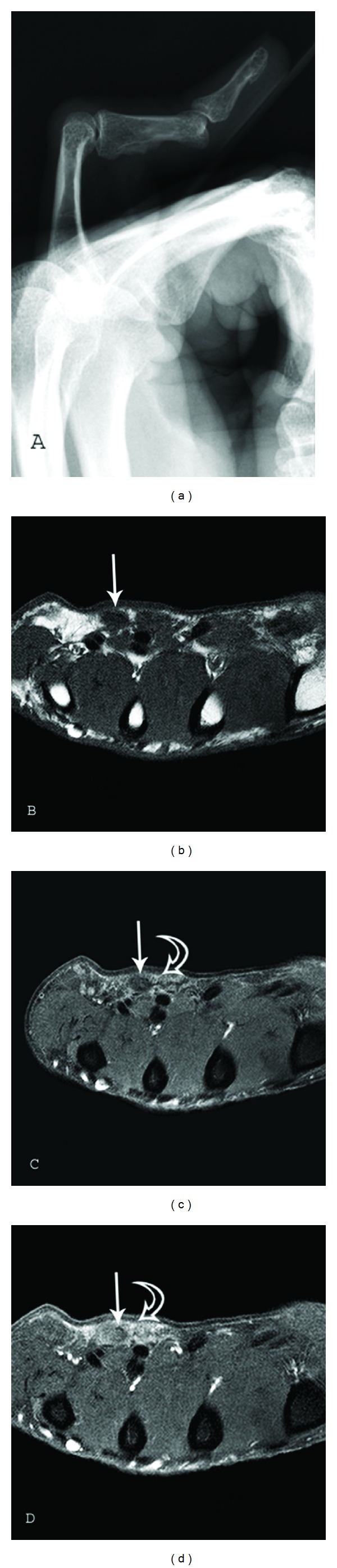
MRILateral radiograph: flexion contracture of the proximal interphalangeal joint of the fifth finger. Axial MRI of the hand: nodules of low signal intensity in the palmar subcutaneous fat, superficial to the flexor tendons.
Source : Walker et al., Sarcoma, 2012, figure 1 · CC BY
The diagnosis of Dupuytren's disease is essentially clinical : it rests on inspection, palpation and goniometric measurement, without routine recourse to imaging 10. The therapist recognises a fibroproliferative condition of the palmar aponeurosis that gradually turns painless palmar nodules into retractile fibrous cords responsible for fixed flexion of the fingers 2. The point of assessment is therefore not to make a difficult diagnosis (the palpable cord and the extension deficit speak for themselves) but to quantify the involvement objectively, to localise it joint by joint, and to follow its course over time. It is this quantification that guides the decision to intervene and that serves as the reference for peri-interventional rehabilitation.
Involvement predominates on the 4th and 5th rays (ring and little fingers), even though any finger can be affected 3. The first clinical reflex is to locate the nodules and the cords along these rays, to distinguish purely palmar involvement from digital extension, and to specify which joints are stiff. This mapping shapes both the prognosis, because not all joints correct with the same ease, and the choice of therapeutic measures.
Hueston's table test (tabletop test)
The Hueston table test, or tabletop test, is the simplest and most reproducible screening manoeuvre. The patient is asked to place the hand flat, palm against the surface of a table. The test is positive when the patient can no longer lay the hand flat, the flexion contracture of the fingers preventing full contact of the palm and the finger pulps with the plane of the table 3. Its value lies in its simplicity: it needs no equipment, it is immediately understandable to the patient, and it provides a telling functional marker.
A tabletop test that turns positive marks a threshold in practice: it signals that the contracture has reached a degree likely to hamper grip and weight-bearing activities, and it is traditionally taken as one of the criteria for discussing interventional management. The hand therapist also uses it as a follow-up marker, since a test that becomes positive again after treatment reflects a return of the extension deficit. The tabletop test remains, however, a binary test, positive or negative: it says that a significant contracture exists, but it does not measure it. It therefore never removes the need for the goniometric measurement it complements 10.
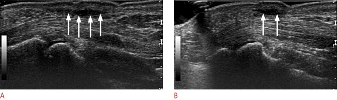
UltrasoundLongitudinal ultrasound of the palm: fusiform hypoechoic thickening of the palmar aponeurosis, arrows on the lesion, separated from the flexor tendons by a thin hyperechoic rim.
Source : Lee et al., Ultrasonography (Seoul, Korea), 2016, figure 26 · CC BY-NC
Key points: the tripod of clinical assessment. Three tools combine and do not replace one another: the Hueston table test screens for and flags a significant contracture (a hand that no longer lies flat), the Tubiana staging grades the overall severity of the finger, and the goniometric measurement of the extension deficit joint by joint (MCP, PIP, DIP) provides the objective reference figure. The diagnosis is clinical; imaging is not routine 10.
Tubiana staging
The Tubiana classification introduces a grading based on the total flexion deformity of the affected finger 10. In practice, the extension deficits of the different joints of a single ray are added together to give a total deficit, which is then classified into stages of increasing severity. This summation approach has the advantage of summarising the whole deformity of a finger in a single figure, then a single stage, which makes communication between professionals and longitudinal follow-up easier.
For the therapist, Tubiana staging plays a double role. It first places the patient on a shared severity scale, a common language with the hand surgeon. It then serves as a basis for follow-up : comparing stages before and after intervention, or over the course of spontaneous progression, demonstrates the progression or the correction obtained. But an overall stage can mask very different joint situations, since the same total deficit may result from isolated metacarpophalangeal involvement or from predominantly interphalangeal involvement, whose prognosis is not the same. That is why staging only makes sense alongside a joint-by-joint measurement.
Measuring the extension deficit with a goniometer (MCP, PIP, DIP)
The quantitative heart of the assessment is the goniometric measurement of the extension deficit at each of the finger joints: metacarpophalangeal (MCP), proximal interphalangeal (PIP) and distal interphalangeal (DIP) 10. One measures how many degrees each joint lacks to reach full extension (neutral position at 0°). This measurement, taken under standardised conditions, is the reference datum that feeds Tubiana staging, guides the therapeutic decision and serves as the yardstick for following rehabilitation.
The joint-level distinction is not an academic refinement: the location of the deficit largely determines the prognosis. Long-term data show that the proximal interphalangeal (PIP) joint recurs markedly more than the metacarpophalangeal (MCP) joint after treatment: in the CORDLESS follow-up after collagenase, recurrence affected 66% of PIP joints against 39% of MCP joints successfully treated at five years 11, and already 56% against 27% at three years 12. PIP involvement is known to be harder to correct and more liable to a return of the extension deficit. Distinguishing with the goniometer the MCP share and the PIP share of a contracture is therefore not cosmetic: it directly informs the patient about the expected difficulty of correction and about the risk of recurrence in their own case.
| Joint | Recurrence at 3 years | Recurrence at 5 years | Clinical implication |
|---|---|---|---|
| Proximal interphalangeal (PIP) | 56 % 12 | 66 % 11 | Harder to correct, frequent return of the extension deficit |
| Metacarpophalangeal (MCP) | 27 % 12 | 39 % 11 | Correction generally better maintained |
In practical terms, the goniometer must be applied in the same way at every assessment (same axis, same bony landmarks, same instruction for active then passive extension) so that the figures are comparable from one session to the next. It is the regularity of the measurement, more than the absolute precision of any single reading, that gives follow-up its value: a worsening of several degrees from one session to the next, or conversely a gain maintained after intervention, can only be read reliably across a series of consistent measurements.
What the hand therapist assesses beyond the angles
Measuring degrees does not sum up the assessment. The hand therapist palpates the nodules and the cords to specify their consistency, mobility and extent, distinguishes palmar from digital involvement, and identifies the rays involved, chiefly the ring and little fingers 3. They also assess the state of the skin over the cords, the tissue trophicity, and the real functional impact: difficulty with grip, with resting the hand flat, with everyday and work tasks. These qualitative elements complete the figures and shape the care plan.
Lastly, the assessment takes in the notion of constitutional background, or « Dupuytren's diathesis »: the presence of certain factors (bilateral involvement, family history, associated involvement at other sites, early onset) markedly increases the risk of recurrence after treatment 9. More generally, the disease is robustly associated with older age, male sex, family history, diabetes, heavy alcohol consumption, smoking and manual work 6. Collecting these elements at the initial assessment is not incidental: they weigh on the prognosis and on the expectations that can reasonably be set with the patient.
Assessing Dupuytren's disease is not only measuring an angle: it is localising each deficit, recognising a background, and setting a numerical marker that will make any change visible.
One last reminder of what assessment cannot promise: Dupuytren's disease is a progressive condition, and conservative measures alone (massage, splinting) are unlikely to prevent or correct flexion contractures; physiotherapy does not cure the disease 3. Rigorous clinical assessment therefore does not aim to substitute rehabilitation for intervention, but to document the involvement precisely, to identify the joints and the backgrounds at risk, and to provide the objective markers (tabletop test, Tubiana stage, MCP/PIP/DIP goniometric deficits) on which the therapeutic decision and the follow-up of peri-interventional management will rest.
🔁 Needle, collagenase, surgery: what about recurrence?
🔁 The real limit of every treatment: recurrence
No technique erases the disease for good. At 5 years, the more minimally invasive the procedure, the more it recurs: that is the central trade-off to explain to the patient.
Recurrence rates at 5 years. Needle vs fasciectomy: randomised trial at 5 years, same definition 16. Collagenase: CORDLESS follow-up at 5 years, 291/623 joints 11. Definitions of « recurrence » vary from one study to another, so the figures call for caution, but the hierarchy is constant: minimally invasive procedures recur more, at the price of a longer recovery for surgery.
Three main options are available today to release the retractile cord of Dupuytren's disease: needle aponeurotomy, injection of Clostridium histolyticum collagenase (CCH) and surgical fasciectomy. None of them cures the disease. The myofibroblastic proliferation that turns palmar nodules into contractile cords is a chronic fibroproliferative process 21 : the deformity is treated, not the background. That is why the true criterion for comparison is not initial success, which is often equivalent, but the durability of the correction, that is, recurrence. For the hand therapist, this is the information that shapes the patient's expectations and the follow-up schedule.
Key points. The three techniques correct the deformity comparably in the short term, but the disease recurs in every case. The minimally invasive techniques (needle, collagenase) expose the patient to recurrence that is more frequent and above all earlier than after surgery; fasciectomy offers the most durable correction at the price of a longer recovery. The published recurrence figures are widely scattered, because the definition of « recurrence » varies from one study to another: that is the central message of this section.
The three techniques in brief
Percutaneous needle aponeurotomy (or fasciotomy) divides the cord with a needle, in the clinic, under local anaesthetic: a quick, minimally invasive procedure with almost immediate recovery. Collagenase injection enzymatically lyses the collagen of the cord, which is then ruptured by an extension manipulation; it gives better results in mild to moderate contractures, with fewer complications than open surgery, but its recurrence rate appears to rise over time 13. The partial (limited) fasciectomy remains the surgical reference: it resects the pathological tissue, requires an operating theatre and rehabilitation, and maintains the correction best over time.
Initial success: the three techniques are level
The most counter-intuitive point for a patient is that, in the short term, the three options are equivalent. In a multicentre randomised controlled trial of 302 treatment-naive patients, success rates at 3 months were almost superimposable: 71% for surgery, 73% for needle aponeurotomy and 73% for collagenase 14. In other words, at three months the techniques cannot be told apart on the correction obtained alone. The difference appears only with longer follow-up.
At 2 years: surgery takes the lead
In that same trial, the gap widens markedly at two years. Limited fasciectomy maintained a success rate of 78 %, against 50 % for needle aponeurotomy and 65 % for collagenase, and this despite retreatments in the minimally invasive arms 14. Surgical correction « holds » better: the loss of benefit is faster after percutaneous procedures. This is the underlying trade-off that peri-interventional management accompanies: durability against the weight of the procedure and the length of recovery.
All three techniques succeed in the short term; at two years, only the question of recurrence really separates them.
Recurrence rates compared, and why they contradict one another
This is where the literature becomes frankly heterogeneous, and it should be said plainly. A first landmark systematic review reports very wide ranges: 12–39 % recurrence after open partial fasciectomy (mean follow-up 1.5 to 7.3 years), 50–58 % after needle aponeurotomy (follow-up 3 to 5 years) and 10–31 % after collagenase injection (follow-up 120 days to 4 years) 15. Taken at face value, this synthesis paradoxically places collagenase as the least recurrent option, but with the shortest follow-up, which biases the comparison.
| Technique | Reported recurrence rate | Follow-up / source |
|---|---|---|
| Partial fasciectomy (surgery) | 12–39 % | follow-up 1.5–7.3 years 15 |
| Needle aponeurotomy | 50–58 % | follow-up 3–5 years 15 |
| Collagenase injection (CCH) | 10–31 % | follow-up 120 days–4 years 15 |
| Needle vs limited fasciectomy | 84.9 % vs 20.9 % | RCT at 5 years 16 |
| Collagenase (CORDLESS) | 47% (66% at the PIP, 39% at the MCP) | follow-up at 5 years 11 |
| Collagenase / needle / fasciectomy | 41 % / 33 % / 10 % | individual patient data meta-analysis, 36 months 17 |
Two longer-term findings overturn the optimistic picture of collagenase. First, a randomised controlled trial at 5 years finds a recurrence rate of 84.9 % after percutaneous needle aponeurotomy against 20.9 % after limited fasciectomy, at the price, for surgery, of a longer recovery 16. Second, the most recent individual patient data meta-analysis, which directly compares limited fasciectomy, needle fasciotomy and collagenase, arrives at the reverse ranking to Chen's: recurrence there is most frequent after collagenase (41%), followed by the needle (33%) then fasciectomy (10%), and above all it occurs far earlier after minimally invasive procedures, about 5.8 times earlier after the needle and 6.5 times earlier after collagenase than after surgery, over 36 months of follow-up 17.
How can these contradictory figures be reconciled? The main cause is methodological: there is no single consensus definition of recurrence, and follow-up differs from one study to another. Depending on whether a threshold worsening of the angle is used (for example ≥ 20°), the reappearance of a palpable cord or the need for retreatment, and depending on whether measurement is at 2, 3 or 5 years, the same treatment may show 10% or 40% recurrence. The clinical lesson is therefore not to hold on to an isolated figure, but to understand the robust trend : the more minimally invasive the procedure, the more frequent and the earlier the recurrence; the longer the follow-up, the more the advantage of surgery stands out.
Location counts as much as technique: the PIP recurs more
The long-term follow-up of the CORDLESS study, after collagenase injection, adds a decisive detail for the therapist. At 5 years, overall recurrence reached 47 % (291 of the 623 successfully treated joints, the threshold set at a worsening ≥ 20° with a palpable cord or a further intervention), a figure the authors judged comparable with the rates published after surgery 11. Above all, recurrence is not distributed at random: it affected 66% of proximal interphalangeal (PIP) joints against 39% of metacarpophalangeal (MCP) joints. This gradient was already clear at 3 years 12.
This point is crucial in rehabilitation. PIP involvement is known to be harder to correct and more liable to a return of the extension deficit than the MCP. In practice, a patient treated for a predominantly PIP cord must be told of a higher risk of recurrence and followed more closely on that finger, whatever the technique used.
Levels of evidence and clinical reading
The hierarchy of evidence is more solid here than elsewhere in Dupuytren's disease. Several randomised controlled trials are available 1416 along with an individual patient data meta-analysis 17, which is the best available level of evidence for comparing the techniques. The weak point is not the quality of the studies taken individually, but the heterogeneity of the definitions and of the follow-up durations, which rules out a single recurrence figure. The Chen 2011 review, older and descriptive, should be read as a snapshot of ranges, not as a definitive ranking.
What the therapist takes away. (1) In the short term, needle, collagenase and surgery correct the deformity equivalently (≈ 71–73% at 3 months). (2) In the medium and long term, fasciectomy maintains the correction better (78% vs 50–65% at 2 years); recurrence after minimally invasive techniques is more frequent and earlier. (3) The published recurrence rates (from ~10% to ~85%) diverge above all because recurrence has no single definition, so reason in terms of trend, not of an isolated figure. (4) The PIP recurs markedly more than the MCP: that finger deserves close monitoring. (5) No option is curative; the role of hand therapy is essentially peri-interventional, supporting the correction and screening for a returning extension deficit.
In practice, the choice between the three techniques does not rest on the recurrence rate alone: it takes in the location and the severity of the cord, the patient's age and expectations, tolerance of a longer or shorter recovery, and the number of rays involved. The message to convey to the patient is honest and stable despite the scatter of the figures: the deformity is corrected effectively, but the disease can come back, all the sooner if the initial procedure was light, and structured follow-up, particularly of PIP extension, is an integral part of the treatment.
🎯 What place for hand therapy?
This is the question almost every patient asks, and many colleagues starting out in hand therapy: can a nodule or a cord be « melted away » by rehabilitation? The answer, as it emerges from the literature, is clear and sometimes frustrating. Dupuytren's disease is a fibroproliferative condition of the palmar aponeurosis, in which contractile myofibroblasts gradually turn painless nodules into retractile cords responsible for fixed flexion of the fingers 21. This process is tissue-based and progressive : no mobilisation, no massage, no splint dissolves an established cord. Hand therapy does not cure the disease and does not on its own correct an established contracture; its main field of action lies after the intervention, supporting the surgical or minimally invasive procedure.
Key points. Hand therapy is not a treatment for Dupuytren's disease: it does not reduce the nodules or the cords and it never replaces the interventional procedure. Its role is essentially postoperative namely recovering and maintaining the extension gained, managing the scar, the oedema and the pain. Given the progressive nature of the disease, conservative measures alone are unlikely to prevent or correct flexion contractures 3.
Why physiotherapy does not act on the disease itself
One must be clear with the patient from the first assessment: a Dupuytren cord is organised fibrous tissue, not joint stiffness or a simple adhesion to be loosened. The authors who have reviewed the place of conservative treatments say it plainly: given the progressive nature of the condition, it is unlikely that massage or splinting alone will be enough to prevent or reverse a flexion contracture 3. In other words, a purely rehabilitative approach is not expected to « straighten » a finger already bent by a cord. This message matters so that false hopes do not delay referral to the hand surgeon once the extension deficit becomes functionally troublesome, typically when Hueston's table test turns positive.
This reframing takes nothing away from the therapist's usefulness: it simply moves their input to where it is genuinely effective, that is, the phase following treatment of the cord, whatever that treatment may be: fasciectomy, needle aponeurotomy or collagenase injection.
The real playing field: the post-interventional phase
After the procedure, the finger has just been « reopened »: the skin, the scar, the oedema and the fear of pain all work, mechanically, to bring the finger back into flexion. This is precisely where hand therapy comes into its own. The classic objectives are ranked:
- Maintain and recover the extension gained in theatre, through progressive active and passive mobilisation of the metacarpophalangeal (MCP) and proximal interphalangeal (PIP) joints.
- Restore full flexion and overall grip function; a finger stiff in extension is not a success either.
- Manage the scar (scar massage, desensitisation) and oedema after surgery, two factors which, left to themselves, stiffen the hand.
- Support the wearing of an extension orthosis where indicated and the return to function, grading the effort so as not to reawaken an inflammatory reaction.
This management sits in a context where the major issue, common to every technique, remains recurrence. The correction is maintained unevenly according to the procedure: in a randomised trial comparing the three options, success rates were similar at 3 months (71% surgery, 73% needle, 73% collagenase) but diverged at 2 years, surgery doing better (78%) than the needle (50%) and collagenase (65%) 14. The therapist is therefore supporting a correction whose durability is variable, and this reality must be explained to the patient so as to anchor long-term monitoring rather than the illusion of a definitive cure.
The postoperative splint: a low level of evidence, to be stated clearly
This is where the message must be at its most honest. The night extension splint (or orthosis) has long been prescribed almost routinely after Dupuytren's surgery, by analogy with other hand operations. Yet the available data do not support this habit, and that must be acknowledged explicitly: the level of evidence for the postoperative splint is low and debated.
The most cited trial is a multicentre randomised controlled trial of 154 patients, randomised after fasciectomy or dermofasciectomy between hand therapy alone and hand therapy plus a night splint. At one year, no statistically significant difference appeared in self-reported function (DASH questionnaire), in the extension deficit of the operated fingers, or in satisfaction 18. The authors conclude that routinely adding a night splint for all patients is not recommended, and propose reserving it for cases where an extension deficit reappears 18.
| Study | Design / population | Result on the extension splint |
|---|---|---|
| Jerosch-Herold 2011 | Multicentre RCT, 154 patients, after fasciectomy / dermofasciectomy | No difference in DASH, extension deficit or satisfaction; routine splinting not recommended |
| Collis 2013 | Single-centre RCT, 56 patients, after surgical release | No difference in total active extension or in the secondary outcomes; an orthosis for everyone is not justified |
| Samargandi 2017 | Systematic review | The literature does not support the static night orthosis in addition to therapy; no significant improvement in range of motion |
| Tam 2016 | Retrospective chart review, 53 patients, after needle aponeurotomy | Night splinting does not improve active MCP or PIP range |
A second randomised controlled trial, single-centre this time (56 patients), confirms this finding after surgical release: adding a night extension orthosis to hand therapy made no statistically significant difference to total active extension or to the secondary outcomes, the authors judging it unjustified to provide an orthosis to every patient 19. A systematic review reaches the same conclusion: the current literature does not support the use of a static night orthosis in addition to hand therapy after surgical correction, with no significant improvement in joint range of motion 20. And on the minimally invasive side, a retrospective review of 53 patients after needle aponeurotomy likewise finds no improvement in active MCP or PIP mobility from night splinting 21.
Key points on splinting. Two randomised trials and a systematic review converge: the routine night extension splint after Dupuytren's treatment improves neither function, nor the extension deficit, nor satisfaction 181920. The reasonable course is therefore to target it in particular when an extension deficit reappears, rather than prescribing it for everyone, while keeping in mind that this level of evidence remains low.
Individualise rather than systematise
« Low evidence » does not mean « useless for everyone »: it means that the prescription must rest on individual clinical reasoning, not on an automatic protocol. Some situations point towards a targeted splint, first among them the reappearance of an extension deficit, a case explicitly retained by the authors as an exception to the « no routine splinting » rule 18. The location of the involvement also counts: follow-up data show that recurrence affects the PIP far more than the MCP, 56% against 27% of successfully treated joints in the three-year data after collagenase 12, and 66% against 39% at five years 11. Now the PIP is known to be harder to correct and more liable to a return of the extension deficit: a predominantly PIP finger justifies close monitoring and, where appropriate, a targeted orthosis, without the evidence allowing this to become a general rule.
This figure sums up the spirit in which to work: whatever the technique, Dupuytren's disease tends to recur and no treatment is definitively curative 11. The hand therapist is therefore not a one-off « repairer » but a follow-up partner: they optimise the immediate recovery, teach the patient to spot early any return of the extension deficit, and adapt the orthosis to the real trajectory of the finger rather than to a fixed protocol.
What can be promised, and what cannot
To conclude on a clinical footing and honestly towards the patient: hand therapy can speed up and secure recovery after a procedure, maintain the extension gained, soften a scar, reduce oedema and restore functional grip. It cannot make the disease regress, dissolve a cord, or correct an established contracture on its own, which is the province of surgery or of a minimally invasive procedure 3. And the emblematic tool of postoperative rehabilitation, the night splint, does not deserve to be imposed on everyone: its routine benefit is not demonstrated, its level of evidence is low, and the reasonable use is to reserve it for situations where an extension deficit threatens to return 1820. Saying this clearly protects the therapeutic relationship: referral to the surgeon is not left too late, the orthosis is not oversold, and the logic of long-term follow-up is set up from the outset in the face of a disease that, by its nature, tends to come back.
🗂️ What do concrete case reports teach us?
The case that follows is published and carries its PubMed identifier. It is reported here as its authors described it, and every decision commented on rests on confirmed, referenced data. The aim is not to supply a protocol to copy, but to make visible how one assesses, refers and supports, from Hueston's table test through to managing expectations around a treatment which, let us remember, does not cure the disease 3.
A published case: bilateral plantar fibromatosis associated with Dupuytren's disease
The case. Okano and colleagues report a bilateral plantar fibromatosis accompanied by Dupuytren's disease of the right palm22. MRI was useful in diagnosing the plantar fibromatosis, but a biopsy was needed to rule out a haemangioma22. Local excision with a one-centimetre margin was performed, with no complication and no recurrence at six months22.
A hypothesis the authors put forward with caution. The patient had been receiving feminising hormone therapy since an orchidectomy, and the authors raise the possibility that oestrogens accelerated the growth of the plantar fibromatosis22. It is a hypothesis, presented as such, in a single case.
What this brings to the physiotherapist. First, the reminder that Dupuytren's disease belongs to a family of fibromatoses : finding a plantar nodule in a patient followed for a hand, or the reverse, is not a coincidence to be brushed aside. Second, that the diagnosis of a nodule is not made by palpation alone, even when it looks typical: here, MRI and then a biopsy were needed.
What this published case teaches. First, that structured assessment (Hueston, goniometer, Tubiana) guides everything else, starting with the joint prognosis (an affected PIP is not an affected MCP). Second, that the interventional choice is an explicit trade-off between the speed of the procedure and the durability of the correction, to be set out clearly with the patient. Finally, that managing expectations is a care act in its own right: saying that the disease is not cured 3, that recurrence is the common rule 11, and that the night splint is prescribed on indication and not out of habit 18. This case is a single published report: on its own it proves nothing, but the reasoning does stand on the evidence.
🧭 How is this applied in practice?
The clinical difficulty of Dupuytren's disease lies not in the complexity of the procedures, but in the exact place hand therapy occupies in a pathway dominated by the surgeon and by one inescapable feature: recurrence. The disease is a fibroproliferative condition of the palmar aponeurosis, which turns painless nodules into retractile cords responsible for fixed flexion of the fingers 2. Understanding that physiotherapy does not reverse this fibrotic process shapes the whole of practice: given the progressive nature of the disease, conservative measures such as massage or splinting are unlikely on their own to prevent or correct flexion contractures 3. The role of hand therapy is therefore mainly post-interventional 18. This section offers a decision algorithm, referral criteria, and a realistic reading of the expectations to share with the patient.
A practical three-step algorithm
The logic of management reads as three linked sequences: spot and assess, refer at the right time, support around the intervention.
1. Spot and stage. The diagnosis is essentially clinical; it does not require imaging. Palpation identifies nodules and cords, predominating on the 4th and 5th rays, the ring and little fingers 3. Severity is quantified with a goniometer, by measuring the extension deficit at each finger joint (MCP, PIP, DIP) according to Tubiana's staging, completed by Hueston's table test: this is positive when the patient can no longer place the hand flat on a table 103. These two markers, a numerical extension deficit and the tabletop test, form your common language with the surgeon and your reference for objective follow-up.
2. Sort by background and rate of progression. Some patients accumulate factors that accelerate the disease and increase the risk of recurrence. A systematic review of non-genetic factors shows a strong association with older age, male sex, family history, diabetes, heavy alcohol consumption, smoking and manual work 6. Alcohol and tobacco act independently and in a dose-dependent way, their combination conferring a very high risk 8. Diabetes and alcohol stand out as major factors in a Swedish cohort of nearly 30,000 people followed for more than 20 years 7. This « background », summed up by the notion of Dupuytren's diathesis, is not a detail: the presence of all the diathesis factors increases the risk of recurrence by 71 % relative to a reference baseline 9. Spotting this profile steers the conversation from the outset towards a potentially more difficult trajectory.
3. Support the intervention. This is where hand therapy shows its real usefulness: preparing, rehabilitating and following up around the corrective procedure, without claiming to replace it.
When should you refer to the hand surgeon?
Since no conservative measure durably corrects an established contracture 3, referral is not a last-resort option but a structuring step. Hueston's table test offers a simple, shareable clinical threshold: a hand that no longer lies flat marks a functionally significant contracture justifying a surgical opinion 3. Involvement of the proximal interphalangeal (PIP) joint deserves particular vigilance: it is known to be harder to correct and more liable to a return of the extension deficit than metacarpophalangeal (MCP) involvement 12.
The patient will inevitably ask about the « best » treatment. Your role is not to decide, that is for the surgeon and the patient, but to shed honest light on the trade-off between immediate effectiveness and durability. A randomised trial comparing the three options in treatment-naive patients shows almost identical initial success rates at 3 months (surgery 71 %, needle 73 %, collagenase 73 %), but a drop-off at 2 years: fasciectomy maintains the correction better (78 %) than needle aponeurotomy (50 %) and than collagenase (65 %), despite retreatments 14. The minimally invasive techniques are lighter but expose the patient to more frequent or earlier recurrence.
| Technique | Reported recurrence rates | Durability marker | Source |
|---|---|---|---|
| Open partial fasciectomy | 12–39 % | Most durable correction; longer recovery | Chen 2011; van Rijssen 2012 |
| Collagenase injection | 10–31% (up to 47% at 5 years, CORDLESS) | Fewer complications in mild to moderate contractures; recurrence increasing over time | Chen 2011; Peimer 2015; Smeraglia 2016 |
| Needle aponeurotomy / fasciotomy | 50–58% (84.9% at 5 years in an RCT) | Most frequent and earliest recurrence | Chen 2011; van Rijssen 2012 |
A recent individual patient data meta-analysis confirms the hierarchy and its timing: recurrence is most frequent after collagenase (41 %), then needle aponeurotomy (33 %), then limited fasciectomy (10 %), and it occurs far earlier after the minimally invasive techniques, about 5.8 times earlier after the needle and 6.5 times earlier after collagenase than after surgery, over 36 months 17.
Key messages and managing expectations
Recurrence is not a failure of rehabilitation: it is the major limit common to all interventional treatments. Five years after successful treatment with collagenase, 47 % of the successfully treated joints had recurred, a figure judged comparable with the rates published after surgery, a reminder that, whatever the technique, the disease tends to recur and that no treatment is definitively curative 11. This recurrence affects the PIP (66 %) more than the MCP (39 %) 11.
Three messages structure the dialogue with the patient. First, put the role of rehabilitation back in its place : it optimises and protects the result of the corrective procedure, without acting on the fibrotic process itself 3. Second, name the risk of recurrence from the start, linking it to the technique chosen and to the background (diathesis, tobacco, alcohol, diabetes): a forewarned patient experiences a returning deficit as an expected course of their disease, not as a fault. Finally, act on the modifiable factors : without promising a cure, advice about alcohol and tobacco rests on a documented dose-dependent relationship 8 and legitimately belongs in that advice.
On the night splint, the message must be honest: its level of evidence is low 18. A multicentre randomised trial (154 patients) found no difference in function (DASH), extension deficit or satisfaction between hand therapy alone and therapy plus a night splint after fasciectomy or dermofasciectomy; routine splinting for everyone is therefore not recommended, except where an extension deficit reappears 18. This finding is convergent: a second single-centre RCT (56 patients) shows no benefit on total active extension 19, a systematic review concludes that there is no significant improvement in range of motion 20, and after needle aponeurotomy the night splint likewise does not improve active mobility 21. The splint keeps a targeted place, the return of an extension deficit, not a routine one.
Common mistakes to avoid
Promising a patient on conservative management that massage and splinting will « avoid surgery ». The progressive nature of the disease makes that outcome unlikely 3 ; such a promise delays referral and undermines trust.
Prescribing a night splint to every operated patient « on principle ». The data do not support this routine use 18192021 : reserve it for situations where an extension deficit reappears.
Underestimating the PIP. Treating proximal interphalangeal involvement as equivalent to metacarpophalangeal involvement, when it recurs more and corrects less easily 1211, leads to unrealistic expectations.
Reading a recurrence as a failure of rehabilitation. Recurrence is inherent in the disease and in the technique, not in the quality of the follow-up 11 ; presenting it otherwise wrongly makes the patient feel at fault.
Neglecting the background. Ignoring the diathesis and the risk factors means missing a lever for information 976 and a chance to anticipate a more complex trajectory.
Key points
- Post-interventional role. Hand therapy supports the surgical correction; it does not cure the fibrosis and it does not replace the corrective procedure 318.
- Refer on a clear criterion. A positive tabletop test and a numerical goniometric extension deficit (Tubiana staging) justify a hand surgeon's opinion 103.
- Inform the choice, do not make it. Comparable initial success, but at 2 years fasciectomy maintains the correction better (78%) than the needle (50%) and collagenase (65%) 14.
- Name recurrence from the outset. An issue common to every treatment (up to 47% at 5 years after collagenase), more marked at the PIP than at the MCP 11.
- Night splint: targeted, not routine. Low evidence; to be reserved for a returning extension deficit 181920.
- Act on the modifiable factors. Tobacco and alcohol are independent, dose-dependent risks that belong in the advice given 8.
Bibliography
Every reference verified individually on PubMed (clickable PMID). 21 sources. Click a superscript note marker in the text: the bibliography opens and highlights the source.
- Pirri C, et al. (2025). International Journal of Molecular Sciences. PMID 41516259. doi:10.3390/ijms27010382.
- Boe C, Blazar P, Iannuzzi N (2021). The Journal of Hand Surgery (American volume). PMID 34452797. doi:10.1016/j.jhsa.2021.07.005.
- Almadani YH, et al. (2021). Seminars in Plastic Surgery. PMID 34526871. doi:10.1055/s-0041-1731631.
- Salari N, Heydari M, Hassanabadi M, et al. (2020). Journal of Orthopaedic Surgery and Research. PMID 33115483. doi:10.1186/s13018-020-01999-7.
- Hindocha S, McGrouther DA, Bayat A (2009). Hand (New York, N.Y.). PMID 19145463. doi:10.1007/s11552-008-9160-9.
- Alser OH, Kuo RYL, Furniss D (2020). Plastic and Reconstructive Surgery. PMID 32970002. doi:10.1097/PRS.0000000000007146.
- Rydberg M, Zimmerman M, Gottsäter A, Nilsson PM, Melander O, Dahlin LB (2021). Scientific Reports. PMID 34282190. doi:10.1038/s41598-021-94025-7.
- Godtfredsen NS, Lucht H, Prescott E, Sørensen TIA, Grønbaek M (2004). Journal of Clinical Epidemiology. PMID 15485739. doi:10.1016/j.jclinepi.2003.11.015.
- Hindocha S, Stanley JK, Watson S, Bayat A (2006). The Journal of Hand Surgery (American volume). PMID 17145383. doi:10.1016/j.jhsa.2006.09.006.
- Hindocha S, Stanley JK, Watson JS, Bayat A (2007). Hand (New York, N.Y.). PMID 18780081. doi:10.1007/s11552-007-9071-1.
- Peimer CA, Blazar P, Coleman S, Kaplan FTD, Smith T, Lindau T (2015). The Journal of Hand Surgery (American volume). PMID 26096221. doi:10.1016/j.jhsa.2015.04.036.
- Peimer CA, Blazar P, Coleman S, et al. (2013). Journal of Hand Surgery (American Volume). PMID 23200951. doi:10.1016/j.jhsa.2012.09.028.
- Smeraglia F, Del Buono A, Maffulli N (2016). British Medical Bulletin. PMID 27151958. doi:10.1093/bmb/ldw020.
- Räisänen MP, et al. (2024). Annals of Internal Medicine. PMID 38346307. doi:10.7326/M23-1485.
- Chen NC, Srinivasan RC, Shauver MJ, Chung KC (2011). Hand (New York, N.Y.). PMID 22942847. doi:10.1007/s11552-011-9326-8.
- van Rijssen AL, ter Linden H, Werker PMN (2012). Plastic and Reconstructive Surgery. PMID 21987045. doi:10.1097/PRS.0b013e31823aea95.
- van den Berge BA, et al. (2025). Journal of Hand Surgery (European Volume). PMID 40391547. doi:10.1177/17531934251338349.
- Jerosch-Herold C, Shepstone L, Chojnowski AJ, Larson D, Barrett E, Vaughan SP (2011). BMC Musculoskeletal Disorders. PMID 21693044. doi:10.1186/1471-2474-12-136.
- Collis J, Collocott S, Hing W, Kelly E (2013). The Journal of Hand Surgery (American volume). PMID 23790420. doi:10.1016/j.jhsa.2013.04.012.
- Samargandi OA, Alyouha S, Larouche P, Corkum JP, Kemler MA, Tang DT (2017). The Journal of Hand Surgery (American Volume). PMID 28867249. doi:10.1016/j.jhsa.2017.06.100.
- Tam L, Chung YY (2016). Plastic Surgery (Oakville, Ont.). PMID 27054134. doi:10.4172/plastic-surgery.1000951.
- Okano J, Arakawa A, Ogino S, Suzuki Y (2020). Bilateral plantar fibromatosis complicated by Dupuytren's contracture. Journal of Surgical Case Reports. PMID 32128110. doi:10.1093/jscr/rjz402.
❓ Frequently asked questions
What is Dupuytren's disease?
It is a palmar fibromatosis: a chronic fibroproliferative disorder of the palmar aponeurosis of the hand. Fibroblasts undergo a lasting transformation into contractile myofibroblasts, which leads to the formation of painless palmar nodules and then of retractile fibrous cords responsible for fixed flexion of the fingers and disabling digital contractures. Involvement predominates on the 4th and 5th rays, the ring and little fingers 213.
How common is it and who is affected?
A worldwide meta-analysis estimates overall prevalence at 8.2% (95% CI 5.7 to 11.7%), with wide geographical variation. Frequency rises with age (around 12%, 21% and 29% at 55, 65 and 75 years) and reaches 30% among Norwegians over 60. There is a marked male predominance, with a male-to-female ratio of about 5.9:1, and an over-representation in populations of northern European origin 435.
What are the risk factors?
A systematic review shows a strong association with older age, male sex, family history, diabetes, heavy alcohol consumption, smoking and exposure to manual work. Alcohol and tobacco are independent factors, with a dose-dependent relationship, and their combination confers a very high risk. In a Swedish cohort of about 30,000 people followed for more than 20 years, diabetes (men HR 2.23; women HR 2.69) and alcohol (men HR 2.46; women HR 3.56) emerge as major factors 687.
How is the diagnosis made and severity assessed?
The diagnosis is essentially clinical. Severity is assessed with a goniometer by measuring the extension deficit at the finger joints (metacarpophalangeal MCP, proximal interphalangeal PIP and distal DIP) according to Tubiana's staging, which is based on the total flexion deformity of the affected finger. This is completed by Hueston's table test, or tabletop test, positive when the patient can no longer place the hand flat on a table 103.
What treatments exist and which one lasts longest?
The three main options are surgery (fasciectomy), needle aponeurotomy and collagenase injection. Initial success rates at 3 months are similar, 71 to 73%, but at 2 years surgery maintains the correction better (78%) than the needle (50%) and collagenase (65%). Recurrence rates differ markedly: 12 to 39% after open fasciectomy, 50 to 58% after needle aponeurotomy and 10 to 31% after collagenase, with recurrence occurring earlier after minimally invasive techniques 1415.
Are physiotherapy and night splinting useful?
The role of hand therapy is mainly post-interventional, and physiotherapy does not cure the disease: given its progressive nature, conservative measures alone are unlikely to prevent or correct flexion contractures. A multicentre randomised trial (154 patients) found no difference in function (DASH), extension or satisfaction when routine night splinting was added after fasciectomy; a systematic review confirms this low level of evidence. Splinting should be reserved for cases where an extension deficit reappears 31820.

