














Online

Lower limb
Heel spur, plantar heel pain and tibialis posterior dysfunction
Anthony Baillon
Go further
A hard lump under the arch of the foot, which does not always hurt, which grows slowly, and which almost everyone first takes for plantar fasciitis.
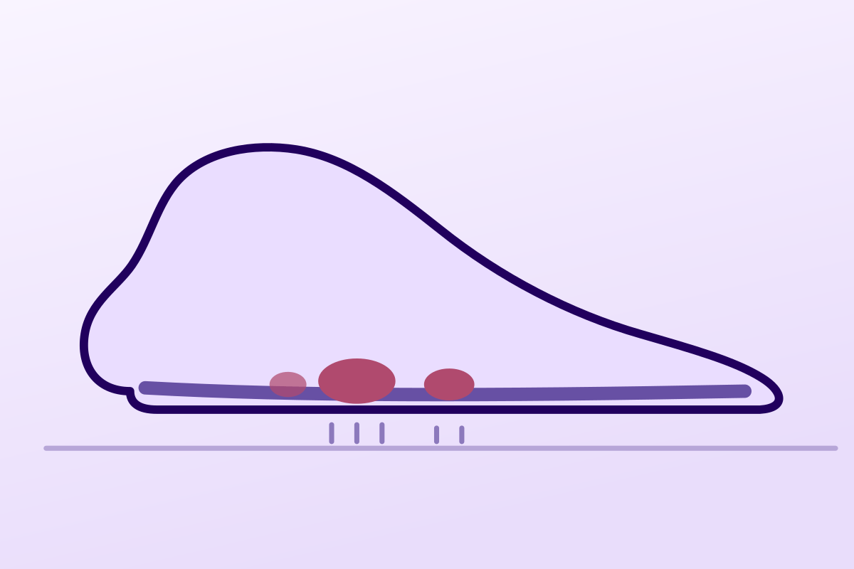
A mass that hurts on weight bearing, under the arch at the medial border, points to a benign fibromatosis with no causal treatment: the first step is mechanical offloading of the nodule.
15.2%of men with Dupuytren's disease who also have plantar nodules
What follows covers each of these points in detail, with sources. It is there if you need it.
This topic is taught in a course: Heel spur, plantar heel pain and tibialis posterior dysfunction, online with Anthony Baillon.
1 / 11
10 article chapters · 33 min in total
Understanding◔ 2 min
It is the same process as Dupuytren's disease, but in a foot that carries the body's weight.
Who is affected◔ 4 min
A man with Dupuytren's disease has a 15.2 % risk of plantar nodules, against 3.9 % in controls.
Multiple sites◔ 4 min
Three sites, one process: palmar, plantar and penile forms share the same biology.
Recognising it◔ 3 min
The nodule is part of the fascia: it does not roll like a lipoma and hardens on the windlass manoeuvre.
Imaging◔ 2 min
A genuine plantar fibroma can look like a sarcoma on MRI, a real diagnostic trap.
Physiotherapy's role◔ 5 min
Of 213 patients studied, only 35 received conservative treatment: most came from surgery.
Radiotherapy◔ 3 min
The LedRad trial shows relief in 74 % of those irradiated against 56 % under placebo.
Surgery◔ 2 min
Recurrence depends on the width of resection, and on nothing else: 67 % locally, 27 % after fasciectomy.
Case reports◔ 3 min
A 48-year-old man achieved complete resolution of pain after three shockwave sessions.
In practice◔ 5 min
The toe contracture the patient fears is the exception, not the rule, unlike in the hand.
Train on this
Lower limb
Anthony Baillon
Next comes practice: the course that teaches this topic, with Anthony Baillon.
Our courses on this topic may be covered by DPC or FIFPL, the French continuing-education funding schemes, depending on your profession and situation.
A hard lump under the arch of the foot, which does not always hurt, which grows slowly, and which almost everyone first takes for plantar fasciitis. It is in fact another disease, a first cousin of Dupuytren's disease, of which only one treatment has been validated against placebo, and it is neither surgery nor physiotherapy.
Summary written from primary sources verified one by one on PubMed: identifier resolved, journal and year checked, abstract read before citation. Full bibliography at the end of the article.
Ledderhose disease in three figures
A strong familial link with the hand, one treatment that has passed the placebo test, and surgery that recurs
Sources: Gudmundsson KG, Jónsson T, Arngrímsson R. Foot Ankle Int 2013;34(6):841-845 (PMID 23386754); de Haan A et al. Radiother Oncol 2023;185:109718 (PMID 37211283); Anwander H et al. Foot Ankle Spec 2024;17(2):109-116 (PMID 34369197).
What to take away before reading the rest
Understanding what this disease makes saves a long detour: it is the same tissue as in the palm, at the same cellular stage, but in a foot that carries the body's weight, and that last point changes almost everything else.
Ledderhose disease belongs to the group of superficial fibromatoses, alongside Dupuytren's disease (palmar fibromatosis) and Peyronie's disease (penile involvement). All rest on the same process: a benign proliferation of fibroblasts and myofibroblasts, producing excess collagen.
A pathology review of the two sites describes a shared histology: an unremarkable spindle-cell proliferation, bluish in appearance, with a variable amount of background collagen depending on the age of the lesion1. The authors stress above all the decisive clinical difference: palmar fibromatosis leads to nodules and then to digital contracture, whereas plantar fibromatosis produces nodules that are often painful but does not usually end in contracture1.
That is the nuance to explain to the patient who has a relative operated on for the hand and fears “no longer being able to straighten their toes”. Toe contracture does exist, but it is rare2 ; what is troublesome is the lump under the foot.
The topography is stereotyped and is a diagnostic argument. The nodules sit in the medial part of the plantar fascia, on the central or medial band. An ultrasound series of 57 fibromas in 43 patients measured their distribution: 84 % were at the middle third of the plantar fascia3.
That detail explains two things. First, why the pain is not in the heel: it sits under the arch, in front of the heel, exactly where plantar fasciitis does not hurt. Second, why footwear and weight bearing govern the complaint: a nodule in the middle of the arch is crushed at every step between the ground and the bone.
No aetiology is established, and the reviews say so plainly: there is no causal treatment, management remains symptomatic2. The associations reported in the literature are, for their part, fairly consistent from one review to another: trauma, diabetes, anticonvulsant use, frozen shoulder, alcohol consumption and liver disease4.
They have to be handled with the caution appropriate to associations drawn from case series: they guide the history, they do not explain the disease and they cannot be treated. The only link whose size has been measured in a cohort with a control group is that with Dupuytren's disease, and that is the subject of the next chapter.
Ledderhose disease is not a plantar fasciitis that has gone wrong: it is a disease of the hand that has come out on the wrong floor.
Key point
This is the most useful chapter in the clinic, because it turns a curiosity into an examination reflex: two areas to look at in the same patient, in both directions.
We should start with a methodological honesty: no general population study has measured the prevalence of Ledderhose disease. The reviews quote incidence figures of the order of one to two cases per hundred thousand people, but those values come from hospital series and not from a representative sample4. What is established, on the other hand, is the profile: a middle-aged or older adult, with a male predominance.
The surgical series give consistent markers. In a British series of 18 patients operated on with partial fasciectomy, the mean age was 41.3 (range 20 to 57), with 12 men out of 18, and 17 % of patients had multiple nodules at their first consultation5. Cohen's ultrasound series covered 43 patients aged 26 to 77, mean age 543.
This is the most solid datum in the whole chapter, and it comes from the Reykjavík study. One hundred and twenty-two men with Dupuytren's disease were invited back eighteen years after their original enrolment, along with the same number of controls matched for age and smoking. Ninety-two patients and 101 controls attended and were examined for plantar nodules6.
Ledderhose disease was identified in 14 of the 92 men with Dupuytren's (15.2 %), against 4 of the 101 controls (3.9 %), an odds ratio of 4.35 (95 % confidence interval: 1.3 to 16.7; p < 0.01). Two secondary results deserve just as much attention:
The converse was observed in a Dutch prevalence study of 763 randomly selected subjects aged 50 to 89: the prevalence of Dupuytren's disease there reached 22.1 %, and the presence of Ledderhose disease was among the significant risk factors for Dupuytren's disease, alongside a history of hand trauma, excessive alcohol consumption and family history7.
Plantar nodules: Dupuytren's disease against matched controls
Reykjavík cohort, clinical examination of the feet at 18 years of follow-up: 92 affected men, 101 controls matched for age and smoking
Source: Gudmundsson KG, Jónsson T, Arngrímsson R. Association of Morbus Ledderhose with Dupuytren's contracture. Foot Ankle Int 2013;34(6):841-845 (PMID 23386754). Confidence intervals: 1.3-16.7 for the first ratio, 1.8-27.1 for the second.
The notion of a Dupuytren's diathesis denotes the clinical features that identify aggressive forms at high risk of recurrence. Its historical version counted ectopic involvement (that is, outside the palm, so plantar or penile), among its four factors.
A reappraisal in 322 patients modified that definition, and the detail matters: the factors were widened to male sex and onset before the age of 50, but ectopic involvement was restricted to knuckle pads over the interphalangeal joints (Garrod's pads) alone. The presence of all the revised factors takes the risk of recurrence to 71 %, against 23 % in a patient with none of them8.
In other words: Ledderhose disease is not, in the revised version, a formal diathesis criterion. But the Reykjavík study shows that it preferentially accompanies the most severe palmar forms6. The practical course is the same on both readings, look at the other site, but the exact wording avoids making a score say what it does not.
Four practical markers
Values taken from the series cited; in the absence of a general population study, these are clinical orders of magnitude and not population rates
Sources: Kadir HKA, Chandrasekar CR. Foot 2017;31:31-34 (PMID 28334642); Cohen BE, Murthy NS, McKenzie GA. J Ultrasound Med 2018;37(11):2725-2731 (PMID 29603334).
Key point
Some patients do not have Ledderhose disease: they have a fibromatous disease expressing itself in several places. That subgroup is worth identifying, because it changes the examination, the outlook and what you say.
The superficial fibromatoses form a coherent group histologically. A pathology review of the palmar and plantar forms describes in both cases the same proliferation of fibroblasts and myofibroblasts, with a shared microscopic appearance: unremarkable bluish spindle cells, with a background amount of collagen varying with the age of the lesion1. The clinical reviews add the third site, penile, under the name Peyronie's disease4.
What distinguishes these three expressions is the mechanical behaviour of the tissue concerned, not the nature of the lesion:
| Site | Name | What the lesion does | Dominant clinical consequence |
|---|---|---|---|
| Palmar fascia | Dupuytren's disease | Nodules then longitudinal cords that contract1 | Digital contracture, loss of finger extension |
| Plantar fascia | Ledderhose disease | Nodules of the medial border, middle third in 84 % of cases3 ; contracture unusual1 | A mass that hurts on weight bearing, difficulty with footwear |
| Penile tunica albuginea | Peyronie's disease | A fibrous plaque of the tunica albuginea4 | Deformity on erection, pain |
The difference between the hand and the foot is worth explaining to the patient, because it explains why the treatment differs. In the palm, the cord pulls on a mobile finger and surgery has a geometric aim: to restore extension. Under the foot, the nodule is crushed against the ground and surgery has an analgesic aim: to remove the compression. The two diseases share a histology and not a therapeutic logic.
Three things are enough, and none takes more than a minute:
The strongest signal, however, is not simply the presence of palmar involvement: it is its severity. Men operated on for their Dupuytren's disease, or with an established digital contracture, were six times more likely to have plantar nodules than those who had only nodules or cords (odds ratio 6.1; 1.8 to 27.1; p < 0.001)6. In other words, the patient whose shoes should come off first is the one who has already been operated on for the hand.
What it changes. The examination becomes routinely bilateral and multi-site. Follow-up lengthens, because one site can appear years after the other: the Icelandic cohort re-examined its patients eighteen years after enrolment6. And what you say changes: you no longer talk about an isolated nodule but about a predisposition, which helps the patient understand why the disease comes back after surgery.
What it does not change. The treatment of each site remains the treatment of that site. No data indicate that a systemic treatment acts on all of them, and the drugs cited in the reviews (verapamil, tamoxifen, imatinib, sorafenib, mitomycin C) are cited at descriptive levels of evidence, with no established indication49.
Quoting the diathesis without making it say more than it does
The revised version of the Dupuytren's diathesis, established in 322 patients, restricted the ectopic involvement criterion to knuckle pads over the interphalangeal joints (Garrod's pads), and added male sex and onset before the age of 50. The presence of all the revised factors takes the risk of recurrence to 71 %, against 23 % in their absence8. Ledderhose disease therefore does not feature there by name as a criterion, which takes nothing away from its measured association with severe palmar forms6. Saying “your plantar involvement is part of your diathesis” is an approximation; saying “it more often goes with the forms of hand disease that recur” is accurate.
Key point
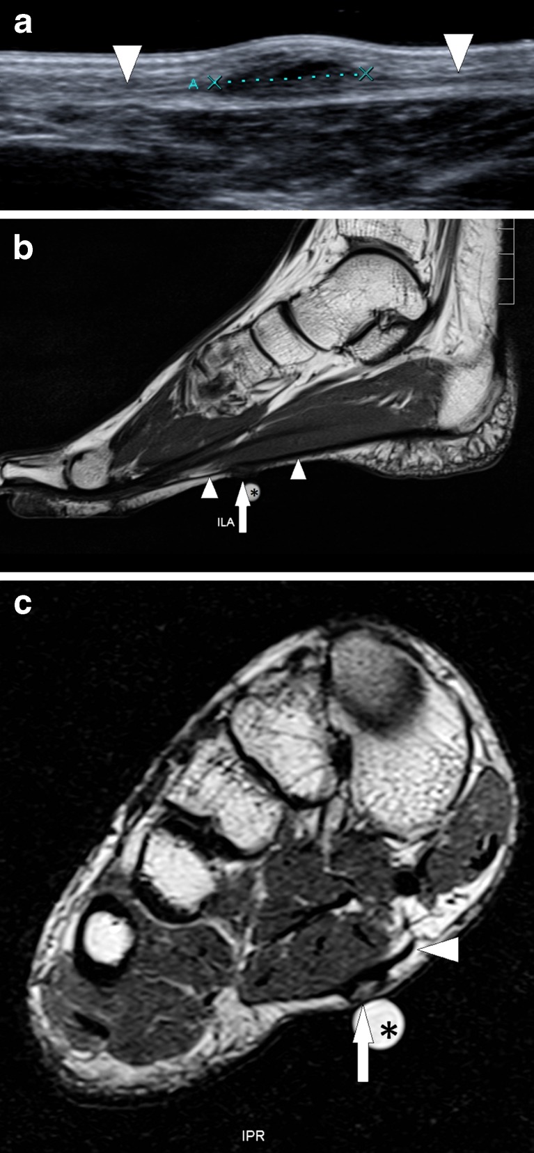
UltrasoundUltrasound and MRI of the foot: nodule of the plantar fascia delineated by arrows, plantar fibromatosis.
Source : Van Hul et al., Insights into imaging, 2011, figure 16 · CC BY
The diagnosis is clinical. It comes down to a well-conducted palpation and three questions: provided you have in mind what it must on no account be confused with.
Three complaints recur, in this order of frequency: a lump under the foot that the patient discovered while washing or walking barefoot, pain on weight bearing that increases with prolonged standing and walking barefoot on hard ground, and difficulty with footwear. Kadir's surgical series sums up the reasons for consulting in three items: pain, swelling, and functional difficulty5.
What is not the picture: pain on the first steps in the morning, maximal under the heel, easing after a few minutes of walking. That is the picture of plantar fasciopathy, and the distinction is the first one to make.
| Diagnosis | What suggests it | What distinguishes it from a plantar fibroma |
|---|---|---|
| Plantar fasciopathy | First-step pain, maximal at the calcaneal insertion, easing as walking continues | No palpable mass; heel pain rather than mid-plantar pain |
| Tendon sheath fibroma | A firm mass in contact with a flexor tendon | It is not part of the fascia; it does not harden on the windlass manoeuvre |
| Ganglion cyst | A fluctuant, transilluminable mass, often of rapid onset | Anechoic fluid contents on ultrasound |
| Morton's neuroma | Forefoot pain between the metatarsal heads, radiating to the toes, Mulder's sign | An anterior location, no fascial mass; see the dedicated article on the site |
| Plantar bursitis or callus | A pressure point, overlying hyperkeratosis | A superficial skin lesion, mobile with the skin |
| Foreign body, granuloma | A history of skin penetration, local inflammation | Context, inflammatory signs, imaging |
| Soft tissue sarcoma | Rapid growth, size greater than 5 cm, night pain, a deep and fixed mass | An absolute red flag: sarcomas have been taken for plantar fibromas19 |
Red flags: do not treat, refer
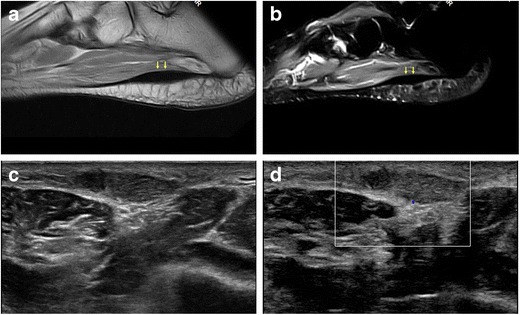
MRIMRI (a, b) and ultrasound (c, d) of the foot: fusiform thickening of the distal plantar fascia, low signal on MRI and hypoechoic on ultrasound, with hypervascularity on Doppler, Ledderhose disease.
Source : Draghi et al., Insights into Imaging, 2017, figure 5 · CC BY
Imaging does not make the diagnosis, it confirms it and above all it rules things out. Two publications are enough to know what to request and how to read the report.
The reviews agree: the diagnosis is clinical, ultrasound and MRI are confirmatory adjuncts, with MRI reserved for aggressive or advanced forms49. Ultrasound has the advantage of being dynamic, comparative and immediate.
The reference series described 57 fibromas in 43 patients and quantified the ultrasound features3 :
What ultrasound shows in plantar fibromatosis
57 nodules in 43 patients: frequency of each feature
Source: Cohen BE, Murthy NS, McKenzie GA. Ultrasonography of Plantar Fibromatosis: Updated Case Series, Review of the Literature, and a Novel Descriptive Appearance Termed the “Comb Sign”. J Ultrasound Med 2018;37(11):2725-2731 (PMID 29603334).
Two points of interpretation deserve emphasis, to the patient as much as to the requesting doctor. First, intralesional vascularity on Doppler is not a sign of malignancy here : it is present in more than half the benign fibromas in that series3. Second, the comb sign, that alternation of hypoechoic and isoechoic bands described by these authors, appears all the more readily the larger the nodule, which makes it a useful but not an essential sign.
MRI is requested when the lesion is large, deep, atypical, or when doubt about a tumour persists. It does however have a shortcoming worth knowing before reading an alarming report.
A case published in The Foot reports a 42-year-old man with a histologically proven plantar fibromatosis, whose MRI showed a so-called “brain gyriform” pattern: an appearance until then described as specific to low-grade fibromyxoid sarcoma. Posterior acoustic enhancement on ultrasound, T2 hyperintensity and contrast enhancement were all atypical, and corresponded histologically to areas of high cellularity alternating with cell-poor fibrous areas20.
The practical message runs in both directions: a genuine plantar fibroma can look like a sarcoma on MRI, and a sarcoma can be taken for a plantar fibroma19. No imaging replaces histopathology when the presentation falls outside the usual frame. It is not for the physiotherapist to decide, it is for them to know when not to treat.
Key point
This is a disease in which the physiotherapist is often the first to be consulted and rarely the one who will treat it. Knowing what can be offered, and when to hand over, is better than multiplying sessions on a nodule that will not move.
The most complete systematic review of treatments gathered 25 studies and 233 patients. Across that whole set, only five studies concerned a conservative treatment, totalling 35 patients, against 178 patients operated on15. The authors conclude that clinical trials with standardised outcomes are still needed to evaluate the success rate and the complications of each procedure seriously.
An older review already wrote it: no causal treatment exists, management is symptomatic and graded by severity, and no long-term randomised controlled study was available for the conservative options2. Since then one exception has appeared, and only one: radiotherapy, which has its own chapter below.
Aim no. 1, take the load off the nodule. This is the measure whose effect is the most immediate and the most reproducible in practice. A foot orthosis with a hollow over the nodule, a cushioning insole, footwear with a thick, rigid sole: anything that stops the nodule being crushed between the ground and the bone reduces weight-bearing pain. No trial has measured that effect in this condition: it is mechanical reasoning, and it must be presented as such.
Aim no. 2: maintain the flexibility of the posterior chain. A short triceps surae increases the load on the fascia. Stretching of the gastrocnemius and soleus, and specific plantar fascia stretching, are commonly proposed. Here again, no data specific to Ledderhose disease: the transposition comes from plantar fasciopathy, which is another disease of the same tissue.
Aim no. 3: monitor. This is a physiotherapist's job in its own right and it is underrated. Mapping the nodules, measuring them, recording their number: that is what will make it possible to say in six months whether the disease is progressing, and that is what will trigger referral.
What to avoid. Aggressive techniques applied directly to the nodule (heavy deep transverse friction, prolonged ischaemic pressure, radial shockwaves aimed at “breaking it up” without an established protocol) have no data in their favour and risk aggravating the pain of a lesion which, for its part, does not dissolve under the finger. That point is reasoning, not a trial: there is no trial.
Shockwaves: a special case, not to be confused with massage
High-energy focused shockwaves have been the subject of a small prospective series: six patients, two sessions seven days apart, 2,000 impulses at 3 Hz and 1.24 mJ/mm². Pain fell from 6 ± 2 before treatment to 2 ± 1 at fourteen days and then 1 ± 1 at three months, with all patients reporting softening of the nodules and no adverse effects12. A case report published in 2024 describes a comparable result in a 48-year-old man with bilateral disease, with a different protocol: three weekly sessions, 2,000 impulses at 5 Hz and 0.20 mJ/mm²13. Six patients plus one case, with no control group, do not make evidence: they make a serious lead, with published parameters, and the authors themselves call for larger trials12.
The ranking below applies GRADE principles to the precise question of symptomatic treatment of Ledderhose disease. It is an editorial appraisal : no published GRADE assessment and no learned society guideline exists for this condition.
Radiotherapy to symptomatic nodules. The only treatment evaluated by a double-blind randomised phase 3 trial against placebo: pain relief in 74 % against 56 %, a significant difference at 12 and 18 months, with improved quality of life and barefoot walking speed10. Downgraded from “high” for the sample size (84 patients) and the absence of independent replication.
Mechanical offloading : a foot orthosis hollowed over the nodule, a cushioning insole, adapted footwear. Consistent with the mechanism of the pain and without risk, but no study has measured it in this indication. Recommended by the reviews as part of graded symptomatic treatment2.
Surgical excision, reserved for failure of conservative treatment. Effective on symptoms, but recurrence depends directly on the extent of the resection: 67 % after local excision, 42 % after wide excision, 27 % after fasciectomy16. Retrospective series only.
High-energy focused shockwaves. A prospective series of six patients12 and one case report13, both without a control group. Published parameters, no adverse effects reported, but the sample size rules out any conclusion about efficacy.
Corticosteroid injection, with or without intralesional fenestration. Two case reports describe a reduction in pain and volume at twelve months after two triamcinolone injections combined with fenestration14. The authors themselves stress that this option has received only marginal attention in the literature.
Collagenase from Clostridium histolyticum. . Described only in isolated case reports, including an ultrasound-guided injection into a recurrence1718. No comparative series, no validated indication in the foot.
Topical verapamil, tamoxifen, imatinib, sorafenib, mitomycin C. Cited by the reviews as options documented at varying levels of evidence, essentially descriptive49. None of them falls to physiotherapy and none has an established indication.
Stretching and flexibility work for the posterior chain in this indication: no study at all. Transposed from plantar fasciopathy, which is another disease of the same tissue. Without risk, but to be announced for what it is.
Heavy deep transverse friction and ischaemic pressure on the nodule. No data, no plausible mechanism for resorbing mature fibrous tissue, and a risk of aggravating the pain of a lesion already crushed at every step.
Key point
It is the only question in this article that a randomised trial against placebo answers. It deserves detailed treatment, because it is also the treatment patients are most afraid of.
The LedRad study is a prospective multicentre double-blind randomised phase 3 trial. Patients were allocated between radiotherapy and sham radiotherapy: a placebo, therefore, which is exceptional in this field. The primary outcome was the reduction in pain at twelve months on a numerical rating scale10.
Eighty-four patients were included. The results:
LedRad: radiotherapy against sham radiotherapy
Double-blind randomised phase 3 trial, 84 patients: mean pain on a numerical rating scale
Source: de Haan A, van Nes JGH, Kolff MW et al. Radiotherapy for Ledderhose disease: results of the LedRad-study. Radiother Oncol 2023;185:109718 (PMID 37211283).
The same team surveyed by questionnaire all the patients treated with radiotherapy in their centre between 2008 and 2017. One hundred and two feet were irradiated in 67 patients (28 men, 39 women), on a protocol of two series of five daily fractions of 3 Gy11.
Mean pain fell from 5.7 before treatment to 1.7 at the time of assessment (p < 0.001). The detail of the response is more instructive than the mean: a complete response, pain gone, for 42 feet (41 %), a partial response for 38 feet (37 %), no change for 22 feet (22 %), and worsening for none. Seventy-eight per cent of patients said they were satisfied, 57 % did not find the treatment burdensome, and quality of life scores were comparable to those of the Dutch population of the same age. The most frequent residual adverse effect was dry skin, in 10 patients (15 %)11.
One patient in five gets nothing from radiotherapy, none comes out worse, and two in five have no pain at all: it is the only treatment for this disease of which that can be said with a figure.
What the physiotherapist can say about it to the patient
The word “radiotherapy” evokes cancer, and many patients refuse before listening. Three factual points are enough to restore proportion: these are low doses (two series of five sessions of 3 Gy in the published protocol11), the adverse effects reported are cutaneous and mild in 95 % of cases and resolved in 87 % of patients at eighteen months10, and it is the only treatment for this disease to have been compared with a placebo in double blind. The decision rests with the doctor; the factual information can be given in a session.
Key point
Surgery relieves, and it recurs. Those two facts do not contradict each other: they are explained by a single variable, the extent of what is removed.
This is the most useful result in the whole surgical literature on this disease. A systematic review coupled with a personal series compiled six studies covering 109 feet in 92 patients, and classified recurrence by the procedure performed16 :
Recurrence after surgery, by extent of resection
Systematic review of six studies, 109 feet in 92 patients
Source: Anwander H, Weichsel F, Cullmann J et al. Recurrence Rate After Wide Resection of Plantar Fibromatosis: A Case Series and Systematic Literature Review. Foot Ankle Spec 2024;17(2):109-116 (PMID 34369197).
The authors draw an explicit recommendation from it: prefer wide excision or fasciectomy to local excision, because of the markedly lower recurrence rate16. Their own series of 12 patients reviewed between 2 and 13 years after a wide excision shows 2 recurrences (17 %) at a mean follow-up of 7.8 years, with a median Foot Functional Index (Foot Functional Index) of 1 and a median AOFAS score of 95, in other words excellent functional results in those who do not recur.
A British series offers a useful counterpoint. Eighteen patients, nineteen partial fasciectomies, with up to five years of follow-up: a single recurrence (6 %), but 17 % scar problems: two cases of scar pain and one hypersensitive scar that resolved at twelve months5. That last point is not trivial in a region that carries the body's weight: a painful plantar scar can be more disabling than the nodule it replaced.
The rule accepted by all the reviews is simple: surgery comes after conservative treatment has failed, never before915. The criteria that tip the balance:
And a common-sense contraindication that the literature suggests through its figures: an asymptomatic nodule is not operated on. Two thirds recurrence after local excision is not a reasonable price for removing a lump that does not hurt.
Key point
Four published situations, taken as they stand with their identifiers. Two show what treatment can do, two show what imaging cannot.
Case 1: Bilateral disease treated with shockwaves
Fulceri F, Ryskalin L, Morucci G et al. Life 2024;14(2):16913: free full text (PMC10889909).
A 48-year-old man has bilateral Ledderhose disease. The protocol applied comprised three sessions one week apart, 2,000 impulses at 5 Hz, energy flux density of 0.20 mJ/mm², with ultrasound follow-up. The reported result is complete resolution of the pain, restoration of full functional activity and a clear improvement in quality of life.
What this case teaches, and what it does not. It documents precise, reproducible parameters, which this field badly lacks: the authors themselves note that the literature on shockwaves in this indication is “extremely limited” and that no optimal parameters have been defined. But a single case with no control group, in a disease that fluctuates, does not demonstrate efficacy.
Case 2: Two nodules treated with fenestration and corticosteroids
Flanagan G, Burt N, Reilly IN. SAGE Open Med Case Rep 2021;9:2050313X21101181314: free full text (PMC8107658).
Two patients with painful plantar nodules each received, on two occasions, intralesional fenestration combined with an injection of triamcinolone acetonide mixed with a local anaesthetic. At twelve months, the authors report a significant reduction in pain and in lesion volume.
What this case teaches. The authors open their article by noting that corticosteroid injection has received, in this disease, only a passing mention in the scientific literature. That is exactly the problem in this field: widespread practices, almost never evaluated. Two patients do not change that, but they describe a protocol others can reproduce.
Case 3: A sarcoma taken for a plantar fibroma
Lockyer MG, Rosen DG. Anticancer Res 2015;35(11):6171-617419.
A 44-year-old man presents with a mass of the medial arch of the foot, initially considered a plantar fibroma. MRI showed a lobulated subcutaneous mass, inseparable from the fascia. Microscopic examination revealed an extraskeletal myxoid chondrosarcoma: a rare tumour, indolent in its course but with a high potential for local recurrence and metastasis.
What this case teaches. The authors conclude that this diagnosis must always be considered in a mass of the plantar fascia. For the physiotherapist, the translation is direct: a plantar mass that is growing, that exceeds a few centimetres or that comes with night pain is not treated: it is referred.
Case 4: A plantar fibroma that, for its part, looked like a sarcoma
Touraine S, Bousson V, Kaci R et al. The Foot 2013;23(2-3):88-9220.
A 42-year-old man with a histologically proven plantar fibromatosis showed on MRI a “brain gyriform” pattern until then described as exclusive to low-grade fibromyxoid sarcoma, with posterior acoustic enhancement on ultrasound, T2 hyperintensity and contrast enhancement: all atypical. Histology showed that this pattern corresponded to the juxtaposition of highly cellular areas and cell-poor fibrous areas.
What this case teaches. It is the exact mirror of the previous one, and the two go together. Imaging is wrong in both directions; only histopathology settles it. An alarming MRI report no more condemns the patient than a reassuring one keeps them safe.
Key point
A rare disease, with no guideline, whose best treatment does not fall to us: the physiotherapist's role in it is real, but it has to be defined precisely so as not to be lost in pointless sessions.
| What you measure | Why | How often |
|---|---|---|
| Number and size of nodules | The only objective marker of progression; multiple nodules affect 17 % of patients at the first consultation5 | Every 2 to 3 months |
| Weight-bearing pain on a numerical rating scale | It is the primary outcome of the LedRad trial, which makes the values comparable with the literature10 | At every assessment |
| Walking distance and tolerance of standing | A direct functional translation; barefoot walking speed was a secondary outcome in LedRad10 | At every assessment |
| Examination of the palms | Screening for an associated Dupuytren's disease, whose prevalence is four times higher in this context6 | Initially, then annually |
| Toe mobility | Contracture is rare but it marks an advanced form1 | At every assessment |
| Tolerance of the orthosis | A badly placed hollow shifts the load instead of removing it | At 3 weeks, then at every assessment |
That is unlikely. Ledderhose disease is a fibrous proliferation; no regular spontaneous regression is described, and no causal treatment exists2. On the other hand, a nodule can stay stable and painless for years, in which case there is nothing to do, and that is good news which should be delivered as such.
The link exists and it has been quantified: in the Reykjavík cohort, 15.2 % of the men with Dupuytren's disease had plantar nodules against 3.9 % of controls6, and the presence of Ledderhose disease is among the risk factors for Dupuytren's in a Dutch population study7. That justifies examining the palms, not being alarmed: most patients will not have both.
No: it is a benign lesion1. But the question is legitimate, because rare malignant tumours can present in exactly the same way19. That is precisely why a mass that grows quickly, that exceeds a few centimetres or that wakes you at night is investigated before it is treated.
Only after conservative treatment has failed915, and never on a painless nodule. The point to know before deciding: recurrence depends on the extent of the resection: 67 % after removing the nodule alone, 42 % after wide excision, 27 % after fasciectomy16. A partial fasciectomy also led to 17 % scar problems in a series of 18 patients5, which is no small thing under a foot.
No study has measured it in this disease. The mechanical reasoning is solid (a nodule at the middle third of the arch is compressed at every step between the ground and the bone, and a hollow removes that compression) and the risk is nil. It is a common-sense intervention, to be offered as such and not as a validated treatment.
Nothing forbids it and nothing encourages it: the question has not been studied. The practical marker is weight-bearing pain, which governs. Change the surface, the shoe and the volume before stopping, and adapt the orthosis to the sports shoe, which does not have the same internal volume as a street shoe.
In the randomised phase 3 trial, the adverse effects (erythema, dry skin, burning sensations, a transient increase in pain) were mild in grade in 95 % of cases and resolved in 87 % of patients at eighteen months10. In the long follow-up cohort, the most frequent residual effect was dry skin in 15 % of patients11. The decision rests with the doctor; the factual information can be given in a session.
The available literature is a prospective series of six patients and one case report, both without a control group1213. The reported results are favourable and the parameters are published (high-energy focused waves, not radial ones), but the sample size rules out any conclusion. To be offered, where appropriate, as a therapeutic trial whose level of evidence is stated.
On the same subject
Every reference was verified individually in PubMed: identifier resolved, journal, year, volume and author list checked, abstract read to make sure the source does establish what is attributed to it.
Method and limitations
Sources searched in PubMed through the NCBI E-utilities services, crossing plantar fibromatosis and Ledderhose disease with epidemiology, imaging, radiotherapy, shockwaves, injections, collagenase and surgery. Every identifier was resolved and every abstract read before citation. Main limitation: only one randomised trial exists in this disease, and it concerns radiotherapy; everything else rests on retrospective series and case reports. The levels of evidence in the table of modalities are an editorial appraisal applying GRADE principles, and not a published GRADE assessment. One reference was also set aside during the work: an article whose title announced a case of bilateral plantar fibromatosis but whose indexed abstract described an entirely different patient: inconsistent metadata, therefore not cited. Article written on 14 August 2026.
Behind this article
An author who explains, a reviewer who checks.
How we write and check our content

Physiotherapist · co-founder of Physio Learning
Marked for life by his first four-hour lecture without a single image, he took a master’s in instructional design so that it would never happen to anyone again. He hunts down publication bias and unreadable slides with the same intransigence.

Head of scientific content
Physiotherapist specialising in neuro-musculoskeletal practice and holder of a master’s in public health. He checks the methodological rigour of every article: primary sources, levels of evidence, no exceptions.