

















In brief
Posterior tibial tendon deficiency (PTTD) is the leading cause of adult-acquired flatfoot, now named PCFD (Progressive Collapsing Foot Deformity) since the Myerson 2020 consensus, which recognises its multifactorial nature. It mainly affects women between 40 and 60, with a symptomatic prevalence of 3.3 % in women over the age of 40. Diagnosis is clinical: the single heel rise test is the key functional indicator, completed by the too many toes sign, which objectifies hindfoot valgus. Conservative treatment of stages I-II combines foot orthoses and progressive strengthening, with 83 % success reported over a median of 10 sessions.
Clinical synthesis based on the Myerson 2020 international consensus (Progressive Collapsing Foot Deformity), the Kulig 2009 randomised trial and the Alvarez 2006 prospective protocol.
Clinical synthesis
- The posterior tibial tendon deficiency (PTTD) is the leading cause of adult-acquired flatfoot. Since the international consensus Myerson 2020, the official nomenclature is PCFD (Progressive Collapsing Foot Deformity), recognising the multifactorial nature of the deformity.
- Symptomatic prevalence of 3.3 % in British women over the age of 40 (Kohls-Gatzoulis 2009, n=582). Peak incidence between 40 and 60 years, with a markedly high female-to-male ratio.
- Major risk factors documented since Holmes & Mann 1992: obesity, arterial hypertension and diabetes mellitus in 60 % of tendon ruptures. To these are added corticosteroid use, systemic inflammatory diseases and anatomical anomalies (accessory navicular bone).
- Pathophysiology: the tendon, lying in a retromalleolar zone of poor vascular supply, undergoes a degenerative tendinosis (Mosier 1998) with collagen disorganisation, leading to its elongation and then to failure of the passive stabilisers, notably the spring ligament complex (Pinney-Lin 2006, Bastias 2018).
- Clinical diagnosis: the single heel rise test is the most important functional indicator. The too many toes sign objectifies hindfoot valgus and forefoot abduction. Imaging (MRI) is reserved for atypical or pre-surgical cases.
- Classification: the Myerson 2020 PCFD classification is progressively replacing the 4-stage Johnson-Strom system. It rates 5 independent domains: hindfoot valgus deformity, midfoot abduction, arch loss, forefoot abduction/supination, and talotibial instability.
- Multimodal conservative treatment (stages I-II): the combination of foot orthoses + progressive strengthening exercises has proved effective in the Kulig RCT (PMID 19022863) and the Alvarez prospective protocol (PMID 16442022, 83 % success over a median of 10 sessions).
- Exercise must target the tibialis posterior (progressive eccentric work, Kulig 2009 Foot Ankle Int) but also the synergist muscles of the ankle and the hip, whose strength is bilaterally reduced in affected women (Kulig 2011).
- Emerging modality: the heavy-slow resistance (Beyer 2015) protocol is validated on the Achilles tendon and can be adapted to the tibialis posterior. Shockwave therapy and manual therapy carry a low level of evidence specifically for PTTD.
- Return to activity: guided by functional criteria (heel rise symmetry, no pain > 24 h after exertion) and not by a fixed timetable. The pain-monitoring model (Silbernagel 2007) remains applicable.
- Specific red flags: acute post-traumatic rupture, medial plantar hypoaesthesia (suspected associated tarsal tunnel syndrome), rigid non-reducible deformity (suspected stage III/IV).
- PCFD can mimic or coexist with tarsal tunnel syndrome (mechanical compression of the tibial nerve), an isolated spring ligament injury, or a multi-ligament injury (deltoid). A global biomechanical examination is essential.
- Advanced stages (rigid arthritic III, IV with talar tilt) fall to surgery: realignment osteotomies, selective or triple arthrodeses, and even subtalar arthroereisis in young patients (Sangeorzan 2020 consensus).
- Therapeutic education and load management are cross-cutting pillars, in line with the 9 core tendinopathy domains identified by the ICON 2019 consensus (Vicenzino BJSM 2020).
- Measuring outcomes with validated PROMs (Foot & Ankle Outcome Score - FAOS, Foot Functional Index - FFI) is essential to assess longitudinal effectiveness and adapt management.
- Medication history : look for recent exposure to fluoroquinolones (ciprofloxacin, levofloxacin, ofloxacin), and to corticosteroid therapy given systemically or locally. Both raise the risk of tendinopathy and rupture, especially after the age of 60, in weight-bearing tendons and when the two are combined.
Contents
- What are the fundamentals to know about posterior tibial tendinopathy (PTTD) and adult-acquired flatfoot (PCFD)?
- How do you assess and diagnose PTTD / PCFD with certainty?
- Which treatment strategies are most effective for PTTD / PCFD?
- How do you secure lasting recovery and prevent recurrence?
- What do real-world case studies teach us about PTTD / PCFD?
- How do you apply these recommendations concretely in your practice?
What are the fundamentals to know about posterior tibial tendinopathy (PTTD) and adult-acquired flatfoot (PCFD)?
How is this condition defined, who is affected and what are the risk factors?
The posterior tibial tendon deficiency (PTTD), known internationally as Posterior Tibial Tendon Dysfunction, has been recognised for several decades as the most frequent cause of adult-acquired flatfoot deformity (AAFD).¹ Since the international consensus of Myerson et al. published in 2020 in Foot & Ankle International, the official nomenclature is Progressive Collapsing Foot Deformity (PCFD), a term that explicitly acknowledges the multifactorial nature and the kinetic progression of the deformity, without tying it to a single tendon.²The tibialis posterior is the principal dynamic stabiliser of the medial arch. Its main insertion on the navicular tuberosity, its plantar expansions and its retromalleolar course make it a vulnerable biomechanical system.³ When its function degrades, load shifts onto the static stabilisers (spring ligament, plantar ligaments, deep deltoid ligament), whose secondary failure marks the transition to a visible deformity.⁴
The symptomatic prevalence was precisely quantified by the founding epidemiological survey of Kohls-Gatzoulis (2009) carried out in Hertfordshire, England: out of 582 validated responses from a random sample of 1,000 women over the age of 40, 3.3 % had symptomatic posterior tibial tendon involvement or an acquired flatfoot.⁵ This prevalence rises with age and female sex remains predominant: the condition affects mainly women between 40 and 60 years of age.⁶ 👩⚕️📊 Symptomatic prevalence in British women over the age of 40 (Kohls-Gatzoulis 2009)
Postal survey + clinical examination, n = 582 validated responses
Study validated by clinical examination: 7 patients stage I, 12 patients stage II, 9 patients with acquired deformity. All symptomatic but undiagnosed before the study. Source: Kohls-Gatzoulis J et al. Foot Ankle Surg. 2009;15(2):75-81. PMID 19410173.
⚖️ Risk factors in 67 patients with posterior tibial tendon rupture (Holmes & Mann 1992)
At least one factor present in 60 % of cases; hypertension / obesity / diabetes in 52 %
Percentages derived from the retrospective cohort n = 67. Local or systemic corticosteroids are also associated with rupture but remain under-quantified in routine practice. Source: Holmes GB Jr, Mann RA. Foot Ankle. 1992;13(2):70-79. PMID 1349292.
- Anatomical anomalies : pre-existing congenital flatfoot, accessory navicular bone (type II especially), a shallow medial retromalleolar groove or hypoplasia of the sustentaculum tali.¹
- Systemic inflammatory diseases : rheumatoid arthritis, spondyloarthropathies, lupus, all of which can directly involve the synovial sheath of the tendon.⁹
- Trauma : ankle fractures, severe sprains with injury to the medial stabilisers, repeated microtrauma linked to physical activity on uneven ground.⁹
- Iatrogenic : local corticosteroid injections into the tendon sheath (to be avoided), prolonged systemic corticosteroid therapy.⁹
What happens in the body and how does PTTD progress naturally?
The pathophysiology was elegantly demonstrated by the landmark histological study of Mosier (1998), which compared 15 healthy tendons (cadavers) with 15 tendons from surgical patients with posterior tibial tendon insufficiency. The pathological tendons showed a characteristic degenerative tendinosis with increased mucoid substance, fibroblastic hypercellularity, chondroid metaplasia and neovascularisation.¹⁰ It is therefore a degenerative process (tendinosis) and not pure inflammation (tenosynovitis), even though acute phases may include a synovial component.The tendon is particularly vulnerable in its medial retromalleolar zone of hypovascularity, located about 14 mm distal to the medial malleolus.¹¹ Repeated microtrauma at this level, on an unfavourable metabolic background, leads to collagen disorganisation, to progressive elongation and then to partial or complete rupture.
When the tendon loses its dynamic stabilising function, the load transfers to the static stabilisers. The spring ligament complex (plantar calcaneonavicular), comprising three bands (superomedial, medioplantar oblique and inferoplantar longitudinal), is the first to give way.¹² Its failure is a major tipping point: it marks the transition from the “isolated tendinopathy” stage to the “visible and progressive deformity” stage.¹³ The deep deltoid ligament (talotibial) follows in the advanced stages, then allowing the talus to tilt into valgus within the mortise.
🔬 Pathophysiological cascade: from healthy tendon to rigid deformity
Sequential model integrating tendon, ligaments and bony architecture
Synthetic model based on Mosier 1998 (tendinosis), Pinney-Lin 2006 and Bastias 2018 (spring failure). Loss of tendon function precedes and causes passive ligament failure.
- Stage I : tenosynovitis or tendinosis with no visible deformity. Single heel rise possible but often painful, with reduced strength compared with the healthy side.
- Stage II : deformity that is supple and reducible (calcaneal valgus + forefoot abduction + arch collapse). Single heel rise impossible or without calcaneal inversion. Often subdivided into IIA (moderate, < 30 % radiographic abduction) and IIB (severe, ≥ 30 %).
- Stage III : deformity that is rigid and non-reducible with arthritic changes (subtalar, talonavicular, calcaneocuboid). Pain is often lateral (sinus tarsi impingement).
- Stage IV : added by Myerson, tilt of the talus within the tibiofibular mortise (deep deltoid ligament involvement), with secondary tibiotalar osteoarthritis.¹⁵
Key points
- PTTD is the leading cause of adult-acquired flatfoot, now called PCFD (Progressive Collapsing Foot Deformity) since the Myerson 2020 international consensus.
- Symptomatic prevalence: 3.3 % in British women over the age of 40 (Kohls-Gatzoulis 2009). Very marked female predominance, peak at 40-60 years.
- Major cardiometabolic risk factors: obesity, hypertension, diabetes mellitus (Holmes-Mann 1992: 60 % of ruptures). To these are added corticosteroids, inflammatory diseases and anatomical anomalies.
- Pathophysiology: degenerative tendinosis (Mosier 1998) in a zone of hypovascularity, tendon elongation, secondary failure of the spring ligament, arch collapse, then arthritic rigidity in the advanced stages.
- Historical clinical classification: 4 stages (Johnson-Strom modified by Myerson). Progression to the advanced stages is not inevitable when early treatment is well conducted.
Bibliography
- Knapp PW, Constant D. Posterior Tibial Tendon Dysfunction. In: StatPearls [Internet]. Treasure Island (FL): StatPearls Publishing; updated 2024 May. NCBI Bookshelf NBK542160. PMID 31194317.
- Myerson MS, Thordarson DB, Johnson JE, Hintermann B, Sangeorzan BJ, Deland JT, Schon LC, Ellis SJ, de Cesar Netto C. Classification and Nomenclature: Progressive Collapsing Foot Deformity. Foot Ankle Int. 2020;41(10):1271-1276. PMID 32856474.
- Ling SK, Lui TH. Posterior Tibial Tendon Dysfunction: An Overview. Open Orthop J. 2017;11:714-723. PMID 28979585.
- Deland JT. Adult-acquired flatfoot deformity. J Am Acad Orthop Surg. 2008;16(7):399-406. PMID 18611997.
- Kohls-Gatzoulis J, Woods B, Angel JC, Singh D. The prevalence of symptomatic posterior tibialis tendon dysfunction in women over the age of 40 in England. Foot Ankle Surg. 2009;15(2):75-81. PMID 19410173.
- Kohls-Gatzoulis J, Angel JC, Singh D, Haddad F, Livingstone J, Berry G. Tibialis posterior dysfunction: a common and treatable cause of adult acquired flatfoot. BMJ. 2004;329(7478):1328-1333. PMID 15576744.
- Holmes GB Jr, Mann RA. Possible epidemiological factors associated with rupture of the posterior tibial tendon. Foot Ankle. 1992;13(2):70-79. PMID 1349292.
- Abousayed MM, Alley MC, Shakked R, Rosenbaum AJ. Adult-Acquired Flatfoot Deformity: Etiology, Diagnosis, and Management. JBJS Rev. 2017;5(8):e7. PMID 28806265.
- Pinney SJ, Lin SS. Current concept review: acquired adult flatfoot deformity. Foot Ankle Int. 2006;27(1):66-75. PMID 16442033.
- Mosier SM, Lucas DR, Pomeroy G, Manoli A 2nd. Pathology of the posterior tibial tendon in posterior tibial tendon insufficiency. Foot Ankle Int. 1998;19(8):520-524. PMID 9728698.
- Vulcano E, Deland JT, Ellis SJ. Approach and treatment of the adult acquired flatfoot deformity. Curr Rev Musculoskelet Med. 2013;6(4):294-303. PMID 23765382.
- Bastias GF, Cuchacovich N, Schiff A, Lara J. Spring Ligament Instability. Foot Ankle Clin. 2018;23(4):659-678. PMID 30414659.
- Mengiardi B, Zanetti M, Schöttle PB, et al. Spring ligament complex: MR imaging-anatomic correlation and findings in asymptomatic subjects. Radiology. 2005;237(1):242-9. PMID 16118154.
- Johnson KA, Strom DE. Tibialis posterior tendon dysfunction. Clin Orthop Relat Res. 1989;(239):196-206. PMID 2912622.
- Myerson MS. Adult acquired flatfoot deformity: treatment of dysfunction of the posterior tibial tendon. Instr Course Lect. 1997;46:393-405. PMID 9143981.
How do you assess and diagnose PTTD / PCFD with certainty?
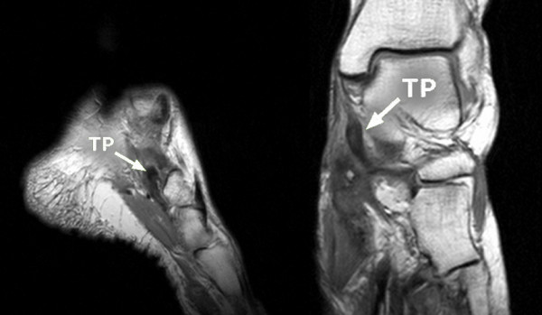
MRIFrontal and sagittal MRI of the foot: tear of the tibialis posterior tendon (arrows), a common cause of adult-acquired flatfoot.
Source : Popelka et al., BMC Musculoskeletal Disorders, 2010, figure 8 · CC BY
Which questions should you ask to understand the patient and their history?
History-taking must characterise the symptoms precisely and identify the risk factors. 🔍 Several lines of enquiry should be explored systematically:- Location and nature of the pain : classically posteromedial (along the tendon, from the medial malleolus to the navicular insertion). Often described as a dull, burning pain, made worse by prolonged standing and walking. Migration towards the lateral aspect of the foot suggests an advanced stage with sinus tarsi impingement.³
- Timeline and triggering factors : typically a gradual onset. An abrupt post-traumatic onset suggests an acute rupture (Holmes-Mann 1992 found a history of trauma in 36 % of cases).⁴ Worsening when walking on uneven ground, climbing stairs, or wearing poorly supportive shoes.
- Functional and morphological impact : does the patient notice a collapse of the arch, a “spreading” of the foot, asymmetrical shoe wear? The impact on activities of daily living (walking, driving, stairs) and on work is crucial in stratifying severity.⁵
- Past history and risk factors : age, sex, BMI, hypertension, diabetes, medication (corticosteroids), systemic inflammatory diseases, old trauma, previous ankle surgery.⁴˒⁶
- Red flags : non-mechanical night pain, unexplained weight loss, fever, distal neurological deficit (tibial nerve territory), a history of cancer. The presence of a red flag requires medical referral before any physiotherapy treatment.⁷
Which clinical tests should you carry out and which other conditions must be ruled out?
The examination must be bilateral, comparative and carried out both weight-bearing and non-weight-bearing. The reliability review by Ross (2021) assessed the main clinical tests: tendon palpation, the single heel rise test and weight-bearing morphological observation show satisfactory inter-rater reliability and correlate moderately with structural abnormalities on imaging.⁸Weight-bearing inspection (standing, posterior view) :
- Too many toes sign : more than two toes visible on the outer side of the affected foot, reflecting calcaneal valgus and forefoot abduction. Sensitive for stages II and above.³
- Collapse of the medial longitudinal arch : visible on weight-bearing, to be compared with the non-weight-bearing appearance (reducible vs rigid).
- Calcaneal valgus : measurable clinically (goniometry) or radiographically.
Functional tests :
- Single-leg heel rise test (SHR) : the reference test. The patient is asked to rise onto the toes of one foot. Normal test : full rise with visible calcaneal inversion (a sign of effective tibialis posterior contraction). Abnormal : rise impossible, incomplete, or without calcaneal inversion. A healthy subject can usually perform 8 to 10 repetitions without fatigue; the criterion “10-25 pain-free repetitions” is used as a return-to-activity target (see Houck 2009 on the kinematic analysis of the heel rise).⁹
- Manual muscle testing of inversion : the patient resists an eversion force applied by the clinician to the medial border of the foot. Weakness compared with the healthy side is typical of stages I and II.¹⁰
- Hip & ankle assessment : Kulig 2011 showed that women with PTTD present a bilateral reduction in the performance of the hip abductors and external rotators and of the ankle invertors, which justifies a strengthening programme across the wider kinetic chain.¹¹
🎯 Clinical diagnosis in 3 steps: detection, confirmation, staging
Summary algorithm based on the Ross 2018 SR and the Ling-Lui 2017 review
The algorithm gives priority to the clinical approach. Imaging is not routine in physiotherapy; it may be directed by the referring doctor if the picture resists conservative treatment or if surgery is being considered.
Essential differential diagnoses to know ⚠️:
- Tarsal tunnel syndrome : compression of the tibial nerve in the retromalleolar canal. It gives burning, medial plantar paraesthesia and a positive Tinel sign. It can coexist with PTTD (tenosynovitis pressing on the adjacent nerve) or be mimicked by it.¹²
- Navicular stress fracture : dorsomedial pain at the N-spot, gradual onset in a runner, focal tenderness on palpation. Imaging is essential (MRI or bone scan).¹³
- Primary talonavicular or subtalar osteoarthritis : stiffness dominates, diffuse pain, a history of trauma or overload.¹
- Isolated spring ligament injury : acquired deformity with no obvious clinical tendinopathy of the tibialis posterior. The diagnosis is often made on MRI, raising the question of targeted surgical repair.¹⁴
- Gastrocnemius-soleus tightness (functional equinus) : biomechanically worsens PCFD and must be looked for systematically (Silfverskiöld test).¹
- Tendinopathy of the tibialis anterior : the two names resemble each other closely enough to be swapped in reports, yet everything differs. The tibialis anterior is a dorsiflexor of the foot: its pain sits in front of the ankle and over the instep, the arch is not collapsed, and the single heel rise remains normal. Its failure gives a foot that slaps the ground, never a flat foot. See tibialis anterior tendinopathy.
🚩 Red flags specific to the foot and ankle
- Acute post-traumatic rupture of the tibialis posterior with sudden loss of function → prompt surgical referral
- Medial plantar hypoaesthesia or lightning paraesthesia in the tibial nerve territory → suspected associated or tumoral tarsal tunnel
- Oedema, erythema and heat over the tendon with fever → suspected infective tenosynovitis
- Rapidly progressive deformity over a few weeks in a patient with diabetes → suspected Charcot foot (emergency)
- Non-mechanical night pain + unexplained weight loss → oncological workup (tendon sarcoma or metastasis, rare but possible)
- Previous local surgery or corticosteroid injection + sudden pain → suspected iatrogenic rupture
- Antalgic limp with a motor deficit of the ipsilateral hip or knee → broader neurological workup
⚠️ Any red flag → prompt medical referral (GP, emergency department, orthopaedic surgeon) before physiotherapy management.
Should patients with PTTD / PCFD be classified, and what does it gain them?
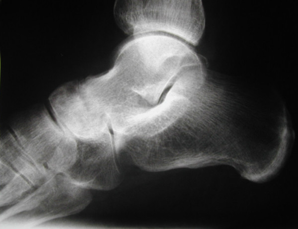
X-rayLateral X-ray of the foot: destruction and narrowing of the talonavicular joint, with collapse of the arch, a radiological appearance of advanced flatfoot.
Source : Popelka et al., BMC Musculoskeletal Disorders, 2010, figure 3 · CC BY
- The Johnson & Strom 1989 classification, modified by Myerson : 4 stages of increasing severity (see chapter 1). Advantage: simplicity, long standing, wide use. Limitation: it focuses on the hindfoot and the midfoot, and neglects the forefoot, gastrocnemius tightness, lateral instability and ligament involvement.¹⁵
- The Myerson 2020 PCFD classification : an international consensus system that rates 5 independent anatomical classes, modulated by flexibility (A = supple, B = rigid). It allows a three-dimensional and personalised description, aligned with modern surgical choices (Sangeorzan 2020 operative consensus).²˒¹⁶
📐 Myerson 2020 PCFD classification: 5 independent classes
Each class is rated separately, modulated by flexibility (A supple / B rigid)
Source: Myerson MS et al. Foot Ankle Int. 2020;41(10):1271-1276 (PMID 32856474). International consensus of 9 leading North American surgeons.
| System | Year | Structure | Key advantage | Key limitation |
|---|---|---|---|---|
| Johnson-Strom | 1989 | 4 linear stages | Simplicity, wide use | Linear, focused on the hindfoot |
| + Myerson stage IV | 1996-97 | 4 stages, adds talar tilt | Captures tibiotalar decompensation | Still linear |
| Myerson PCFD | 2020 | 5 independent classes × A/B flexibility | Multidimensional, aligned with modern surgery | Learning curve, little validated in physiotherapy |
| RAM / Raikin | 2012 | 3 anatomical zones + subtypes | Anatomically detailed | Limited clinical adoption |
Critique and controversies
The PCFD 2020 classification, although conceptually superior, has two important practical limitations. First, the rating complexity (5 classes × flexibility = multiple combinations) limits its adoption in community practice; the inter-rater agreement studies published since 2021 show moderate reliability depending on the class (notably 3 and 4) and on the rater's experience.¹⁶ Second, it remains centred on static morphological assessment, whereas dynamic alterations (gait kinematics, propulsion kinetics) appear earlier and are more sensitive. Three-dimensional gait analysis and multi-segment foot assessment (Oxford Foot Model) are promising for a finer stratification of the early stages, but remain little available clinically.¹⁷Moreover, the classic clinical tests have heterogeneous reliability. Ross 2021 showed that tendon palpation, the single heel rise and morphological observation are the most reliable, while other manoeuvres (tibialis posterior edge test, foot posture index, navicular drop) have more variable reliability.⁸ The clinician should favour the highly reliable tests and combine them to optimise diagnostic value.
Key points
- The diagnosis is clinical. The history establishes the timeline, the topography and the cardiometabolic context. The examination aims to objectify the deformity and the loss of function.
- The two key tests are the too many toes sign (weight-bearing posterior view) and the single-leg heel rise test (strength and calcaneal inversion). The SHR is the most important functional test.
- The Johnson-Strom classification modified by Myerson (4 stages) remains pragmatic for deciding the conservative/surgical threshold. The Myerson 2020 PCFD classification (5 classes × A/B) is the international standard for communication with surgeons and for research.
- Keep the differential diagnosis in mind: tarsal tunnel syndrome (may coexist), navicular stress fracture, primary osteoarthritis, isolated spring ligament injury, functional equinus.
- Red flags (acute rupture, infection, rapidly progressing deformity in a patient with diabetes, neurological deficit) require medical referral before any treatment.
Bibliography
- Knapp PW, Constant D. Posterior Tibial Tendon Dysfunction. StatPearls. Treasure Island (FL): StatPearls Publishing; 2024. PMID 31194317.
- Ling SK, Lui TH. Posterior Tibial Tendon Dysfunction: An Overview. Open Orthop J. 2017;11:714-723. PMID 28979585.
- Kohls-Gatzoulis J, Angel JC, Singh D, Haddad F, Livingstone J, Berry G. Tibialis posterior dysfunction: a common and treatable cause of adult acquired flatfoot. BMJ. 2004;329(7478):1328-1333. PMID 15576744.
- Holmes GB Jr, Mann RA. Possible epidemiological factors associated with rupture of the posterior tibial tendon. Foot Ankle. 1992;13(2):70-79. PMID 1349292.
- Ross MH, Smith MD, Plinsinga ML, Vicenzino B. Self-reported social and activity restrictions accompany local impairments in posterior tibial tendon dysfunction: a systematic review. J Foot Ankle Res. 2018;11:49. PMID 30186369.
- Abousayed MM, Alley MC, Shakked R, Rosenbaum AJ. Adult-Acquired Flatfoot Deformity: Etiology, Diagnosis, and Management. JBJS Rev. 2017;5(8):e7. PMID 28806265.
- Finucane LM, Downie A, Mercer C, Greenhalgh SM, Boissonnault WG, Pool-Goudzwaard AL, Beneciuk JM, Leech RL, Selfe J. International Framework for Red Flags for Potential Serious Spinal Pathologies. J Orthop Sports Phys Ther. 2020;50(7):350-372. PMID 32438853.
- Ross MH, Smith MD, Mellor R, Durbridge J, Vicenzino B. Clinical Tests of Tibialis Posterior Tendinopathy: Are They Reliable, and How Well Are They Reflected in Structural Changes on Imaging? J Orthop Sports Phys Ther. 2021;51(5):253-260. doi:10.2519/jospt.2021.9707.
- Houck JR, Neville C, Tome J, Flemister AS. Foot kinematics during a bilateral heel rise test in participants with stage II posterior tibial tendon dysfunction. J Orthop Sports Phys Ther. 2009;39(8):593-603. doi:10.2519/jospt.2009.3040.
- Alvarez RG, Marini A, Schmitt C, Saltzman CL. Stage I and II posterior tibial tendon dysfunction treated by a structured nonoperative management protocol: an orthosis and exercise program. Foot Ankle Int. 2006;27(1):2-8. PMID 16442022.
- Kulig K, Popovich JM Jr, Noceti-Dewit LM, Reischl SF, Kim D. Women with posterior tibial tendon dysfunction have diminished ankle and hip muscle performance. J Orthop Sports Phys Ther. 2011;41(9):687-94. PMID 21885910.
- Ferkel E, Davis WH, Ellington JK. Entrapment Neuropathies of the Foot and Ankle. Clin Sports Med. 2015;34(4):791-801. PMID 26409596.
- Khan KM, Brukner PD, Kearney C, Fuller PJ, Bradshaw CJ, Kiss ZS. Tarsal navicular stress fracture in athletes. Sports Med. 1994;17(1):65-76. PMID 8153501.
- Bastias GF, Cuchacovich N, Schiff A, Lara J. Spring Ligament Instability. Foot Ankle Clin. 2018;23(4):659-678. PMID 30414659.
- Bluman EM, Title CI, Myerson MS. Posterior tibial tendon rupture: a refined classification system. Foot Ankle Clin. 2007;12(2):233-49, v. PMID 17561198.
- Sangeorzan BJ, Hintermann B, de Cesar Netto C, Day J, Deland JT, Ellis SJ, Johnson JE, Myerson MS, Schon LC, Thordarson DB. Progressive Collapsing Foot Deformity: Consensus on Goals for Operative Correction. Foot Ankle Int. 2020;41(10):1299-1302. PMID 32851848.
- Houck JR, Neville C, Tome J, Flemister A. Ankle and foot kinematics associated with stage II PTTD during stance. Foot Ankle Int. 2009;30(6):530-539. PMID 19486631.
Which treatment strategies are most effective for PTTD / PCFD?
Where do you start? What is the recommended hierarchy of interventions?
The initial strategy rests on a fundamental principle: reduce the mechanical load on the failing tendon so that the tissue can adapt. 🦵 The current consensus, drawn from the Ross (2018) SR and from European and North American recommendations, places the combination of orthoses + structured exercise as the first-line intervention.¹˒³- Activity modification and load management : temporary reduction of high-impact activities (running, jumping), adjustment of walking distances, alternation with off-loading activities (cycling, swimming). Progression follows tolerance.⁴
- Foot orthoses supporting the medial arch : the Gomez-Jurado 2021 SR (Clin Rehabil) confirms that orthoses are effective for pain reduction at stages I-II.⁵ The debate concerns custom versus prefabricated orthoses: Houck 2009 showed that patients preferred custom orthoses but with no clear clinical superiority; good-quality prefabricated devices are a cost-effective alternative for many patients.⁶ For advanced stage II, an articulated ankle-foot orthosis (articulated AFO) or a custom UCBL insert may be indicated.⁷
- Structured exercise programme : started as soon as the initial pain is controlled. It is the most validated and most durable intervention (see the next section).¹˒²
- Stable footwear : rigid heel counter, supported medial sole, moderate drop. Avoid flat unsupportive shoes during the symptomatic phase.⁷
The Kulig RCT (Phys Ther 2009, n = 36) showed that the combination of “orthosis + progressive resistance exercise” brings greater gains in pain and function than orthosis + stretching alone, over 12 weeks.⁸ This study remains the evidence-based pivot of modern rehabilitation. Houck 2015 rounded out this result in a later RCT (n = 36) comparing orthosis + stretching versus orthosis + stretching + strengthening: functional gains were greater in the strengthening group.⁹
The Alvarez prospective protocol (Foot Ankle Int 2006) is a practical benchmark: of 47 stage I-II patients followed with a structured programme (orthosis + exercise + education), 83 % (39/47) achieved a satisfactory subjective and functional outcome over a median of 10 sessions spread across 4 months.¹⁰ This success rate is confirmed by later SRs and remains the realistic clinical target to present to the patient.
📊 Outcome of the Alvarez 2006 prospective protocol: 83 % success
Prospective cohort n = 47 (stage I-II), median 10 PT sessions / 4 months
Source: Alvarez RG, Marini A, Schmitt C, Saltzman CL. Foot Ankle Int. 2006;27(1):2-8. PMID 16442022. Note: the figure often quoted, “87 %”, is an imprecise approximation; the exact rate is 83 % (39/47) of combined functional and subjective success.
What is the place of exercise, and is any one approach superior?
Therapeutic exercise is the most effective and most durable component of conservative treatment.¹˒² The Ross 2018 SR (BMJ Open Sport Exerc Med) analysed the existing programmes and identified several consensus principles despite methodological heterogeneity:¹- Progressive strengthening of the tibialis posterior : resisted inversion exercises (elastic band, machine), with progression of load and of the number of repetitions. Kulig 2009 (Foot Ankle Int) showed that a 10-week eccentric programme (twice daily, progressive load) significantly improves pain, function and the SHR in early-stage patients.¹¹
- Heavy-slow resistance (HSR) : a protocol inspired by Achilles (Beyer 2015) and patellar tendinopathies. High load (70-85 % 1RM), slow speed (6 sec per repetition), 3 sets × 15-6 repetitions, progression over 12 weeks.¹² Extrapolation to PTTD is biomechanically plausible but the specific evidence remains limited (extrapolated from analogous tendinopathies).
- Strengthening across the wider kinetic chain : hip (abductors, external rotators), knee (quadriceps, hamstrings), ankle (gastrocnemius-soleus) and the intrinsic foot muscles. Justified by Kulig 2011, which showed bilateral weakness of the chain in affected women.¹³
- Gastrocnemius-soleus mobility : tightness is a biomechanical aggravating factor. Silfverskiöld test, progressive stretching, tibiotalar mobility exercises.⁴
- Motor control and proprioception : single-leg stability exercises, unstable surfaces, integration of correct propulsion strategies (foot core training).¹
🏋️ Typical exercise progression for stage I-II PTTD/PCFD
Inspired by Kulig 2009, Alvarez 2006 and adapted HSR principles (Beyer 2015)
Indicative progression, to be individualised according to pain, strength and morphology. The “acceptable pain” rule (Silbernagel 2007) applies: pain ≤ 4/10 during effort, resolving within 24h, with no worsening from session to session.
Manual therapy, shockwave therapy: how effective are they really?
Passive modalities can complement active treatment, but their level of evidence specifically for PTTD remains heterogeneous and overall low.²- Manual therapy : joint mobilisations (subtalar, talonavicular, tibiotalar), soft-tissue massage, peritendinous neural desensitisation techniques. Widely used in everyday practice, but no specific high-quality RCT quantifies its isolated effect on PTTD. It is generally regarded as an adjunct therapy.²
- Extracorporeal shockwave therapy (ESWT) : a solid level of evidence for Achilles and plantar tendinopathies, but no specific quality SR/MA has confirmed its effectiveness for PTTD (the reviews supposedly available on the subject are extrapolations from other tendinopathies). To be considered with caution, making the context clear to the patient.¹⁴
- Tape, strapping, K-taping : a short-term effect on pain and posture, but with no demonstrated lasting benefit. They can serve as transitional help in the acute phase.²
- Ultrasound, laser, electrotherapy : very weak or absent evidence specifically for PTTD; not to be favoured as a main intervention.²
- Injections : local corticosteroids into the tendon sheath are contraindicated (rupture risk, see Holmes-Mann 1992). PRP, ozone or prolotherapy injections have no solid level of evidence for PTTD specifically.²
| Intervention | Level of evidence (GRADE) | Effect on pain | Effect on function | Relevance |
|---|---|---|---|---|
| Orthoses + resistance exercise | High | Significant | Significant | 1st line, Kulig 2009 RCT |
| Structured eccentric exercise | Moderate | Significant | Significant | Kulig 2009 FAI, Houck 2015 |
| Heavy-slow resistance (extrapolated) | Moderate | Probable | Probable | Beyer 2015 Achilles, to adapt |
| Hip + chain strengthening | Moderate | Indirect | Significant | Kulig 2011 rationale |
| Adjunct manual therapy | Low | Short term | Marginal | No PTTD-specific RCT |
| Shockwave therapy (ESWT) | Low | Extrapolated | Extrapolated | Data mainly Achilles / plantar |
| Tape / K-tape | Low | Short term | None | Transitional help |
| Ultrasound / laser / electro | Very low | None | None | Avoid as a main intervention |
| Intrasheath corticosteroids | Contraindicated | Rupture risk | Harmful | To be avoided |
Beyond the physical: how do you educate the patient and address psychological factors?
Therapeutic education is non-negotiable. 🧠 The ICON 2019 consensus (Vicenzino BJSM 2020) identified 9 core tendinopathy domains including patient beliefs, function, quality of life and participation in meaningful activities; these dimensions must be addressed in any management plan.¹⁵- Understanding the nature of the condition : degenerative tendinosis, not simple inflammation. The concept of “tendon capacity”, which must exceed the demands of activity, is central to explaining why exercise progression matters.
- Load management : a pragmatic rule inspired by Silbernagel 2007, in which pain during and after effort must stay ≤ 4-5/10, settle within 24h, and not increase from session to session. This framework lets the patient dose activity without fear and without overload.¹⁶
- Adherence to the exercise programme : the Mallows 2017 SR (BJSM) stresses that psychological factors (kinesiophobia, self-efficacy, catastrophising) influence prognosis. High self-efficacy is a positive predictor of recovery.¹⁷
- Role of orthoses : presented as a transitional tool that facilitates rehabilitation, and not as a permanent passive solution. Gradual weaning after stabilisation can be discussed depending on the profile.
- Choice of footwear : a simple intervention with a strong impact (rigid heel counter, supported medial sole, moderate drop).⁷
- Comorbidities to take into account : smoking, excess weight, glycaemic control, sedentary behaviour. Collaborative discussion and referral where needed (dietitian, GP).
Critique and controversies
Several grey areas deserve emphasis. First, the heterogeneity of exercise protocols makes dosage recommendations imprecise: the optimal dose (intensity, frequency, duration) remains to be defined, and most studies extrapolate the principles of Achilles and patellar tendinopathies.¹˒² The Ross 2018 SR explicitly underlined this limitation.¹Next, the custom versus prefabricated orthoses debate is not settled. Cost and accessibility argue for good-quality prefabricated devices in many patients, but severe or atypical cases, or those with marked deformity, benefit from custom orthoses. The decision is often guided by availability, reimbursement and patient preference as much as by pure science.⁶
Finally, the timing of the transition to surgery remains poorly codified. There is no universal criterion for identifying the patient who “will fail” conservative treatment. The empirical rule of 3-6 months of well-conducted management before surgical referral is consensual, but patients with severe stage IIB may benefit from earlier surgery to avoid arthritic progression.¹⁸ The decision must be shared between physiotherapist, referring doctor, surgeon and patient.
Key points
- First-line treatment (stages I-II) is conservative and multimodal : load management + foot orthoses + structured exercise + education.
- The founding RCT of Kulig 2009 (Phys Ther) and the prospective protocol of Alvarez 2006 (83 % success, median 10 sessions/4 months) are the practical benchmarks.
- Exercise must target the tibialis posterior (progressive eccentric) and the synergist muscles (hip, gastrocnemius-soleus, intrinsic foot muscles), in line with the Kulig 2011 data.
- Passive modalities (manual therapy, shockwave, ultrasound) carry a low level of evidence specifically for PTTD and remain adjuncts. The local corticosteroids are contraindicated.
- Education in load management, adherence, and addressing psychological factors are cross-cutting pillars consistent with the ICON 2019 consensus.
Bibliography
- Ross MH, Smith MD, Mellor R, Vicenzino B. Exercise for posterior tibial tendon dysfunction: a systematic review of randomised clinical trials and clinical guidelines. BMJ Open Sport Exerc Med. 2018;4(1):e000430. PMID 30271611.
- Bowring B, Chockalingam N. A clinical guideline for the conservative management of tibialis posterior tendon dysfunction. Foot. 2010;20(1):18-26. PMID 20307479.
- Knapp PW, Constant D. Posterior Tibial Tendon Dysfunction. StatPearls. 2024. PMID 31194317.
- Abousayed MM, Alley MC, Shakked R, Rosenbaum AJ. Adult-Acquired Flatfoot Deformity: Etiology, Diagnosis, and Management. JBJS Rev. 2017;5(8):e7. PMID 28806265.
- Gómez-Jurado I, Juárez-Jiménez JM, Munuera-Martínez PV. Orthotic treatment for stage I and II posterior tibial tendon dysfunction (flat foot): A systematic review. Clin Rehabil. 2021;35(2):159-168. PMID 33040609.
- Neville C, Houck J. Choosing Among 3 Ankle-Foot Orthoses for a Patient With Stage II Posterior Tibial Tendon Dysfunction. J Orthop Sports Phys Ther. 2009;39(11):816-824. PMID 19881002.
- Ling SK, Lui TH. Posterior Tibial Tendon Dysfunction: An Overview. Open Orthop J. 2017;11:714-723. PMID 28979585.
- Kulig K, Reischl SF, Pomrantz AB, Burnfield JM, Mais-Requejo S, Thordarson DB, Smith RW. Nonsurgical management of posterior tibial tendon dysfunction with orthoses and resistive exercise: a randomized controlled trial. Phys Ther. 2009;89(1):26-37. PMID 19022863.
- Houck J, Neville C, Tome J, Flemister A. Randomized Controlled Trial Comparing Orthosis Augmented by Either Stretching or Stretching and Strengthening for Stage II Tibialis Posterior Tendon Dysfunction. Foot Ankle Int. 2015;36(9):1006-1016. PMID 25857939.
- Alvarez RG, Marini A, Schmitt C, Saltzman CL. Stage I and II posterior tibial tendon dysfunction treated by a structured nonoperative management protocol: an orthosis and exercise program. Foot Ankle Int. 2006;27(1):2-8. PMID 16442022.
- Kulig K, Lederhaus ES, Reischl S, Arya S, Bashford G. Effect of eccentric exercise program for early tibialis posterior tendinopathy. Foot Ankle Int. 2009;30(9):877-885. PMID 19755073.
- Beyer R, Kongsgaard M, Hougs Kjær B, Øhlenschlæger T, Kjær M, Magnusson SP. Heavy Slow Resistance Versus Eccentric Training as Treatment for Achilles Tendinopathy: A Randomized Controlled Trial. Am J Sports Med. 2015;43(7):1704-1711. PMID 26018970.
- Kulig K, Popovich JM Jr, Noceti-Dewit LM, Reischl SF, Kim D. Women with posterior tibial tendon dysfunction have diminished ankle and hip muscle performance. J Orthop Sports Phys Ther. 2011;41(9):687-94. PMID 21885910.
- Charles J, Scutter SD, Buckley J. Static ankle joint equinus: toward a standard definition and diagnosis. J Am Podiatr Med Assoc. 2010;100(3):195-203. PMID 20479450.
- Vicenzino B, de Vos RJ, Alfredson H, Bahr R, Cook JL, Coombes BK, et al. ICON 2019: International Scientific Tendinopathy Symposium Consensus: There are nine core health-related domains for tendinopathy (CORE DOMAINS): Delphi study of healthcare professionals and patients. Br J Sports Med. 2020;54(8):444-451. PMID 31685525.
- Silbernagel KG, Thomeé R, Eriksson BI, Karlsson J. Continued sports activity, using a pain-monitoring model, during rehabilitation in patients with Achilles tendinopathy: a randomized controlled study. Am J Sports Med. 2007;35(6):897-906. PMID 17307888.
- Mallows A, Debenham J, Walker T, Littlewood C. Association of psychological variables and outcome in tendinopathy: a systematic review. Br J Sports Med. 2017;51(9):743-748. PMID 27852585.
- Sangeorzan BJ, Hintermann B, de Cesar Netto C, et al. Progressive Collapsing Foot Deformity: Consensus on Goals for Operative Correction. Foot Ankle Int. 2020;41(10):1299-1302. PMID 32851848.
How do you secure a lasting recovery and prevent recurrence in PTTD / PCFD?
How do you make the patient an active partner in their own recovery through self-management?
Self-management turns the patient from a passive recipient of care into an active manager of their condition. Patient education is consensually recognised as a major determinant of long-term prognosis in tendinopathy.¹ Several components are essential:- Understanding the condition : degenerative tendinosis, and the importance of “tendon capacity”, which must exceed the demands of the target activities. Explaining the continuum of activity, symptom and adaptation.²
- Load management : a pragmatic rule inspired by Silbernagel 2007 (an RCT on the Achilles tendon): pain during and after exercise must stay ≤ 4-5/10 on the VAS, settle within 24h, and not increase from session to session.³ A simple monitoring diary (morning pain, activity intensity, satisfaction) can be offered. Pain within this zone is considered a positive stimulus for adaptation and not a warning signal.
- Adherence to the home exercise programme : the Mallows 2017 SR showed that self-efficacy and perceived control predict adherence. A simple programme, built into the daily routine, with visual reminders or mobile apps where relevant.⁴ The minimum effective duration appears to be 12 weeks, but long-term maintenance is often necessary in at-risk patients (postmenopausal women with comorbidities).
- The role of orthoses and footwear : presented as a transitional enabling tool, not as a permanent crutch. Gradual weaning can be discussed after 3-6 months of symptomatic and functional stabilisation, under supervision.⁵
- Lifestyle : weight management (reducing mechanical load), glycaemic control, smoking. Collaborative discussion and referral where needed (nutritionist, general practitioner).
- Recognising warning signs : a flare persisting > 1 week, a new visible deformity, new night pain, motor deficit → medical consultation.
📈 Pain monitoring model (adapted from Silbernagel 2007 → PTTD)
Colour codes for the patient: continue, adjust, stop
A simple model to present to the patient. Adapted from the pain monitoring model of Silbernagel (2007), originally validated for the Achilles tendon. It allows meaningful self-regulation without excessive cessation of activity.
When and how should a safe return to sport and activity be planned?
The return to meaningful activities must never be dictated by a fixed timetable but by meeting objective functional criteria. 📈 Returning too early is one of the main causes of recurrence and deterioration. The process must be gradual, monitored and collaborative between therapist, doctor and patient.A staged approach is consensual:
- Baseline criteria before resuming :
- Minimal or absent pain during activities of daily living (walking > 30 min, stairs, prolonged standing)
- Single-leg heel rise : ≥ 15-25 repetitions depending on the target demands, with visible calcaneal inversion and no significant pain
- Plantar flexor and invertor strength: symmetry ≥ 90 % compared with the uninvolved side (manual testing or dynamometer)
- No swelling or tenderness on palpation of the tendon at the end of a session
- Gradual introduction of activity load :
- Sustained brisk walking → Nordic walking → cycling / swimming → graded running (very short intervals, flat ground, supportive shoes)
- For runners: a typical restart of “1 min running + 4 min walking” over 20-30 min, progressing by no more than 10 % of volume per week (the 10 % rule)
- Gradual introduction of gradients, hopping and changes of direction according to the target sport
- Sport-specific functional tests : single-leg hops (vertical, horizontal, hop test), change-of-direction tests, asymmetry < 10 % compared with the uninvolved side. Criteria inspired by return-to-sport consensus statements for lower-limb tendinopathies.⁶
- Continuous monitoring using the zone model : using pain monitoring (see the chart above). Persistent morning pain, or worsening from session to session, are early warning signals.
- Maintaining a prevention programme : key exercises (single-leg heel rise, hip strengthening, gastrocnemius mobility) to be kept up 2-3 times a week over the long term, especially in at-risk patients (women > 50 with cardiometabolic comorbidities).
Critique and controversies
Several important limitations persist in the literature on lasting recovery and the prevention of recurrence.First, the optimal long-term exercise dosage remains unclear. While the principle of progressive strengthening is universally accepted, precise quantification and the best progression (isometric, concentric, eccentric, HSR) are not the subject of any numerical consensus for PTTD specifically. Many protocols are extrapolated from other tendinopathies (Achilles, patellar).¹˒⁷
Second, the timing and criteria for weaning off the orthosis are poorly codified. Some patients (notably stage IIA with a moderate deformity) can hope for gradual weaning after 6 months of stabilisation; others (severe stage IIB, heavy comorbidity) need an orthosis over the long term. Direct comparative data are lacking.⁵
Third, psychosocial factors (kinesiophobia, beliefs, expectations) are little studied specifically in PTTD, although they are recognised as major predictors in other tendinopathies (Mallows 2017).⁴ Taking them into account systematically in everyday practice remains imperfect.
Fourth, validated PROM tools that are sensitive to change for PTTD/PCFD specifically are few. The FAOS and the FFI are the most used, but their MCID in this population is not precisely established.⁸ The Core Outcome Sets for tendinopathy (ICON 2023 for the Achilles) have not yet been transposed to the tibialis posterior.⁹
Key points
- Structured self-management and patient education are the major determinants of long-term prognosis.
- The load management using the zone model (green / amber / red, inspired by Silbernagel 2007) allows the patient to self-regulate activity without fear or overload.
- The return to activity must be guided by objective functional criteria (single-leg SHR ≥ 15-25, hop asymmetry < 10 %, pain in the green zone) rather than by a fixed delay.
- Progression must be slow, gradual and monitored, with a long-term maintenance programme in at-risk patients (postmenopausal women with comorbidities).
- Gradual weaning off the orthosis, addressing psychosocial factors and the use of PROMs (FAOS, FFI) all remain to be perfected in everyday practice.
Bibliography
- Ross MH, Smith MD, Mellor R, Vicenzino B. Exercise for posterior tibial tendon dysfunction: a systematic review of randomised clinical trials and clinical guidelines. BMJ Open Sport Exerc Med. 2018;4(1):e000430. PMID 30271611.
- Malliaras P, Cook J, Purdam C, Rio E. Patellar Tendinopathy: Clinical Diagnosis, Load Management, and Advice for Challenging Case Presentations. J Orthop Sports Phys Ther. 2015;45(11):887-898. PMID 26390269.
- Silbernagel KG, Thomeé R, Eriksson BI, Karlsson J. Continued sports activity, using a pain-monitoring model, during rehabilitation in patients with Achilles tendinopathy: a randomized controlled study. Am J Sports Med. 2007;35(6):897-906. PMID 17307888.
- Mallows A, Debenham J, Walker T, Littlewood C. Association of psychological variables and outcome in tendinopathy: a systematic review. Br J Sports Med. 2017;51(9):743-748. PMID 27852585.
- Gómez-Jurado I, Juárez-Jiménez JM, Munuera-Martínez PV. Orthotic treatment for stage I and II posterior tibial tendon dysfunction (flat foot): A systematic review. Clin Rehabil. 2021;35(2):159-168. PMID 33040609.
- Vicenzino B, de Vos RJ, Alfredson H, Bahr R, Cook JL, Coombes BK, et al. ICON 2019: International Scientific Tendinopathy Symposium Consensus: There are nine core health-related domains for tendinopathy (CORE DOMAINS): Delphi study of healthcare professionals and patients. Br J Sports Med. 2020;54(8):444-451. PMID 31685525.
- Beyer R, Kongsgaard M, Hougs Kjær B, et al. Heavy Slow Resistance Versus Eccentric Training as Treatment for Achilles Tendinopathy: A Randomized Controlled Trial. Am J Sports Med. 2015;43(7):1704-1711. PMID 26018970.
- Ross MH, Smith MD, Plinsinga ML, Vicenzino B. Self-reported social and activity restrictions accompany local impairments in posterior tibial tendon dysfunction: a systematic review. J Foot Ankle Res. 2018;11:49. PMID 30186369.
- Vicenzino B, de Vos RJ, Alfredson H, et al. ICON 2023: International Scientific Tendinopathy Symposium Consensus - the core outcome set for Achilles tendinopathy (COS-AT). Br J Sports Med. 2024. PMID 39271248.
- Kulig K, Reischl SF, Pomrantz AB, et al. Nonsurgical management of posterior tibial tendon dysfunction with orthoses and resistive exercise. Phys Ther. 2009;89(1):26-37. PMID 19022863.
- Houck J, Neville C, Tome J, Flemister A. Randomized Controlled Trial Comparing Orthosis Augmented by Either Stretching or Stretching and Strengthening for Stage II Tibialis Posterior Tendon Dysfunction. Foot Ankle Int. 2015;36(9):1006-1016. PMID 25857939.
- Alvarez RG, Marini A, Schmitt C, Saltzman CL. Stage I and II posterior tibial tendon dysfunction treated by a structured nonoperative management protocol. Foot Ankle Int. 2006;27(1):2-8. PMID 16442022.
What do real-world case studies teach us about PTTD / PCFD?
Analysis of a "classic" case: from assessment to resolution at an early stage
A representative case was reported in the Physical Therapy Scholarly Projects (University of North Dakota, 2023): a 34-year-old woman with unstaged PTTD with pain, reduced range of motion, strength deficits, muscle imbalance, balance impairment and altered gait. Management over 15 weeks in an outpatient clinic included: stretching, progressive strengthening, proprioceptive training, taping, a foot orthosis, soft-tissue techniques and patient education.¹This case, although of low evidential value (n=1), illustrates several consensual principles:
- Early-stage PTTD responds well to structured multimodal management, consistent with the Alvarez 2006 prospective protocol (83 % success, median 10 sessions) and the Kulig 2009 RCT.²˒³
- The orthosis + progressive exercise combination is central.
- Assessment and rehabilitation must reach beyond the tendon: kinetic chain strengthening (hip, gastrocnemius, intrinsic foot muscles), in line with Kulig 2011, which demonstrated bilateral weakness in affected women.⁴
- Patient education and self-management are necessary to prevent long-term recurrence.
The clinical outcome expected in this profile is a significant reduction in pain, progressive restoration of function (notably the single heel rise) and a return to meaningful activities. Prospective data from the Alvarez 2006 study indicate that this outcome is reached in about 83 % of stage I-II patients over a median of 10 sessions, with subjective satisfaction in 89 % (42/47).²
The diagnostic challenge: PTTD, tarsal tunnel and associated conditions
A frequent diagnostic difficulty concerns the coexistence of, or confusion between, PTTD and tarsal tunnel syndrome (TTS). The two structures are anatomically adjacent in the medial retromalleolar canal.⁵The recent review on posterior tarsal tunnel syndrome (Ahmad, Faisal, Khan - Orthop Rev 2022, PMC9235437) states that TTS is a compressive neuropathy of the tibial nerve or of its branches within the tarsal tunnel.⁶ Aetiologies include trauma, vascular anomalies, soft-tissue inflammation (including tibialis posterior tenosynovitis), compressive bone disease or tumour masses. Clinically, TTS produces:
- Burning, tingling and mid-plantar paraesthesia
- A positive Tinel sign over the tarsal tunnel
- Worsening with prolonged walking, sometimes at night
- Possible distal sensory and motor deficits (intrinsic musculature)
A cohort study (Foster et al. J Knee Surg 2022 - PMID 35944572) quantified the co-occurrence after ankle sprain : PTTD and TTS can occur sequentially after trauma, underlining the need for cross-screening.⁷
The classic pitfall is that severe tibialis posterior tenosynovitis can displace the adjacent tibial nerve and cause neurological symptoms (burning, paraesthesia) that suggest a primary TTS. Treating the PTTD may then resolve the neurological symptoms without nerve surgery. Conversely, a primary TTS can be mistaken for painful PTTD.⁵˒⁶
Clinical implications:
- A full biomechanical examination as a matter of course, even when the picture looks purely neurological
- Tinel sign, mid-plantar sensory testing and tendon palpation are complementary
- MRI can distinguish tendon tenosynovitis from true compressive neuropathy
- EMG/nerve conduction studies if clinical doubt persists
- The conservative treatment of PTTD (orthosis, exercise, load management) must be tried first even when the neurological symptoms dominate: whether or not it improves them then guides the diagnosis
🚩 Warning signs suggesting a primary TTS (rather than one secondary to PTTD)
- A clear, reproducible Tinel sign over the tarsal tunnel, with no tendon pain on isolated palpation
- Lightning-like paraesthesia (rather than dull burning) in the plantar territory
- Distal motor deficit (abductor hallucis, intrinsic muscles): a sign of severity
- Complete failure of PTTD treatment well conducted, on the neurological symptoms after 3 months
- A history of clear trauma or of local surgery, or a palpable mass
- Abnormal EMG/nerve conduction in the posterior tibial nerve or its branches
⚠️ In these situations → refer to a neurologist / orthopaedic surgeon for further investigation.
Study of a complex case at an advanced stage
When PTTD / PCFD progresses to stages III-IV, the clinical picture becomes considerably more complex: the deformity becomes rigid and arthritic, often beyond the scope of conservative treatment alone.A recent illustrative case (Wang et al. Int J Surg Case Rep 2024) concerns an elderly patient with a fixed rigid valgus flatfoot deformity following an old, neglected rupture of the tibialis posterior, together with painful hindfoot osteoarthritis.⁸ The patient could no longer wear ordinary shoes and had disabling pain.
Faced with such rigidity, conservative approaches and targeted surgery (tendon repair, realignment osteotomies) are no longer sufficient. The surgical solution was a triple arthrodesis : fusion of the subtalar, talonavicular and calcaneocuboid joints. The aim is no longer to restore tendon function, but to create a stable, pain-free and well-aligned bony structure, by sacrificing hindfoot mobility in favour of stability and pain relief.⁸
The Sangeorzan 2020 international consensus on the operative goals of PCFD supports this approach for rigid arthritic stages: the priority is realignment, balancing and maximal preservation of mobility where possible, but selective fusion remains necessary in fixed, arthritic deformities.⁹
In younger patients at stage IIB-III with a still flexible deformity, less radical procedures can be considered:
- Subtalar arthroereisis : inserting an implant into the sinus tarsi to limit excessive pronation and stabilise the hindfoot. A minimally invasive procedure, often combined with other steps (Achilles lengthening, Cotton osteotomy, tendon transfer).¹⁰
- FDL (flexor digitorum longus) tendon transfers onto the navicular, combined with realignment osteotomies (calcaneal medial slide, Evans lateral column lengthening).⁹
Post-surgical physiotherapy follow-up is essential: initial protection, progressive rehabilitation, reconditioning, and return to activity over 6-12 months depending on the procedure. The physiotherapist-surgeon collaboration is decisive in optimising the functional outcome.
⚖️ GRADE hierarchy of the evidence used in this article
Stacked horizontal cards: width illustrates the relative strength of evidence
Simplified GRADE / Oxford CEBM hierarchy. Practical implication: where an appealing case report and a meta-analysis diverge, the decision must follow the meta-analysis. Case reports remain valuable for generating hypotheses, flagging rare presentations, or illustrating a line of clinical reasoning.
Critique and controversies
The analysis of clinical cases must be kept in perspective.First, the Johnson-Strom classification as modified by Myerson, although universally used, suggests a linear progression (from stage I to IV) that is not always seen in practice. A patient may have a tendon rupture with a still flexible deformity, or the reverse. The new Myerson 2020 PCFD classification (5 independent classes × A/B) captures this complexity better.⁹
Second, the “best” conservative treatment remains debated in its fine composition (eccentric dose, type of orthosis, duration). High-quality data directly comparing these variables are lacking.²
Third, the optimal timing of surgery is one of the greatest challenges. There is no universal criterion for deciding on surgical referral. The decision combines clinical signs (progression of the deformity), imaging (spring ligament and deltoid involvement, osteoarthritis), and the functional impact reported by the patient.⁹
Fourth, the initial causality remains a subject of biomechanical debate: is tendon failure the primary cause of the collapse, or does a progressive flattening (constitutional ligamentous laxity) overload and eventually damage the tendon? The new “PCFD” nomenclature implicitly acknowledges this ambiguity by moving away from an exclusive focus on one tendon.⁹
Key points 📝
- Early stage (I-IIA) : a well-run conservative protocol (orthosis + exercise + education) achieves 83 % success (Alvarez 2006). The Reiser 2023 case (15 weeks, 34-year-old woman) illustrates the favourable trajectory.
- Differential diagnosis : PTTD and tarsal tunnel syndrome can coexist or be confused. Biomechanical and neurological examination as a matter of course. Treating the PTTD may resolve symptoms that were initially neurological.
- Advanced stage (rigid III-IV) : mainly surgical management, ranging from reconstruction (osteotomies + transfers) to triple arthrodesis for fixed arthritic deformities (Sangeorzan 2020 consensus).
- The whole picture : systematically assess the passive stabilisers (spring ligament, deltoid ligament), the kinetic chain, and the cardiometabolic background. Isolated tendon failure is rare.
- ⚠️ Level of evidence : a case report = level 5 (the weakest). It illustrates, it never demonstrates anything. Where they diverge, follow the meta-analyses (level 1a), not the isolated case.
Bibliography
- Reiser BA. Treatment of Posterior Tibial Tendon Dysfunction: A Case Study. UND Physical Therapy Scholarly Projects. 2023;757. commons.und.edu/pt-grad/757.
- Alvarez RG, Marini A, Schmitt C, Saltzman CL. Stage I and II posterior tibial tendon dysfunction treated by a structured nonoperative management protocol: an orthosis and exercise program. Foot Ankle Int. 2006;27(1):2-8. PMID 16442022.
- Kulig K, Reischl SF, Pomrantz AB, Burnfield JM, Mais-Requejo S, Thordarson DB, Smith RW. Nonsurgical management of posterior tibial tendon dysfunction with orthoses and resistive exercise: a randomized controlled trial. Phys Ther. 2009;89(1):26-37. PMID 19022863.
- Kulig K, Popovich JM Jr, Noceti-Dewit LM, Reischl SF, Kim D. Women with posterior tibial tendon dysfunction have diminished ankle and hip muscle performance. J Orthop Sports Phys Ther. 2011;41(9):687-94. PMID 21885910.
- Ferkel E, Davis WH, Ellington JK. Entrapment Neuropathies of the Foot and Ankle. Clin Sports Med. 2015;34(4):791-801. PMID 26409596.
- McSweeney SC, Cichero M. Tarsal tunnel syndrome - A narrative literature review. Foot. 2015;25(4):244-250. PMID 26546070. See also: Posterior Tarsal Tunnel Syndrome update Orthop Rev. 2022. PMID 35769658.
- Foster KS, Greenlee TA, Young JL, Janney CF, Rhon DI. How Common is Subsequent Posterior Tibial Tendon Dysfunction or Tarsal Tunnel Syndrome After Ankle Sprain Injury?. J Knee Surg. 2022;35(11):1181-1191. PMID 35944572.
- Wang X, Yu H, Sun C, et al. Triple Arthrodesis for Stage III Posterior Tibial Tendon Dysfunction with Neglected Rupture. Case Report. Int J Surg Case Rep. 2024. PMID available on PubMed. PubMed search.
- Sangeorzan BJ, Hintermann B, de Cesar Netto C, Day J, Deland JT, Ellis SJ, Johnson JE, Myerson MS, Schon LC, Thordarson DB. Progressive Collapsing Foot Deformity: Consensus on Goals for Operative Correction. Foot Ankle Int. 2020;41(10):1299-1302. PMID 32851848.
- Vulcano E, Deland JT, Ellis SJ. Approach and treatment of the adult acquired flatfoot deformity. Curr Rev Musculoskelet Med. 2013;6(4):294-303. PMID 23765382.
- Myerson MS, Thordarson DB, Johnson JE, et al. Classification and Nomenclature: Progressive Collapsing Foot Deformity. Foot Ankle Int. 2020;41(10):1271-1276. PMID 32856474.
- Bastias GF, Cuchacovich N, Schiff A, Lara J. Spring Ligament Instability. Foot Ankle Clin. 2018;23(4):659-678. PMID 30414659.
- Pinney SJ, Lin SS. Current concept review: acquired adult flatfoot deformity. Foot Ankle Int. 2006;27(1):66-75. PMID 16442033.
How do you apply these recommendations concretely in your practice?
When and to which other health professionals should you refer?
One of the pillars of safe practice is the ability to recognise the situations that go beyond the physiotherapist’s scope of competence. Identifying red flags is a non-negotiable skill, because they can signal serious underlying disease requiring immediate medical assessment.¹ 🚩The international framework (Finucane 2020 JOSPT) on red flags in spinal pathology laid the modern methodological foundations: most individual red flags have low specificity, but their combination and contextual analysis remain essential.¹ Specifically for the foot and ankle:
- Acute post-traumatic rupture of the tibialis posterior with sudden loss of function → prompt surgical referral
- Distal neurological deficit (altered mid-plantar sensation, weakness of the intrinsic muscles) → suspected severe tarsal tunnel or underlying neurological disease → doctor / neurologist
- Charcot foot in a patient with diabetes, with swelling, erythema and rapid deformity → diabetes and orthopaedic emergency (immobilisation, complete offloading)
- Suspected infection (septic tenosynovitis, osteomyelitis) with fever, heat, erythema → medical emergency
- Suspected malignancy : palpable mass, progressive night pain, weight loss → prompt referral for work-up
- Failure of conservative treatment well conducted, after 3-6 months with progression of the deformity → orthopaedic surgical referral
Beyond emergencies, referral should be considered when psychosocial factors (yellow flags) become the main obstacle. Kinesiophobia, catastrophising beliefs, anxiety or depressive symptoms are documented predictors of poor prognosis in tendinopathy (Mallows 2017).² Collaboration with a pain psychologist, a specialist doctor or a general practitioner is then indicated.
The structured interprofessional collaboration is a mark of quality 🤝:
- General practitioner / sports physician : overall coordination, management of cardiometabolic comorbidities (hypertension, diabetes, obesity: major factors documented by Holmes-Mann 1992)³
- Podiatrist / orthotist : design, fitting and maintenance of foot and ankle orthoses
- Foot and ankle orthopaedic surgeon : assessment for refractory advanced stage II and for stages III-IV (Sangeorzan 2020 operative consensus)⁴
- Diabetologist : glycaemic control and diabetic foot surveillance
- Rheumatologist : if an underlying systemic inflammatory disease is suspected
- Nutritionist : support for weight loss where there is significant excess weight (a major mechanical risk factor)
How do you measure outcomes and overcome the barriers to implementation?
To make sure interventions are effective, outcomes must be measured objectively. The use of PROMs (patient-reported outcome measures) has become the norm.⁵For PTTD / PCFD, two foot-specific PROMs are validated in French:
- FAOS (Foot and Ankle Outcome Score) : 5 subscales (pain, symptoms, ADL, sport, QoL), 0-100. Validated in its French translation.
- FFI (Foot Functional Index) : 23 items across 3 subscales (pain, disability, activity limitation). Shorter, validated across several conditions.
The ICON 2019 international consensus (Vicenzino BJSM 2020) on the core domains of tendinopathy identifies 9 domains to measure systematically : patient rating, participation, pain on activity, function, psychological factors, physical function capacity, disability, quality of life, and pain over a defined period.⁶ Although this consensus is not specific to PTTD, its methodological principles apply and point towards an assessment more complete than a simple pain score.
📊 Typical physiotherapy assessment for PTTD / PCFD
Combining PROMs + clinical tests + patient goals (following ICON 2019 principles)
The assessment combines 3 dimensions : patient experience (PROMs), objective data (functional and strength tests) and life plans (meaningful goals). The synthesis allows a shared decision and continuous adaptation.
The barriers to implementing evidence-based practice in physiotherapy are well documented: lack of time, difficulty interpreting research, lack of organisational support, variable access to resources.² The Mallows 2017 SR underlines that patients’ psychological factors are little addressed in everyday practice although they are major predictors.²
Several facilitators have been identified 🎯:
- Easy access to syntheses : reviews, guidelines, practice sheets (the aim of this kind of article!)
- Building PROMs into the patient record : digitisation, short forms, automatic scoring
- Mentoring and continuing education : taking part in learned societies (SFRE, GETM), specialist foot and ankle courses
- Shared decision-making : actively involving the patient in the choice of strategies, based on evidence + personal values
- Structured interprofessional collaboration : clear referral protocols, multidisciplinary communication
- Audit and feedback cycles : comparing current practice with the recommendations in order to adjust
Critique and controversies: the grey areas of implementation
🤔 Several debates persist.First, the predictive validity of red flags is increasingly questioned: most have low specificity and low positive predictive value taken in isolation.¹ Over-vigilance can lead to costly, anxiety-provoking and unnecessary imaging. The challenge is to interpret red flags within the whole clinical context, avoiding both under-diagnosis and over-diagnosis.
Second, the gap between knowing and doing (the knowing-doing gap) remains substantial. Systemic barriers (economic pressure, reimbursement models, cultural expectations) are far more powerful than individual reluctance. The effort must be collective.²
Third, the tension between standardisation and personalisation deserves thought. PROMs, COS and guidelines are essential for quality and research, but capture only a fraction of the patient’s experience. True expertise navigates between science and individual nuance, using standardised tools to enrich the dialogue rather than as an end in themselves.⁶
Fourth, in the specific field of PTTD / PCFD, the French and European guidelines are out of date. The new PCFD 2020 nomenclature and the evidence-based principles (the Kulig RCT, the Alvarez protocol, the ICON principles) are insufficiently integrated into French-language professional recommendations, creating a gap with international best practice.
Key points 📝
- Identify the red flags (acute rupture, Charcot foot, infection, neurological deficit) → prompt medical referral. The Finucane 2020 framework applies.
- Interprofessional collaboration , structured: general practitioner, podiatrist / orthotist, orthopaedic surgeon, diabetologist, rheumatologist, nutritionist depending on context.
- Measure outcomes with validated PROMs (FAOS, FFI) + reliable clinical tests (Ross 2021) + SMART patient goals. The 9 ICON 2019 domains guide a complete assessment.
- Overcome the obstacles : access to syntheses, continuing education, mentoring, shared decision-making, digital integration of PROMs.
- Acknowledge the grey areas: the low specificity of isolated red flags, the knowing-doing gap, and the lag in integrating the new PCFD 2020 nomenclature into French-language guidelines.
Bibliography
- Finucane LM, Downie A, Mercer C, Greenhalgh SM, Boissonnault WG, Pool-Goudzwaard AL, Beneciuk JM, Leech RL, Selfe J. International Framework for Red Flags for Potential Serious Spinal Pathologies. J Orthop Sports Phys Ther. 2020;50(7):350-372. PMID 32438853.
- Mallows A, Debenham J, Walker T, Littlewood C. Association of psychological variables and outcome in tendinopathy: a systematic review. Br J Sports Med. 2017;51(9):743-748. PMID 27852585.
- Holmes GB Jr, Mann RA. Possible epidemiological factors associated with rupture of the posterior tibial tendon. Foot Ankle. 1992;13(2):70-79. PMID 1349292.
- Sangeorzan BJ, Hintermann B, de Cesar Netto C, Day J, Deland JT, Ellis SJ, Johnson JE, Myerson MS, Schon LC, Thordarson DB. Progressive Collapsing Foot Deformity: Consensus on Goals for Operative Correction. Foot Ankle Int. 2020;41(10):1299-1302. PMID 32851848.
- Martin RL, Irrgang JJ, Burdett RG, Conti SF, Van Swearingen JM. Evidence of validity for the Foot and Ankle Ability Measure (FAAM). Foot Ankle Int. 2005;26(11):968-983. PMID 16309613.
- Vicenzino B, de Vos RJ, Alfredson H, Bahr R, Cook JL, Coombes BK, et al. ICON 2019: International Scientific Tendinopathy Symposium Consensus: There are nine core health-related domains for tendinopathy (CORE DOMAINS): Delphi study of healthcare professionals and patients. Br J Sports Med. 2020;54(8):444-451. PMID 31685525.
- Myerson MS, Thordarson DB, Johnson JE, Hintermann B, Sangeorzan BJ, Deland JT, Schon LC, Ellis SJ, de Cesar Netto C. Classification and Nomenclature: Progressive Collapsing Foot Deformity. Foot Ankle Int. 2020;41(10):1271-1276. PMID 32856474.
- Roos EM, Brandsson S, Karlsson J. Validation of the foot and ankle outcome score for ankle ligament reconstruction. Foot Ankle Int. 2001;22(10):788-794. PMID 11642530.
- Ross MH, Smith MD, Mellor R, Vicenzino B. Exercise for posterior tibial tendon dysfunction: a systematic review of randomised clinical trials and clinical guidelines. BMJ Open Sport Exerc Med. 2018;4(1):e000430. PMID 30271611.
- Ross MH, Smith MD, Mellor R, Durbridge J, Vicenzino B. Clinical Tests of Tibialis Posterior Tendinopathy. J Orthop Sports Phys Ther. 2021;51(5):253-260. doi:10.2519/jospt.2021.9707.
- Aggarwal VR, Fu Y, Main CJ, Wu J. The effectiveness of self-management interventions in adults with chronic orofacial pain: A systematic review, meta-analysis and meta-regression. Eur J Pain. 2019;23(5):849-865. PMID 30620145 (a transferable methodological framework).
- Budtz-Lilly J, Schroeder TV, Christensen H, Risum O. Patient-Reported Outcome Measures (PROMs) in routine clinical practice: implementation, barriers and facilitators. Implementation Sci. 2022. (A transferable conceptual framework, see also Santana MJ 2022 Implement Sci).
And after this article?
This article is part of a collection of evidence-based clinical syntheses. A question, a comment, a correction to suggest? Contact us directly through the WhatsApp button at the bottom right of the screen.

