














Online

Lower limb
Heel spur, plantar heel pain and tibialis posterior dysfunction
Anthony Baillon
Go further
Tibialis anterior tendinopathy: pain at the front of the ankle in runners and hikers, palpation and tests, load management and return to activity.
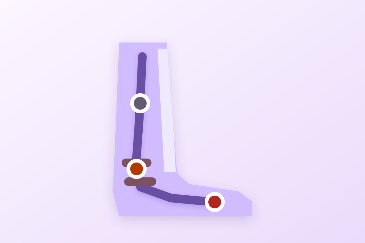
Anterior ankle pain, worse on downhill walking, points to tibialis anterior tendinopathy; no protocol has been tested, progressive loading remains the reference.
What follows covers each of these points in detail, with sources. It is there if you need it.
This topic is taught in a course: Heel spur, plantar heel pain and tibialis posterior dysfunction, online with Anthony Baillon.
1 / 12
11 article chapters · 40 min in total
Fundamentals◔ 4 min
The retinacula turn any external compression into tendon strain: footwear counts as a mechanical factor.
Two presentations◔ 3 min
This form is degenerative: aiming for a return to running makes no sense for most patients.
Diagnosis◔ 4 min
No clinical test has published sensitivity or specificity for this tendon.
Differentials◔ 4 min
Sorting is done first on the rhythm of the pain, before even its site.
Rupture◔ 4 min
Rupture typically occurs after 45, often without notable trauma.
Risk factors◔ 4 min
No risk factor for this tendinopathy has been measured prospectively.
Rehabilitation◔ 6 min
The only intervention specific to this tendon is correcting the external constraint, and also the least risky.
Footwear◔ 3 min
A relacing trial in the session, tested before and after, gives the patient an immediate answer.
Case reports◔ 3 min
The published cases are ruptures or an infection, never an overload tendinopathy treated in physiotherapy.
In practice◔ 2 min
The first examination looks for rupture, not the tendinopathy itself.
FAQ◔ 3 min
No study has measured the healing time: any precise duration given is an invention.
Train on this
Lower limb
Anthony Baillon
Next comes practice: the course that teaches this topic, with Anthony Baillon.
Our courses on this topic may be covered by DPC or FIFPL, the French continuing-education funding schemes, depending on your profession and situation.
Pain at the front of the ankle and over the instep, which wakes up on walking downhill and on running, in a runner, a hiker, or just after a change of footwear. The picture is easy to recognise. What is less easy is that the literature devoted to this tendon fits into a few dozen papers, without a single randomised controlled trial, and that most of the recommendations you read about it are borrowed from the Achilles tendon without saying so.
Tibialis anterior or tibialis posterior? They are not the same tendons, nor the same picture. The two names resemble each other to the point of being swapped in reports, and that is the first cause of confusion on this subject.
What to have in mind before opening the clinic door. The rest of the article sets out each of these points and gives the source of every figure.
A muscle that works twice per step, a tendon that crosses two fibrous tunnels, and a literature that fits in a folder. Those three facts explain almost everything that follows.
Tibialis anterior occupies the anterior compartment of the leg, with extensor digitorum longus, extensor hallucis longus and fibularis tertius. It arises from the lateral surface of the tibia and the interosseous membrane, descends in front of the ankle, then inserts on the medial aspect of the medial cuneiform and on the base of the first metatarsal. That insertion on the inner border of the foot gives it its dual action: it lifts the foot, and it carries it into supination.
It is the bulkiest of the foot dorsiflexors, described as the powerful dorsiflexor of the ankle, and no other really replaces it when it fails. Its function comes into play at two moments of the step, and it is the first that counts here:
The clinical consequence is direct: this tendon suffers from braking work, not from propulsion. Anything that increases the demand for braking increases its load, and going downhill is the situation par excellence.
The course of the tendon and its three zones of trouble
The clinical pictures differ according to the level involved
Course and insertion after classical descriptive anatomy. Distribution of the pictures after Beischer et al. 2009 (PMID 19912714) for the distal form, Levitsky et al. 2020 (DOI 10.1002/tsm2.152) for compression under the retinaculum, and Santos-Faria et al. 2022 (PMID 36582552) for involvement of the muscle belly.
Before reaching the foot, the tendon passes under two thickenings of the fascia: the superior extensor retinaculum, then the inferior retinaculum, in front of the talocrural joint. These structures hold the tendon pressed against the skeleton during dorsiflexion, which stops it bowstringing.
That arrangement has a price. In dorsiflexion, the tendon becomes prominent and finds itself caught between the bone behind and the retinaculum in front. Add external compression, tight lacing, a rigid upper, the edge of a skate, and you get a zone of mechanical conflict reproducible at every step. That is the logic of the picture the English-language literature calls lace bite, described in ice hockey players by Levitsky and co-authors in 2020.
This is the point that governs the reading of all the rest of this article. A PubMed search run on 15 August 2026 returns 53 references linking tibialis anterior to a tendinopathy, a tendinosis or a tendinitis. The same search applied to the Achilles tendon returns 2,480. Restricting to randomised controlled trials, tibialis anterior returns two, and checking them one by one shows that neither of the two covers this tendon : one studies the patellar tendon, the other the Achilles.
There is, to this day, no randomised controlled trial on the treatment of tibialis anterior tendinopathy. Every protocol proposed for this tendon is a transposition from another.
What the literature actually contains
Number of PubMed references, search of 15 August 2026
Measurement carried out by us on PubMed on 15 August 2026: this is a count of references, not published data. The two randomised trials returned by the tibialis anterior search are Holden et al. 2020 (PMID 31735531, patellar tendon) and Sobhani et al. 2015 (PMID 24636129, Achilles tendon): neither of them studies tibialis anterior tendinopathy.
That scarcity is not a methodological detail, it is a clinical fact. It means that the practitioner treating this tendon works by analogy, and that it is better to know it than to believe you are applying a validated protocol. It also explains why so many pages devoted to the subject copy out, word for word, the Alfredson protocols designed for the Achilles.
Most pages devoted to this subject describe a runner. The only published clinical series describes an overweight woman of 62 who hurts at night. Both exist, but confusing their management is the most costly error on this tendon.
In 2009, Beischer and co-authors published in Foot & Ankle International the first clinical description of distal tibialis anterior tendinosis without rupture. Twenty-nine patients, thirty-two feet. The characteristics of that group do not resemble those of a sporting population:
The commonest symptom is described there as a burning pain of the inner border of the midfoot, often worst at night. Swelling over the tendon was frequently observed. On MRI, the tendon was thickened in all the patients, and degenerative involvement of the first tarsometatarsal or the medial naviculocuneiform joint coexisted in eleven feet.
In other words: the best-described form of this tendinopathy is a distal degenerative tendinosis of the older person, readily associated with midfoot osteoarthritis, and not an overload condition of the runner. It is a fact of the literature, and it deserves to be known before modelling sports management on a patient for whom it is not appropriate.
The other picture, the one the presenting complaint most often suggests in a sports physiotherapy clinic, sits higher: in front of the ankle, under the retinacula. It rarely comes from tendon degeneration and far more often from an external compression constraint, or from an abrupt increase in the demand for braking.
Levitsky, Vosseller and Popkin published a review of it in 2020 centred on the ice hockey player, where the mechanism is particularly clear: a rigid skate upper, tight lacing, and the tendon finds itself compressed against the bone at every dorsiflexion. They describe pain, swelling and tenderness over the tendon, worsened by dorsiflexion, and a treatment that goes first through modifying the footwear, padding the area and adapting the activity.
The same mechanism is found, without a skate, in:
Two pictures not to be treated in the same way
What the literature documents for each
Left-hand column after Beischer et al. 2009 (PMID 19912714). Right-hand column after Levitsky et al. 2020 (DOI 10.1002/tsm2.152), a narrative review with no numerical series: the level of evidence of the two columns is not equivalent.
A word of honesty about this comparative table: the left-hand column rests on a series of twenty-nine patients with imaging and operative confirmation for some of them; the right-hand one on a narrative review with no sample size. That is not the same solidity. The distinction remains clinical and useful, but it has not been validated by a study that would have compared the two populations.
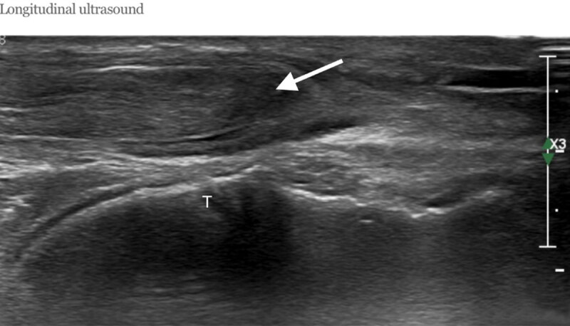
UltrasoundLongitudinal ultrasound of the tibialis anterior tendon: retracted, irregular, hypoechoic and swollen proximal part on a background of pre-existing tendinopathy, with focal discontinuity indicating rupture, arrow.
Source : Al-Musaedi et al., Journal of the Belgian Society of Radiology, 2023, figure 1 · CC BY
The diagnosis is clinical. It rests on a precise location, a tensioning manoeuvre, and a manoeuvre too rarely done: repeating the examination with the shoe on.
The approach comes in three stages, and the first already does most of the work.
1. Situate the pain along the course. The tendon is easily palpated, especially if you ask the patient to lift the foot against resistance: it becomes prominent from the lower quarter of the leg to the inner border of the foot. Follow the course with a finger and note the most painful point. Pain in front of the ankle points to the retinacular zone; pain at the inner border of the midfoot, to the insertion.
2. Put the tendon under passive tension. This is the test described in the American Family Physician review of 2022: the practitioner passively takes the foot into plantarflexion and eversion, which stretches tibialis anterior. Reproduction of the usual pain along the course of the tendon supports the diagnosis. That same review is a reminder that tendinopathies of the foot and ankle are “commonly overlooked” in primary care, tibialis anterior foremost.
3. Test under load and against resistance. Resisted dorsiflexion, then heel walking, then walking down a slope or a few steps. The aim is not to grade strength but to find the situation that hurts, in gradation.
If the suspected mechanism is compression by footwear, the barefoot examination may be unrevealing while the patient hurts on every outing. Three manoeuvres make up for that, and they take two minutes:
This last point has not been the subject of a validity study. It is a manoeuvre of mechanical common sense, consistent with the mechanism described by Levitsky and co-authors, and we present it as such: an aid to reasoning, not a validated test.
Imaging does not make the diagnosis of tendinopathy, which remains clinical. It serves three precise purposes:
Ultrasound has the advantage of being dynamic and available: it shows thickening, hypoechogenicity and hypervascularity of the tendon and its sheath, and allows comparison with the healthy side at the same time. One reservation concerns it, however, for this particular tendon: the variation in the cross-sectional area of the ankle tendons depends on age and on the level measured along the tendon, and there is no published cut-off value that would define a “pathologically thickened” tibialis anterior. Mansur and co-authors, measuring these cross-sections by MRI in sixty healthy adults, in fact showed that age-related differences were clear for the Achilles but not for tibialis anterior, and that there was no difference between the sexes.
Approach to pain at the front of the ankle or over the instep
Sorting is done first on the red flag, then on the rhythm of the pain
Reasoning tree built from the pictures described by Deu et al. 2022 (PMID 35559641), Aweid et al. 2012 (PMID 22627653) for compartment syndrome and Harkin et al. 2017 (PMID 29078830) for rupture. This tree has not been validated prospectively: it is an order of questions, not a diagnostic score.
Five diagnoses compete for this territory. Each has a sign that belongs to it, and it is on that sign that you should ask, not on the location alone.
The front of the ankle and the lower third of the leg receive pains of very different origins. Sorting is done better on the rhythm of the pain and on what reproduces it than on its site, because the territories overlap widely.
| Diagnosis | Site and rhythm | What distinguishes it | The manoeuvre that helps |
|---|---|---|---|
| Tibialis anterior tendinopathy | Course of the tendon, from the instep to the inner border of the midfoot. Warm-up pain, worse downhill. | Pain reproduced over the tendon, not over the bone or in the compartment. No strength deficit. | Passive tensioning: plantarflexion and eversion. |
| Chronic exertional compartment syndrome | Anterior compartment, at an almost constant exertion threshold. Eases within a few minutes of rest, returns at the same point. | A sensation of tightness, of the leg “swelling up”, sometimes paraesthesiae or foot drop at the end of a run. Examination at rest is normal. | Repeat the examination immediately after the triggering effort. |
| Medial tibial stress syndrome | The posteromedial border of the tibia, over several centimetres. Pain at the start of effort that fades then returns afterwards. | Pain diffuse on bony palpation, over a length, and not focal. | Palpation along the tibial border, compared with the healthy side. |
| Stress fracture of the tibia | Bony point that is focal, pain increasing from one outing to the next, sometimes at night and at rest. | Pain reproduced over a point less than three centimetres across, on single-leg hopping or on percussion. | Imaging without delay if the point is focal, especially on the anterior cortex. |
| Talocrural osteoarthritis | Anterior joint line, morning stiffness, pain on prolonged weight bearing. | Limited dorsiflexion, frequent history of trauma, joint and not tendon pain. | Comparative measurement of dorsiflexion, weight-bearing radiograph. |
| Anterior ankle impingement | Anterior joint line, pain at the end of dorsiflexion, a sensation of a block. | Pain reproduced by forced dorsiflexion with anterior pressure. Frequent after repeated sprains. | Look for any associated chronic ankle instability as well. |
| Tibialis posterior dysfunction | The inner border, below the medial malleolus, behind the tibialis anterior tendon. | Collapse of the arch, the “too many toes” sign, difficulty lifting the heel on one foot. | Single-leg heel raise test. |
This is the trickiest differential, because it shares the exact territory of tibialis anterior: the anterior compartment. The distinction lies in the rhythm. Tendinopathy hurts at the start of the effort, sometimes warms up, and wakes up on cooling down. Compartment syndrome, for its part, appears at a reproducible threshold, often to within a few hundred metres, forces the person to slow down, and eases within a few minutes of rest to return at exactly the same point when they start again.
Measurement of intracompartmental pressures, long presented as the reference test, does not decide the matter as well as is believed. The systematic review by Aweid and co-authors, covering thirty-two studies, concludes that the pressure criteria in common use rest on weak evidence, and that the emphasis should stay on the history. Those authors note that at one minute after exertion, and at that moment only, the values of patients and controls did not overlap: from 34 to 55.4 mmHg in patients against 9 to 19 mmHg in controls, which leads them to consider that a value exceeding 27.5 mmHg at that interval, together with a suggestive history, is strongly suggestive.
This diagnosis is dealt with in detail in its own article: chronic exertional compartment syndrome. We do not develop it further here.
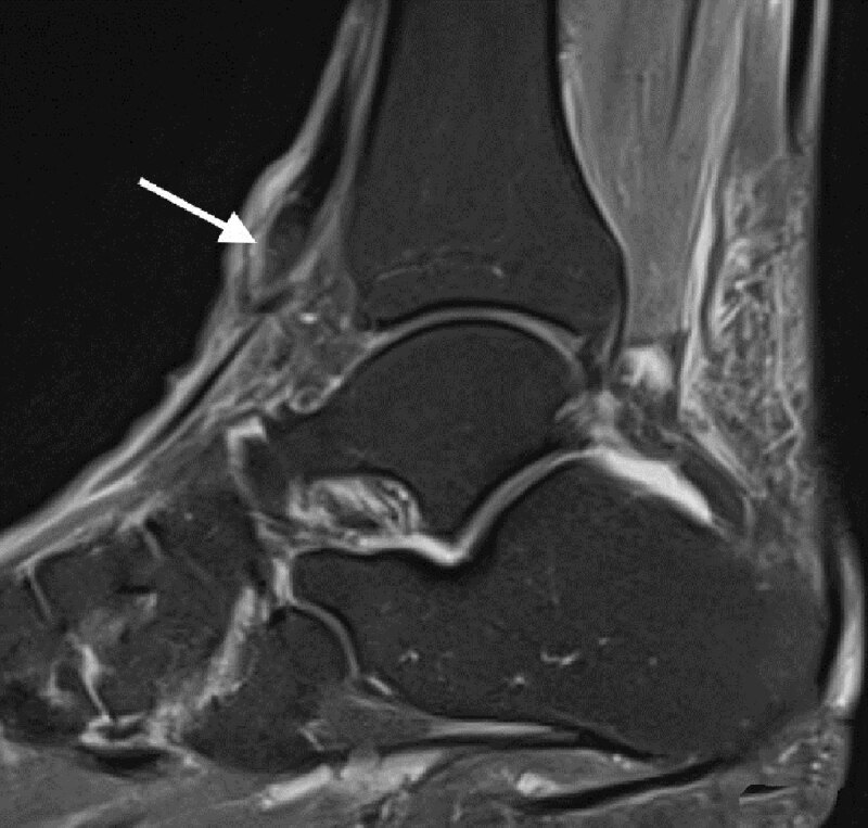
MRISagittal ankle MRI, fat-saturated T2 sequence: full-thickness rupture of the tibialis anterior tendon with slight retraction, arrow pointing to the tendon stump.
Source : Al-Musaedi et al., Journal of the Belgian Society of Radiology, 2023, figure 2 · CC BY
Because it hurts little, because it happens without frank trauma, and because the patient is still walking. It is the only chapter of this article where a diagnostic error really changes the prognosis.
Rupture of the tibialis anterior tendon is described as the third commonest tendon rupture of the lower limb, after that of the Achilles tendon and that of the patellar tendon. It remains rare in absolute terms: the literature documenting it is made of isolated cases and small series, which makes it impossible to give a reliable incidence.
The background is fairly constant from one publication to another: a person over 45 to 50, with rupture occurring after minimal trauma, or even with no identifiable trauma. In the series of seven patients by Gwynne-Jones and co-authors, published in 2009, ages ranged from 43 to 82, and above all four of the seven patients were on systemic corticosteroid therapy, a fifth having received a local injection. The authors conclude explicitly to an association with corticosteroid therapy, systemic as well as local.
Other backgrounds have been reported in documented cases: diabetes (Siang et al. 2019, in a 73-year-old patient whose rupture had been developing for six months with a tendon gap of 4.2 cm), and gout, with an intratendinous tophaceous deposit (Jerome et al. 2008). In the sportsperson, the lesion takes another form: Santos-Faria and co-authors reported in 2022 the rupture of the muscle belly of tibialis anterior, and not of the tendon, in a 47-year-old ultramarathon runner.
The trap lies in the fact that the rupture is not very painful. The patient does not describe the abrupt episode of the Achilles tendon; they sometimes come weeks later, for difficulty walking. The signs to look for are mechanical:
A patient over 50 whose foot slaps the ground and in whom the prominence of the tendon has disappeared on resisted dorsiflexion has a tibialis anterior rupture until proven otherwise, even if they have never had pain.
The Gwynne-Jones series is the only one comparing the two options on functional scores, and its sample is small: seven patients, five operated on and two treated without surgery. At follow-up, ranging from one to twelve years, the mean FAOS score was 85 in the operated group against 52 in the non-operated group, and one of the two non-operated patients had to be taken back to surgery four years after the rupture. The authors conclude in favour of surgical repair, including in the older person, and stress that an early diagnosis allows direct repair.
This result has to be read for what it is: a series of seven patients, not randomised, in which the operated and non-operated patients were not comparable at the outset. It does not demonstrate the superiority of surgery; it indicates a trend, in the same direction as the published cases as a whole.
For the physiotherapist, the practical consequence is simple and does not depend on that debate: faced with a suspected rupture, the question is not what rehabilitation to undertake, but who to refer to. The delay counts, because a tendon retracted for months can no longer be repaired directly and requires a graft or a tendon transfer, as in the cases reported by Siang and by Zmerly and their co-authors.
On this tendon, almost no risk factor has been measured. This chapter therefore distinguishes what is documented from what is reasoned, because the two are too often read in the same tone.
There is no prospective cohort study that has looked for risk factors for tibialis anterior tendinopathy. What we have is limited to the characteristics of the patients in the published series, which describes a population of attenders, not a risk:
These figures describe surgical series of a few dozen patients. They do not allow a relative risk to be calculated, and no odds ratio has been published for this tendon. That is why this article contains no chart of risk factors as odds ratios: there is nothing to put in it that would not be invented.
The rest is a matter of mechanism, not of measurement. It is useful clinically, provided it is not presented to the patient as established fact. Three families of constraint increase the load on tibialis anterior:
The third point deserves a clarification. The idea that too rapid a progression of load favours injury is solidly established for other structures, in particular bone: the 2024 scoping review on return to running after tibial stress fracture stresses that too rapid a progression amplifies the risk of recurrence. Nothing of the kind has been demonstrated for the tibialis anterior tendon. We apply the principle because it is physiologically coherent, knowing that it is borrowed.
The honest answer is that nobody has measured it. No study has followed a cohort of untreated patients. What can be said comes down to three observations, each with its limit:
What is known, what is supposed
Level of certainty of the common claims about this tendon
Classification established from the sources cited in this article, and from the PubMed search of 15 August 2026 reported above. The sample sizes given are those of the series by Beischer 2009 (PMID 19912714), Gwynne-Jones 2009 (PMID 19735632) and Grundy 2010 (PMID 20230699).
No randomised trial has tested a protocol on this tendon. This chapter therefore proposes a reasoned approach, indicating for each modality where the evidence comes from and what it is worth once transposed.
Rehabilitation of a tendinopathy rests today on progressive loading : exposing the tendon to an increasing, tolerated, regular constraint, rather than resting it. That principle is solidly supported, but it is supported elsewhere.
The trial by Breda and co-authors, published in the British Journal of Sports Medicine in 2021, compared in 76 patients with patellar tendinopathy a progressive loading programme with a classical eccentric programme: the gain on the VISA-P score at 24 weeks was 28 points against 18, that is an adjusted difference of 9 points (95 % confidence interval 1 to 16, p = 0.023). On the Achilles tendon, Beyer and co-authors had shown in 2015 that heavy slow resistance and eccentric work gave equivalent results at twelve weeks.
These results are good. They do not concern tibialis anterior. The large-scale synthesis commissioned by the British Health Technology Assessment programme, published in 2023 by Cooper and co-authors, in fact confirms that exercise is the first-line treatment of tendinopathies, while stressing the heterogeneity of the protocols and the uncertainty that persists about the optimal modality.
Transposing an Achilles protocol to tibialis anterior is defensible: both tendons undergo cyclical eccentric loading in walking. Presenting it to the patient as a treatment validated for their tendon is not.
Here is a usable framework, built on load-based reasoning applied to the other tendons and on the mechanical particularities of tibialis anterior. It is offered as a reasoned approach, not as a proven protocol.
Before any exercise, look for and remove the mechanical cause: lacing, shoe, gradient, recent volume. In the compression form, that step is sometimes enough. The American Family Physician review mentions an initial immobilisation followed by physiotherapy for this tendon; that is a described practice, not compared with an early return to loading, and it must stay brief so as not to decondition a muscle whose work is precisely to brake.
Isometrics have their place here, for their analgesic effect: Rio and co-authors showed, in patellar tendinopathy, that isometric work produced immediate analgesia. On tibialis anterior: isometric dorsiflexion against manual or elastic resistance, ankle in neutral, sets of 30 to 45 seconds, repeated several times a day, at an intensity that does not wake the pain.
The basic exercise is resisted dorsiflexion, with elastic or pulley, performed slowly. The logic of heavy slow resistance consists of favouring slowness and load over the number of repetitions: three to four sets of six to fifteen repetitions, three seconds up and three down, every other day.
The tolerance marker, borrowed from the Achilles and patellar tendinopathy protocols, is as follows: pain that is acceptable during the exercise, that comes down within twenty-four hours and does not worsen from one session to the next.
This is the step specific to this tendon, and the one most often forgotten when strengthening it as a dorsiflexor. Tibialis anterior works above all to hold back the forefoot. The work must therefore become eccentric and move closer to the step:
Progression is by intervals of walking and running, volume before intensity, reintroducing downhill gradient last, because it is the constraint most specific to this tendon. These principles come from the recommendations for return after tibial bone stress injury: the scoping review published in 2024 in Sports Medicine formulates them from fifty studies, but concludes that the resulting recommendations rest on level IV evidence. Caution is therefore doubly in order here, since the principle is both of low evidential level and borrowed from another structure.
No passive modality has been evaluated on this tendon. Two remarks, however, are important in practice:
| Modality | Where the evidence comes from | GRADE level for THIS tendon | What can be said about it to the patient |
|---|---|---|---|
| Correction of footwear and lacing | Narrative review on the ice hockey player (Levitsky 2020), direct mechanical coherence | Very low, but directly applicable to the described mechanism | First move, without risk, sometimes enough on its own. |
| Adaptation of load and gradient | General principle of load management; recommendations for return after tibial bone injury, level IV (2024) | Very low, transposed | Reasonable and safe, but the precise timetable is not validated. |
| Progressive loading of the tendon | Randomised trial on the patellar tendon (Breda 2021), HTA synthesis 2023 | Very low here, moderate on the tendon of origin | The reference approach to tendinopathies, never tested on this tendon. |
| Eccentric and heavy slow resistance | Randomised trial on the Achilles tendon (Beyer 2015) | Very low here | Two equivalent options on the Achilles; choose the one the patient will stick to. |
| Analgesic isometrics | Trial on patellar tendinopathy (Rio 2015) | Very low here | Useful for getting through a painful patch, effect mainly immediate. |
| Brief initial immobilisation | Practice described in a synthesis review (Deu 2022) | Very low, never compared | May relieve an acute phase; do not prolong it. |
| Shockwave therapy | 2023 meta-analysis on the Achilles, the patellar tendon and plantar fasciitis | No data for this tendon | Do not present it as a treatment for this tendon. |
| Corticosteroid injection | Series of ruptures associated with corticosteroid therapy (Gwynne-Jones 2009) | Risk signal, no demonstrated benefit | Particular caution: reported association with rupture. |
| Surgery after conservative failure | Series of 11 operated patients (Grundy 2010) | Very low (series without a comparator) | An option in case of prolonged failure, after several months of well-conducted treatment. |
The level of evidence, modality by modality
Stacked cards: on the left the original evidence, on the right what remains of it once transposed to this tendon
Rating applied by us following GRADE logic, downgrading the level for indirectness of the evidence: data obtained on another tendon cannot keep its original level. Sources: Breda 2021 (PMID 33219115), Beyer 2015 (PMID 26018970), Rio 2015 (PMID 25979840), Cooper 2023 (PMID 37929629), Charles 2023 (PMID 37662911), Levitsky 2020 (DOI 10.1002/tsm2.152) and Gwynne-Jones 2009 (PMID 19735632).
This is the most useful chapter of this article, and paradoxically the one that rests on the most trivial cause: a lace too tight in the wrong place.
The mechanism described in the ice hockey player holds for any closed shoe: in dorsiflexion, the tendon becomes prominent and finds itself caught between the bone and what covers it. Lacing tightened over the instep turns every step into compression. The correction is mechanical, free, and its effect is judged in a single outing:
Three elements deserve examination, and it is better to look at them than to assume them:
A word on foot orthoses: the 2022 synthesis review mentions lateral wedge orthoses in the management of fibularis tendinopathies, not of tibialis anterior. Nothing allows one to state that a foot orthosis modifies the constraint on this tendon, and we do not claim so.
Two levers are consistent with the function of the muscle, and both belong to mechanical reasoning more than to evidence:
Reintroducing load: in what order
Downhill gradient is reintroduced last, because it is the constraint specific to this tendon
Order built from the return-to-running principles described in the 2024 scoping review on tibial bone stress injury (PMID 39141251), whose authors state that their recommendations rest on level IV evidence, and adapted to the constraint specific to tibialis anterior. This is not a validated protocol.
Four real cases, all indexed, chosen because each corrects a false idea about this tendon. None is a vignette reconstructed for the demonstration.
A 47-year-old ultra-endurance runner presents with persistent discomfort at the front of the left leg. Four weeks earlier, during an ultramarathon, he had felt a snapping sensation, followed the next day by bruising and functional difficulty. Examination finds a palpable area of fibrosis at the front of the leg. Ultrasound shows a partially organised rupture of the distal third of the muscle belly of tibialis anterior, measured at 36 by 10 by 27 millimetres, with muscle oedema.
The patient was treated without surgery, with an individualised rehabilitation programme, and returned to ultra-endurance competition.
What the case corrects : the lesion in the endurance athlete readily affects the muscle belly and not the tendon, and its prognosis has nothing in common with that of tendon rupture in the older person. It is also a reminder that a four-week delay between the accident and the consultation is commonplace with this muscle, because the pain remains bearable.
Santos-Faria J, Malta JN, Coelho AP, Branco JP. Running on the Edge: Rupture of the Tibialis Anterior Muscle During an Ultramarathon. Cureus 2022;14(11):e31930. PMID 36582552
A 73-year-old woman, a known diabetic, presents with progressive pain and swelling of the right ankle developing over six months, with no trauma or awkward movement found. Examination notes anterior tenderness, an erythematous cystic soft tissue mass, weakness of dorsiflexion, loss of the normal prominence of the tendon in front of the ankle, and marked hyperextension of the first metatarsophalangeal joint on dorsiflexion. MRI concludes to a complete rupture with a tendon gap of 4.2 cm, requiring reconstruction with an allograft.
What the case corrects : rupture can set in with no acute episode and present as a swelling. Six months of evolution turn a direct repair into a graft reconstruction. Hyperextension of the big toe on dorsiflexion is a compensation sign worth knowing.
Siang DKT, Pitarini A, Koo K. Allograft Reconstruction of Spontaneous Tibialis Anterior Tendon Rupture in a Diabetic Patient. J Orthop Case Rep 2019;9(3):57-60. PMID 31559229
A case of spontaneous rupture of the tibialis anterior tendon occurring on an intratendinous gouty tophaceous deposit. The authors are a reminder that the lesion affects the middle-aged or older person, often after a movement of plantarflexion and eversion of the foot, and that a careful history and examination lead to the diagnosis. After repair with non-absorbable suture, cast immobilisation and resumption of assisted active exercises at three weeks, the patient had recovered full strength and range at six months.
What the case corrects : faced with an apparently spontaneous rupture, general illness is not a detail of the history. Gout, diabetes, rheumatoid arthritis and corticosteroid therapy change how the file reads.
Jerome JT, Varghese M, Sankaran B, Thomas S, Thirumagal SK. Tibialis anterior tendon rupture in gout: case report and literature review. Foot Ankle Surg 2008;14(3):166-9. PMID 19083637
The authors report a case of isolated pyogenic tenosynovitis of tibialis anterior, published in BMJ Case Reports. The location is unusual, the presentation misleading for anyone who thinks first of an overload condition.
What the case corrects : anterior ankle pain of rapid onset, with redness, heat or fever, is not a mechanical tendinopathy. It is rare, but it is the kind of picture a practitioner sees once in their career and must not treat with progressive loading.
Greenhalgh MS, Iyengar KP, Sangani C, Toh EM. Isolated pyogenic tenosynovitis of tibialis anterior. BMJ Case Rep 2020;13(6):e236368. PMID 32532897
A first session that comes down to four manoeuvres, and three sentences to say to the patient that avoid selling them a certainty the literature does not have.
1. Situate, then tension. Follow the course of the tendon with a finger, from the lower quarter of the leg to the inner border of the midfoot, and note the painful point. Then passive plantarflexion and eversion. Pain reproduced along the course points; pain over the bone or in the compartment points elsewhere.
2. Rule out the flag before everything else. Look at the prominence of the tendon on resisted dorsiflexion, compared with the healthy side. Watch the gait, especially the landing of the foot at heel strike. A foot that slaps, a steppage gait, an absent prominence: you refer, you do not rehabilitate.
3. Ask about the footwear, and test it. Ask for the shoe of the activity that triggers it. Look for the mark of the lacing on the instep. Relace skipping the painful eyelet, repeat the test. It is diagnostic and therapeutic in the same movement.
4. Set the starting load. Isometric dorsiflexion if the pain is sharp, slow resisted dorsiflexion otherwise, with the tolerance marker: pain acceptable during, come down the next day, not increasing from one session to the next.
Three formulations, which respect the real state of knowledge without discouraging:
They are two different tendons, in two different compartments. Tibialis anterior lifts the foot: its pain sits in front of the ankle and on the inner border of the midfoot, and its failure gives a foot that slaps the ground. Tibialis posterior supports the medial arch: its pain sits on the inner side, below the medial malleolus, and its failure gives progressive collapse of the foot with difficulty rising onto the toes. The second is dealt with in its own article.
No study has measured the time to recovery of this tendinopathy, and any duration announced with precision is an invention. Two indirect markers exist: in the form of compression by footwear, the authors of the review on the ice hockey player describe episodes often easing within one to two weeks after the cause is corrected. In the distal degenerative form, the patients who reached surgery had on average a year of symptoms, which describes the failures, not the usual course.
Not necessarily. The logic applied to the other tendinopathies is to reduce the constraint without removing the load: reduce the volume, take out the downhill gradient, correct the footwear, and keep whatever remains pain-free. Complete rest deconditions a muscle whose function is precisely to brake at every step. This approach has not been tested on this tendon.
It is the best-described mechanism for the anterior form. In dorsiflexion, the tendon becomes prominent and finds itself caught between the bone and what covers it; lacing tightened over the instep adds compression at every step. Levitsky and co-authors described it in the ice hockey player, where the rigid skate upper worsens the phenomenon. The test is simple: relace skipping the painful eyelet and compare.
Not to establish a tendinopathy, whose diagnosis is clinical. It becomes necessary to confirm a suspected rupture, to rule out a stress fracture when there is focal bony pain, or to look for associated midfoot involvement. There is no thickness cut-off value that would define a pathological tibialis anterior on ultrasound.
No benefit has been demonstrated on this tendon, and a risk signal exists: in the Gwynne-Jones series of seven ruptures, four patients were on systemic corticosteroid therapy and one had received a local injection, which led the authors to retain an association between corticosteroids and rupture. Caution is therefore in order, and the decision rests with the doctor.
By the rhythm. Compartment syndrome appears at a reproducible exertion threshold, forces the person to slow down, eases within a few minutes of rest and returns at exactly the same point. Tendinopathy hurts at the start, sometimes warms up, and wakes up on cooling down or the next day. The subject has its own dedicated article: chronic exertional compartment syndrome.
It is the sign that should prompt consultation without delay, especially after the age of 50. It reflects a loss of forefoot braking, which may come from a tendon rupture or from a neurological cause. Tibialis anterior rupture is often not very painful, which explains the late diagnoses, and the delay determines the repair technique.
Every reference has been verified by its PubMed identifier or its DOI, including its author list. The sources marked “another tendon” are those this article uses by transposition, and which do not concern tibialis anterior.
PubMed counts cited in the article: searches run on 15 August 2026 on the title and abstract fields. These are measurements carried out for this article, and not published data.
Behind this article
An author who explains, a reviewer who checks.
How we write and check our content

Physiotherapist · co-founder of Physio Learning
Marked for life by his first four-hour lecture without a single image, he took a master’s in instructional design so that it would never happen to anyone again. He hunts down publication bias and unreadable slides with the same intransigence.

Head of scientific content
Physiotherapist specialising in neuro-musculoskeletal practice and holder of a master’s in public health. He checks the methodological rigour of every article: primary sources, levels of evidence, no exceptions.