


















In brief
Leg pain that appears at an exertion threshold which is almost always the same, felt as tightness or burning, forces the patient to stop, eases within a few minutes of rest, and returns at exactly the same point when running resumes. Sometimes pins and needles, sometimes a foot that slaps at the end of a run. Between episodes the examination finds nothing, and that is precisely what misleads: a patient examined at rest looks perfectly well. The classic error is to treat medial tibial stress syndrome or a stress fracture for months. Intracompartmental pressure measurement remains the reference standard, but its historical thresholds overlap the values of healthy subjects, and the debate on their validity has been open since 2012. On the treatment side, the best-documented conservative option is modifying running technique, and fasciotomy, often presented as the solution, leaves more than four runners in ten without a return to running.
Clinical synthesis based on the historical criteria of Pedowitz (AJSM 1990), the two 2012 systematic reviews that challenged their validity (Roberts, Aweid), the diagnostic series of Roscoe (AJSM 2015), the largest series of measurements published to date (Lindorsson, 864 patients) and the review article in the British Medical Bulletin 2026: 55 references checked one by one on PubMed and cross-checked on CrossRef.
Clinical synthesis
The diagnosis is made from the history, not from the examination. This is the most important sentence in this article, and it is also the hardest to apply, because it contradicts every clinician's reflex: to look for a sign. Here, the sign does not exist at rest. What does exist is an account, and that account has such a constant shape that it makes up, on its own, most of the evidence. The review in the British Medical Bulletin 2026 sets it out as one of the field's rare points of agreement: “the history remains central”, supported by a symptom provocation test (Vogels and Zimmermann, PMID 42478572).
The exertion threshold is reproducible, and it is the best clue. The patient does not say “it hurts when I run”. They say “it hurts after ten minutes”, or “at the third kilometre”, and if asked whether it is always at the same moment, they answer yes without hesitating. This regularity is the most discriminating feature of the picture. In a direct comparison between patients with compartment syndrome and patients over 50 with arterial disease, the median time to symptom onset was 15 minutes for compartment syndrome versus 4 minutes for arterial disease (de Bruijn, J Vasc Surg 2021, PMID 33278541).
The two competing diagnoses are medial tibial stress syndrome and stress fracture, and they compete in the strong sense: they are what gets diagnosed instead, often for months. They differ on three points, which the differential chapter sets out: the location of the pain (bone versus muscle), its time course within the session (present from the outset versus triggered at a threshold), and what is found on palpation (painful over several centimetres of tibial border versus silent). The site publishes a dedicated synthesis for each: medial tibial stress syndrome (MTSS) and stress fracture in the athlete.
Pressure measurement is the reference standard, and that standard is contested. The three Pedowitz thresholds (1990) are still the ones most series use. Two systematic reviews published in the same year showed that, apart from the relaxation pressure, these thresholds overlap the range of healthy subjects, and that several studies report, in asymptomatic people, mean values that would trigger a positive diagnosis (Roberts and Franklyn-Miller, PMID 22092446 ; Aweid, PMID 22627653). Fourteen years later, the 2026 review still concludes that “current thresholds lack robust validation”.
The pathophysiology is not established. The usual explanation, a pressure that strangles muscle perfusion, is presented in textbooks as a fact. The 2026 review places it among the open questions: the syndrome “has traditionally been seen as a perfusion disorder, which remains unproven”. And the fascia, the target of the operation, does not look guilty: neither its thickness nor its stiffness differs between patients and healthy subjects on tibialis anterior biopsies (Dahl, CORR 2011, PMID 21948310). So a structure is being operated on that has not been shown to be abnormal, with results that are, for their part, real.
There is a serious conservative option, and it is mechanical. Switching from a rearfoot strike to a forefoot strike dropped post-run pressure from 78.4 to 38.4 mmHg in ten patients who already had a surgical indication, while more than tripling the distance run without pain. None of them was operated on (Diebal, AJSM 2012, PMID 22427621). The sample is small, the design is a case series, and the result has since been replicated in 19 and then in 75 patients.
Fasciotomy works, but less well than is often claimed. Across 1,596 operated patients, success is achieved in about two thirds and satisfaction in 84%, with 13% complications and 6% revisions (Campano, Arthroscopy 2016, PMID 27020462). Among runners followed for a mean of 66 months, 84% returned to a sport, but only 56% to running (Salzler, Sports Health 2020, PMID 32163722). It is this difference between “returning to a sport” and “returning to their sport” that has to be spelled out before surgery.
- The diagnosis is history-based. A normal examination between episodes does not refute it, it almost confirms it.
- The question to ask is not “where does it hurt?” but “at exactly what moment?” and “what do you have to do to make it stop?”.
- Pressure thresholds are not a test of truth. A value below the threshold in a patient with a typical account does not close the case; a value above it in a patient with an atypical account does not open it.
- Before surgery is raised, running technique must have been modified and assessed. It is the only conservative modality with several converging series behind it.
What are the fundamentals to know about chronic exertional compartment syndrome?
A muscle compartment is a volume closed off by bone and fascia. During exercise, the working muscle swells. If the container does not yield, pressure rises. That is the theory, it fits in three sentences, and it is probably incomplete: this chapter sets out the anatomy and the population figures, then says plainly what is not known.
Four compartments, only one usually implicated
The leg is divided into four osteofascial compartments, bounded by the tibia, the fibula, the interosseous membrane and intermuscular septa. Each contains its own muscles and its own nerve, and it is that nerve which explains the sensory signs when they are present.
- Anterior compartment : tibialis anterior, extensor digitorum longus, extensor hallucis longus, fibularis tertius. Deep fibular nerve. It is the stiffest compartment, wedged between the tibia, the fibula and the interosseous membrane.
- Lateral compartment : fibularis longus and brevis. Superficial fibular nerve.
- Superficial posterior compartment : gastrocnemius, soleus, plantaris. Sural nerve (sensory).
- Deep posterior compartment : tibialis posterior, flexor digitorum longus, flexor hallucis longus, popliteus. Tibial nerve.
Involvement is not evenly distributed, and the distribution is known with unusual precision thanks to Campano's systematic review, which pooled 24 surgical series representing 1,596 patients.
Which compartment is involved, and in whom
Distribution of decompressed compartments across 24 pooled surgical series, 1,596 patients
Source: Campano D, Robaina JA, Kusnezov N, Dunn JC, Waterman BR. Surgical Management for Chronic Exertional Compartment Syndrome of the Leg: A Systematic Review of the Literature. Arthroscopy 2016;32(7):1478-86 (PMID 27020462). Note what the denominator is: these are patients who have had surgery, so the distribution also reflects surgical indications and not only the disease itself.
Two practical lessons follow. First, if only one compartment were to be explored, it would be the anterior one: it accounts for half of all involvement, and it is also the one whose clinical picture is the most characteristic, with the slapping foot and the loss of dorsiflexion at the end of a run. Second, the deep posterior compartment is the hardest: it is deep, measuring it is technically delicate, and it is the one whose presentation most resembles medial tibial stress syndrome, since it hurts on the same side of the tibia.
- Involvement of the lateral compartment alone exists and is not anecdotal: it has been the subject of dedicated series, and its presentation, lateral leg pain sometimes with sensory discomfort over the dorsum of the foot, is readily confused with superficial fibular nerve entrapment.
- The fact that the anterior compartment dominates in surgery does not mean it dominates in clinic. In de Bruijn's cohort of 698 patients, involvement of the deep flexors affected 53% of those under 50 versus 26% of older patients (PMID 29531960): in the young runner, the deep posterior compartment is far from marginal.
Who is affected, and how often
The incidence in the general population is poorly known. The review article in Current Sports Medicine Reports puts forward an estimate of roughly 1 case per 2,000 people per year (Velasco and Leggit, 2020, PMID 32925373). This figure circulates widely and deserves to be handled with caution: it is an estimate quoted in a review article, not the result of a population survey.
The solid figures come from closed populations, where the denominator is known. Among 294 elite cross-country skiers on the French national teams followed from 1994 to 2014, eighteen confirmed cases were identified, giving a prevalence of 6.1 % and an incidence of 13 per 1,000 skier-years, higher in biathletes, with skate skiing and roller skiing as the main triggering conditions (Calvelli, Int J Sports Med 2020, PMID 31935775). The military is the other major source: in Campano's review, 54% of operated patients were service members and 29% were athletes, 83% of them recreational.
These four figures deserve to be read together, because they overturn three received ideas at once.
This is not a man's disease. The systematic review devoted to sex differences pooled 24 studies providing a breakdown: 51% of diagnosed patients were women (Rothman, Phys Sportsmed 2024, PMID 36698053). The contrast with the 70% of men in Campano's surgical series is not a contradiction, it is a recruitment effect: operative series are dominated by military cohorts, which are themselves mostly male. In outpatient practice, the split is even. The same review also finds an unfavourable signal in women: male athletes returned to their sport after surgery more often than female athletes.
This is not an adolescent's disease only. Of 698 patients at a Dutch referral centre, 98 were 50 or older, that is one in seven. These older patients were less sporty (62% did nothing or only walked), had more comorbidities, and above all presented far more often with unilateral symptoms (45% versus 22%). In them, the anterior compartment clearly dominated (82% versus 59%) (de Bruijn, OJSM 2018, PMID 29531960).
It is most often bilateral. Among 155 patients aged 18 or under operated on at a paediatric centre, 131 (85%) had bilateral symptoms requiring release on both sides, 88% were girls, and the main sport was running (25%), football (23%) or field hockey (12%) (Beck, AJSM 2016, PMID 27365374). Strictly unilateral pain in an adolescent should therefore prompt closer attention to other causes.
What we think we know about the pathophysiology, and what we do know
The standard account is a loop: exercise makes the muscle swell, the inextensible compartment does not adapt, pressure rises, capillary perfusion falls, the muscle becomes ischaemic, pain appears, and stopping the effort reverses the process. It is coherent, it is taught everywhere, and at least two findings suggest it should not be taken for granted.
The fascia is not abnormal. Dahl took fascial biopsies of tibialis anterior from six healthy subjects, eleven affected patients and ten affected patients who also had diabetes, then measured stiffness on a tensile testing machine and thickness under the microscope. Neither weight-normalised stiffness nor thickness differed between the groups. The authors conclude that “structural and mechanical properties are unlikely to explain the syndrome” and that the cause must be sought elsewhere (Clin Orthop Relat Res 2011, PMID 21948310). That is an awkward finding for a treatment that consists precisely of opening this fascia.
Ischaemia has not been demonstrated. The 2026 review article explicitly places aetiology among the open questions, in terms that leave no room for interpretation: future research “should address the central question of the aetiology of the symptoms”, the syndrome having “traditionally been seen as a perfusion-related disorder, which remains unproven” (Vogels and Zimmermann, PMID 42478572).
An alternative hypothesis has been explored on a large scale: functional venous obstruction. In 284 patients with exertional leg pain investigated by provocative CT angiography, functional venous compression reproducing the symptoms was found in 91.5% of them, and targeted treatment with botulinum toxin reduced or abolished the symptoms in 79.9% at four weeks (McGinley, Clin J Sport Med 2022, PMID 34009799). The design is a single-centre retrospective series without a control group, the authors' conclusion (“the syndrome results from venous obstruction”) goes beyond what that design allows, and the result has not been independently replicated. It deserves to be known, not to be taught as the mechanism.
One clinical observation points to mechanics rather than to simple ischaemia: patients change the way they walk before they feel any pain. Roscoe measured plantar pressures in 20 affected service members and 20 controls, walking and marching in step, before symptoms appeared. Patients brought the foot flat sooner after heel strike, with an area under the ROC curve of 0.75 to 0.77, which the authors call “fair predictive validity”. Their interpretation is a mechanical disadvantage of tibialis anterior (Gait Posture 2018, PMID 29702369). This result matters for what follows: if running mechanics come first, changing them can be a treatment, and not merely an accommodation.
- Four compartments, one of which dominates operative series by a wide margin: the anterior one, half the cases.
- Populations at risk: runners, service members, and endurance sports that get talked about less, such as skate-style cross-country skiing.
- The male-to-female split is even when you look at diagnosed patients and not at operated patients.
- Neither the fascia nor ischaemia has been shown to be the cause. Saying so to the patient is more honest than serving up the textbook diagram.
How do you recognise the picture, once you know it?
This is the chapter that makes this article useful. Chronic exertional compartment syndrome is extremely recognisable, provided you have the exact shape of the account in mind. What follows is that shape, broken down into its elements, each with what is known about it.
Five cardinal symptoms, and an order of importance
The literature agrees on five symptoms: pain, a sense of tightness, cramping, weakness and sensory disturbance, appearing on exertion and disappearing on stopping (Ding, J Vasc Surg 2020, PMID 32473344 ; Dunn and Waterman, Clin Sports Med 2014, PMID 25280617). They do not carry the same weight. In de Bruijn's cohort of 698 patients, pain was present in 94 to 96% of patients depending on the age band, and a sense of tightness in 57 to 62% (PMID 29531960). Pain and tightness are therefore the core; the other three are additional arguments when they are there, and their absence rules nothing out.
The word patients use spontaneously deserves attention. “It squeezes”, “it swells”, “it feels as if the skin is going to split”, “it burns” come up more often than “it hurts”. This quality of tightness, distinct from a pinpoint pain, is one of the features that points away from bone straight away.
The reproducible exertion threshold: the most discriminating feature
The patient is able to say at what moment it happens, and that moment does not move. Ten minutes, three kilometres, the second climb of the route. This regularity is no mere comfort detail: it is what distinguishes compartment pain from bone pain, which sets in more gradually and depends above all on the cumulative load of the preceding weeks.
The only direct measurement of this delay comes from an unexpected comparison. Seeking to help vascular surgeons avoid confusing compartment syndrome with arterial disease in patients over 50, de Bruijn compared 43 patients with compartment syndrome and 41 newly diagnosed patients with peripheral arterial disease. The time to symptom onset during exercise was 15 minutes in the compartment group versus 4 minutes in the arterial group (p < 0.01) (J Vasc Surg 2021, PMID 33278541).
The shape of the episode: three time courses that look nothing alike
Schematic diagram, anchored on the delays measured by de Bruijn 2021 (15 min versus 4 min)
The delays plotted on the axis come from de Bruijn JA, Wijns KCA, van Kuijk SMJ, Hoogeveen AR, Teijink JAW, Scheltinga MRM. Chronic exertional compartment syndrome in the differential diagnosis of peripheral artery disease in older patients with exercise-induced lower limb pain. J Vasc Surg 2021;73(6):2114-21 (PMID 33278541). The shape of the curves is a teaching representation of clinical descriptions, it does not come from a continuous measurement of pain.
What eases with rest, and what does not always ease
The third element of the account is recovery: the patient stops, walks for a few minutes, and it passes. Then they set off again, and it comes back at the same place. This rapid reversibility is what gives the picture its name and separates it from an established tissue lesion.
One should nevertheless resist turning this into an absolute criterion, because the only study that measured it head-on says something else. In de Bruijn's series, stopping the effort relieved all 41 patients with arterial disease completely, without exception, but 73% of patients with compartment syndrome kept some residual discomfort. This result inverts the usual use of the criterion: it is not the disappearance that marks out compartment syndrome, it is the persistence of discomfort after stopping that distinguished it best from arterial disease, with a specificity of 100% and a positive predictive value of 100% in this comparison. Pain persisting beyond 4.5 minutes after stopping had the best sensitivity (95%), for a specificity of 54%.
Two cautions before generalising. First, the population is unusual: patients aged 50 and over on both sides, compared with arterial patients whose median age was 72. Second, the comparison does not set compartment syndrome against medial tibial stress syndrome or stress fracture, but against arterial disease. What the study establishes solidly is that relief which is immediate and complete on stopping points more towards vascular claudication than towards compartment syndrome. What it does not establish is a threshold applicable to the young runner.
- The textbook formula, “the pain eases with rest within a few minutes”, is true as a tendency : the pain drops markedly on stopping. It is false as a binary criterion: nearly three patients in four keep something.
- A useful question in clinic: “when you stop, does it go away completely, or is some discomfort left?” Residual discomfort is not an argument against the diagnosis.
Paraesthesia and the foot that gives way
When the affected compartment contains a motor nerve, the story may include a transient neurological component. The most characteristic one involves the anterior compartment and the deep fibular nerve: numbness of the first intermetatarsal space, and above all a foot that slaps at the end of a run, because tibialis anterior no longer controls the lowering of the forefoot. Some patients describe tripping on the edge of a kerb in the last minutes of the outing, never at the start.
The frequency of paraesthesia has been measured in a surgical cohort of 78 legs: 33 patients out of 78, that is 42.3%, reported it (Shankar, Sports Health 2024, PMID 36951383). So it is neither rare nor constant. And this symptom carries a prognostic value that matters for what follows: in that same series, the presence of paraesthesia predicted poorer results after fasciotomy, with more severe rest pain and pain during daily activity, a smaller gain in activity and a lower probability of return to sport. The least favourable subgroup was the one with paraesthesia in the tibial nerve territory.
In other words, a patient who describes pins and needles gives both a diagnostic argument and a prognostic signal. That deserves to be recorded in the notes before, and not after, the surgical discussion.
The one examination sign that has proved itself: muscle herniation
Pedowitz compared, across 131 records, patients whose diagnosis was confirmed by pressure measurement with those in whom it was refuted. The conclusion is striking and has never been contradicted since: the only significant difference found on history and physical examination was the presence of muscle herniations, in 45.9% of affected patients versus 12.9% of the others (AJSM 1990, PMID 2301689).
It is a small subcutaneous bulge, often on the anterolateral aspect of the middle or distal third of the leg, at the point where a perforating branch leaves the fascia, best seen on contraction or after exercise. It is painless in most cases. Looking for it means examining the leg standing, with the dorsiflexors contracting, and not just palpating the patient lying down.
Diagnostic delay, and why it is the rule
In Beck's paediatric series, symptoms had been going on for more than a year in 63% of the 155 patients at the time of surgery (PMID 27365374). This delay is not an accident: it is produced by the very structure of the picture. The patient comes in for a pain they do not have at the time of the consultation, to a practitioner who examines them at rest and finds nothing, with normal plain imaging. Everyone logically concludes that it is something benign, and the most available conclusion is medial tibial stress syndrome.
The practical consequence is simple: it is the history that must be systematic, not the examination. Three questions are enough to point the way:
- “At what point in the session, exactly?” A precise and stable answer points to the compartment.
- “What do you have to do to make it stop?” Having to stop, and that being enough within a few minutes, points to the compartment. Having to wait several days points to bone.
- “Does it come back in the same place if you set off again?” A return at the same threshold, within the same session, is highly suggestive.
Red flags in exertional leg pain
- Pain out of proportion, not eased by stopping, with a tense compartment and a progressive sensorimotor deficit : think of acute-on-chronic compartment syndrome. It is a surgical emergency. The acute-on-chronic form is rare but described, including bilateral and without trauma, and the published case report stresses that “pain out of proportion to the examination is the most sensitive sign” (Schwartz, Bull Hosp Jt Dis 2017, PMID 28583063).
- Night pain, rest pain, non-mechanical, waking the patient, or exquisite fixed pain over a bony point with inability to hop on one leg: think of a high-risk stress fracture before anything else.
- A unilateral calf that is tense, warm and swollen, with no triggering exercise, especially after immobilisation, a long journey or surgery: deep vein thrombosis, urgent exclusion.
- Claudication at a short, fixed distance in a patient over 50 with vascular risk factors, disappearing completely and immediately on stopping: peripheral arterial disease, measure the ankle-brachial index.
- Symptoms triggered by resisted plantarflexion or forced dorsiflexion, loss of a distal pulse in the provoked position in a young athlete: popliteal artery entrapment syndrome, dynamic vascular imaging.
- A background of low energy availability, amenorrhoea, previous stress fracture : the leg pain is no longer a local problem, it is the symptom of a general one.
- Pain and a sense of tightness are the core of the picture; cramping, weakness and paraesthesia are supporting arguments.
- The reproducible threshold is the most useful element in the whole history.
- The muscle hernia is the only physical sign whose discriminating value has been measured. You look for it standing, on contraction.
- The paraesthesias are present in about 4 patients in 10 and herald a poorer surgical result: it is not only a symptom, it is a prognostic factor.
Why is the examination at rest normal, and how do you make the leg speak?
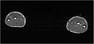
MRIAxial MRI of both forearms before exercise, baseline image: muscle compartments of normal appearance, without hyperintensity, in a pitcher with chronic exertional compartment syndrome.
Source : Cole et al., Sports Medicine - Open, 2017, figure 1 · CC BY
This is the central trap of this condition: the patient comes to see you in exactly the state in which they have nothing. A normal examination is not an argument against the diagnosis, it is an expected consequence of the mechanism. The answer comes down to one principle: do not examine the leg at rest, but after putting it in difficulty.
A normal resting examination is the rule, not the exception
Between two episodes there is nothing to find, and there should be nothing to find. Range of motion is free, manually tested strength is normal, palpation of the muscle belly is painless, neurodynamic tension tests are negative. The only thing that can be picked up when cold is the muscle hernia described above, present in fewer than one patient in two.
This normality has a documented perverse effect: it steers towards a diagnosis that does give something to palpate. Medial tibial stress syndrome offers reproducible pain on pressure over the posteromedial border of the tibia, over several centimetres. The clinician who palpates finds something, and what they find becomes the diagnosis. Yet nothing forces the two to exclude each other: some of the patients managed in a military centre for compartment syndrome also had associated medial tibial stress syndrome, and their conservative treatment gave the same results as that of patients without it (Zimmermann, BMJ Open Sport Exerc Med 2019, PMID 31191976).
The symptom provocation test
The 2026 review article places the provocation test among the few consensus elements: the history “remains central, supported by a positive symptom provocation test” (Vogels and Zimmermann, PMID 42478572). The principle is to reproduce, in the clinic, the effort that triggers the symptoms, then to examine immediately.
In practice, this is done without any special equipment:
- Reproduce the triggering effort, not a generic one. If the patient hurts while running, then they have to run. A treadmill, an outdoor loop, or failing that a sequence of heel raises and resisted dorsiflexion until the symptom is reproduced.
- Go all the way to the threshold. A test stopped before symptoms appear proves nothing. This is the commonest error: the test is interrupted at the first discomfort, whereas the picture reveals itself at the moment the patient says “there, that's it”.
- Examine immediately afterwards. Within the minute, not after undressing and taking the blood pressure.
What to look for in that short window:
- A compartment that feels tense on comparative palpation, sometimes with a clear difference between the two legs or between two compartments of the same leg.
- A muscle hernia that becomes visible when it was not visible when cold.
- A weakness of dorsiflexion, tested manually and functionally (heel walking). Its presence, transient, points strongly to the anterior compartment.
- Reduced sensation over the dorsum of the foot or the first web space, compared with the opposite side.
- Reproduction of the patient's own symptom, which is the most important point: they must recognise their pain, not describe a new one.
- None of these elements has a published, quantified diagnostic value. They are classical clinical observations, not validated tests, and that has to be known when using them.
- Their real value lies elsewhere: they document what happens on exertion, and that documentation makes the difference between “the patient says it hurts” and “I saw the leg tighten and the dorsiflexion weaken”. It changes the conversation with the referring doctor.
What the clinical approach cannot do
It is worth being clear about the limits. No clinical test for chronic exertional compartment syndrome has a published sensitivity or specificity. The systematic review devoted to non-invasive diagnostic tools reviewed 25 studies covering 1,257 participants and retained, among the modalities assessed, no tool validated as a substitute for pressure measurement, including a clinical prediction model, present in a single study (van der Kraats, OJSM 2023, PMID 36655016).
The physiotherapist's role is therefore not to make a definitive diagnosis, but to do three things that nobody else will do as well:
- Recognise the pattern in an account that would otherwise be filed as medial tibial stress syndrome.
- Actively rule out the competitors, in particular stress fracture, which allows no delay.
- Start conservative treatment, whose best-documented modality falls squarely within their competence, and which does not require a pressure to have been measured before it can begin. This point is established: in 108 service members, the anterior compartment pressure value was not associated with the outcome of the conservative programme, and the authors explicitly conclude that measurement “can safely be deferred” (Vogels, Arch Rehabil Res Clin Transl 2022, PMID 35282146).
- A normal resting examination is expected. Do not take it as an argument against the diagnosis.
- The provocation test consists of reproducing the real effort up to the threshold, then examining within the minute.
- What to look for: a tense compartment, a hernia that appears, weak dorsiflexion, reduced sensation over the dorsum of the foot, and above all the patient recognising the symptom.
- None of these manoeuvres has a published diagnostic value. They document, they do not prove.
Medial tibial stress syndrome, stress fracture or compartment syndrome: how do you decide?
The two diagnoses made instead are medial tibial stress syndrome and stress fracture. That is no accident: these are the three commonest causes of leg pain in the runner, they affect the same population, and two of them give something to palpate. This chapter separates them on criteria usable in the clinic, then widens out to the rest of the differential diagnosis.
The runner's trio, line by line
Three questions are enough to separate the three entities in the great majority of cases: where, when within the session, and what palpation gives.
Where. Medial tibial stress syndrome hurts over the bone, precisely over the posteromedial border of the tibia, in its distal or middle third. A stress fracture hurts over the bone as well, but at a point, not along a line. Compartment syndrome hurts inside the muscle, at the front or on the outer side in half of cases, in a location the patient points out with the flat of the hand and not with a fingertip.
When. Medial tibial stress syndrome is often present from the warm-up, may ease during the run, and comes back at the end or afterwards. A stress fracture worsens as the session goes on and above all as the weeks go by, with a tolerated load that falls from outing to outing. Compartment syndrome is silent up to a threshold, then becomes intolerable within a few minutes, and that threshold does not deteriorate session after session.
Palpation. This is the most workable criterion. Medial tibial stress syndrome gives reproducible pain on pressure over several centimetres of tibial border. A stress fracture gives pain that is exquisite and focal, over two or three centimetres at most, often with pain on hopping on one leg. Compartment syndrome gives nothing at all at rest.
| Entity | Where | When within the session | Palpation at rest | What decides it |
|---|---|---|---|---|
| Chronic exertional compartment syndrome | In the muscle. Anterior or lateral in 84% of operated compartments | Silent, then a reproducible threshold (median delay measured at 15 min versus 4 min for arterial disease) | Normal. Muscle hernia in 45.9% versus 12.9% of those unaffected | The account of a reproducible threshold and rapid recovery; pressure measurement if doubt persists |
| Medial tibial stress syndrome (MTSS) | Posteromedial border of the tibia, distal or middle third | Often from the warm-up, may ease while running | Painful over several centimetres of tibial border | Palpation. See our dedicated synthesis |
| Stress fracture of the tibia | Focal bony point. Tibia in 99% of leg lesions, distal third in 57% | Progressive worsening within the session and from week to week | Exquisite focal pain, painful hopping on one leg | MRI. See our dedicated synthesis |
| Popliteal artery entrapment | Calf, sometimes a cold or pale foot | Intense exercise, sometimes very early | Normal at rest; a pulse may be lost on plantarflexion or active dorsiflexion | Dynamic vascular imaging (MR angiography or CT angiography in the provoked position) |
| Superficial fibular nerve entrapment | Lateral aspect of the leg, dorsum of the foot | On exertion, sometimes on prolonged walking | Possible Tinel's sign at the fascial exit point, 10 to 12 cm above the lateral malleolus | Sensory symptoms predominate over muscle pain; diagnostic anaesthetic block |
| Endofibrosis of the external iliac artery | Thigh and buttock more than leg, dominant side | At maximal intensities only, in the cyclist and the triathlete | Normal | Ankle-brachial pressure index after maximal exercise, arterial imaging |
| Peripheral arterial disease | Calf, fixed walking distance | Short, constant delay (median 4 min) | Diminished or absent pulses, trophic changes | Complete and immediate relief on stopping in 100% of arterial patients; ankle-brachial index |
| Deep vein thrombosis | Unilateral calf, tense, warm | Constant, not linked to exercise | Swollen, doughy, painful calf | Clinical score, D-dimer, urgent duplex ultrasound |
| L5 or S1 radiculopathy | Radicular distribution, often from the spine to the foot | Variable, often worsened by sitting or coughing | Spinal pain, positive neural tension tests | Radicular topography, neurological examination, absence of a reproducible exertion threshold |
Sources of the figures quoted in the table: Campano 2016 (PMID 27020462) for the distribution of compartments; de Bruijn 2021 (PMID 33278541) for the delays and the relief on stopping; Pedowitz 1990 (PMID 2301689) for muscle herniations; Ruohola 2006 (PMID 16523142) for the topography of bony lesions. The other rows draw on the review articles on exertional leg pain by Rajasekaran and Finnoff (PMID 26616179) and Burrus (PMID 25157051); the thresholds they contain are clinical landmarks, with no published quantified diagnostic value.
An order of exclusion, not a list
The differential diagnosis does not unfold on a flat plane: some hypotheses cost more than others if they are missed. Stress fracture therefore comes first, not because it is the most likely, but because continuing to run on it can turn a crack into a complete fracture. The order that follows reflects that cost.
Order of exclusion in exertional leg pain
The hypotheses that are costliest to miss are ruled out first, not the most likely ones
Sequence built from the review articles on exertional leg pain (Rajasekaran and Finnoff 2016, PMID 26616179 ; Burrus 2015, PMID 25157051) and from the 2026 review (PMID 42478572). This is not a validated algorithm: it is a priority order based on the cost of the error.
Two diagnoses can coexist, and often do
The tree above has a flaw that every tree shares: it suggests you stop at the first positive answer. Two findings argue against doing so.
First, medial tibial stress syndrome and compartment syndrome live side by side. In Zimmermann's military cohort, the 75 patients treated were split into two groups by design: those with an anterior compartment syndrome on positive pressures, and those who had in addition an associated medial tibial stress syndrome. Notable for practice: the outcome of conservative treatment did not differ between the two groups (PMID 31191976). Finding a medial tibial stress syndrome therefore excuses neither looking for a compartment syndrome nor treating running mechanics.
Second, compartment syndrome and popliteal entrapment can coexist. A college football player followed for two years for bilateral exertional calf pain had been diagnosed with compartment syndrome on pressure measurements; persisting symptoms and a suggestive examination led to MR angiography, which showed severe arterial spasm on plantarflexion on both sides, establishing an associated type VI popliteal artery entrapment syndrome. She underwent a popliteal release. The authors draw the lesson that counts: “the successful diagnosis of one condition does not exclude the possibility of a second, independent pathology” (Bellomo, Diagnostics 2024, PMID 39202313).
What each hypothesis weighs in a running population
Two figures give the order of magnitude. Of 154 consecutive service members referred for anterior leg pain and investigated by MRI over five years, 86 (56%) had a bone stress injury, 99% of which were in the tibia and 57% in its distal third (Ruohola, Clin Orthop Relat Res 2006, PMID 16523142). In other words, in a service member in training whose anterior leg pain warrants an MRI, more than half the time the answer is bone.
On the other side, medial tibial stress syndrome is commonplace: the most recent scoping review reports prevalences of up to 69.5% in recreational marathon runners in an Indian study, and incidences of up to 35.7 % in a German study, with considerable heterogeneity between populations (Saad, Cureus 2025, PMID 40171337). These extreme values say above all this: medial tibial stress syndrome is so common that it will almost always be the most likely hypothesis a priori, which mechanically explains why compartment syndrome is missed.
- Three questions separate the runner's trio: where (bone versus muscle), when (from the outset versus at a threshold), palpation (painful along a line, exquisite at a point, or silent).
- Stress fracture is ruled out first, by cost of the error and not by probability.
- Medial tibial stress syndrome is so common that it captures the diagnosis by default. There has to be a positive reason to settle on it, not merely the absence of another idea.
- Two entities can coexist. Finding the first does not license you to stop looking.
Does pressure measurement really make the diagnosis?
This is the question that has structured the whole field for fifteen years. Intracompartmental pressure measurement is presented everywhere as the reference standard, including in the articles that demonstrate its weaknesses. This chapter sets out where the thresholds come from, what has been held against them, what has been proposed to replace them, and what that changes in the clinic.
Where the 1990 thresholds come from
The three values most departments still use today come from a 1990 article. Pedowitz and colleagues reviewed 131 records of patients referred between 1978 and 1987 for chronic exertional leg pain, of whom 45 received the diagnosis of chronic compartment syndrome and 75 in whom it was ruled out by measurement. They then built criteria from the pressures recorded with a slit catheter in 210 muscle compartments without the syndrome. The result is the triplet that has become canonical: in the presence of concordant clinical signs, a resting pressure of 15 mmHg or more, or a pressure at 1 minute after exercise of 30 mmHg or more, or a pressure at 5 minutes of 20 mmHg or more is diagnostic (AJSM 1990, PMID 2301689).
Two remarks on how these thresholds were built, which explain the whole later debate. First, they are derived from the distribution of subjects without the syndrome, not from a comparison of diagnostic performance against an independent reference standard. Second, the article itself contains a figure that is rarely quoted: at six months to nine years of follow-up, a third of affected patients and more than half of the unaffected patients still reported persistent moderate to severe pain. So the series that founded the criteria already described a population whose outcome was mediocre on both sides of the threshold.
The 2012 challenge, in two systematic reviews
Two systematic reviews were published in the same year, independently, and reached the same conclusion by two different routes.
Roberts and Franklyn-Miller compared anterior compartment intramuscular pressures in healthy subjects, reported in 38 studies, with the diagnostic criteria in use. Their conclusion is unambiguous: apart from the relaxation pressure, “current criteria, held to be the reference standard, overlap the range found in healthy subjects ”. Several studies reported mean pressures that would trigger a positive diagnosis when not one subject reported the slightest symptom. They add that intramuscular pressure varies with many factors other than the presence of the disease, and that these data “have major implications for the ability to use these published criteria” (Scand J Med Sci Sports 2012, PMID 22092446).
Aweid and colleagues, for their part, pooled the values measured in 32 studies, in patients and in controls, at the three classic time points. The result deserves a detailed look, because it is more nuanced than a plain rejection.
Where patients and controls separate, and where they merge
Range of the means reported in 32 studies, in mmHg, at the three measurement time points
Source: Aweid O, Del Buono A, Malliaras P, Iqbal H, Morrissey D, Maffulli N, Padhiar N. Systematic review and recommendations for intracompartmental pressure monitoring in diagnosing chronic exertional compartment syndrome of the leg. Clin J Sport Med 2012;22(4):356-70 (PMID 22627653). The bars represent the range of the means reported per study, not the scatter of individual measurements.
The authors' conclusion follows this figure exactly: the pressure criteria in use are “unreliable”, the emphasis must stay on a good history, and if a measurement is taken, it is at one minute after exercise that it should be taken, the only time point where the ranges did not overlap in the studies analysed. They add a rarely quoted operational recommendation: a value above 27.5 mmHg, the highest value recorded in a control across all the studies, combined with a suggestive history, should be considered highly suggestive.
- Aweid does not say “measurement is useless”. He says: measure at 1 minute, not during exercise and not at rest, and read the result alongside the history, not in its place.
- Pedowitz's 30 mmHg threshold at 1 minute and Aweid's 27.5 mmHg landmark are close. It is not that threshold that is most contested, it is the other two: the resting pressure at 15 and the 5-minute pressure at 20.
The 2015 proposal: measure during, not after
Roscoe took the problem from the other end: rather than refining a post-exercise threshold, measure continuously during exercise. Forty men aged 21 to 40, twenty with symptoms of anterior compartment syndrome and twenty asymptomatic controls, ran on a treadmill carrying a 15 kg load and wearing identical footwear, with pressure recorded continuously before, during and after.
Three results. First, patients already had a higher pressure on simply moving into standing : 35.5 versus 23.8 mmHg (p = 0.006). Second, the gap widened at the point of maximum tolerable pain: 114 versus 68.7 mmHg (p < 0.001). Third, a threshold of 105 mmHg during the exercise phase gave a sensitivity of 63%, a specificity of 95% and a positive likelihood ratio of 12.5 (95% CI: 3.2 to 49), “consistently superior to the existing criteria” (AJSM 2015, PMID 25406302).
These figures have to be read for what they are. A specificity of 95% and a likelihood ratio of 12.5 make a positive test highly informative: above 105 mmHg under load, the diagnosis becomes likely. But a sensitivity of 63% means that slightly more than one patient in three will fall below the threshold. A negative test rules nothing out. And the confidence interval of the likelihood ratio, from 3.2 to 49, reflects the uncertainty of a sample of forty subjects.
What happened next is worth knowing, because it is rare. The same team followed what became of the patients operated on after being selected on these stricter criteria: 46% improved their occupational fitness grading in the military. The authors say so themselves: these results “lie at the lower end of the range reported in civilian populations” (Simpson, BMJ Mil Health 2020, PMID 30992340). More specific diagnostic criteria therefore did not produce better surgical results, which raises questions about the surgery as much as about the criteria.
What the largest published series of measurements shows
Lindorsson measured consecutively, from 2009 to 2018, the intramuscular pressures of all four compartments in every patient referred for exertional leg pain to a Swedish orthopaedic department. 864 patients: 442 with the diagnosis confirmed, 422 with the diagnosis ruled out. It is by far the largest dataset available, and it lets you see two things at once: that the compartments do not have the same values, and that the extremes overlap.
Pressure at 1 minute after exercise, by compartment, in 864 patients
Light bar: observed range. Dark line: median. At the top of each pair, the 442 patients with the diagnosis confirmed; below, the 422 in whom it was ruled out
Source: Lindorsson S, Zhang Q, Brisby H, Rennerfelt K. Significantly lower intramuscular pressure in the posterior and lateral compartments compared with the anterior compartment suggests alterations of the diagnostic criteria for chronic exertional compartment syndrome in the lower leg. Knee Surg Sports Traumatol Arthrosc 2021;29(4):1332-9 (PMID 32642913). Level of evidence 2. The diagnosis was confirmed or ruled out on the history, the examination and the measurement: the groups are therefore not independent of the test being evaluated.
Two lessons. The first is explicit in the article's title: the posterior and lateral compartments have significantly lower pressures than the anterior compartment, in patients as in non-patients. Applying a single 30 mmHg threshold to every compartment therefore amounts to demanding more of the posterior compartments, and probably to underdiagnosing deep involvement. The authors explicitly recommend lowering the threshold for those compartments.
The second leaps out of the figure: in all four compartments the ranges overlap. A patient with compartment syndrome may have 24 mmHg in the anterior compartment, a patient without the syndrome may have 34. The median separates the two groups very well; the individual case, far less.
| Proposed threshold | Origin | Published performance | Status in 2026 |
|---|---|---|---|
| Rest ≥ 15 mmHg | Pedowitz 1990, derived from 210 compartments without the syndrome | No sensitivity or specificity published in the original article | Contested : overlaps the range of healthy subjects (Roberts 2012) |
| 1 min ≥ 30 mmHg | Pedowitz 1990 | Sensitivity measured at 77% (CI 67 to 86) for a 35 mmHg threshold in a prospective series of 50 patients | The least contested : the only time point without overlap in Aweid's review, which proposes 27.5 mmHg as a landmark |
| 5 min ≥ 20 mmHg | Pedowitz 1990 | No diagnostic performance published separately | Contested : overlaps the range of healthy subjects |
| Exercise ≥ 105 mmHg (continuous measurement) | Roscoe 2015, 20 patients and 20 controls | Sensitivity 63%, specificity 95%, LR+ 12.5 (CI 3.2 to 49) | Promising but not independently replicated; requires a dynamic device |
| Lowered thresholds for the posterior and lateral compartments | Lindorsson 2021, 864 patients | Significant difference between compartments, no validated numerical threshold proposed | Expert recommendation not yet translated into operational criteria |
What is actually done, and what that implies
Despite fifteen years of challenge, practice has barely moved. Dean's systematic review classified 29 studies by their diagnostic modality: 24 used a static pressure measurement, 5 a dynamic measurement, and only 2 a strictly clinical diagnosis, for a total of 1,270 patients (J ISAKOS 2024, PMID 37778507). Static measurement, the one whose thresholds are most disputed, therefore remains the de facto standard.
The 2026 review article states the end point: “the diagnostic value of pressure measurement remains highly debated. Current thresholds lack robust validation and show limited sensitivity and specificity, with considerable methodological variability” (PMID 42478572).
For a physiotherapist, three practical consequences follow, and they are liberating rather than paralysing.
- A negative result does not close the case. With a sensitivity of 63% for the best published threshold, one patient in three with a typical account will have a measurement below the threshold. Management does not change for all that.
- A positive result does not excuse you from the rest of the differential. The football player in the case reported by Bellomo had a positive measurement and an entrapped popliteal artery.
- There is no need to wait for the measurement before starting. This is established by a study designed for the purpose: in 108 service members who completed a 6 to 12 week conservative programme, the absolute anterior compartment pressure value was not associated with the outcome (odds ratio 1.01; p = 0.64), and the authors' conclusion is that measurement “can safely be deferred” (Vogels, Arch Rehabil Res Clin Transl 2022, PMID 35282146). No acute-on-chronic compartment syndrome occurred in this cohort.
- The 1990 thresholds derive from the distribution of subjects without the disease, not from a comparison against an independent reference standard.
- Two systematic reviews from 2012 showed their overlap with the values of healthy subjects. The most solid landmark remains the measurement at 1 minute.
- Roscoe's dynamic threshold of 105 mmHg is the best performing one published: very good for confirming, not good enough for ruling out (sensitivity 63%).
- The measurement does not need to precede the start of conservative treatment: it does not predict its outcome.
What are the non-invasive alternatives to the needle worth?
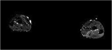
MRIAxial MRI of both forearms just after exercise: marked hyperintensity of the muscle compartments, reflecting the oedema characteristic of chronic exertional compartment syndrome, invisible at rest.
Source : Cole et al., Sports Medicine - Open, 2017, figure 2 · CC BY
Since pressure measurement is invasive, painful and contested, several teams have tried to replace it. Three avenues have been seriously explored: MRI, near-infrared spectroscopy and scintigraphy. A fourth, ultrasound, is more accessible and much less studied. Here is what each has demonstrated, and why none has yet replaced the needle.
MRI after exercise
The principle is simple: if the muscle suffers on exertion, its T2 signal should rise more than that of a healthy muscle. Verleisdonk tested this in 21 patients (41 anterior compartments) and 12 controls, with MRI before and after exercise, then again in 13 patients after fasciotomy. The results are clear: the T2 signal of the anterior compartment rose by 27.5% (range 13.6 to 38.6%) in patients, versus 7.6 % (0-9.1 %) in controls and 4.25 % in their own posterior compartment. After fasciotomy, the rise fell back to 4.1 % (Skeletal Radiol 2001, PMID 11465772).
This result is elegant: the affected compartment stands apart from the healthy compartment of the same patient, from the control, and from itself once operated on. Yet it has two limitations that explain why it has not replaced measurement. The first is logistical: an MRI has to be available immediately after an effort sufficient to trigger the symptoms, which is not the usual organisation of a radiology department. The second is one of performance: when MRI and pressure were compared head to head in a larger population, MRI had comparable sensitivity but a lower specificity at equal sensitivity.
Near-infrared spectroscopy
Near-infrared spectroscopy measures tissue oxygen saturation through the skin, without a needle. Van den Brand showed that affected patients desaturated far more: mean saturation at peak exercise of 27 in patients versus 56 in healthy subjects (p < 0.05), with a swing between rest and peak exercise that was also more marked. The values returned to the normal range after fasciotomy (AJSM 2004, PMID 14977673).
The same team then ran the most useful study in the field: a prospective comparison of the three modalities in 50 patients (100 legs), with the disappearance of symptoms after fasciotomy as the retrospective reference standard. The diagnosis was confirmed in 42 of the 45 patients reviewed, and refuted in 3.
Measured sensitivity, needle versus light
Prospective study in 50 patients, reference standard: disappearance of symptoms after fasciotomy
Source: van den Brand JG, Nelson T, Verleisdonk EJ, van der Werken C. The diagnostic value of intracompartmental pressure measurement, magnetic resonance imaging, and near-infrared spectroscopy in chronic exertional compartment syndrome: a prospective study in 50 patients. Am J Sports Med 2005;33(5):699-704 (PMID 15722275). Level of evidence 2. The design includes only operated patients: specificity therefore cannot be estimated as solidly as sensitivity.
The landmark result is the one in the purple bar: the sensitivity of pressure measurement as measured in this study, 77% (95% CI: 67 to 86), was lower than the 93% the literature credited it with. And spectroscopy, non-invasive, reached 85% (CI: 76 to 92), exactly the earlier estimate. The authors' conclusion is explicit: “the sensitivity of non-invasive near-infrared spectroscopy is clinically equivalent to that of invasive pressure measurements”.
- It is the only work that compares the three modalities in the same patients. It does two things at once: it validates spectroscopy and it deflates the sensitivity of the reference measurement.
- Twenty years on, near-infrared spectroscopy is still not in routine use. Not for want of a result, but for want of large-scale replication and a standardised protocol.
Ultrasound, the most accessible and the least studied
Measuring the thickening of the anterior compartment on ultrasound before and after a run is appealing: the machine is in the practice, the examination is immediate, it costs the patient nothing. A pilot study compared 4 patients and 9 controls after ten minutes on a treadmill. The thickness of the anterior compartment increased by 21.3% versus 6.32% at 30 seconds, 24.6% versus 4.22% at 2 minutes 30, and 24.9% versus 5.08% at 4 minutes 30, all differences significant (Rajasekaran, Clin J Sport Med 2013, PMID 23558330).
Four patients. The authors themselves conclude that “further studies are needed to validate these findings, with the aim of developing ultrasound diagnostic criteria”. Thirteen years on, that validation work has not been published on a scale that makes it usable. It is a lead, not a tool.
The verdict of the systematic reviews
Two reviews took stock a year apart, and they agree.
Ritchie included 28 studies assessing tests other than pressure measurement: MRI in 8, SPECT scintigraphy in 6, spectroscopy in 4, MRI and spectroscopy together in 1, various modalities in 9. Promising results were reported for MRI, spectroscopy and scintigraphy, but “the diagnostic tools and protocols were varied”, five studies explored these modalities as an adjunct rather than as a replacement, and “high-quality validation studies were absent” (Int J Sports Med 2023, PMID 35649437).
Van der Kraats covered 25 studies and 1,257 participants, assessing the risk of bias with the QUADAS tools. Only four studies had a low risk of bias; 21 were at risk. The conclusion is blunt: “despite the need to replace the controversial use of pressure measurement, our review indicates a lack of validity of all the non-invasive tools discussed as a replacement” (OJSM 2023, PMID 36655016).
Ritchie adds a sentence that shows where the field is heading, and that chimes with the thread of this article: optimising precise clinical criteria based on “the patient's history, physical examination and symptom provocation” could in time “make intracompartmental pressure measurement redundant”. In other words, the most serious lead for replacing the needle may not be a machine, but a better structured history.
- MRI : T2 signal of the affected compartment clearly higher after exercise, with normalisation after surgery. Sensitivity comparable to pressure, specificity lower.
- Near-infrared spectroscopy : sensitivity 85%, higher than the 77% measured for pressure in the same study. Non-invasive. Little used.
- Ultrasound : plausible and accessible, but a single pilot study in 4 patients. No diagnostic use today.
- Two systematic reviews conclude that no non-invasive tool is validated as a replacement. The most promising lead remains a well-built clinical algorithm.
What can modifying running technique achieve?
It is the best-documented conservative option, and by a wide margin. It rests on a simple idea: if the compartment suffers because tibialis anterior works too hard to control the foot at every heel strike, then changing the way the foot lands changes the demand. Four independent series have tested this hypothesis, with converging results. Here is what they show, and what they do not.
The founding study: ten patients who were due for surgery
Diebal prospectively recruited ten patients with an established surgical indication for compartment syndrome, and offered them six weeks of forefoot strike retraining before operating. The choice of this population is what gives the study its strength: these are not mild cases, they are patients for whom surgery had already been decided.
Six weeks of forefoot strike, in ten patients who were due for surgery
Light dot: before. Solid dot: after six weeks. Each row has its own scale
Source: Diebal AR, Gregory R, Alitz C, Gerber JP. Forefoot running improves pain and disability associated with chronic exertional compartment syndrome. Am J Sports Med 2012;40(5):1060-7 (PMID 22427621). Prospective case series, level of evidence 4, with no control group and no randomisation.
Three important results come on top of the four measures in the figure. The vertical ground reaction forces and impulses were significantly reduced, which gives the benefit a plausible mechanism. The global rating of perceived change was between 5 and 7 for every patient, that is between “a good deal better” and “a great deal better”. And above all, at one year the scores were better still than at six weeks, with two-mile (3.2 km) times significantly faster than before the intervention. None of the ten patients was operated on.
What replication confirmed, and at what cost
A case series of ten patients is not enough. Three later studies tested the robustness of the result.
Helmhout repeated the protocol in 19 patients, comparing a centre-based supervised programme with a home programme supervised less intensively. The gains are reproduced: running distance +43 %, post-run pressure −36 %, SANE +36 %, Lower Leg Outcome Survey +18 %, patient-specific complaints +60 %. The global rating of change was between +4 and +5, so “a little better” to “moderately better”, that is more modest than in Diebal's series. Two details matter: the scores were still improving four months after the end of the programme (SANE +48%, patient-specific complaints +81%, perceived change +6, “a great deal better”), and centre-based supervision did no better than the home programme (OJSM 2015, PMID 26665032). This last point is decisive in community practice: the benefit does not depend on specialised facilities.
Breen worked with 10 patients using a slightly different approach, four coaching cues rather than the forefoot strike alone: increase hip flexion, increase cadence, keep the trunk upright, adopt a midfoot strike. At one year, 70% of patients were running without pain, the exertional leg pain questionnaire score had improved by 40.3% at six weeks and by 49.2% at one year, and three of the four cues had produced lasting kinematic changes (Int J Sports Phys Ther 2015, PMID 25709867).
Zimmermann assessed scaling up in a military setting, in 75 patients eligible for surgery. After a programme lasting 145 days on average, centred on retraining running and marching in step, 65% (49 of 75) returned to active duty without surgery, 28% were referred to the surgeon and 7% left the army. Two-year follow-up, obtained in 42 of the 50 patients contacted, shows some erosion: 57% had continued in service without surgery, but among them 57% held a physically less demanding post (BMJ Open Sport Exerc Med 2019, PMID 31191976).
This last nuance is the one to pass on to the patient. A successful conservative outcome is not always a return to exactly the same thing: it is sometimes a return with an adjusted load. That is already considerable against the alternative, but it is not the same promise.
The programme, in the published detail
The published protocols resemble one another closely enough for a common framework to emerge. It is described most precisely in Vogels's cohort: 4 to 6 individual gait retraining sessions over 6 to 12 weeks, with uniform home exercises stressing acquisition of the new technique (PMID 35282146). In this cohort of 108 patients, 69% returned to active duty.
| Element | What the published protocols say | Measured effect |
|---|---|---|
| Foot strike point | Switch from a rearfoot strike to a forefoot strike (Diebal) or a midfoot strike (Breen) | Post-run pressure halved in Diebal, reduced by 36% in Helmhout |
| Cadence | Increase in step frequency, with a reduction in stride length | Stride length, impulse and peak vertical force all reduced (Diebal) |
| Trunk posture | Trunk kept upright, without excessive forward lean | One of the three cues that produced a lasting kinematic change at one year (Breen) |
| Hip flexion | Increase in hip flexion during the swing phase | Kinematic change maintained at one year (Breen) |
| Volume and progression | 4 to 6 individual sessions over 6 to 12 weeks, plus home work | 69% return to active duty in 108 patients (Vogels) |
| Place of care | Specialist centre or supervised home programme | No significant difference between the two (Helmhout) |
- The switch to a forefoot strike increases the demand on triceps surae and the Achilles tendon. The published protocols spread the transition over six weeks, and that gradual build is not decorative: it is what stops a compartment syndrome being traded for an Achilles tendinopathy.
- None of these studies compared gait retraining with a randomised control group. The level of evidence remains that of case series and cohorts.
Why it should work
The proposed mechanism stands up and rests on measurements. With a heel strike, tibialis anterior works eccentrically to control the lowering of the forefoot. Roscoe showed that affected patients bring the foot flat sooner than controls, and do so even before pain appears, which he interprets as a mechanical disadvantage of that muscle (PMID 29702369). With a forefoot strike, this braking phase largely disappears, and with it the eccentric demand on the anterior compartment. Alongside the fall in pressure, Diebal measured a reduction in vertical forces and in impulse, which is consistent.
An honest limitation has to be stated: this reasoning explains the benefit well for the anterior compartment. It explains far less well what might be expected in deep posterior involvement, where a forefoot strike increases the demand instead. The published series in any case deal overwhelmingly with anterior involvement. Transposing the protocol to deep posterior involvement is supported by no data at all.
What does not predict the outcome
Two studies have tackled the question of prognosis, and their answer is useful because it is negative.
Meulekamp tested thirteen prognostic factors in 45 service members in rehabilitation: age, body mass index, body fat percentage, self-efficacy, foot malalignment, intramuscular pressure, comorbidities, protein and creatine use, smoking, alcohol, symptom duration, physical demands of the post, length of service. None reached significance (Mil Med Res 2017, PMID 29502519). And Vogels showed that pressure was not associated with the outcome, whereas a low self-assessment score at entry was, unfavourably (odds ratio 0.95; p = 0.01).
The practical conclusion is simple: you cannot select in advance the patients who will respond. There is no profile to whom a conservative trial can be refused on objective grounds. That argues for offering it to everyone, emergencies aside.
- Four converging series: modifying running technique reduces pressure and increases the distance run, with gains that hold or improve further at one year.
- Order of magnitude of success: 65 to 70% return to activity without surgery in the two largest cohorts.
- The programme fits into 4 to 6 sessions over 6 to 12 weeks, and home does as well as the centre.
- The mechanical reasoning holds for the anterior compartment. Nothing supports it for the deep posterior compartment.
- No factor allows you to predict who will respond. Do not sort before trying.
What are the real results of fasciotomy, and its failures?
Fasciotomy is presented as the reference treatment, and it works in a majority of patients. It does not work in all of them, its success rates vary enormously from one series to another, and the difference between “returning to an activity” and “returning to their sport” is the one that matters to the patient. This chapter sets out the figures with their spread, because the spread is the information.
The principle, and its variants
The operation consists of opening the fascia of the affected compartment or compartments, to remove the constraint of the container. It is performed open, through mini-incisions, endoscopically, and, when it fails, as a fasciectomy, that is resection of a strip of fascia rather than a simple incision. The number of compartments opened varies: two (anterior and lateral) or four.
On this last question, Weiss's systematic review compared the two strategies across 7 studies and 194 athletes and service members of mean age 24. Return-to-activity rates were 90 to 100% for two compartments and 50 to 100% for four compartments, with no significant difference, and the authors conclude that the two approaches “appear equally effective”. They immediately add the necessary caveat: every included study carried a high risk of bias, with small samples, selection bias and no uniform outcome measure (J Foot Ankle Surg 2022, PMID 35337738).
Two series nevertheless suggest the opposite, and they need to be known. In 155 adolescents, legs that had only an anterior or lateral release carried a 3.4 times higher risk of reoperation (95% CI: 1.29 to 9.14; p = 0.01) than those in which all four compartments were opened (Beck, PMID 27365374). And in a small series of 21 patients reviewed, 91% of those who had all four compartments released regained their desired level of exercise, versus 66.7 % of the others (Maher, Foot Ankle Spec 2018, PMID 29931999). The debate is therefore not settled.
The figures, and above all their spread
What the literature reports, as ranges rather than means
Each bar covers the gap between the lowest and the highest value reported by the systematic reviews
Sources: Dean RS, Farley KX, Waterman BR, Guettler J, Bicos J. Chronic exertional compartment syndrome is frequently diagnosed through static compartment pressure measurements and managed with fasciotomy: A systematic review. J ISAKOS 2024;9(1):71-8 (PMID 37778507); Ding A, Machin M, Onida S, Davies AH. J Vasc Surg 2020;72(5):1802-12 (PMID 32473344); Shankar DS et al. Sports Health 2024;16(3):396-406 (PMID 36951383). These ranges pool different populations, different definitions of success and different follow-up durations: they describe uncertainty, not biological variability.
- The “return to sport, conservative” row does not contradict the 65 to 70% return to activity in the military cohorts: these are neither the same populations nor the same criteria. Dean pools heterogeneous conservative studies, often the control arms of surgical series, where “conservative” treatment sometimes amounts to a reduction in activity with no structured programme.
- The lesson to draw is not “conservative treatment works less well” but “ not everything called conservative is the same treatment ”. Relative rest and twelve weeks of gait retraining are not comparable.
The most complete series, and the most measured
Campano pooled 24 studies bringing together 1,596 patients, with a mean follow-up of 48.8 months. The verdict: primary surgery was a success in about two thirds of young, athletic patients, 84% declared themselves satisfied, 6% had revision surgery and the overall complication rate stood at 13 %, mainly postoperative neurological dysfunction and infections (Arthroscopy 2016, PMID 27020462).
Two thirds success and 84% satisfaction: the gap between those two figures is not a contradiction, it says something important. Patients can be satisfied with a partial improvement. That is exactly what Oliver's mini-fasciotomy series confirms: at a mean of 3.7 years, 30% of patients were asymptomatic and a further 56% reported improved symptoms, with pain during sport falling from 9.1 to 4.7 out of 10. 74% were satisfied and 81% would recommend the operation, while only 64% had returned to their sport (Eur J Orthop Surg Traumatol 2022, PMID 33675406).
The figure to give a runner
If the patient runs, one series deserves to be quoted as it stands. Salzler surveyed 32 runners who had had surgery, at a mean follow-up of 66 months. The results:
- 84 % returned to a sport.
- only 56% went back to running competitively, and among them the mean weekly distance had fallen.
- 28 % turned to a sport without running, 16% of them because of recurrent pain.
- 19 % had a recurrence, and all were reoperated.
- 78.1 % declared themselves satisfied, with pain during activity falling from 7.9 to 1.7 out of 10 (Sports Health 2020, PMID 32163722).
Not all populations are alike. Among 18 elite French cross-country skiers operated on bilaterally, 94% reported no further pain or only sporadic pain and nearly 90% had returned to competition at the same level or higher (Calvelli, PMID 31935775). At the other extreme, in service members, “only about half achieve complete resolution of symptoms and at least 25% are unable to return to full duty” (Dunn and Waterman, PMID 25280617). The same operation, two different worlds.
Complications, and what becomes of the failures
Ding's review gives the most complete ranges: haematoma 2.7 to 22.5%, nerve injury 2.0 to 18.6%, deep vein thrombosis 2.7%, recurrence of symptoms 0.65 to 8.4%, and up to 10.4% revision surgery (PMID 32473344). Oliver's mini-fasciotomy series reports 16% complications, including 11% superficial infections.
When surgery fails, revision is possible and documented. Vogels followed 24 patients, out of 958 assessed for exertional leg pain, who underwent a fasciectomy after a failed minimally invasive fasciotomy. Intraoperatively, the surgeons found fibrous bands, pseudo-fascia or complete fusion of the fascial edges. After rehabilitation, exertional symptom scores were cut to a third, and at a median follow-up of 12 months 79% reported a satisfactory result and 75% had resumed physical activity (Med Sci Sports Exerc 2021, PMID 33731658).
This result throws light on a mechanism of failure: the fascia closes up again. It also justifies not treating a first-line failure as the end of the road.
Surgery versus conservative care: what the direct comparison says
There is no randomised trial comparing fasciotomy with conservative treatment. Ding puts it bluntly: “there is insufficient evidence in the literature to support either conservative or surgical management over the other”, and calls for randomised trials (PMID 32473344). This absence can be measured: on 15 August 2026, a Europe PMC query crossing “chronic exertional compartment syndrome” with the publication type “Randomized Controlled Trial” returns only three records, only one of which is genuinely on the subject, and it compares compartment pressures between two roller-skiing techniques in skiers, not two treatments (PMID 26090709). This is a gap in indexing, which a poorly indexed trial could mask, but the order of magnitude leaves no room for doubt.
A meta-analysis has nevertheless pooled the four comparative cohort studies available. Surgery did better on pain (standardised mean difference −0.46; 95% CI: −0.74 to −0.17; p = 0.002, with low heterogeneity, I² = 13%) and on satisfaction (odds ratio 3.51; CI: 2.19 to 5.60; p < 0.00001). On the other hand, no significant difference in return to activity (odds ratio 3.70; CI: 0.53 to 25.96; p = 0.19), with massive heterogeneity (I² = 88%) (Elsenosy, Cureus 2024, PMID 39759644).
This confidence interval has to be read: from 0.53 to 25.96, it contains both no effect and an enormous effect. Across four non-randomised cohort studies, with 88% heterogeneity, this result allows no conclusion either way. What the meta-analysis reasonably establishes is an advantage for surgery on pain and satisfaction. What it does not establish is an advantage on what interests the runner: getting back to running.
- Success in about two thirds, satisfaction 84 %, complications 13 %, revisions 6 % in 1,596 patients.
- In runners: 84% return to a sport, 56% to running. That is the figure to give.
- Opening two or four compartments: no difference in the systematic review, but two series suggest an advantage for four compartments on recurrence.
- The preoperative paraesthesias herald a poorer result, especially in the tibial nerve territory.
- No randomised trial has compared surgery with rehabilitation. The available meta-analysis shows a gain on pain, not on return to activity.
Which modalities, at what level of evidence?
The table that follows ranks each modality by the confidence that can be placed in its effect. A warning is in order before reading it: none of these modalities has been the subject of a published GRADE rating for this condition. The level-of-evidence column is a judgement, built along GRADE lines, with the reason for downgrading given on each row so that the reader can contest it.
Where the whole available literature stands
Stacked cards: no modality reaches a high or moderate level of evidence, for want of a randomised trial
Judgement built along GRADE lines from the sources cited in the table below. No published GRADE rating exists for this condition; this ranking is our own, and the reason for downgrading each row appears in the table so that it can be discussed.
| Modality | What is measured | Evidence base | Level | Reason for downgrading |
|---|---|---|---|---|
| Running technique retraining | Post-run pressure halved; distance run tripled; 65 to 70% return to activity without surgery | Diebal 2012 (n = 10), Helmhout 2015 (n = 19), Breen 2015 (n = 10), Zimmermann 2019 (n = 75), Vogels 2022 (n = 108) | Low | No randomisation, no control group, limited samples. Not downgraded further because of the convergence of five independent studies and the size of the effect |
| Fasciotomy | Success in about two thirds, satisfaction 84%, complications 13% | Campano 2016 (1,596 patients), Ding 2020, Dean 2024, Salzler 2020 | Low | No randomised trial; heterogeneous definitions of success; rates ranging from 42 to 94% across series |
| Fasciectomy after failed fasciotomy | 79% satisfactory result, 75% return to activity | Vogels 2021 (n = 24, out of 958 patients assessed) | Very low | Single series, very small sample, highly selected population |
| Botulinum toxin | Initial efficacy in 68.75%; recurrence in 100% of partial responders and 57% of complete responders | Charvin 2022 (n = 16), McGinley 2022 (n = 284, no control group) | Very low | Retrospective series with no control group, documented transient effect, induced muscle weakness not quantified in the sporting context |
| Training load adjustment alone | Symptom relief reported, without quantification | Mentioned as clinical common sense in Ding 2020 and the review articles | Very low | No dedicated study. Effective by construction if the triggering activity is given up, which is acceptable for only some patients |
| Opening four compartments rather than two | No difference in the systematic review; 3.4 times more revisions after anterior or lateral release alone in adolescents | Weiss 2022 (7 studies, 194 patients), Beck 2016 (n = 155), Maher 2018 (n = 21) | Very low | High risk of bias in the 7 included studies; discordant results between the review and the series |
| Pressure measurement before conservative treatment | No association with the outcome of the programme (odds ratio 1.01; p = 0.64) | Vogels 2022 (n = 108, 200 legs) | Low, and against | Single retrospective study, but designed for this precise question and of good size |
| Stretching, manual therapy, foot orthoses | No results specific to this condition | No study identified assessing these modalities in isolation in compartment syndrome | Not assessable | No data. Do not confuse absence of evidence of effect with evidence of absence of effect |
Botulinum toxin, a lead that deserves a careful reading
The idea is counter-intuitive: deliberately weakening the muscle to reduce the pressure in its compartment. Two studies exist, very different from each other.
Charvin retrospectively followed 16 patients treated first line with botulinum toxin A injections, upper and lower limbs together, median age 25.5 years. 68.75% reported an initial benefit (7 complete, 4 partial), and 8 of the 16 patients were able to resume the triggering activity. Then comes the decisive part: every patient with a partial benefit relapsed, with a median delay of 2.25 months, and 57.14% of those with a complete benefit relapsed, with a median delay of 5 months. Adverse effects were minor and without functional consequences (Clin J Sport Med 2022, PMID 34282061).
McGinley published a much larger series, 284 patients, but in a setting that limits its reach: single-centre, retrospective, without a control group, with a primary outcome resting on imaging and a strong causal conclusion drawn from a design that does not allow it (PMID 34009799).
What can be said to a patient: the toxin may relieve symptoms for a few months, in about two patients in three, and the relief fades. In an athlete, the induced weakness is an effect to be weighed and not a mere side effect, since it is the very mechanism of the treatment.
A shift in the discourse worth noting
In 2014, a reference review on compartment syndrome in the military wrote that “non-operative management is generally unsuccessful, although gait retraining may have benefits in selected individuals” (Dunn and Waterman, PMID 25280617).
Twelve years later, the review in the British Medical Bulletin places among the points of agreement the statement that “conservative management, in particular structured rehabilitation with activity modification and gait retraining, is increasingly regarded as the first-line treatment” (PMID 42478572).
This reversal happened without a single randomised trial being published. It rests on the accumulation of converging series and on the demonstration, in the military, that a structured programme avoids surgery in two patients out of three. It is a low level of evidence that has changed a practice, which happens, and which it is better to know and to say to the patient.
- No modality reaches a high or moderate level of evidence, for want of a randomised trial in this condition.
- Gait retraining and fasciotomy sit at the same level, low, for different reasons: convergence of small series for one, large volume without a comparator for the other.
- Botulinum toxin temporarily relieves about two patients in three, with systematic recurrence in partial responders.
- Pressure measurement does not guide conservative treatment: it is the only point where the evidence runs against a widespread practice.
What do real clinical cases teach us?
The four observations that follow are published, indexed cases, quoted with their identifier. None is a reconstructed vignette. They were chosen because each illustrates a different error or decision: the missed diagnosis, the surgery avoided, the second diagnosis hidden behind the first, and the emergency.
Case 1. A runner treated for months for something else
The patient is a 24-year-old distance runner who presents with acute-on-chronic pain in the right leg. Her history includes two earlier diagnoses, both wrong according to the authors: medial tibial stress syndrome, then a tibial stress fracture. The history brings out the expected picture, pain and numbness on exertion, relieved by rest.
MRI shows no tibial abnormality. Pressure measurements find a rise in all four compartments, confirming the diagnosis. A minimally invasive four-compartment fasciotomy is performed. Two months after running is resumed, the symptoms in the right leg are improved, but symptoms appear on the left. Measurements there confirm the same diagnosis, a four-compartment fasciotomy is performed on the left side, and the patient returns to running pain-free on both sides at six weeks.
Truex N, Menge TJ. Bilateral Chronic Exertional Compartment Syndrome of the Leg: A Rare but often Debilitating Condition in Athletes. S D Med 2018;71(7):310-314 (PMID 30005541). What the case teaches: the two diagnoses made instead are exactly the ones the differential chapter names; and involvement that presents as unilateral can turn out to be bilateral as soon as the painful side is treated and the load redistributes.
Case 2. Surgery called off after six weeks of retraining
The patient is a 34-year-old sportswoman who describes pain, a sense of tightness and altered sensation in the legs and feet coming on after about 1.6 kilometres of running. A wick catheter test is performed; an orthopaedic surgeon makes the diagnosis of chronic exertional compartment syndrome and declares her eligible for a fasciotomy.
The patient chooses to try first a programme of six weeks of gait retraining, on the hypothesis that a forefoot strike gait delays the onset of symptoms and reduces discomfort. At the end of the six weeks, she runs with no symptoms at all, and her compartment pressures are reduced, which leads the surgeon to no longer recommend the fasciotomy.
Allison AK, Ishikawa KL, Gerber JP, Dewing C. Chronic Exertional Compartment Syndrome Resolved With Running Gait Retraining: A Case Report. J Athl Train 2023;58(4):345-348 (PMID 37418562). What the case teaches: an established surgical indication is not a sentence. The threshold described, around 1.6 km, also illustrates the reproducibility characteristic of the picture.
Case 3. The right diagnosis, and the one hiding behind it
The patient is a college football player who has had two years of progressive bilateral exertional calf pain, with ankle weakness during activity. Pressure measurements lead to the diagnosis of chronic exertional compartment syndrome. But her presentation remains suspicious for a type VI popliteal artery entrapment syndrome in addition.
MR angiography is requested. It shows severe arterial spasm on plantarflexion, on both sides, confirming the second diagnosis. As the symptoms worsened, the patient underwent an open popliteal release on the right.
Bellomo TR, Hsu C, Bolla P, Mohapatra A, Kotler DH. Concurrent Chronic Exertional Compartment Syndrome and Popliteal Artery Entrapment Syndrome. Diagnostics (Basel) 2024;14(16):1825 (PMID 39202313). What the case teaches: the authors put it themselves: “the successful diagnosis of one condition does not exclude the possibility of a second, independent pathology”. A positive test closes a case; it should close only a hypothesis.
Case 4. When the chronic form tips into an emergency
The patient is a 17-year-old boy who is admitted to a paediatric ward for bilateral anterior compartment leg pain of undetermined origin. This is an acute-on-chronic compartment syndrome, a rare and severe form of decompensation of a chronic exertional compartment syndrome. The authors stress that the non-specific nature of the pain demands a high index of suspicion if the diagnosis is to be made in time and compartment ischaemia with irreversible soft tissue and nerve damage is to be avoided.
They recall that, as in any compartment syndrome, pain out of proportion to the examination findings is the most sensitive sign, and that the shortage of literature on this entity itself contributed to the diagnostic delay.
Schwartz A, Poole C, Schleien C. Characterization of the Development of Acute-on-Chronic Exertional Compartment Syndrome: A Case Report of Symmetric Compartment Syndromes and Review of the Literature. Bull Hosp Jt Dis 2017;75(2):148-152 (PMID 28583063). What the case teaches: chronic compartment syndrome is a benign condition until the moment when it no longer is. Pain that does not ease on stopping, with a tense compartment and a deficit setting in, falls outside the scope of this article and is an emergency.
An open question, raised by a fifth case
One last reported case deserves a cautious mention. It describes a distance runner who developed a leg compartment syndrome after creatine use, and discusses the risk factors and the differential diagnoses (Bruneau, Curr Sports Med Rep 2025, PMID 40627559). The authors themselves note that research on the relationship between compartment syndrome and supplementation is “sparse”.
A single case establishes no causation, and it would be wrong to draw a rule from it. The reason for quoting it is different: it is a question patients ask, and it is useful to know that it has been raised in the literature, with exactly this level of evidence, that is none. It can be noted in passing that creatine use was among the thirteen prognostic factors tested by Meulekamp, without reaching significance (PMID 29502519).
- Case 1: the two wrong diagnoses found are indeed medial tibial stress syndrome and stress fracture, and the involvement turned out to be bilateral after the first side was treated.
- Case 2: an established surgical indication was lifted by six weeks of gait retraining.
- Case 3: a positive pressure measurement masked for two years an entrapped popliteal artery as well.
- Case 4: the acute-on-chronic form exists, it is an emergency, and its most sensitive sign is pain out of proportion to the examination.
How do you apply all this in practice?
This chapter condenses the article into a way of running a consultation. It does not replace the previous ones, it orders them: what you ask, what you do, what you refer on, and what you tell the patient.
First consultation: the history makes the diagnosis
Five questions, in this order, frame the essentials:
- “At what point in the session, exactly?” A precise and stable moment points to the compartment. Pain present from the outset points to bone or tendon.
- “Is it always at the same moment?” Reproducibility is the most discriminating feature of the picture.
- “How would you describe the sensation?” Tightness, swelling, burning, cramping point to the compartment. Pinpoint pain on the bone points elsewhere.
- “What do you have to do to make it stop, and how long does it take?” Stopping and waiting a few minutes points to the compartment. Waiting several days points to bone.
- “Does it come back at the same point if you set off again in the same session?” A return at the same threshold is highly suggestive.
Two further questions deserve to be asked routinely, for their prognostic and safety value: “do you get pins and needles or a numb patch?” and “does the foot give way, or slap, at the end of a run?”.
Examination: at rest, then after provocation
| Stage | What you do | What you look for | Interpretation |
|---|---|---|---|
| At rest | Palpation of the tibial border along its whole length, palpation of the muscle bellies, hop test on one leg, neurological examination of the leg and foot | Exquisite focal pain, linear pain along the posteromedial border, fixed neurological deficit | Focal bone pain stops the process and calls for imaging. A normal examination is expected and refutes nothing |
| At rest, standing | Inspection under load and with the dorsiflexors contracting | Anterolateral muscle hernia | Present in 45.9% of affected patients versus 12.9% of those unaffected: it is the only sign whose value has been measured |
| Provocation | Reproduce the triggering effort, up to the threshold and not before | The patient recognising the symptom | A test interrupted before the threshold concludes nothing |
| Immediately afterwards | Comparative palpation of the compartments, inspection, dorsiflexion testing, heel walking, sensation over the dorsum of the foot | Tense compartment, hernia now visible, weak dorsiflexion, reduced sensation | No published diagnostic value. These elements document and point the way, they do not prove |
What you start without waiting
If the account is typical and no red flag is present, conservative treatment can begin immediately. Pressure measurement is not a prerequisite: it does not predict the outcome of the programme and can be deferred safely, which has been demonstrated in 108 military patients (PMID 35282146).
The programme, as described in the published work:
- Format : 4 to 6 individual sessions spread over 6 to 12 weeks, supplemented by home work. Centre-based follow-up did no better than home follow-up in the only published comparison.
- Content : switch to a forefoot or midfoot strike, increased cadence with reduced stride length, upright trunk, increased hip flexion.
- Gradual build : the transition is spread over six weeks in every protocol. That slowness protects triceps surae and the Achilles tendon, whose load mechanically increases.
- Outcome measurement : distance run without pain before and after, and a self-reported score. The published work uses the SANE, the Lower Leg Outcome Survey or the patient-specific complaints scale. A systematic review of the questionnaires available for exertional leg pain concluded that the Lower Extremity Functional Scale was the best validated, ahead of the medial tibial stress syndrome score and the questionnaire dedicated to exertional leg pain (Castillo-Domínguez, Medicina 2022, PMID 35888560).
- Retraining is not just saying “forefoot strike”. The series that get results use explicit cues, repeated, and checked on video, with home work between sessions.
- Measuring the distance run without pain before and after is the outcome that speaks most to the patient, and it is the one that moved most in the studies: from 1.4 to 4.8 km in Diebal.
When to refer, and to whom
- As an emergency : pain that does not ease on stopping, a tense compartment, a sensorimotor deficit setting in. Acute-on-chronic compartment syndrome until proved otherwise.
- Without delay : suspected stress fracture, particularly at a high-risk site, or a background of low energy availability.
- Promptly : vascular signs in the provoked position in a young athlete, for dynamic vascular imaging.
- After failure of the conservative programme : refer for discussion of a pressure measurement and a possible fasciotomy. Three months of a well-run programme is consistent with the published protocols, which last 6 to 12 weeks.
What to tell the patient, in figures
Patients ask for probabilities. Here are four, with their source, that can be given without overstating in either direction.
Three sentences to avoid, because they are not supported by what we have read:
- “The pressure measurement will say whether you have it or not.” With a sensitivity of 63% for the best published threshold, it will not say so in a third of cases.
- “It is the fascia that is too tight, the operation corrects that.” Neither the thickness nor the stiffness of the fascia differs from that of healthy subjects.
- “After the operation, you will run as before.” Slightly more than one operated runner in two returns to running, and often with a reduced volume.
- The diagnosis is made from the history, in five questions.
- Provocation followed by immediate examination documents; it does not prove.
- Conservative treatment starts without waiting for the pressure measurement.
- Three months of a well-run programme before considering surgery is consistent with the published protocols.
- The figures to give: two thirds avoid surgery; among those operated on, slightly more than one runner in two returns to running.
Frequently asked questions
Can chronic exertional compartment syndrome resolve without surgery?
In some patients, yes. The best-evaluated conservative programme, built around gait retraining, returned 65% of 75 service members to active duty without surgery, and 57% were still there two years later, some of them in a physically less demanding post (PMID 31191976). A later cohort of 108 patients found 69% returning to duty (PMID 35282146). This is not a guarantee, and it does not apply to everyone: these are military populations, mostly with anterior compartment involvement.
Should pressures be measured before starting rehabilitation?
No. This is the one point where the evidence runs squarely against widespread practice. In 108 service members who completed a 6 to 12 week programme, the anterior compartment pressure value was not associated with the outcome (odds ratio 1.01; p = 0.64), and the authors conclude that measurement “can safely be deferred”. No acute-on-chronic compartment syndrome occurred in this cohort (PMID 35282146).
What is the difference from medial tibial stress syndrome?
Three things separate them. Location : medial tibial stress syndrome hurts over the posteromedial border of the tibia, compartment syndrome inside the muscle. The time course : medial tibial stress syndrome is often present from the warm-up and may ease while running, compartment syndrome is silent up to a threshold and then becomes intolerable. And palpation : medial tibial stress syndrome is painful over several centimetres of tibial border, compartment syndrome gives nothing at rest. The two can coexist, and in Zimmermann's cohort conservative treatment gave the same results with or without an associated medial tibial stress syndrome.
Does a normal examination rule out the diagnosis?
No, quite the opposite. The examination between episodes is normal by construction: the mechanism only operates on exertion. The only physical sign whose discriminating value has been measured is the muscle hernia, present in 45.9% of affected patients versus 12.9% of those unaffected (PMID 2301689). So it is missing more than half the time.
Does fasciotomy cure the condition for good?
Not always. Across 1,596 pooled patients, success is achieved in about two thirds, with 84% satisfaction, 13% complications and 6% revision procedures (PMID 27020462). Among 32 runners followed for a mean of 66 months, 84% returned to a sport but only 56% to running, and 19% had a recurrence and were reoperated (PMID 32163722). When it fails, revision fasciectomy gives 79% satisfactory results in a series of 24 patients (PMID 33731658).
How many compartments should be opened?
The question is not settled. The systematic review of 7 studies and 194 patients finds no difference between two and four compartments, but every included study carried a high risk of bias (PMID 35337738). Two series suggest the opposite: in 155 adolescents, a release limited to the anterior or the lateral compartment multiplied the risk of reoperation by 3.4 (PMID 27365374).
Does it affect only young runners?
No. Of 698 patients at a referral centre, one in seven was 50 or older ; these patients were less sporty, more often unilateral (45% versus 22%) and anterior involvement dominated in them (82%) (PMID 29531960). And the male-to-female split is even: 51% women among diagnosed patients across 24 studies (PMID 36698053).
Can you keep running with this diagnosis?
No data show that continuing to run makes the disease worse or creates a structural risk, apart from the acute-on-chronic form, which is rare. The limit is therefore mainly functional: pain caps the distance. The reasonable approach is to keep running below the threshold while working on running technique, rather than stopping altogether. Pain that no longer eases on stopping, on the other hand, means stopping and seeking advice the same day.
Is botulinum toxin an option?
It exists, it relieves symptoms temporarily, and it does not last. Of 16 patients treated first line, 68.75% had an initial benefit, but every partial responder relapsed (median delay 2.25 months) and so did 57% of the complete responders (median delay 5 months) (PMID 34282061). In an athlete, the induced muscle weakness is not an incidental side effect: it is the very mechanism of the treatment.
Why does this diagnosis take so long to be made?
Because the patient comes in for a pain they do not have at the time of the consultation, to a practitioner who examines them at rest and finds nothing, with normal plain imaging. Medial tibial stress syndrome, far more common, then captures the diagnosis by default. In a paediatric series, symptoms had been going on for more than a year in 63% of patients at the time of surgery (PMID 27365374).
Bibliography
The 55 references cited in this article, in alphabetical order of first author. Each was retrieved through the PubMed E-utilities API (full author list, journal, volume, pagination) then cross-checked on CrossRef for title, journal, year and author list. Four references have no registered DOI and are identified by their PMID alone.
- Allison AK, Ishikawa KL, Gerber JP, Dewing C. Chronic Exertional Compartment Syndrome Resolved With Running Gait Retraining: A Case Report. J Athl Train 2023;58(4):345-348. PMID 37418562 · DOI
- Aweid O, Del Buono A, Malliaras P, Iqbal H, Morrissey D, Maffulli N, Padhiar N. Systematic review and recommendations for intracompartmental pressure monitoring in diagnosing chronic exertional compartment syndrome of the leg. Clin J Sport Med 2012;22(4):356-70. PMID 22627653 · DOI
- Baumfeld D, Silva M, Simões AP, Baumfeld T. Mini Open Fasciotomy for Chronic Exertional Compartment Syndrome: A Prospective Case Series. Foot Ankle Spec 2025;18(6):606-612. PMID 38084741 · DOI
- Beck JJ, Tepolt FA, Miller PE, Micheli LJ, Kocher MS. Surgical Treatment of Chronic Exertional Compartment Syndrome in Pediatric Patients. Am J Sports Med 2016;44(10):2644-2650. PMID 27365374 · DOI
- Bellomo TR, Hsu C, Bolla P, Mohapatra A, Kotler DH. Concurrent Chronic Exertional Compartment Syndrome and Popliteal Artery Entrapment Syndrome. Diagnostics (Basel) 2024;14(16). PMID 39202313 · DOI
- Breen DT, Foster J, Falvey E, Franklyn-Miller A. Gait re-training to alleviate the symptoms of anterior exertional lower leg pain: a case series. Int J Sports Phys Ther 2015;10(1):85-94. PMID 25709867
- Bruneau MM, Sirls ER, Corrado GD, Theodore GH, Tenforde AS. Association of Creatine Use with Chronic Exertional Compartment Syndrome in a Distance Runner: A Case Report. Curr Sports Med Rep 2025;24(7):194-197. PMID 40627559 · DOI
- Buerba RA, Fretes NF, Devana SK, Beck JJ. Chronic exertional compartment syndrome: current management strategies. Open Access J Sports Med 2019;10:71-79. PMID 31213933 · DOI
- Burrus MT, Werner BC, Starman JS, Gwathmey FW, Carson EW, Wilder RP, Diduch DR. Chronic leg pain in athletes. Am J Sports Med 2015;43(6):1538-47. PMID 25157051 · DOI
- Calvelli N, Vergès S, Rousseaux-Blanchi MP, Edouard P, Guinot M. Features of Chronic Exertional Compartmental Syndrome of the Leg in Elite Nordic Skiers. Int J Sports Med 2020;41(3):196-202. PMID 31935775 · DOI
- Campano D, Robaina JA, Kusnezov N, Dunn JC, Waterman BR. Surgical Management for Chronic Exertional Compartment Syndrome of the Leg: A Systematic Review of the Literature. Arthroscopy 2016;32(7):1478-86. PMID 27020462 · DOI
- Castillo-Domínguez A, García-Romero JC, Alvero-Cruz JR, Ponce-García T, Benítez-Porres J, Páez-Moguer J. Systematic Review of Patient-Reported Outcome Measures for Patients with Exercise-Induced Leg Pain. Medicina (Kaunas) 2022;58(7). PMID 35888560 · DOI
- Charvin M, Orta C, Davy L, Raumel MA, Petit J, Casillas JM, Gremeaux V, Gouteron A. Botulinum Toxin A for Chronic Exertional Compartment Syndrome: A Retrospective Study of 16 Upper- and Lower-Limb Cases. Clin J Sport Med 2022;32(4):e436-e440. PMID 34282061 · DOI
- Dahl M, Hansen P, Stål P, Edmundsson D, Magnusson SP. Stiffness and thickness of fascia do not explain chronic exertional compartment syndrome. Clin Orthop Relat Res 2011;469(12):3495-500. PMID 21948310 · DOI
- de Bruijn JA, van Zantvoort APM, Winkes MB, van der Cruijsen-Raaijmakers M, Hoogeveen AR, Teijink JAW, Scheltinga MRM. Lower Leg Chronic Exertional Compartment Syndrome in Patients 50 Years of Age and Older. Orthop J Sports Med 2018;6(3):2325967118757179. PMID 29531960 · DOI
- de Bruijn JA, Wijns KCA, van Kuijk SMJ, Hoogeveen AR, Teijink JAW, Scheltinga MRM. Chronic exertional compartment syndrome in the differential diagnosis of peripheral artery disease in older patients with exercise-induced lower limb pain. J Vasc Surg 2021;73(6):2114-2121. PMID 33278541 · DOI
- Dean RS, Farley KX, Waterman BR, Guettler J, Bicos J. Chronic exertional compartment syndrome is frequently diagnosed through static compartment pressure measurements and managed with fasciotomy: A systematic review. J ISAKOS 2024;9(1):71-78. PMID 37778507 · DOI
- Diebal AR, Gregory R, Alitz C, Gerber JP. Effects of forefoot running on chronic exertional compartment syndrome: a case series. Int J Sports Phys Ther 2011;6(4):312-21. PMID 22163093
- Diebal AR, Gregory R, Alitz C, Gerber JP. Forefoot running improves pain and disability associated with chronic exertional compartment syndrome. Am J Sports Med 2012;40(5):1060-7. PMID 22427621 · DOI
- Ding A, Machin M, Onida S, Davies AH. A systematic review of fasciotomy in chronic exertional compartment syndrome. J Vasc Surg 2020;72(5):1802-1812. PMID 32473344 · DOI
- Elsenosy AM, Elnewishy A, Hassan E, Delewar RA. Outcomes of Fasciotomy Versus Conservative Management for Chronic Exertional Compartment Syndrome: A Systematic Review and Meta-Analysis. Cureus 2024;16(12):e75803. PMID 39759644 · DOI
- Helmhout PH, Diebal AR, van der Kaaden L, Harts CC, Beutler A, Zimmermann WO. The Effectiveness of a 6-Week Intervention Program Aimed at Modifying Running Style in Patients With Chronic Exertional Compartment Syndrome: Results From a Series of Case Studies. Orthop J Sports Med 2015;3(3):2325967115575691. PMID 26665032 · DOI
- Lindorsson S, Zhang Q, Brisby H, Rennerfelt K. Significantly lower intramuscular pressure in the posterior and lateral compartments compared with the anterior compartment suggests alterations of the diagnostic criteria for chronic exertional compartment syndrome in the lower leg. Knee Surg Sports Traumatol Arthrosc 2021;29(4):1332-1339. PMID 32642913 · DOI
- Maher JM, Brook EM, Chiodo C, Smith J, Bluman EM, Matzkin EG. Patient-Reported Outcomes Following Fasciotomy for Chronic Exertional Compartment Syndrome. Foot Ankle Spec 2018;11(5):471-477. PMID 29931999 · DOI
- Malik GR, Vyas KA. The Pressure Is Rising: Evaluation and Treatment of Chronic Exertional Compartment Syndrome. Curr Sports Med Rep 2023;22(6):204-209. PMID 37294195 · DOI
- McGinley JC, Thompson TA, Ficken S, White J. Chronic Exertional Compartment Syndrome Caused by Functional Venous Outflow Obstruction. Clin J Sport Med 2022;32(4):355-360. PMID 34009799 · DOI
- Meulekamp MZ, Sauter W, Buitenhuis M, Mert A, van der Wurff P. Short-Term Results of a Rehabilitation Program for Service Members With Lower Leg Pain and the Evaluation of Patient Characteristics. Mil Med 2016;181(9):1081-7. PMID 27612357 · DOI
- Meulekamp MZ, van der Wurff P, van der Meer A, Lucas C. Identifying prognostic factors for conservative treatment outcomes in servicemen with chronic exertional compartment syndrome treated at a rehabilitation center. Mil Med Res 2017;4(1):36. PMID 29502519 · DOI
- Oliver WM, Rhatigan D, Mackenzie SP, White TO, Duckworth AD, Molyneux SG. Outcome following mini-open lower limb fasciotomy for chronic exertional compartment syndrome. Eur J Orthop Surg Traumatol 2022;32(1):27-36. PMID 33675406 · DOI
- Pedowitz RA, Hargens AR, Mubarak SJ, Gershuni DH. Modified criteria for the objective diagnosis of chronic compartment syndrome of the leg. Am J Sports Med 1990;18(1):35-40. PMID 2301689 · DOI
- Rajasekaran S, Beavis C, Aly AR, Leswick D. The utility of ultrasound in detecting anterior compartment thickness changes in chronic exertional compartment syndrome: a pilot study. Clin J Sport Med 2013;23(4):305-11. PMID 23558330 · DOI
- Rajasekaran S, Finnoff JT. Exertional Leg Pain. Phys Med Rehabil Clin N Am 2016;27(1):91-119. PMID 26616179 · DOI
- Ritchie ED, Vogels S, van Dongen TTCF, van der Burg BLSB, Scheltinga MRM, Zimmermann WO, Hoencamp R. Systematic Review of Innovative Diagnostic Tests for Chronic Exertional Compartment Syndrome. Int J Sports Med 2023;44(1):20-28. PMID 35649437 · DOI
- Roberts A, Franklyn-Miller A. The validity of the diagnostic criteria used in chronic exertional compartment syndrome: a systematic review. Scand J Med Sci Sports 2012;22(5):585-95. PMID 22092446 · DOI
- Roscoe D, Roberts AJ, Hulse D. Intramuscular compartment pressure measurement in chronic exertional compartment syndrome: new and improved diagnostic criteria. Am J Sports Med 2015;43(2):392-8. PMID 25406302 · DOI
- Roscoe D, Roberts AJ, Hulse D, Shaheen A, Hughes MP, Bennett A. Barefoot plantar pressure measurement in Chronic Exertional Compartment Syndrome. Gait Posture 2018;63:10-16. PMID 29702369 · DOI
- Rothman R, Berke C, Jivanelli B, Casey E, Cheng J. Sex and gender differences in lower limb chronic exertional compartment syndrome: a systematic review. Phys Sportsmed 2024;52(1):1-11. PMID 36698053 · DOI
- Ruohola JP, Kiuru MJ, Pihlajamäki HK. Fatigue bone injuries causing anterior lower leg pain. Clin Orthop Relat Res 2006;444:216-23. PMID 16523142 · DOI
- Saad MA, Jamal JM, Aldhafiri AT, Alkandari SA. Medial Tibial Stress Syndrome: A Scoping Review of Epidemiology, Biomechanics, and Risk Factors. Cureus 2025;17(3):e81463. PMID 40171337 · DOI
- Salzler M, Maguire K, Heyworth BE, Nasreddine AY, Micheli LJ, Kocher MS. Outcomes of Surgically Treated Chronic Exertional Compartment Syndrome in Runners. Sports Health 2020;12(3):304-309. PMID 32163722 · DOI
- Schwartz A, Poole C, Schleien C. Characterization of the Development of Acute-on-Chronic Exertional Compartment Syndrome A Case Report of Symmetric Compartment Syndromes and Review of the Literature. Bull Hosp Jt Dis (2013) 2017;75(2):148-152. PMID 28583063
- Shankar DS, Blaeser AM, Gillinov LA, Vasavada KD, Fariyike BB, Mojica ES, Borowski LE, Jazrawi LM, Cardone DA. Paresthesia Is Predictive of Symptom Recurrence After Fasciotomy for Exertional Compartment Syndrome of the Leg. Sports Health 2024;16(3):396-406. PMID 36951383 · DOI
- Simpson C, Roscoe D, Hughes S, Hulse D, Guthrie H. Surgical outcomes for chronic exertional compartment syndrome following improved diagnostic criteria. BMJ Mil Health 2020;166(E):e17-e20. PMID 30992340 · DOI
- Truex N, Menge TJ. Bilateral Chronic Exertional Compartment Syndrome of the Leg: A Rare but often Debilitating Condition in Athletes. S D Med 2018;71(7):310-314. PMID 30005541
- van den Brand JG, Verleisdonk EJ, van der Werken C. Near infrared spectroscopy in the diagnosis of chronic exertional compartment syndrome. Am J Sports Med 2004;32(2):452-6. PMID 14977673 · DOI
- van den Brand JG, Nelson T, Verleisdonk EJ, van der Werken C. The diagnostic value of intracompartmental pressure measurement, magnetic resonance imaging, and near-infrared spectroscopy in chronic exertional compartment syndrome: a prospective study in 50 patients. Am J Sports Med 2005;33(5):699-704. PMID 15722275 · DOI
- van der Kraats AM, Winkes M, Janzing HMJ, Eijkelenboom RPR, de Koning MTG. Review of Reliable and Valid Noninvasive Tools for the Diagnosis of Chronic Exertional Compartment Syndrome. Orthop J Sports Med 2023;11(1):23259671221145151. PMID 36655016 · DOI
- Velasco TO, Leggit JC. Chronic Exertional Compartment Syndrome: A Clinical Update. Curr Sports Med Rep 2020;19(9):347-352. PMID 32925373 · DOI
- Verleisdonk EJ, van Gils A, van der Werken C. The diagnostic value of MRI scans for the diagnosis of chronic exertional compartment syndrome of the lower leg. Skeletal Radiol 2001;30(6):321-5. PMID 11465772 · DOI
- Vogels S, VAN Ark W, Janssen L, Scheltinga MRM. Fasciectomy for Recurrent Chronic Exertional Compartment Syndrome of the Anterior Leg. Med Sci Sports Exerc 2021;53(8):1549-1554. PMID 33731658 · DOI
- Vogels S, Bakker EWP, O'Connor FG, Hoencamp R, Zimmermann WO. Association Between Intracompartmental Pressures in the Anterior Compartment of the Leg and Conservative Treatment Outcome for Exercise-Related Leg Pain in Military Service Members. Arch Rehabil Res Clin Transl 2022;4(1):100171. PMID 35282146 · DOI
- Vogels S, Zimmermann WO. Diagnostic and therapeutic dilemmas in chronic exertional compartment syndrome. Br Med Bull 2026;159(1). PMID 42478572 · DOI
- Weiss C, Vogels S, Wee L, Janssen L, de Bie RA, Scheltinga MR. Two-Compartment or 4-Compartment Fasciotomy for Lower Leg Chronic Exertional Compartment Syndrome: A Systematic Review. J Foot Ankle Surg 2022;61(5):1124-1133. PMID 35337738 · DOI
- Woods KM, Petron DJ, Shultz BB, Hicks-Little CA. Lower Leg Anterior and Lateral Intracompartmental Pressure Changes Before and After Classic Versus Skate Nordic Rollerskiing. J Athl Train 2015;50(8):812-8. PMID 26090709 · DOI
- Zimmermann WO, Hutchinson MR, Van den Berg R, Hoencamp R, Backx FJG, Bakker EWP. Conservative treatment of anterior chronic exertional compartment syndrome in the military, with a mid-term follow-up. BMJ Open Sport Exerc Med 2019;5(1):e000532. PMID 31191976 · DOI
- What this article covers: chronic compartment syndrome brought on by exertion, the reversible, exercise-related form.
- What it does not cover: compartment syndrome that is acute, post-traumatic or from a crush injury, which is a surgical emergency of a quite different nature. It is covered, as a complication, in our syntheses on muscle contusion and on midfoot sprain.
Method
The 55 references in this article were searched on PubMed, retrieved through the E-utilities API (full author list, journal, volume, issue, pagination, digital object identifier) then cross-checked one by one on CrossRef for title, journal, year and number of authors. One candidate reference was dropped from the base because its digital identifier was not registered with CrossRef. Every figure in the text carries its source at the point where it is written. The level-of-evidence judgements in the “modalities” chapter are our own and are argued row by row: no published GRADE rating exists for this condition.

