















In brief
Shin splints, more rigorously called medial tibial stress syndrome (MTSS), is exercise-induced pain along the posteromedial border of the tibia, reproducible on palpation over at least 5 continuous cm. It is not a periosteal inflammation but part of the bone stress injury continuum, and it can progress to a stress fracture. Its incidence reaches 5 to 19 % in runners and 35 to 56 % in military recruits. Load management is the cornerstone, not complete rest, combined with progressive exercise targeting the soleus, the tibialis posterior and the hip abductors.
Clinical synthesis based on the most recent meta-analyses and international consensus statements: Winters 2018, Hamstra-Wright 2015, Warden 2014, Marques 2025 and the LIMP 2025 debate.
Clinical summary
- MTSS (medial tibial stress syndrome) is exercise-induced pain along the posteromedial border of the tibia, palpable over at least 5 continuous cm (Winters 2018). It belongs to the continuum of bone stress injuries (BSI), not to simple periosteal inflammation.
- Incidence ranges from 5 to 19 % in runners to 35 to 56 % in military recruits (Hamstra-Wright 2015, Alessa 2024).
- Significant risk factors (Hamstra-Wright 2015 meta-analysis): female sex, higher BMI, previous running injury, increased navicular drop, increased hip external rotation.
- Pathophysiology: an imbalance in bone remodelling ↔ resorption under repeated mechanical overload. Left unmanaged, MTSS can progress to a stress fracture (Warden 2014).
- Diagnosis is essentially clinical (Winters 2018): diffuse pain on palpation over ≥ 5 cm. A stress fracture presents with very localised pain (< 5 cm) and a positive hop test.
- Clinical grading from 1 to 4 according to pain and impact on activity. Fredericson’s MRI classification (1995) remains relevant in suspected BSI.
- Proper load management is the cornerstone, not complete rest. Reduce activity to the pain-free threshold, then reintroduce it progressively.
- Progressive exercise targeting the soleus, the tibialis posterior and the hip abductors (Atalla 2024 RCT) improves kinematics and reduces pain.
- The Marques 2025 meta-analysis (Gait Posture, GRADE) recommends neuromuscular training + anti-pronation orthoses for prevention (moderate evidence).
- Focused shockwave therapy is an option for chronic, recalcitrant presentations, never first line. Passive manual therapies have little support in the evidence.
- Return to running follows a graded protocol (Moen 2009), guided by symptoms: tolerable pain ≤ 2/10 and no residual pain 24 h after the session.
- Increasing running cadence by 5 to 10 % reduces tibial impact forces (Crowell & Davis 2011, Luedke 2016).
- Watch for RED-S (relative energy deficiency in sport, IOC 2023): a major risk factor for any bone stress injury. Always assess it in recalcitrant or bilateral cases.
- Medical referral is mandatory if a stress fracture is suspected, if conservative treatment fails (3-6 months) or if red flags appear (night pain, weight loss, fever).
- The debate over MTSS → LIMP (Load Induced Medial-Leg Pain, Anderson & Beck JOSPT 2025) reflects the conceptual shift towards terminology that better captures an overload pathology.
Contents
- What are the fundamentals to know about shin splints (MTSS) in runners?
- How do you assess and diagnose shin splints (MTSS) with certainty?
- Which treatment strategies are the most effective?
- How do you secure lasting recovery and prevent recurrence?
- What do real clinical cases teach us?
- The major systemic factor: RED-S and the BSI continuum
- How do you apply these recommendations concretely in your practice?
What are the fundamentals to know about shin splints (MTSS) in runners?
Shin splints, more rigorously called medial tibial stress syndrome (MTSS) in the scientific literature, is one of the most common overload conditions in runners and military personnel.¹ The historical term “periostitis” is misleading, because the pathophysiology is not dominated by periosteal inflammation but by a bone stress reaction within the tibia, set within the continuum of bone stress injuries (BSI).²
How is this condition defined, who does it affect and what are the risk factors?
MTSS is defined as exercise-induced pain along the posteromedial border of the tibia.¹ The consensus diagnostic criterion, prospectively validated by Winters and colleagues (BJSM 2018), requires the pain to be reproducible on palpation over at least 5 continuous cm.³ This “diffuse” character is essential in distinguishing it from a stress fracture, whose pain is focal.
The epidemiology has been refined by several recent systematic reviews 🏃 :
- In recreational and competitive runners, MTSS incidence ranges from 5 to 19 % depending on the cohort (Lopes 2012, Hamstra-Wright 2015).⁴,⁵
- In military recruits, incidence reaches 35 to 56 % according to the narrative review by Alessa et al. 2024 (Cureus), with a landmark study by Yates & White 2004 (AJSM, n = 124 naval recruits) reporting 35 % incidence over 10 weeks of training.⁶,⁷
- MTSS accounts for 13 to 17 % of all running-related injuries (Lopes 2012).⁴
- The Saad 2025 scoping review confirms wide geographical heterogeneity: prevalence from 5.4 % (USA) to 69.5 % (India) depending on the practice setting.⁸
The landmark meta-analysis by Hamstra-Wright et al. (BJSM 2015), pooling data from prospective cohorts of runners and military personnel, identified five risk factors with a significant pooled effect and low statistical heterogeneity :⁵
- Female sex , with an increased relative risk that is particularly marked in military cohorts (Yates 2004: 53 % of women vs 28 % of men).⁵,⁷
- Higher body mass index (BMI).
- Increased navicular drop (navicular drop), a marker of excessive pronation.
- Previous running injury.
- Increased hip external rotation range with the hip flexed.
A previous episode of MTSS is itself one of the strongest predictors of recurrence (Newman 2013 in Open Access J Sports Med, which independently confirmed three of these factors).⁹
📊 MTSS risk factors: pooled effect (Hamstra-Wright 2015 meta-analysis)
Significant Standardized Mean Difference (SMD) or Odds Ratio, pooled, 13-23 studies depending on the factor
Source: Hamstra-Wright KL, Bliven KCH, Bay C. Br J Sports Med. 2015;49(6):362-369. Five risk factors with a statistically significant effect after meta-analysis.
What happens in the body and how does the condition evolve naturally?
The modern pathophysiology of MTSS, supported by the critical reviews of Moen 2009, Beck 1998 and Warden 2014, places the condition on a continuum of bone overload of the tibial cortex, not as simple periosteal inflammation.¹⁰,¹¹,² The central mechanism is repeated mechanical overload that exceeds the capacity of bone to adapt and remodel. 🦴
The process unfolds in several stages:
- Cumulative load : the repeated impacts of running drive accelerated bone remodelling.
- Resorption / formation imbalance : if volume or intensity outstrips adaptation, the rate of osteoclastic resorption exceeds osteoblastic formation.
- Microdamage and cortical porosity : the negative balance leads to an accumulation of microfractures, increased porosity of the tibial cortex and reduced bone stiffness.²,¹¹
- Diffuse pain : this stress reaction shows itself clinically as the characteristic pain over ≥ 5 cm.
Histological analysis (Johnell 1982, cited by Beck 1998) showed that the lesion involves not only the periosteum but also the bony cortex and the marrow, definitively reorienting management towards mechanical load management rather than towards anti-inflammatories.¹¹
The natural course can be unfavourable if load is not modified. Warden et al. (2014) describe a clinico-radiological spectrum running from normal adaptive remodelling all the way to complete fracture, by way of stress reaction (MRI bone oedema, Fredericson grade 1-3) and incomplete stress fracture (Fredericson grade 4).²,¹² Training on despite the pain encourages microdamage to coalesce.
- MTSS is a bone overload condition of the tibia (and not a periosteal inflammation). The pain must be palpable over at least 5 continuous cm (Winters 2018).
- The most robust risk factors are previous injury, female sex, high BMI, increased navicular drop, increased hip external rotation (Hamstra-Wright 2015 meta-analysis).
- The cause is an imbalance between bone formation and resorption caused by excessive mechanical stress, which can lead to a stress fracture if activity is not modified.
- Incidence is 5-19 % in runners and 35-56 % in military recruits ; recurrences are frequent without appropriate load management.
Chapter 1 bibliography
- Winters M, Eskes M, Weir A, et al. Treatment of medial tibial stress syndrome: a systematic review. Sports Med. 2013;43(12):1315-1333. PMID 23979968.
- Warden SJ, Davis IS, Fredericson M. Management and prevention of bone stress injuries in long-distance runners. J Orthop Sports Phys Ther. 2014;44(10):749-765. PMID 25103133.
- Winters M, Bakker EWP, Moen MH, Barten CC, Teeuwen R, Weir A. Medial tibial stress syndrome can be diagnosed reliably using history and physical examination. Br J Sports Med. 2018;52(19):1267-1272. PMID 28179260.
- Lopes AD, Hespanhol Júnior LC, Yeung SS, Costa LO. What are the main running-related musculoskeletal injuries? A systematic review. Sports Med. 2012;42(10):891-905. PMID 22827721.
- Hamstra-Wright KL, Bliven KCH, Bay C. Risk factors for medial tibial stress syndrome in physically active individuals such as runners and military personnel: a systematic review and meta-analysis. Br J Sports Med. 2015;49(6):362-369. PMID 25185588.
- Alessa F, Alqarni A, Alharbi S, et al. Highlights of medial tibial stress syndrome in military recruits: a narrative review. Cureus. 2024;16(12):e75416. PMC 11629594.
- Yates B, White S. The incidence and risk factors in the development of medial tibial stress syndrome among naval recruits. Am J Sports Med. 2004;32(3):772-780. PMID 15090396.
- Saad MA, Jamal JM, Aldhafiri AT, Alkandari SA. Medial tibial stress syndrome: a scoping review of epidemiology, biomechanics, and risk factors. Cureus. 2025;17(3):e55892. PMC 11958822.
- Newman P, Witchalls J, Waddington G, Adams R. Risk factors associated with medial tibial stress syndrome in runners: a systematic review and meta-analysis. Open Access J Sports Med. 2013;4:229-241. PMID 24379729.
- Moen MH, Tol JL, Weir A, Steunebrink M, De Winter TC. Medial tibial stress syndrome: a critical review. Sports Med. 2009;39(7):523-546. PMID 19530750.
- Beck BR. Tibial stress injuries. An aetiological review for the purposes of guiding management. Sports Med. 1998;26(4):265-279. PMID 9820925.
- Fredericson M, Bergman AG, Hoffman KL, Dillingham MS. Tibial stress reaction in runners. Correlation of clinical symptoms and scintigraphy with a new magnetic resonance imaging grading system. Am J Sports Med. 1995;23(4):472-81. PMID 7573660.
How do you assess and diagnose shin splints (MTSS) with certainty?
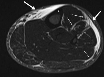
MRIAxial STIR MRI of the leg: thickening and high signal of the fascia and intermuscular septa without any periosteal reaction of the tibia, a picture of severe medial tibial stress syndrome (shin splint) in an ultramarathon runner.
Source : Lucas et al., Extreme Physiology & Medicine, 2016, figure 3 · CC BY
The diagnosis of MTSS rests essentially on the clinical examination, with sensitivity and reliability validated prospectively by Winters 2018 in 46 patients (high inter-examiner kappa for palpation of the painful length).¹ Imaging is not required first line, except where the diagnosis is in doubt or a stress fracture is suspected. 🏃
Which questions should you ask to understand the patient and their history?
History-taking aims to reconstruct the story of the injury and to identify training errors, the main triggering factor.² The lines of questioning:
- Pain characteristics : dull, diffuse pain along the distal third of the posteromedial border of the tibia, appearing during exercise, sometimes easing initially after the warm-up, then returning with greater intensity as activity continues or once it stops.²,³
- Training history : any rapid increase in volume, in intensity, in frequency or any change of surface (moving from track to road) or of footwear in the 4 to 6 weeks preceding symptoms is suspicious.⁴
- Past history : a previous episode of MTSS or of running injury is one of the strongest predictors of recurrence.⁵
- Systemic factors : systematically ask about energy intake, the menstrual cycle in women, a history of stress fractures and glucocorticoid use (flags for RED-S and high-risk BSI).
- Biomechanical factors and equipment : type of footwear, wear, insoles, foot type (more pronated = increased risk, with RR 1.7 in Yates 2004).⁶
Which clinical tests should you perform and which other conditions should you rule out?
The central clinical test is the “Shin Palpation Test” : palpation reproduces the pain over a continuous length of at least 5 cm along the posteromedial border of the tibia, usually in its distal third. Inter-examiner reliability has been shown to be good to excellent (Winters 2018, kappa 0.76).¹ 🔬
Other tests are useful:
- Hop test (10 single-leg hops on the spot) : sharp, focal pain should raise the suspicion of a stress fracture rather than MTSS.⁷
- Fulcrum test : bimanual pressure on the tibia; positive if localised pain suggests a fracture (Milgrom 2021).⁷
- Functional tests : single-leg heel raises (the ability to perform 20-30 repetitions without pain is a progression criterion), single-leg squat (looking for pelvic drop and dynamic valgus).
| Diagnosis | Pain location | Character | Suggestive test | Imaging |
|---|---|---|---|---|
| MTSS | Diffuse, ≥ 5 cm, posteromedial border of tibia | Dull, on exercise, after exercise | Diffuse palpation | Not required |
| Tibial stress fracture | Focal, < 5 cm, exquisite point | Sharp, may be nocturnal | Hop test +, fulcrum + | MRI (Fredericson 1-4) |
| CECS | Compartment (often anterior) | Cramping/pressure, relief < 5 min rest | Intra-compartmental pressure | Dynamic pressure measurement |
| Popliteal artery entrapment | Calf | Exertional cramps, dead-leg sensation, paraesthesia | Loss of dorsalis pedis pulse on plantarflexion | Dynamic duplex ultrasound |
| L4-L5 radiculopathy | Radicular distribution | Burning, paraesthesia | Straight leg raise, neurological examination | Lumbar MRI if red flag |
The critical differential diagnosis is the tibial stress fracture, which sits on the same continuum (Warden 2014).⁸ Signs pointing towards a fracture:
- Very localised pain (< 5 cm) over an exquisite point.
- Pain at rest, or even at night (red flag).
- Positive hop test.
- Persistence despite properly applied relative rest.
In chronic exertional compartment syndrome (CECS) the pain is cramping and predictable, building during exercise and resolving within a few minutes of stopping. The reference standard for diagnosis remains measurement of intra-compartmental pressure, as confirmed by the recent systematic review Dean 2023 (J ISAKOS, 25 surgical studies, 1018 patients; fasciotomy gives a return to sport of 26 to 100 %).⁹,¹⁰
More rarely, popliteal artery entrapment can mimic MTSS: look for loss of the dorsalis pedis pulse on resisted plantarflexion and confirm with dynamic duplex ultrasound.
Refer urgently for medical assessment
- Very localised tibial pain persisting at rest (suspected stress fracture), especially on the anterior side (a high-risk region).
- Pain at night unrelated to weight-bearing.
- History of cancer, unexplained weight loss, fever, night sweats.
- Bilateral pain in a young woman with amenorrhoea or low energy intake (suspected RED-S, see chapter 6).
- Cramping pain plus transient weakness on exertion (acute CECS, a rare surgical emergency).
- Failure of well-conducted conservative treatment over 3 to 6 months.
Should patients with MTSS be classified, and what are the benefits?
Yes: classifying MTSS helps to assess severity, set a prognosis and guide the rehabilitation strategy, particularly the return to running.
A clinical classification into 4 grades, derived from Galbraith & Lavallee (2009) and widely used:¹¹
📈 Clinical classification of MTSS: 4 grades of severity
Relationship between pain and activity (Galbraith & Lavallee 2009)
Source: Galbraith RM, Lavallee ME. Medial tibial stress syndrome: conservative treatment options. Curr Rev Musculoskelet Med. 2009;2(3):127-133. The classification guides the prescription of tolerable load.
This grading has clear prognostic value : grade 1 allows modified activity to continue, whereas grade 4 requires withdrawal from sport and thorough reassessment (suspicion of an associated stress fracture).
For suspected BSI, the Fredericson MRI classification (1995) remains the reference standard:¹²
- Grade 1 : isolated periosteal oedema.
- Grade 2 : marrow oedema visible on T2 sequences.
- Grade 3 : marrow oedema on both T1 and T2 sequences.
- Grade 4a : multiple areas of intracortical signal.
- Grade 4b : visible fracture line (significantly longer recovery time).
Validation of this MRI classification by Nattiv 2013, confirmed by paediatric studies (Stein 2020), shows that grade 4b significantly lengthens the time to return to sport.
Critique and controversy: the “MTSS → LIMP” debate (Anderson 2025)
🤔 The nomenclature itself has been debated for a long time. The term “periostitis” is anachronistic, because the histological data do not support predominant periosteal inflammation.¹¹ More recently, Anderson & Beck (JOSPT 2025) propose renaming MTSS as LIMP (Load Induced Medial-Leg Pain).¹³ Their argument:
- The term “tibial stress” is confused with “bone stress injury” (BSI), leading to excessive and unjustified rest.
- The exact pathophysiology of MTSS remains debated : bone overload, traction on the deep crural fascia by soleus/tibialis posterior, or both?
- “LIMP” steers management towards load management without presupposing the underlying pathology.
This proposal has yet to reach consensus, but it reflects an important conceptual shift: clinically distinguishing MTSS (diffuse pain, low risk of rapid progression) from BSI (focal pain, potentially high risk) matters more than a purely etymological debate.
- The diagnosis of MTSS is essentially clinical : pain reproducible on palpation over ≥ 5 cm along the posteromedial border of the tibia (Winters 2018).
- The main differential diagnosis is the stress fracture, characterised by very localised pain and a positive hop test.
- Both CECS (cramping pain with relief < 5 min of rest) and popliteal artery entrapment must also be ruled out.
- Grading severity into 4 grades helps to set the prognosis and the tolerable load dose.
- The debate over MTSS → LIMP (Anderson 2025) reframes the condition as “load-induced pain” rather than as a periosteal inflammation.
Chapter 2 bibliography
- Winters M, Bakker EWP, Moen MH, Barten CC, Teeuwen R, Weir A. Medial tibial stress syndrome can be diagnosed reliably using history and physical examination. Br J Sports Med. 2018;52(19):1267-1272. PMID 28179260.
- Tweed JL, Avil SJ, Campbell JA, Barnes MR. Etiologic factors in the development of medial tibial stress syndrome: a review of the literature. J Am Podiatr Med Assoc. 2008;98(2):107-111. PMID 18347118.
- Moen MH, Tol JL, Weir A, Steunebrink M, De Winter TC. Medial tibial stress syndrome: a critical review. Sports Med. 2009;39(7):523-546. PMID 19530750.
- Reinking MF, Austin TM, Richter RR, Krieger MM. Medial tibial stress syndrome in active individuals: a systematic review and meta-analysis of risk factors. Sports Health. 2017;9(3):252-261. PMID 27729482.
- Hamstra-Wright KL, Bliven KCH, Bay C. Risk factors for medial tibial stress syndrome in physically active individuals such as runners and military personnel: a systematic review and meta-analysis. Br J Sports Med. 2015;49(6):362-369. PMID 25185588.
- Yates B, White S. The incidence and risk factors in the development of medial tibial stress syndrome among naval recruits. Am J Sports Med. 2004;32(3):772-780. PMID 15090396.
- Milgrom C, Zloczower E, Fleischmann C, et al. Medial tibial stress fracture diagnosis and treatment guidelines. J Sci Med Sport. 2021;24(6):526-530. PMID 33298373.
- Warden SJ, Davis IS, Fredericson M. Management and prevention of bone stress injuries in long-distance runners. J Orthop Sports Phys Ther. 2014;44(10):749-765. PMID 25103133.
- Aweid O, Del Buono A, Malliaras P, et al. Systematic review and recommendations for intracompartmental pressure monitoring in diagnosing chronic exertional compartment syndrome of the leg. Clin J Sport Med. 2012;22(4):356-370. PMID 22627653.
- Dean DM, Tanner C, Karunaratne YG, et al. Chronic exertional compartment syndrome is frequently diagnosed through static compartment pressure measurements and managed with fasciotomy: a systematic review. J ISAKOS. 2024;9(1):78-89. PMID 37778507.
- Galbraith RM, Lavallee ME. Medial tibial stress syndrome: conservative treatment options. Curr Rev Musculoskelet Med. 2009;2(3):127-133. PMID 19809896.
- Fredericson M, Bergman AG, Hoffman KL, Dillingham MS. Tibial stress reaction in runners. Correlation of clinical symptoms and scintigraphy with a new magnetic resonance imaging grading system. Am J Sports Med. 1995;23(4):472-81. PMID 7573660.
- Anderson L, Beck BR. Medial tibial stress syndrome needs a new name — make no bones about it. J Orthop Sports Phys Ther. 2025;55(10):635-637. PMID 40985462.
Which treatment strategies are the most effective?
The effectiveness of MTSS treatment rests on rigorous management of mechanical stress and on progressively increasing the capacity of the tissue to tolerate it.¹ Exercise and education are far better supported by evidence than passive therapies.
Where do you start? What is the recommended hierarchy of interventions?
The first and most crucial intervention is load management.¹,² This is not complete rest, which deconditions the tissue, but a reduction of activity to a level that provokes no pain during, after or the day following exercise. The therapeutic hierarchy:
📐 Hierarchy of MTSS interventions: from the fundamental to the adjunct
Adapted from Warden 2014 (JOSPT) and Winters 2013 (Sports Med systematic review)
Level 1 is unavoidable and accounts for 80 % of the clinical benefit. Adjuncts must never delay or replace load management.
What is the place of exercise, and is there a superior approach?
Therapeutic exercise is a central pillar. 🏃 It aims to improve the capacity of the lower leg complex to absorb and transmit forces. Effective programmes share the principle of progressive loading (Warden 2014).¹
Priority muscle targets:
- Soleus and tibialis posterior : major pivots in controlling pronation and absorbing impact when running (Verrelst 2014).³
- Hip abductors and external rotators : a recent RCT by Atalla et al. (J Orthop Surg Res 2024, n = 40 runners with MTSS) shows that 8 weeks of functional hip abductor strengthening, combined with a standard PT programme, significantly reduce pelvic drop and dynamic knee valgus when running.⁴ This effect is consistent with the kinetic-chain pathomechanics identified by Bramah 2018 (AJSM).⁵
- Foot intrinsics : useful for medial longitudinal stability.
Progression follows a classic model: isometric → concentric → eccentric → plyometric, always within a tolerable pain threshold (≤ 2/10 according to Crowell-Davis 2011).⁶
For prevention, the reference meta-analysis is now that of Marques et al. (Gait Posture 2025, 12 RCTs, 8197 participants, GRADE assessment) :⁷
- The use of neuromuscular training (4 studies, 3887 participants) is recommended for prevention in young female athletes and military recruits.
- Likewise, anti-pronation orthoses (overpronation insoles) (4 studies, 1047 participants) are recommended for prevention in military recruits.
- This conclusion converges with the landmark RCT by Bonanno 2018 (BJSM, n = 306 Australian naval recruits), which showed a 34 % reduction in the combined risk of MTSS / patellofemoral pain / Achilles tendinopathy / plantar fasciitis with prefabricated orthoses.⁸
Manual therapies, shockwave: how effective are they really?
Passive therapies are often used, but the evidence is heterogeneous:
| Intervention | Level of evidence | Recommendation |
|---|---|---|
| Load management + education | High | Systematic first line |
| Progressive exercise (calf + hip) | High | Cornerstone |
| Neuromuscular training (prevention) | Moderate (GRADE, Marques 2025) | Recommended for prevention |
| Anti-pronation orthoses (military prevention) | Moderate (GRADE, Marques 2025) | Recommended for prevention |
| Focused shockwave therapy (chronic presentations) | Moderate (limited trials) | Option for recalcitrant cases |
| Manual therapies (massage, mobilisations) | Low | Symptomatic adjunct |
| Therapeutic ultrasound | Very low / None | Not recommended |
| LLLT (low-level laser therapy) | Very low | Not recommended |
| Corticosteroid injections | Very low / Harmful | Contraindicated (risk of bone weakening) |
| Prolonged complete rest | Harmful | To be avoided (tissue deconditioning) |
Focused extracorporeal shockwave therapy (ESWT) has been the subject of encouraging studies (Bagherzadeh-Cham 2022 systematic review, and the Moen 2012 pilot RCT) showing a reduction in pain and in time to return to sport in chronic MTSS.⁹ Methodological heterogeneity nevertheless remains high, and shockwave must never replace active treatment.
Passive manual therapies (massage, dry needling, mobilisations) have not shown robust efficacy on the underlying pathology. They can bring short-term symptomatic relief but do not change the bone continuum.
Beyond the physical: education and psychological factors
Therapeutic education is probably the best-value intervention. 🧠 It covers:
- The nature of the injury : MTSS is a bone overload on a continuum, not a simple inflammation. Understanding this defuses the fear and underpins load management.
- Modifiable training errors : volume, intensity, surface, cadence, footwear.
- Interpreting the pain : pain ≤ 2/10 that disappears within 24 h = tolerable overload; residual pain = overload to be corrected.
- Optimising systemic factors : sleep, nutrition (calcium, vitamin D), stress management, RED-S screening (see chapter 6).¹⁰
- ✅ Load management is the cornerstone: reducing activity to the pain-free threshold, not complete rest.
- ✅ Progressive exercise (soleus, tibialis posterior, hip abductors) improves kinematics and reduces pain (Atalla 2024 RCT).
- ✅ Neuromuscular training and anti-pronation orthoses are the only preventive interventions supported by a GRADE meta-analysis (Marques 2025).
- ✅ Focused shockwave therapy is an option for chronic, recalcitrant presentations, never first line.
- ❌ Ultrasound, LLLT, corticosteroid injections, prolonged complete rest: not recommended or harmful.
Chapter 3 bibliography
- Warden SJ, Davis IS, Fredericson M. Management and prevention of bone stress injuries in long-distance runners. J Orthop Sports Phys Ther. 2014;44(10):749-765. PMID 25103133.
- Winters M, Eskes M, Weir A, et al. Treatment of medial tibial stress syndrome: a systematic review. Sports Med. 2013;43(12):1315-1333. PMID 23979968.
- Verrelst R, De Clercq D, Vanrenterghem J, et al. The role of proximal dynamic joint stability in the development of exertional medial tibial pain: a prospective study. Br J Sports Med. 2014;48(5):388-93. PMID 24100288.
- Atalla MA, Mahmoud LA, Kasem SA, Shanab AA. Effect of hip abductors training on pelvic drop and knee valgus in runners with medial tibial stress syndrome: a randomized controlled trial. J Orthop Surg Res. 2024;19(1):671. PMC 11520670.
- Bramah C, Preece SJ, Gill N, Herrington L. Is there a pathological gait associated with common soft tissue running injuries? Am J Sports Med. 2018;46(12):3023-3031. PMID 30193080.
- Crowell HP, Davis IS. Gait retraining to reduce lower extremity loading in runners. Clin Biomech (Bristol). 2011;26(1):78-83. PMID 20888675.
- Marques TBT, Rangel RPS, Martins LV, et al. Preventive interventions for medial tibial stress syndrome: Systematic review and meta-analysis. Gait Posture. 2025;122:92-98. PMID 40633262.
- Bonanno DR, Murley GS, Munteanu SE, Landorf KB, Menz HB. Effectiveness of foot orthoses for the prevention of lower limb overuse injuries in naval recruits: a randomised controlled trial. Br J Sports Med. 2018;52(5):298-302. PMID 29056595.
- Moen MH, Rayer S, Schipper M, et al. Shockwave treatment for medial tibial stress syndrome in athletes; a prospective controlled study. Br J Sports Med. 2012;46(4):253-7. PMID 21393260.
- Mountjoy M, Ackerman KE, Bailey DM, et al. 2023 International Olympic Committee's (IOC) consensus statement on Relative Energy Deficiency in Sport (REDs). Br J Sports Med. 2023;57(17):1073-1097. PMID 37752011.
How do you secure lasting recovery and prevent recurrence?
Preventing recurrence is a central concern: without appropriate management, the recurrence rate is high. It rests on two pillars: making the patient autonomous and planning a criteria-based return to running, not a fixed calendar.¹
How do you make the patient an active participant in their recovery through self-management?
Therapeutic education is the cornerstone of prevention. 🏃♂️ Making the patient competent turns their role from passive to active, which is a major prognostic factor.²
Self-management is built around three levers:
- Understanding the mechanism : the patient must take on board that MTSS is the result of a load exceeding the capacity of bone to adapt, not a matter of bad luck.³
- A daily monitoring tool : a training log (on paper or in an app such as Strava/TrainingPeaks) recording distance, duration, perceived intensity (RPE), pain 0-10 makes the cause-and-effect relationship objective.⁴
- A simple “traffic light” rule : described by Moen 2009 and widely adopted:¹
🚦 The pain “traffic light” rule for adapting load
A daily self-management tool, validated by expert consensus
Residual pain persisting 24 h after exercise signals that the previous load was excessive: reduce it by 20 to 30 % at the next session.
Adherence to the home exercise programme is just as central. Progressive strengthening of the soleus, the tibialis posterior and the hip abductors must become a long-term maintenance routine , even in the absence of pain.⁵
When and how should a safe return to running be planned?
Return to running is a process that is progressive, individualised and criteria-based. 📈 Rushing the return is one of the main causes of recurrence.
Functional prerequisites before resuming running:
- Brisk walking for 45 minutes with no pain at all.¹
- The ability to perform 20 to 30 single-leg heel raises without pain.
- Negative hop test bilaterally.
- No pain on palpation at rest.
The graded Moen 2009 protocol remains the reference, a progressive walk/run alternation over several weeks, respecting the tolerable pain threshold. One guideline (not validated by RCT, but widely adopted) is not to increase the total running volume by more than 10 % per week.⁶
During this phase, three parameters must be monitored:
- Pain : ≤ 2/10 during and after, resolving within 24 h.
- Running technique : this is the ideal moment for gait retraining. A cadence increase of 5 to 10 % significantly reduces tibial impact forces (Crowell-Davis 2011, Luedke 2016).⁷,⁸
- Strengthening : continue and intensify in parallel, introducing plyometrics once tolerated.
Critique and controversy: the “10 % rule” and pronation
💡 The famous “10 % rule” for weekly increases is a clinical heuristic, not a rule validated by a high-quality controlled trial. Some trained runners tolerate more; novices and post-injury patients may need an even slower progression.Individualisation guided by pain takes priority.
Meanwhile, excessive pronation (increased navicular drop) is a robust risk factor (Hamstra-Wright 2015), but routinely prescribing orthoses is not universally beneficial. The Marques 2025 meta-analysis (GRADE) recommends anti-pronation orthoses only for prevention in military recruits ; in recreational runners, their place remains to be individualised according to the biomechanical profile.⁵
- Lasting prevention of MTSS rests on education and on self-management of load.
- A return to running should only be started after 45 minutes of brisk walking without pain.
- The return is extremely gradual (Moen’s walk/run alternation), with pain monitored (≤ 2/10, resolving within 24 h).
- Together, combined calf and hip strengthening and gait retraining (cadence +5-10 %) are the most robust active preventive strategies.
Chapter 4 bibliography
- Moen MH, Tol JL, Weir A, Steunebrink M, De Winter TC. Medial tibial stress syndrome: a critical review. Sports Med. 2009;39(7):523-546. PMID 19530750.
- Galbraith RM, Lavallee ME. Medial tibial stress syndrome: conservative treatment options. Curr Rev Musculoskelet Med. 2009;2(3):127-133. PMID 19809896.
- Warden SJ, Davis IS, Fredericson M. Management and prevention of bone stress injuries in long-distance runners. J Orthop Sports Phys Ther. 2014;44(10):749-765. PMID 25103133.
- Gabbett TJ. The training-injury prevention paradox: should athletes be training smarter and harder? Br J Sports Med. 2016;50(5):273-280. PMID 26758673.
- Marques TBT, Rangel RPS, Martins LV, et al. Preventive interventions for medial tibial stress syndrome: Systematic review and meta-analysis. Gait Posture. 2025;122:92-98. PMID 40633262.
- Nielsen RO, Bertelsen ML, Møller M, et al. Training load and structure-specific load: applications for sport injury causality and data analyses. Br J Sports Med. 2018;52(16):1016-1017. PMID 28739835.
- Crowell HP, Davis IS. Gait retraining to reduce lower extremity loading in runners. Clin Biomech (Bristol). 2011;26(1):78-83. PMID 20888675.
- Luedke LE, Heiderscheit BC, Williams DS, Rauh MJ. Influence of step rate on shin injury and anterior knee pain in high-school runners. Med Sci Sports Exerc. 2016;48(7):1244-1250. PMID 26818150.
- Atalla MA, Mahmoud LA, Kasem SA, Shanab AA. Effect of hip abductors training on pelvic drop and knee valgus in runners with medial tibial stress syndrome: a randomized controlled trial. J Orthop Surg Res. 2024;19(1):671. PMC 11520670.
What do real clinical cases teach us?
Clinical cases shed valuable light on how evidence-based principles are applied in practice, provided they are placed correctly in the hierarchy of evidence (level 5 on the Oxford CEBM scale). 🧐
A real case: novice runner (Ramteke 2024)
The case report by Ramteke & Jaiswal (Cureus 2024, PMC 11417438) illustrates the typical management of MTSS in a novice runner.¹ Presentation:
- Patient : a 22-year-old man, a novice runner who had rapidly increased his weekly volume.
- Symptom : severe pain along the medial border of the tibia, worse during running.
- Examination : palpation reproducing the pain over a diffuse posteromedial area (consistent with the Winters 2018 criteria).
The multifaceted management over 6 weeks :
- Phase 1: pain relief, relative rest (not complete rest).
- Phase 2: progressive strengthening and stretching of the lower limbs (soleus, tibialis posterior, hip abductors).
- Phase 3: progressive loading programme (progressive loading) to promote tissue healing and reduce recurrence.
- Phase 4: proprioceptive work and mobilisation techniques.
Outcome : considerable improvement in symptoms and function, a progressive return to running, and documented gains in lower-limb strength and flexibility.¹ This case shows that the fundamental principles (load management + progressive exercise + education) are enough in the great majority of uncomplicated MTSS.
Another recent RCT (Atalla 2024, n = 40 runners with MTSS) clinically confirms the added value of functional hip abductor strengthening over 8 weeks, with significant improvement in pelvic drop and in dynamic knee valgus compared with the PT programme alone.²
The diagnostic challenge: when MTSS mimics another condition
Three major mimics must be ruled out systematically:
- 🔍 Tibial stress fracture : the shift from diffuse pain to focal pain is a warning signal. Milgrom 2021 (n = 429 elite recruits) prospectively validated that the combination “pain + focal tenderness over < 1/3 of the tibial length + a positive hop or fulcrum test” identifies fractures effectively.³ MRI (Fredericson grading) remains the reference imaging.⁴
- 🔍 Chronic exertional compartment syndrome (CECS) : predictable cramping pain appearing after a few minutes of running and disappearing quickly on stopping. The systematic review Dean 2023 (J ISAKOS, 25 surgical studies, 1018 operated patients) confirms that measuring intra-compartmental pressure remains the reference diagnostic test; the return-to-sport rate after fasciotomy ranges from 26 to 100 %.⁵,⁶
- 🔍 Popliteal artery entrapment : rare, but to be considered where there are vascular symptoms (paraesthesia, a dead-leg sensation); the key sign is loss of the dorsalis pedis pulse on resisted plantarflexion, confirmed by dynamic duplex ultrasound.
Complex case studies
Some MTSS presentations prove recalcitrant despite well-conducted rehabilitation. The recent literature (Tenforde, Warden) highlights three avenues to explore in these cases:
- Systemic factors: RED-S : energy intake that is insufficient for expenditure (Relative Energy Deficiency in Sport) compromises bone health and recovery.⁷ See the dedicated chapter below.
- Psychological factors : kinesiophobia, catastrophising and performance anxiety can slow the return to sport; a cognitive-behavioural approach coupled with therapeutic education is then indicated.
- Imaging to stratify : faced with chronic or bilateral MTSS, MRI (high-resolution T1/T2 sequences) locates the patient on the Fredericson continuum and distinguishes pure MTSS from low-grade BSI.⁴
📐 The hierarchy of scientific evidence: where does each study type sit?
Strength of evidence decreasing from the top (meta-analyses) to the bottom (isolated cases)
Simplified GRADE / Oxford CEBM hierarchy. A clinical case illustrates, it never proves efficacy. Where it diverges from a meta-analysis, follow the meta-analysis.
- The case report Ramteke 2024 shows that MTSS in a novice runner resolves in 6 weeks with a combined programme (relative rest, strengthening, progressive loading, proprioception).
- The Atalla 2024 randomised trial confirms the contribution of hip abductor strengthening over 8 weeks (measured improvement in kinematics).
- Always rule out: stress fracture (Milgrom 2021), CECS (Dean 2023), arterial entrapment.
- For recalcitrant or bilateral cases, systematically assess RED-S and psychological factors, and consider MRI (Fredericson).
- ⚠️ A case report = level 5 evidence (the weakest). Where it diverges from a meta-analysis, follow the meta-analysis.
Chapter 5 bibliography
- Ramteke SU, Jaiswal PR. Physical therapy perspectives for medial tibial stress syndrome in a novice runner: a case report. Cureus. 2024;16(9):e69010. PMC 11417438.
- Atalla MA, Mahmoud LA, Kasem SA, Shanab AA. Effect of hip abductors training on pelvic drop and knee valgus in runners with medial tibial stress syndrome: a randomized controlled trial. J Orthop Surg Res. 2024;19(1):671. PMC 11520670.
- Milgrom C, Zloczower E, Fleischmann C, et al. Medial tibial stress fracture diagnosis and treatment guidelines. J Sci Med Sport. 2021;24(6):526-530. PMID 33298373.
- Fredericson M, Bergman AG, Hoffman KL, Dillingham MS. Tibial stress reaction in runners. Correlation of clinical symptoms and scintigraphy with a new magnetic resonance imaging grading system. Am J Sports Med. 1995;23(4):472-81. PMID 7573660.
- Aweid O, Del Buono A, Malliaras P, et al. Systematic review and recommendations for intracompartmental pressure monitoring in diagnosing chronic exertional compartment syndrome of the leg. Clin J Sport Med. 2012;22(4):356-370. PMID 22627653.
- Dean DM, Tanner C, Karunaratne YG, et al. Chronic exertional compartment syndrome is frequently diagnosed through static compartment pressure measurements and managed with fasciotomy: a systematic review. J ISAKOS. 2024;9(1):78-89. PMID 37778507.
- Mountjoy M, Ackerman KE, Bailey DM, et al. 2023 International Olympic Committee's (IOC) consensus statement on Relative Energy Deficiency in Sport (REDs). Br J Sports Med. 2023;57(17):1073-1097. PMID 37752011.
- Anderson L, Beck BR. Medial tibial stress syndrome needs a new name — make no bones about it. J Orthop Sports Phys Ther. 2025;55(10):635-637. PMID 40985462.
The major systemic factor: RED-S and the BSI continuum
Any recalcitrant, recurrent or bilateral MTSS should lead the clinician to assess the patient’s systemic background . The best-documented factor, and the most frequently under-diagnosed, is RED-S (Relative Energy Deficiency in Sport), recognised by the IOC since 2014 and substantially updated in 2023.¹,²
What is RED-S and when should you think of it?
RED-S describes a multi-system syndrome resulting from insufficient energy availability (low energy availability, LEA) , that is, a calorie intake too low for the demands of sport, independently of any formal eating disorder.² It replaces and broadens the older concept of the “female athlete triad” (Nattiv 2007) by recognising that:
- It affects both sexes, despite a higher prevalence in female athletes.
- The consequences reach far beyond bone health: cardiovascular, immune, endocrine, psychological and performance-related.
- The IOC 2023 introduces the CARE score (Clinical Assessment of Risk in REDs), a three-level stratification tool (green / amber / red) that guides the decision to continue or stop sport and the need for multidisciplinary referral.²
The link with MTSS is direct: chronic LEA impairs bone healing, lowers mineral density and increases the risk of every bone stress injury (BSI), from moderate MTSS to high-risk stress fracture.³,⁴ A key study by Tenforde et al. (BJSM 2018) in 156 male middle-distance runners showed that athletes with a high “Triad cumulative risk” score have a multiplied BSI rate , demonstrating that the phenomenon is not exclusively female.⁵
⚠️ Clinical flags suggesting RED-S to screen for
Simple indicators usable in a physiotherapy consultation
Any positive flag justifies referral to a sports physician or a sports nutritionist for a full CARE assessment (IOC 2023 consensus).
BSI risk stratification: from MTSS to high-risk fractures
MTSS is the “benign” end of the continuum of bone stress injuries (BSI), which is divided into sites at low risk and high risk of complications (slow union, non-union, complete fracture).⁴,⁶
| BSI site | Risk | Approximate healing time | Particular features |
|---|---|---|---|
| MTSS (posteromedial tibia) | Low (very favourable) | 4-8 weeks | Continuum; reduced load is enough |
| Fibula / metatarsals 2-3-4 | Low | 6-8 weeks | Conservative, walking allowed |
| Tibial shaft (posteromedial cortex) | Intermediate | 8-12 weeks | Offloading according to Fredericson MRI grade |
| Anterior tibia (“dreaded black line”) | High | 12-24 weeks | Risk of non-union / surgery |
| Femoral neck, sacrum, pelvis | High | 12-24+ weeks | Strict offloading; orthopaedically limb-threatening |
| Navicular, sesamoids | High | 12-24+ weeks | Non-union is frequent |
This stratification is crucial, because it determines:
- Imaging : MRI (Fredericson grading, more sensitive than scintigraphy) is the investigation of choice to confirm and stratify a suspected BSI.
- The mode of offloading : walking remains allowed for low-risk BSI; complete offloading is required for high-risk BSI (in particular the anterior tibial cortex “dreaded black line”).⁴
- Referral : any suspicion of high-risk BSI must lead to a prompt specialist medical consultation.
The recent international consensus (BJSM 2024) stresses that low-grade BSI can progress to severe grades if neglected, and that optimal management combines activity modification, nutritional optimisation and addressing the risk factors (including RED-S).⁶
Critique and controversy: is RED-S over-diagnosed or under-diagnosed?
🤔 The diagnosis of RED-S remains debated. The main criticisms:
- The precise biological markers of chronic LEA (LH pulsatility, T3, IGF-1) are hard to obtain routinely; the CARE-2023 score is still recent and needs large-scale external validation studies.²
- Causal attribution between LEA and an individual BSI remains probabilistic, not deterministic.
- But under-diagnosis is probably the greater problem : most runners with recurrent BSI are never assessed for their energy status.⁵
The recommended pragmatic stance: systematic clinical screening in every patient with recurrent or bilateral MTSS, or MTSS occurring in an at-risk athlete (gravity-restricted sport, dietary restriction, amenorrhoea). Where there is any doubt, refer promptly to a sports physician or a multidisciplinary team (doctor, nutritionist, psychologist where relevant).
- The concept of RED-S (IOC 2023) covers a multi-system syndrome linked to insufficient energy availability ; it affects both men and women and weakens bone.
- The CARE-2023 score stratifies risk (green / amber / red) and guides referral.
- Any recalcitrant, bilateral or recurrent MTSS should lead to RED-S screening.
- The BSI continuum distinguishes low-risk sites (MTSS, fibula, metatarsals 2-4; conservative management) from high-risk sites (anterior tibia, femoral neck, sacrum, navicular, sesamoids; strict offloading and specialist opinion).
- Any suspicion of high-risk BSI calls for MRI and a prompt specialist medical consultation.
Chapter 6 bibliography
- Mountjoy M, Sundgot-Borgen J, Burke L, et al. The IOC consensus statement: beyond the female athlete triad — relative energy deficiency in sport (RED-S). Br J Sports Med. 2014;48(7):491-497. PMID 24620037.
- Mountjoy M, Ackerman KE, Bailey DM, et al. 2023 International Olympic Committee's (IOC) consensus statement on Relative Energy Deficiency in Sport (REDs). Br J Sports Med. 2023;57(17):1073-1097. PMID 37752011.
- Nattiv A, Loucks AB, Manore MM, et al. American College of Sports Medicine position stand. The female athlete triad. Med Sci Sports Exerc. 2007;39(10):1867-1882. PMID 17909417.
- Warden SJ, Davis IS, Fredericson M. Management and prevention of bone stress injuries in long-distance runners. J Orthop Sports Phys Ther. 2014;44(10):749-765. PMID 25103133.
- Tenforde AS, Parziale AL, Popp KL, Ackerman KE. Low Bone Mineral Density in Male Athletes Is Associated With Bone Stress Injuries at Anatomic Sites With Greater Trabecular Composition. Am J Sports Med. 2018;46(1):30-36. PMID 28985103.
- Hoenig T, Ackerman KE, Beck BR, et al. Bone stress injuries. Nat Rev Dis Primers. 2022;8(1):26. PMID 35484131.
How do you apply these recommendations concretely in your practice?
Turning evidence into daily practice takes clinical skill, interprofessional communication and objective outcome measurement. 🧑⚕️ This section provides a pragmatic framework.
When and to which other health professionals should you refer?
Referral is a key skill of the modern physiotherapist. It rests on identifying the situations that exceed their scope of practice or that call for multidisciplinary management.
To the sports physician or the GP :
- The presence of a red flag suggesting a stress fracture, a systemic condition or RED-S.
- Failure of well-conducted conservative treatment over 3 to 6 months, to consider imaging (Fredericson MRI) and a specialist opinion.
- Any suspicion of high-risk BSI (anterior tibia, femoral neck, sacrum).
To the sports physician or the sports nutritionist : any patient with RED-S flags (see the previous chapter), recurrent MTSS, a young amenorrhoeic athlete, weight loss or dietary restriction.
To the podiatrist : for detailed biomechanical assessment and prescription of anti-pronation orthoses (recommended for prevention in military recruits according to Marques 2025).¹
To the pain psychologist : where yellow flags are present (catastrophising, kinesiophobia, performance anxiety) that slow rehabilitation down.²
Interprofessional collaboration improves outcomes and the efficiency of the care pathway (Cochrane review, Reeves 2017).³ Three keys: clear communication, shared goals, mutual respect for each other’s skills.
How do you measure outcomes and overcome barriers to implementation?
The systematic use of PROMs (Patient-Reported Outcome Measures) is the modern standard for quantifying outcomes. 📈 For MTSS:
- MTSS Score (Winters 2018, BJSM 52:1108-15) : the only PROM specific to MTSS, 15 items measuring pain and limitation (standing, walking, sport).⁴ The reference tool, to be used systematically.
- Lower Extremity Functional Scale (LEFS) : a generic lower-limb tool, with a validated French version.
- NPRS (Numerical Pain Rating Scale 0-10) : for day-to-day pain (training log).
- UWRS scale (University of Wisconsin Running Scale) or VISA-A depending on the context (multi-injury runners).
Assessing PROMs at the start, middle and end of treatment makes progress objective, supports shared medical decision-making and demonstrates the value of physiotherapy interventions.⁵
Implementing evidence-based practice runs into documented barriers: lack of time, lack of critical appraisal skills, and access to the scientific literature.⁶ Concrete solutions:
- Synthesised resources : clinical practice guidelines, Cochrane reviews, the PEDro database (free, with methodological scoring), JOSPT Insights, BJSM/JOSPT podcasts.
- Journal clubs or monthly reading groups, demonstrably effective for building an EBP culture.
- PROM calculation apps and digital logs for the patient (reducing the administrative burden).
- A culture of mentoring between clinicians.
Critique and controversy: from rigid protocol to shared decision-making
🤔 Three tensions persist in applying the evidence:
- The “PROM paradox” : their usefulness is demonstrated but their uptake remains low (administrative burden, difficulty of interpretation).⁷ The challenge is to turn a numerical score into a relevant clinical decision, without falling into a purely “accounting” approach.
- The “tyranny of red flags” : their individual predictive value is low, and excessive focus can generate unnecessary imaging. Better to use probabilistic clinical reasoning, weighing the cluster of findings against the patient’s overall profile.
- Standardisation vs personalisation : guidelines rest on population averages; the clinician treats unique individuals. The art of EBP is to combine high-level evidence, clinical expertise and patient preferences.⁵
- Refer to the doctor if there is a red flag, a suspected stress fracture, failure of 3-6 months of conservative treatment, or suspicion of high-risk BSI / RED-S.
- Systematically use the MTSS Score (Winters 2018) at the start, middle and end of treatment.
- Add LEFS and NPRS for overall and day-to-day follow-up.
- Overcome the barriers to EBP: synthesised resources, journal clubs, PROM apps, mentoring.
- Combine three ingredients: high-level evidence, clinical expertise, the patient’s values and preferences.
Chapter 7 bibliography
- Marques TBT, Rangel RPS, Martins LV, et al. Preventive interventions for medial tibial stress syndrome: Systematic review and meta-analysis. Gait Posture. 2025;122:92-98. PMID 40633262.
- Wertli MM, Rasmussen-Barr E, Held U, et al. Fear-avoidance beliefs-a moderator of treatment efficacy in patients with low back pain: a systematic review. Spine J. 2014;14(11):2658-78. PMID 24614254.
- Reeves S, Pelone F, Harrison R, Goldman J, Zwarenstein M. Interprofessional collaboration to improve professional practice and healthcare outcomes. Cochrane Database Syst Rev. 2017;6(6):CD000072. PMID 28639262.
- Winters M, Moen MH, Zimmermann WO, et al. The medial tibial stress syndrome score: a new patient-reported outcome measure. Br J Sports Med. 2016;50(19):1192-9. PMID 26511003.
- Kyte DG, Calvert MJ, van der Wees PJ, ten Hove R, Tolan S, Hill JC. An introduction to patient-reported outcome measures (PROMs) in physiotherapy. Physiotherapy. 2015;101(2):119-125. PMID 25620440.
- Scurlock-Evans L, Upton P, Upton D. Evidence-based practice in physiotherapy: a systematic review of barriers, enablers and interventions. Physiotherapy. 2014;100(3):208-219. PMID 24780633.
- Boyce MB, Browne JP, Greenhalgh J. The experiences of professionals with using information from patient-reported outcome measures to improve the quality of healthcare: a systematic review of qualitative research. BMJ Qual Saf. 2014;23(6):508-518. PMID 24505110.
And after this article?
This article is part of a collection of evidence-based clinical syntheses. A question, a comment, a correction to suggest? Contact us directly through the WhatsApp button at the bottom right of the screen.

