















In brief
Patellar tendinopathy (jumper's knee) is a non-inflammatory overuse condition (tendinosis) of the proximal patellar tendon, typical of jumping sports and repeated decelerations. Pain is very localised at the inferior pole of the patella and proportional to load, and is reproduced by the single-leg decline squat; imaging is not routine because structural abnormalities are common in asymptomatic athletes. The foundation of management is education and load management, with progressive exercise (PTLE protocol, Heavy Slow Resistance, isometrics), complete rest being harmful. Overall prevalence in athletes is 18.3%, reaching 24.8% in volleyball.
Clinical synthesis based on the most recent meta-analyses and international consensus statements: SR/MA Nutarelli 2023, NMA Challoumas 2021, RCT Breda 2021 (PTLE), Delphi consensus van Rijn 2022, Cochrane / SR 2022-2025.
Clinical synthesis
- Patellar tendinopathy is a non-inflammatory overuse condition ("tendinosis") of the proximal patellar tendon, typical of jumping sports and repeated decelerations.
- Overall prevalence in athletes: 18.3 % (Nutarelli 2023). Sport-specific: volleyball 24.8%, basketball 20.8%, football 6.1%. It preferentially affects men (17%) and athletes > 18 years (21.3%).
- The diagnosis is clinical : pain very localised at the inferior pole of the patella, proportional to load (jumps, decelerations), reproduced by the single-leg decline squat (Malliaras 2015).
- The main risk factor is poor training-load management (Sprague 2018). An ankle dorsiflexion < 36.5° doubles the risk in junior basketball players (Backman 2011, AJSM, n=90).
- The quadriceps and gluteal strength, landing mechanics and the acute:chronic load ratio are modifiable factors to be assessed systematically (Sprague 2018, McAuliffe 2020).
- The continuum model of Cook-Purdam (reactive → dysrepair → degenerative) remains the dominant teaching framework, but stays conceptual: it guides load modulation more than it formally classifies.
- Imaging is not routine. Structural abnormalities (thickening, hypoechogenicity, neovascularisation) are common in asymptomatic athletes: treat the patient, not the image.
- The VISA-P score (Visentini 1998) remains the reference PROM (0-100). Typical MCID 13-15 points (Hernandez-Sanchez 2014).
- The foundation of treatment is patient education and load management. Complete rest is harmful and must be avoided (Malliaras 2015, Cook 2009).
- Overall, progressive exercise is the most effective intervention. The PTLE of Breda 2021 (4 phases: isometric → isotonic → energy storage → sport-specific) demonstrated its superiority over eccentric decline training at 24 weeks (+28 vs +18 VISA-P points).
- The Heavy Slow Resistance (HSR) protocol of Kongsgaard 2009 and isometrics (Rio 2015, immediate analgesia ≥ 45 min) are complementary components of a progressive rehabilitation programme.
- Extracorporeal shockwave therapy (ESWT) is a valid adjunct for chronic refractory cases after well-conducted exercise. Injections of PRP and corticosteroids are not recommended (Challoumas 2021 NMA, Andriolo 2019 MA).
- Also, psychosocial factors (kinesiophobia, catastrophising, Mallows 2017) must be screened for and addressed; they predict outcome as much as biomechanical factors.
- The return to sport is based on functional criteria: quadriceps strength symmetry > 90%, progressive plyometric tolerance without pain, rising VISA-P.
- Critical differential diagnoses: patellofemoral pain syndrome, fat pad impingement (Hoffa's syndrome), medial synovial plica, anteromedial meniscal lesions, Sinding-Larsen-Johansson (child), Osgood-Schlatter (adolescent), prepatellar bursitis.
Contents
- What are the fundamentals to know about patellar tendinopathy (jumper's knee)?
- How can patellar tendinopathy be assessed and diagnosed with confidence?
- What are the most effective treatment strategies for patellar tendinopathy?
- How can lasting recovery be achieved and recurrence of patellar tendinopathy prevented?
- Kinetic chain & vulnerable subgroup: what to do with recalcitrant tendinopathy?
- What do real-world case studies teach us about patellar tendinopathy?
- How can these recommendations be applied concretely in your practice?
What are the fundamentals to know about patellar tendinopathy (jumper's knee)?
How is this condition defined, who does it affect and what are the risk factors?
The van Rijn 2022 Delphi terminology consensus (Br J Sports Med) recommends using the term "patellar tendinopathy" (in French: tendinopathie rotulienne or patellaire) rather than tendinitis or tendinosis, to convey the non-inflammatory overuse origin and the frequent histological atypia. The classic site of pain is the inferior pole of the patella (proximal insertion, 95% of cases); distal forms (insertion on the tibial tuberosity) are rare in adults and more frequent in paediatrics (Osgood-Schlatter disease).¹,⁴ The prevalence is particularly high in jumping sports. The Nutarelli 2023 meta-analysis (Orthop J Sports Med, 73 studies, n > 11,000 athletes), the most recent and most substantial epidemiological SR/MA, establishes an overall prevalence of 18.3% in athletes, against only 0.1% in the general population.⁵ Sport-specific figures: volleyball 24.8%, basketball 20.8%, football/soccer 6.1%. Prevalence is 2× higher in adults (21.3%) than in minors (10.1%), and higher in men (17%) than in women (11.2%). Incidence per season reaches 7 % in athletes practising a jumping sport.⁵ The historical cohort of Lian 2005 (Am J Sports Med, n = 613 Norwegian elite athletes, 9 sports) already found a point prevalence of 14.2%, reaching 44% in volleyball and 32% in basketball, confirming the robustness of the sport-specific gradient.⁶ In male professional footballers, the UEFA cohort of Hägglund 2011 (n = 2,229 players, 7 seasons) documented a point prevalence of 2.4% and a higher incidence per 1,000 match hours on artificial surfaces.⁷📊 Prevalence of patellar tendinopathy by sport (Nutarelli 2023, SR/MA OJSM)
Point prevalence in athletes: 73 studies, n > 11,000
Source: Nutarelli S, da Lodi CMT, Cook JL, Deabate L, Filardo G. Orthop J Sports Med. 2023;11(6):23259671231173659. PMID 37347023.
- Training load : high weekly jump volume, sudden increase in intensity, high competition frequency. This is the most consistent factor: the condition is load-induced by definition.²,⁸,⁹
- Low ankle dorsiflexion (< 36.5°): Backman & Danielson 2011 (Am J Sports Med, 1-year prospective cohort, 90 junior elite basketball players) showed that participants with more restricted dorsiflexion developed ~2 times more patellar tendinopathy.¹⁰ Mechanism: limited dorsiflexion is compensated by increased knee flexion and anterior tibial translation on landing.
- Quadriceps and gluteal strength reduced: associated with poor shock absorption on landing and excessive demand on the tendon.⁹,¹¹
- Landing mechanics : high peak landing force, dynamic valgus landing, low hip flexion, poor neuromuscular control (Sprague 2018, Theodorou 2023).⁸,¹²
- Anthropometry : high BMI, larger Q angle, particular tendon dimensions, non-modifiable factors.²
- Male sex : OR ≈ 1.5 in adult men (Nutarelli 2023). Probably linked to jump power and cumulative overload.⁵
- Previous tendinopathy or lower-limb pain: a very powerful predictor of recurrence.⁸
- Competition volume : Hägglund 2011 in the UEFA cohort shows that match frequency increases the risk.⁷
⚖️ Hierarchy of modifiable risk factors (identified associations)
Synthesis of the clinical factors identified by the 2018-2023 SR/MAs
Sources: Sprague 2018 (SR), Backman & Danielson 2011 (junior basketball cohort, AJSM 39(12):2626-2633), Nutarelli 2023 (sex), McAuliffe 2022 (prognostic SR).
What happens inside the tendon and how does it evolve naturally?
The most widely used conceptual framework remains the Cook & Purdam 2009 continuum (PMID 18812414), revisited and nuanced in 2016. It describes three states of tendon structure that can coexist, move in either direction, and that guide load modulation:¹³,¹⁴- Reactive tendinopathy : a non-inflammatory proliferative response to acute overload (sudden increase in jump volume, return to sport after a break, etc.). The tendon thickens transiently through an increase in proteoglycans to withstand the stress. A reversible state with load reduction and analgesic isometrics.
- Tendon dysrepair : if overload persists, the matrix becomes disorganised, cellularity increases and angiogenic factors appear (neovessels). A partially reversible state: progressive loading (HSR, PTLE, eccentric) stimulates remodelling.
- Degenerative tendinopathy : focal areas of cell necrosis, severe matrix disorganisation, dense neovascularisation. Often located in the proximal portion of the patellar tendon (enthesis junction) and surrounded by still-healthy tissue. Structural changes are barely reversible; the therapeutic aim becomes to optimise the function of the residual healthy tissue to compensate for the degenerated area.¹⁴
🔄 The Cook-Purdam continuum: 3 states of the patellar tendon
A key model for adapting loading to the clinical stage
Source: Cook JL, Purdam CR. Br J Sports Med. 2009;43(6):409-416. PMID 18812414. Revisited in Cook 2016 Br J Sports Med 50(19):1187-1191 (PMID 27127294).
Key points
- Patellar tendinopathy is a tendinosis (a non-inflammatory overuse condition), with pain localised at the inferior pole of the patella (proximal insertion of the patellar tendon).
- Prevalence in athletes: 18.3% (Nutarelli 2023, SR/MA OJSM). Volleyball 24.8%, basketball 20.8%, football 6.1%. General population 0.1%.
- It preferentially affects men (17% vs 11.2%) and adult athletes (21.3% vs 10.1% in minors).
- Key risk factors : poorly managed training load, low ankle dorsiflexion (Backman 2011, ×2 risk), previous tendinopathy, deficient landing mechanics, reduced quadriceps/hip strength.
- The Cook-Purdam continuum (reactive → dysrepair → degenerative) remains the reference teaching framework for guiding load modulation. The three states can coexist.
- Unfavourable natural history without structured intervention: more than 50% of athletes forced to stop their sport (Kettunen 2002, 15-year follow-up). A progressive conservative approach is required from the first episode.
References
- Malliaras P, Cook J, Purdam C, Rio E. Patellar Tendinopathy: Clinical Diagnosis, Load Management, and Advice for Challenging Case Presentations. J Orthop Sports Phys Ther. 2015;45(11):887-898. PMID 26390269.
- Rudavsky A, Cook J. Physiotherapy management of patellar tendinopathy (jumper's knee). J Physiother. 2014;60(3):122-129. PMID 25092419.
- Khan KM, Bonar F, Desmond PM, et al. Patellar tendinosis (jumper's knee): findings at histopathologic examination, US, and MR imaging. Radiology. 1996;200(3):821-827. PMID 8756939.
- Theodorou A, Komnos G, Hantes M. Patellar tendinopathy: an overview of prevalence, risk factors, screening, diagnosis, treatment and prevention. Arch Orthop Trauma Surg. 2023;143(11):6695-6705. PMID 37542006.
- Nutarelli S, da Lodi CMT, Cook JL, Deabate L, Filardo G. Epidemiology of Patellar Tendinopathy in Athletes and the General Population: A Systematic Review and Meta-analysis. Orthop J Sports Med. 2023;11(6):23259671231173659. PMID 37347023.
- Lian OB, Engebretsen L, Bahr R. Prevalence of jumper's knee among elite athletes from different sports: a cross-sectional study. Am J Sports Med. 2005;33(4):561-567. PMID 15722279.
- Hägglund M, Zwerver J, Ekstrand J. Epidemiology of patellar tendinopathy in elite male soccer players. Am J Sports Med. 2011;39(9):1906-1911. PMID 21642599.
- Sprague AL, Smith AH, Knox P, Pohlig RT, Grävare Silbernagel K. Modifiable risk factors for patellar tendinopathy in athletes: a systematic review and meta-analysis. Br J Sports Med. 2018;52(24):1575-1585. PMID 30054341.
- McAuliffe S, McCreesh K, Culloty F, Purtill H, O'Sullivan K. Can ultrasound imaging predict the development of Achilles and patellar tendinopathy? A systematic review and meta-analysis. Br J Sports Med. 2016;50(24):1516-1523. PMID 27633025.
- Backman LJ, Danielson P. Low range of ankle dorsiflexion predisposes for patellar tendinopathy in junior elite basketball players: a 1-year prospective study. Am J Sports Med. 2011;39(12):2626-2633. PMID 21917610.
- Mendonça LD, Leite HR, Zwerver J, Henschke N, Branco G, Oliveira VC. How strong is the evidence that conservative treatment reduces pain and improves function in individuals with patellar tendinopathy? A systematic review of RCTs. Br J Sports Med. 2020;54(2):87-93. PMID 31391171.
- Bisseling RW, Hof AL, Bredeweg SW, Zwerver J, Mulder T. Are the take-off and landing phase dynamics of the volleyball spike jump related to patellar tendinopathy?. Br J Sports Med. 2008;42(6):483-489. PMID 18385187.
- Cook JL, Purdam CR. Is tendon pathology a continuum? A pathology model to explain the clinical presentation of load-induced tendinopathy. Br J Sports Med. 2009;43(6):409-416. PMID 18812414.
- Cook JL, Rio E, Purdam CR, Docking SI. Revisiting the continuum model of tendon pathology: what is its merit in clinical practice and research? Br J Sports Med. 2016;50(19):1187-1191. PMID 27127294.
- Kettunen JA, Kvist M, Alanen E, Kujala UM. Long-term prognosis for jumper's knee in male athletes. A prospective follow-up study. Am J Sports Med. 2002;30(5):689-692. PMID 12239003.
How can patellar tendinopathy be assessed and diagnosed with confidence?
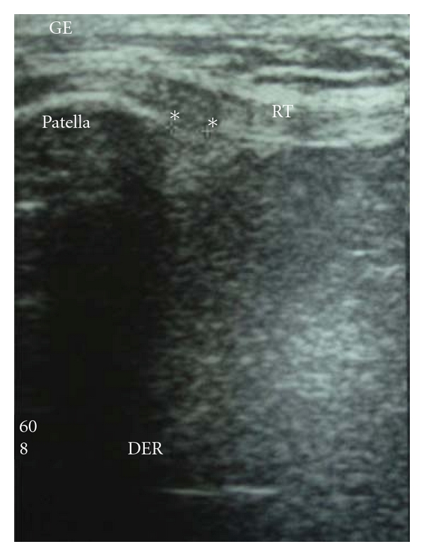
UltrasoundLongitudinal ultrasound of the patellar tendon: hypoechoic area of oedema within the tendon, below the tip of the patella, indicating tendinopathy.
Source : Pascual-Garrido et al., Stem Cells International, 2012, figure 2 · CC BY
Which questions should be asked to fully understand the patient and their history?
The history opens the whole reasoning process. 📝 It must characterise the pain, identify actionable risk factors and confirm the typical load-induced pattern.²,⁵- Precise location of pain : ask the patient to point with one finger. Typical patellar tendinopathy is almost pinpoint at the inferior pole of the patella (95% of cases, proximal insertion of the patellar tendon).¹ Diffuse, medial or retropatellar pain should prompt reconsideration of the diagnosis.
- Mode of onset : insidious and progressive over several weeks, typical of an overuse tendinopathy. Pain of sudden onset after a jump, with a sensation of "snapping", should raise suspicion of an avulsion fracture of the inferior pole or a partial rupture of the patellar tendon: a red flag.
- Dose-response load-pain relationship (pathognomonic): pain appears and intensifies during jumps, landings, decelerations, sprints, stair climbing/descent, deep squats. The “warm-up” pattern is common: initial pain that eases with the warm-up, then returns after the session and the following morning.²
- Recent changes in training load : return after a break, sudden increase in jump volume or match frequency, change of surface (indoor court → tarmac), of footwear, of coach or of programme.⁵
- Sporting profile : volleyball, basketball, jumping sports (gymnastics, diving, athletics: long jump, high jump, triple jump), fast running or changes of direction.⁶,⁷
- Personal history of tendinopathy (patellar or other): the most powerful risk factor for recurrence.⁹
- Psychosocial profile : fear of movement, catastrophising, beliefs about pain ("my tendon is going to rupture, I must stop"), level of self-efficacy, major predictors of chronicity (Mallows 2017).¹⁰
- Baseline VISA-P score : 8 questions (0-100, 100 = asymptomatic), the reference PROM, internationally validated since Visentini 1998.¹¹ It allows severity to be quantified objectively and progress to be tracked.
🚩 Red flags to screen for from the history onwards
- An audible “snap” after a jump + acute pain + inability to fully extend the knee actively → rupture of the patellar tendon or avulsion fracture of the inferior pole. An orthopaedic emergency (ultrasound or MRI).
- Constant night pain, sweats, weight loss → reconsider the diagnosis (systemic inflammatory or tumoral process) → blood tests and MRI.
- Hot swelling, fever, redness → cellulitis, septic prepatellar bursitis, septic arthritis → emergency.
- True joint locking, instability, a sense of giving way → meniscal lesion, ACL injury, symptomatic synovial plica → orthopaedic referral.
- Adolescent < 16 years with pain at the inferior pole of the patella → suspect Sinding-Larsen-Johansson disease (fragmentation/apophysitis of the inferior pole of the patella). Lateral radiograph.
- Adolescent with pain at the tibial tuberosity → Osgood-Schlatter disease. Lateral radiograph.
Which clinical tests should be performed and which other conditions must be ruled out?
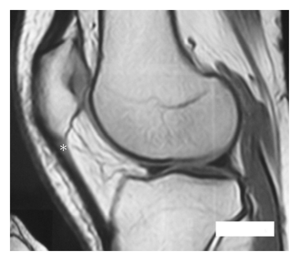
MRISagittal knee MRI: area of low signal in the proximal portion of the patellar tendon, marked with an asterisk, characteristic of patellar tendinopathy.
Source : Pascual-Garrido et al., Stem Cells International, 2012, figure 1 · CC BY
- Ankle dorsiflexion range (knee-to-wall test): a distance < 9-10 cm is a demonstrated risk factor (Backman 2011).¹²
- Quadriceps strength (isometric, Sorensen position or hand-held dynamometry): asymmetry ≥ 10% vs the unaffected side is informative.²
- Strength of the hip abductors and external rotators : weakness is associated with dynamic valgus on landing.¹³
- Qualitative single-leg squat / drop jump : observation of dynamic knee valgus, hip flexion and neuromuscular control.²,¹³
| Clinical test | What it assesses | Performance | Level of evidence |
|---|---|---|---|
| Palpation of the inferior pole of the patella | Reproduction of localised pain | High sensitivity, moderate specificity | High |
| Single-leg decline squat (SLDS) | Reproduction through specific loading | Highest sensitivity of the functional tests | High |
| Passive flexion sign | Distinguishing tendinopathy from other causes | High specificity | Moderate |
| VISA-P (PROM) | Overall severity and longitudinal follow-up | Excellent validity and responsiveness (MCID 13-15 pts) | High |
| Knee-to-wall (dorsiflexion) | Ankle risk factor | Good reliability (Backman 2011) | Moderate |
| Isometric quadriceps strength | Functional indicator for RTS | Predictive of return to sport (symmetry > 90%) | High |
| Ultrasound / MRI | Anatomical work-up as a second-line investigation | Weak correlation between symptoms and images | Low for primary diagnosis |
- Suspicion of patellar tendon rupture or avulsion fracture of the inferior pole (ultrasound or MRI).
- Failure of at least 12 weeks of well-conducted conservative treatment → reassess the diagnosis.
- Suspicion of an alternative diagnosis (meniscal lesion, symptomatic plica, stress fracture, tumoral process).
- A surgical referral, as part of the preoperative work-up.
- Patellofemoral pain syndrome (PFPS) : diffuse retro/peripatellar pain, worsened by prolonged sitting (the theatre sign) and by climbing/descending stairs and deep squatting. No pain localised at the inferior pole of the patella.¹⁵
- Infrapatellar fat pad syndrome (Hoffa's syndrome) : pain on either side of the patellar tendon (and not over the tendon itself), worsened in hyperextension. Hoffa's test (pressure beside the tendon in extension reproduces the pain, unlike the squat).
- Symptomatic medial synovial plica : anteromedial pain, a snapping sensation, sometimes with associated bursitis. Test for the plica snap sign. The Abdou case (probably fabricated), but Sansone 2022 shows that it is possible in the adolescent footballer.¹⁶
- Meniscal lesion (notably the anterior horn of the medial meniscus): pain in deep flexion, locking, positive McMurray. The Sansone 2022 case (PMC9512301) illustrates the typical diagnostic error in an adolescent footballer initially treated for patellar tendinopathy.¹⁶
- Prepatellar bursitis : superficial anterior swelling (above the patella), pain on direct pressure over the bursa.
- Sinding-Larsen-Johansson disease (child aged 10-13): pain at the inferior pole of the patella with radiological fragmentation. Apophysitis of the proximal insertion of the tendon.
- Osgood-Schlatter disease (adolescent aged 11-15): pain at the tibial tuberosity (distal insertion of the tendon). Apophysitis.
- Quadriceps tendinopathy (above the patella): rarer, found in the elite jumper and the weightlifter.
- Injury of the medial patellofemoral ligament / patellar instability : episodes of dislocation/subluxation.
Should patients be classified (VISA-P, clinical stages) and what are the benefits?
Yes, classification is useful for standardising, monitoring and stratifying. Several systems coexist:- The VISA-P score (Victorian Institute of Sport Assessment-Patella) of Visentini 1998: the internationally referenced PROM.¹¹ 8 questions scored from 0 to 100: daily pain, functional capacity, sporting participation. A score of 100 = asymptomatic. MCID 13-15 points. Recommended for baseline measurement, follow-up and return-to-sport criteria.
- Blazina clinical staging (modified): useful for teaching and for adapting load management:¹⁷
- Stage 1 : pain only after activity, with no functional limitation. Sport possible with adjustments.
- Stage 2 : pain at the start of activity (warm-up), during and after, but performance preserved. Training modifications.
- Stage 3 : pain during and after, performance limited. Significant reduction or temporary cessation of high-load activities.
- Stage 4 : pain during activities of daily living, increased risk of rupture. Cessation of sport, intensive management.
- Cook-Purdam continuum (reactive/dysrepair/degenerative): guides load modulation (chapter 1).¹⁵
Key points
- The diagnosis is essentially clinical. Imaging is not needed as a first-line investigation (weak image-symptom correlation, McAuliffe 2016).
- Diagnostic triad : pain very localised at the inferior pole of the patella + a dose-response relationship with load (jumps, decelerations) + reproduction on the single-leg decline squat (SLDS).
- The VISA-P score (Visentini 1998) is the reference PROM (0-100). MCID 13-15 points.
- Systematic complementary assessment of the kinetic chain : ankle dorsiflexion, quadriceps and hip strength, landing mechanics.
- Red flags to screen for: rupture/avulsion fracture (snap + loss of extension), infection/tumour, true joint locking.
- Critical differential diagnoses: patellofemoral pain syndrome, Hoffa's syndrome, plica, meniscal lesion (Sansone 2022), Sinding-Larsen-Johansson in the child, Osgood-Schlatter in the adolescent.
- Useful classifications: VISA-P (severity), Blazina stages (load modulation), Cook-Purdam continuum (tissue state).
References
- Malliaras P, Cook J, Purdam C, Rio E. Patellar Tendinopathy: Clinical Diagnosis, Load Management, and Advice for Challenging Case Presentations. J Orthop Sports Phys Ther. 2015;45(11):887-898. PMID 26390269.
- Rudavsky A, Cook J. Physiotherapy management of patellar tendinopathy (jumper's knee). J Physiother. 2014;60(3):122-129. PMID 25092419.
- Cook JL, Khan KM, Kiss ZS, Coleman BD, Griffiths L. Asymptomatic hypoechoic regions on patellar tendon ultrasound: A 4-year clinical and ultrasound followup of 46 tendons. Scand J Med Sci Sports. 2001;11(6):321-327. PMID 11782263.
- Docking SI, Cook J. How do tendons adapt? Going beyond tissue responses to understand positive adaptation and pathology development: A narrative review. J Musculoskelet Neuronal Interact. 2019;19(3):300-310. PMID 31475937.
- Theodorou A, Komnos G, Hantes M. Patellar tendinopathy: an overview of prevalence, risk factors, screening, diagnosis, treatment and prevention. Arch Orthop Trauma Surg. 2023;143(11):6695-6705. PMID 37542006.
- Nutarelli S, da Lodi CMT, Cook JL, Deabate L, Filardo G. Epidemiology of Patellar Tendinopathy in Athletes and the General Population: A Systematic Review and Meta-analysis. Orthop J Sports Med. 2023;11(6):23259671231173659. PMID 37347023.
- Lian OB, Engebretsen L, Bahr R. Prevalence of jumper's knee among elite athletes from different sports: a cross-sectional study. Am J Sports Med. 2005;33(4):561-567. PMID 15722279.
- Hägglund M, Zwerver J, Ekstrand J. Epidemiology of patellar tendinopathy in elite male soccer players. Am J Sports Med. 2011;39(9):1906-1911. PMID 21642599.
- Sprague AL, Smith AH, Knox P, Pohlig RT, Grävare Silbernagel K. Modifiable risk factors for patellar tendinopathy in athletes: a systematic review and meta-analysis. Br J Sports Med. 2018;52(24):1575-1585. PMID 30054341.
- Mallows A, Debenham J, Walker T, Littlewood C. Association of psychological variables and outcome in tendinopathy: a systematic review. Br J Sports Med. 2017;51(9):743-748. PMID 27852585.
- Visentini PJ, Khan KM, Cook JL, Kiss ZS, Harcourt PR, Wark JD. The VISA score: an index of severity of symptoms in patients with jumper's knee (patellar tendinosis). Victorian Institute of Sport Tendon Study Group. J Sci Med Sport. 1998;1(1):22-28. PMID 9732118.
- Backman LJ, Danielson P. Low range of ankle dorsiflexion predisposes for patellar tendinopathy in junior elite basketball players: a 1-year prospective study. Am J Sports Med. 2011;39(12):2626-2633. PMID 21917610.
- Mendonça LD, Camargo AC, Lima AM, et al. Association of hip and foot factors with patellar tendinopathy (jumper's knee) in athletes. J Orthop Sports Phys Ther. 2018;48(8):676-684. PMID 29792104.
- McAuliffe S, McCreesh K, Culloty F, Purtill H, O'Sullivan K. Can ultrasound imaging predict the development of Achilles and patellar tendinopathy? A systematic review and meta-analysis. Br J Sports Med. 2016;50(24):1516-1523. PMID 27633025.
- Cook JL, Purdam CR. Is tendon pathology a continuum? Br J Sports Med. 2009;43(6):409-416. PMID 18812414.
- Petrolo A. Meniscal lesion or patellar tendinopathy? A case report of an adolescent soccer player with knee pain. J Can Chiropr Assoc. 2022;66(2):157-171. PMID 36275082.
- Blazina ME, Kerlan RK, Jobe FW, Carter VS, Carlson GJ. Jumper's knee. Orthop Clin North Am. 1973;4(3):665-678. PMID 4783891. [Founding article of the clinical classification].
What are the most effective treatment strategies for patellar tendinopathy?
Where to start? What is the recommended hierarchy of interventions?
The Malliaras 2015 synthesis (JOSPT) and the Theodorou 2023 overview (Arch Orthop Trauma Surg) are aligned: two non-negotiable pillars before any other intervention.³,⁴- Patient education and load modulation : explain the non-inflammatory overuse nature of the condition, dismantle the myth of "tendinitis" requiring complete rest, introduce the pain-monitoring model (Silbernagel 2007), set realistic expectations (recovery = 12 weeks to 6 months, non-linear), and make the patient responsible for managing jumping and load.³,⁵
- A progressive exercise programme loading the quadriceps and the patellar tendon: the cornerstone of treatment, high level of evidence (GRADE high). The PTLE of Breda 2021 and the HSR of Kongsgaard 2009 are the best-validated protocols.²,⁶,⁷
🔺 Treatment pyramid for patellar tendinopathy
From the most invasive (apex) to the most universal (base): a hierarchy of intent
⚠️ Critical reading: the percentages are expert clinical estimates illustrating the hierarchy of therapeutic intent according to the SR/MAs (Malliaras 2015, Challoumas 2021 NMA, Theodorou 2023), not drawn from an epidemiological study. The hierarchy itself is solidly established.
What is the role of exercise (PTLE, HSR, isometrics) and is one approach superior?
🏋️ Exercise is the best-documented active treatment, with a high level of evidence.²,³,⁶ Three main protocols have robust data and shape contemporary practice:- Progressive Tendon-Loading Exercise (PTLE). Breda 2021 : pivotal RCT (Br J Sports Med, n=76 athletes aged 18-35 with clinically diagnosed PT confirmed on ultrasound, 24-week follow-up). A protocol in 4 progressive phases : (1) analgesic isometric → (2) isotonic with increasing load → (3) energy storage (progressive plyometric jumps) → (4) sport-specific. PTLE significantly outperformed the classic eccentric decline protocol on the change in VISA-P score at 24 weeks (+28 vs +18 points) and achieved a higher return-to-sport rate (43% vs 27%).⁶ Today it is the reference protocol in sports medicine centres.
- Heavy Slow Resistance (HSR): Kongsgaard 2009 : 3 sets × 6 to 15 RM performed slowly (3 sec concentric + 3 sec eccentric), 3 times a week, for 12 weeks. Exercises: single-leg squat on a machine or with a barbell, heavy leg press, hack squat. External load increased progressively from 15 RM (weeks 1-3) to 6 RM (weeks 9-12). The original RCT (n=39 men, 12 weeks) showed equivalence with eccentric decline training at 12 weeks but superiority at 6 months for patient satisfaction and collagen normalisation.⁷ Advantage: only 3 sessions/week (vs 14 with Alfredson), better adherence in practice.
- Eccentric decline squat (Young/Purdam protocol) : single-leg squat on a board with a 25° decline (which forces knee flexion with the knee ahead of the toes), 3 × 15 repetitions, twice a day, for 12 weeks, progressing by load (weighted backpack). This is the historical protocol for patellar tendinopathy (Visnes & Bahr 2007, critical SR). It has shown positive but inconsistent results as monotherapy; PTLE and HSR have overtaken it.¹⁰
- Analgesic isometrics (Rio 2015) : isometric knee extension 5 × 45 seconds, 70% MVIC, 1-2 times a day. The crossover trial of Rio 2015 (BJSM, n=6 in-season) showed an immediate reduction in pain (~7.0 → 0.17 VAS) persisting ≥ 45 minutes together with an increase in MVIC. A pivotal tool for the acute reactive phase, for in-season management, or as the first phase of PTLE. They do not replace a long-term progressive loading programme.¹¹
⚖️ Comparison of exercise protocols for patellar tendinopathy
Mean change in VISA-P score (landmark trials and NMA)
Sources: Breda 2021 (pivotal RCT, n=76, BJSM, DOI 10.1136/bjsports-2020-103403), Kongsgaard 2009 (RCT, PMID 19793213), Rio 2015 (isometric crossover trial, PMID 25979840), Challoumas 2021 NMA (PMID 34900334). PTLE and HSR are today the first-line protocols because of their documented superiority over eccentric decline training alone and their better adherence (3 sessions/week).
Adjunctive therapies: what is the value of shockwave, PRP and manual therapy?
💡 All adjunctive modalities must be considered as an addition to an exercise programme, never as a substitute for it.²,³ Extracorporeal shockwave therapy (ESWT) : the Charles 2023 SR/MA (Front Immunol, RCTs analysed across 3 tendinopathies) shows that ESWT alone has a modest short-term effect on pain in patellar tendinopathy vs placebo, but that the combination ESWT + eccentric exercise is superior to exercise alone, particularly for chronic cases.¹² The dedicated Stania 2020 SR/MA (J Sports Med Phys Fitness) confirms this trend. Practical recommendation: reserve ESWT for chronic cases (> 3 months of symptoms) who have already been on an exercise programme for at least 12 weeks without sufficient improvement. Always in combination with exercise. Moderate level of evidence. PRP (Platelet-Rich Plasma) : the Andriolo 2019 SR/MA (Am J Sports Med) had suggested some usefulness for multiple PRP injections, though not superior to exercise, and with poor methodological quality.⁸ The Challoumas 2021 NMA and the Everhart 2017 SR confirm the absence of clinically significant superiority of PRP over sham or over progressive exercise alone.²,⁹ The Andia 2020 SR/MA (Br Med Bull) concurs: low-quality evidence, cautious recommendation.¹³ PRP is not recommended routinely ; it may be discussed after failure of at least 6 months of structured conservative treatment, as part of a shared decision. Intratendinous or peritendinous corticosteroids : strongly discouraged. Documented risk of tendon rupture, high recurrence rate, structural alteration of collagen.²,³ Manual therapy (deep transverse friction, joint mobilisations of the ankle/hip): limited evidence as monotherapy. It may be justified to improve limited ankle mobility (dorsiflexion) or hip mobility (internal rotation, abductors) that sustains deficient landing mechanics, but it must not form the core of treatment.² The Acebes-Hueto 2024 SR (Healthcare) on soft-tissue techniques + exercise finds a modest adjunctive effect.¹⁴ Other modalities (low-level laser, therapeutic ultrasound, electrical stimulation, taping, dry needling): insufficient or heterogeneous evidence for a strong recommendation. The Frontiers 2024 SR on dry needling + exercise finds an additional benefit, but the net effect of dry needling as monotherapy remains to be confirmed.| Modality | Main indication | Level of evidence | Expected effect |
|---|---|---|---|
| Education + load modulation | All patients | High | ↗ adherence, ↘ recurrences, ↘ fear of movement |
| PTLE (Breda 2021) | All patients, 24 wks | High | +28 VISA-P pts at 24 wks, RTS 43% |
| HSR (Kongsgaard 2009) | All patients, 12 wks | High | +27 VISA-P pts at 6 months, good adherence |
| Analgesic isometrics (Rio 2015) | Reactive phase, in-season | Moderate | Immediate analgesia ≥ 45 min, MVIC ↑ |
| Eccentric decline alone | Off-season, motivated athletes | Moderate | +18 VISA-P pts (Breda 2021), inferior to PTLE |
| ESWT (chronic cases) | Adjunct after failure of 12 wks of exercise | Moderate | ↘ pain in combination with exercise (Charles 2023) |
| Manual therapy (ankle/hip) | Associated mobility restriction | Low to moderate | Adjunct if mobility is limited |
| Intratendinous PRP | Refractory cases > 6 months | Low (Andriolo 2019, Challoumas 2021) | Not superior to exercise alone |
| Intra/peritendinous corticosteroids | — | Risk of rupture, recurrences ↑ | ⚠️ To be avoided |
| Ultrasound / laser / TENS | Symptomatic adjunct | Low | Insufficient evidence |
| Surgery (debridement, tenotomy) | Failure ≥ 6 months | Exceptional recourse | No superiority vs well-conducted exercise (Everhart 2017) |
Beyond the physical: how to address psychological factors?
🧠 Psychological factors are a major determinant of chronicity and are frequently under-treated. The Mallows 2017 systematic review (BJSM) showed that kinesiophobia (fear of movement), catastrophising (a negative, amplified interpretation of pain) and low self-efficacy are independent predictors of an unfavourable outcome at 12 months in tendinopathy generally: including the patellar form.¹⁵ Patient education must be active and proactive on four points:- Reframe the pain : pain that is tolerable (≤ 5/10 VAS) during the exercises, that does not worsen during effort and that returns to baseline within 24 hours, is acceptable and even productive for adapting the tendon (Silbernagel 2007 model).¹⁶
- Dismantle the myths : “my tendon will not rupture because I do heavy squats”, “complete rest aggravates rather than heals”, “an abnormal scan does not mean serious, lasting pain”.
- Set realistic expectations : recovery = 3 to 6 months for a first episode, 6 to 12 months in chronic cases, non-linear, with expected fluctuations. This information prevents discouragement when a bad day follows a week of improvement.
- Self-efficacy : give the patient concrete tools (a training diary, a daily VAS, simple criteria for adjusting jump load) so that they become an agent of their own recovery rather than a passive recipient.¹⁷
Critique and controversy
The war of the exercise protocols is evolving: although eccentric decline training was long regarded as the reference, the Breda 2021 RCT and the Challoumas 2021 NMA have called that supremacy into question. The PTLE of Breda, integrating the 4 progressive phases (isometric → isotonic → plyometric → sport-specific), is today better supported by the evidence than eccentric decline training alone.²,⁶ The HSR of Kongsgaard remains an excellent compromise between adherence and effect, particularly useful in patients for whom adherence is an issue.⁷ More problematic: the injection industry (PRP, sclerosants, stem cells, polidocanol) continues to grow despite solid scientific data against its superiority over progressive exercise.²,⁸ The clinician must be careful not to give in to commercial pressure or to patient expectations for unvalidated “modern treatments”. Finally, the imaging-pain paradox remains a major clinical trap: tendons that look “healed” on ultrasound can remain painful, and vice versa.¹⁸ Return-to-sport decisions must not be based on follow-up imaging, but on functional criteria and the VISA-P (chapter 4).Key points
- ✅ First line: education + load modulation + a progressive exercise programme over 12+ weeks. High level of evidence.
- ✅ The 4-phase PTLE (Breda 2021) is today the reference protocol: superior to classic eccentric decline training at 24 weeks (+28 vs +18 VISA-P pts, RTS 43% vs 27%).
- ✅ HSR (Kongsgaard 2009) is an excellent alternative: 3 sessions/week, good adherence, an effect equivalent to PTLE in VISA-P terms.
- ✅ Isometrics (Rio 2015) : immediate analgesia ≥ 45 min, a pivotal tool for the acute reactive phase and for in-season management.
- ✅ Shockwave therapy (ESWT) : a valid adjunct for chronic refractory cases after 12 wks of exercise. Always in combination.
- ❌ PRP is not superior to exercise alone (Andriolo 2019, Challoumas 2021 NMA). Not for routine use.
- ❌ Intra/peritendinous corticosteroids are contraindicated (risk of rupture, recurrences).
- ✅ Address psychosocial factors systematically : kinesiophobia and catastrophising are major predictors of failure (Mallows 2017). Pain-monitoring model ≤ 5/10.
References
- Theodorou A, Komnos G, Hantes M. Patellar tendinopathy: an overview of prevalence, risk factors, screening, diagnosis, treatment and prevention. Arch Orthop Trauma Surg. 2023;143(11):6695-6705. PMID 37542006.
- Challoumas D, Pedret C, Biddle M, et al. Management of patellar tendinopathy: a systematic review and network meta-analysis of randomised studies. BMJ Open Sport Exerc Med. 2021;7(4):e001110. PMID 34900334.
- Malliaras P, Cook J, Purdam C, Rio E. Patellar Tendinopathy: Clinical Diagnosis, Load Management, and Advice for Challenging Case Presentations. J Orthop Sports Phys Ther. 2015;45(11):887-898. PMID 26390269.
- Rudavsky A, Cook J. Physiotherapy management of patellar tendinopathy (jumper's knee). J Physiother. 2014;60(3):122-129. PMID 25092419.
- Cook JL, Purdam CR. Is tendon pathology a continuum? Br J Sports Med. 2009;43(6):409-416. PMID 18812414.
- Breda SJ, Oei EHG, Zwerver J, Visser E, Waarsing E, Krestin GP, de Vos RJ. Effectiveness of progressive tendon-loading exercise therapy in patients with patellar tendinopathy: a randomised clinical trial. Br J Sports Med. 2021;55(9):501-509. DOI 10.1136/bjsports-2020-103403.
- Kongsgaard M, Kovanen V, Aagaard P, et al. Corticosteroid injections, eccentric decline squat training and heavy slow resistance training in patellar tendinopathy. Scand J Med Sci Sports. 2009;19(6):790-802. PMID 19793213.
- Andriolo L, Altamura SA, Reale D, Candrian C, Zaffagnini S, Filardo G. Nonsurgical Treatments of Patellar Tendinopathy: Multiple Injections of Platelet-Rich Plasma Are a Suitable Option: A Systematic Review and Meta-analysis. Am J Sports Med. 2019;47(4):1001-1018. PMID 29601207.
- Everhart JS, Cole D, Sojka JH, et al. Treatment Options for Patellar Tendinopathy: A Systematic Review. Arthroscopy. 2017;33(4):861-872. PMID 28110807.
- Visnes H, Bahr R. The evolution of eccentric training as treatment for patellar tendinopathy (jumper's knee): a critical review of exercise programmes. Br J Sports Med. 2007;41(4):217-223. PMID 17261559.
- Rio E, Kidgell D, Purdam C, et al. Isometric exercise induces analgesia and reduces inhibition in patellar tendinopathy. Br J Sports Med. 2015;49(19):1277-1283. PMID 25979840.
- Charles R, Fang L, Zhu R, Wang J. The effectiveness of shockwave therapy on patellar tendinopathy, Achilles tendinopathy, and plantar fasciitis: a systematic review and meta-analysis. Front Immunol. 2023;14:1193835. PMID 37662911.
- Andia I, Martin JI, Maffulli N. Platelet-rich plasma and the conservative management of athletic tendinopathy. Curr Rheumatol Rep. 2018;20(10):63. [Reference for the PRP field, complementing Andriolo 2019].
- Ragone F, Pérez-Guillén S, Carrasco-Uribarren A, et al. The Effects of Soft-Tissue Techniques and Exercise in the Treatment of Patellar Tendinopathy-Systematic Review and Meta-Analysis. Healthcare (Basel). 2024;12(4):427. PMID 38391804.
- Mallows A, Debenham J, Walker T, Littlewood C. Association of psychological variables and outcome in tendinopathy: a systematic review. Br J Sports Med. 2017;51(9):743-748. PMID 27852585.
- Silbernagel KG, Thomeé R, Eriksson BI, Karlsson J. Continued sports activity, using a pain-monitoring model, during rehabilitation in patients with Achilles tendinopathy: a randomized controlled study. Am J Sports Med. 2007;35(6):897-906. PMID 17307888.
- Chimenti RL, Frey-Law LA, Sluka KA. A Mechanism-Based Approach to Physical Therapist Management of Pain. Phys Ther. 2018;98(5):302-314. PMID 29669091.
- Docking SI, Cook J. Pathological tendons maintain sufficient aligned fibrillar structure on ultrasound tissue characterization (UTC). Scand J Med Sci Sports. 2016;26(6):675-683. PMID 26059532.
How can lasting recovery be achieved and recurrence of patellar tendinopathy prevented?
How can the patient become an active participant through the pain-monitoring model?
🎯 The Silbernagel 2007 pain-monitoring model transformed management by demonstrating, in a randomised trial (n = 38), that continuing sport during rehabilitation is not only safe but also effective, provided a simple rule is followed:²- Pain that is tolerable (≤ 5/10 VAS) during effort.
- Pain that does not worsen from session to session.
- Pain that returns to baseline within 24 hours of the effort.
- Morning stiffness that does not worsen week after week.
🚦 The Silbernagel pain-monitoring model
A simple rule for allowing, adjusting or stopping activity
Adapted from Silbernagel KG et al. Am J Sports Med. 2007;35(6):897-906 (PMID 17307888) and from the JOSPT RTS program 2015.
- A weekly training diary : jump volume (number, intensity), morning and post-exercise pain VAS, morning stiffness. It makes trends objective and allows decisions free of emotional bias.³
- Monthly VISA-P : a 0-100 score to make progress objective. An improvement of 13-15 points is clinically significant (MCID).⁴
- Maintaining the strengthening programme in the long term: at least 2-3 sessions of HSR or loaded squats per week, even after complete symptom resolution, to maintain tendon capacity.⁵
- Regulating external load : the +10%/week maximum rule for weekly jump volume, a season plan including 1 deload week every 3-4 weeks, and monitoring of the acute:chronic load ratio in high-level athletes.⁶
When and how should a safe return to sport be planned?
🏃 Return to sport (RTS) must never be based on elapsed time alone or on the absence of pain. The PTLE protocol of Breda 2021 now structures RTS into 4 progressive phases with objective functional criteria.⁷ Minimum criteria to meet before resuming jumping / sport-specific work :- No pain on the single-leg decline squat (SLDS) repeated 5 times consecutively.
- Quadriceps strength symmetry ≥ 90% vs the unaffected side (hand-held dynamometry or isokinetic measurement).⁸
- Hop tests (single-leg hop, triple hop, side hop) with asymmetry < 10% vs the unaffected side.
- VISA-P > 80/100 or a clinically significant improvement of ≥ 15 points from baseline.
- Tolerance of bilateral then unilateral jumping, and drop-jump landing with good neuromuscular control (no dynamic valgus).
- Strength of the hip abductors and external rotators symmetrical: a point that is often neglected (Mendonça 2018).⁹
- Phase 1: Pain management (weeks 0-4): analgesic isometrics (Rio 2015), modulation of sporting load, education about tolerable pain. No jumping. Low-impact activities may be continued if tolerated (cycling, swimming, upper-body pulling work).
- Phase 2: Slow isotonic strength (weeks 4-12): HSR or slow squats with progressive load, 3 sessions/week. Concurrent work on the kinetic chain (ankle, hip). No plyometrics.
- Phase 3, Energy storage (progressive plyometrics) (weeks 8-16): graded introduction of jumping, bilateral static → bilateral dynamic → unilateral static → unilateral dynamic. Monitor pain at 24h.
- Phase 4: Sport-specific (weeks 12-24): reintroduction of specific movements (spike, dunk, running, decelerations, changes of direction), progressive increase in competition volume, maintenance of the strengthening programme.
Critique and controversy
The threshold of “5/10 VAS” in the Silbernagel model is a widely adopted clinical heuristic , but its exact cut-off has never been formally validated on a large scale in patellar tendinopathy (the landmark Silbernagel 2007 trial concerned the Achilles tendon). It may be too permissive for some patients (catastrophisers, kinesiophobes) and too restrictive for others (high-level athletes used to tolerating pain). Personalisation remains necessary according to psychological profile and activity level. The return-to-sport criteria (symmetry ≥ 90%, VISA-P > 80) are logical, but their predictive value for non-recurrence in the long term remains only moderately established. Most studies measure the return-to-sport rate, not the recurrence rate at 2-3 years in patellar tendinopathy. Finally, in everyday practice these criteria require time, equipment (a hand-held dynamometer, a decline board) and specific training: the risk is that they remain theoretical and that the clinician allows return to sport on looser criteria (no pain at rest), which is highly insufficient and increases the risk of recurrence.Key points
- The Silbernagel 2007 pain-monitoring model (transposed to PT) is the pivotal self-management tool: pain ≤ 5/10 VAS, without worsening, back to baseline within 24h → green zone, carry on.
- Concrete tools for the patient: a jump diary, monthly VISA-P, long-term maintenance of the HSR/PTLE programme.
- The return to sport must be based on objective functional criteria : no pain on the SLDS, quadriceps strength symmetry > 90%, hop tests with < 10% asymmetry, VISA-P > 80, neuromuscular control on the drop jump.
- Programming in 4 progressive phases (Breda's PTLE) over 12-24 weeks: pain management → isotonic strength → plyometrics → sport-specific.
- Realistic trajectory: perceived improvement at 4-8 weeks, peak at 12-24 weeks, return to competition between 3 and 12 months depending on the profile. More than 50% of athletes had to stop their sport without structured intervention (Kettunen 2002): maintaining the programme is essential.
References
- Kettunen JA, Kvist M, Alanen E, Kujala UM. Long-term prognosis for jumper's knee in male athletes. A prospective follow-up study. Am J Sports Med. 2002;30(5):689-692. PMID 12239003.
- Silbernagel KG, Thomeé R, Eriksson BI, Karlsson J. Continued sports activity, using a pain-monitoring model, during rehabilitation in patients with Achilles tendinopathy: a randomized controlled study. Am J Sports Med. 2007;35(6):897-906. PMID 17307888.
- Malliaras P, Cook J, Purdam C, Rio E. Patellar Tendinopathy: Clinical Diagnosis, Load Management, and Advice for Challenging Case Presentations. J Orthop Sports Phys Ther. 2015;45(11):887-898. PMID 26390269.
- Visentini PJ, Khan KM, Cook JL, et al. The VISA score: an index of severity of symptoms in patients with jumper's knee (patellar tendinosis). J Sci Med Sport. 1998;1(1):22-28. PMID 9732118.
- Kongsgaard M, Kovanen V, Aagaard P, et al. Corticosteroid injections, eccentric decline squat training and heavy slow resistance training in patellar tendinopathy. Scand J Med Sci Sports. 2009;19(6):790-802. PMID 19793213.
- Gabbett TJ. The training-injury prevention paradox: should athletes be training smarter and harder? Br J Sports Med. 2016;50(5):273-280. PMID 26758673.
- Breda SJ, Oei EHG, Zwerver J, Visser E, Waarsing E, Krestin GP, de Vos RJ. Effectiveness of progressive tendon-loading exercise therapy in patients with patellar tendinopathy: a randomised clinical trial. Br J Sports Med. 2021;55(9):501-509. DOI 10.1136/bjsports-2020-103403.
- Rio E, Kidgell D, Purdam C, et al. Isometric exercise induces analgesia and reduces inhibition in patellar tendinopathy. Br J Sports Med. 2015;49(19):1277-1283. PMID 25979840.
- Mendonça LD, Camargo AC, Lima AM, et al. Association of hip and foot factors with patellar tendinopathy (jumper's knee) in athletes. J Orthop Sports Phys Ther. 2018;48(8):676-684. PMID 29792104.
- Mallows A, Debenham J, Walker T, Littlewood C. Association of psychological variables and outcome in tendinopathy: a systematic review. Br J Sports Med. 2017;51(9):743-748. PMID 27852585.
- Rudavsky A, Cook J. Physiotherapy management of patellar tendinopathy (jumper's knee). J Physiother. 2014;60(3):122-129. PMID 25092419.
Kinetic chain & vulnerable subgroup: what to do with recalcitrant tendinopathy?
Ankle and hip: screening for the deficits that sustain overload
🔄 Three areas must be assessed systematically in the jumping athlete with patellar tendinopathy: 1. Ankle dorsiflexion (knee-to-wall) : Backman & Danielson 2011 (Am J Sports Med, 1-year prospective cohort, 90 junior elite basketball players) demonstrated that athletes with limited dorsiflexion range (< 36.5°) developed ~2× more patellar tendinopathy over the season.³ Mechanism: reduced dorsiflexion is compensated by anterior tibial translation and greater knee flexion on landing, increasing the knee extensor moment and therefore the load on the patellar tendon. Clinical test: knee-to-wall (KTW); target > 10 cm bilaterally or symmetry < 1 cm. Treatment: talocrural mobilisations, specific stretching of the triceps surae (gastrocnemius + soleus), and self-mobilisation at home. 2. Strength and activation of the hip stabilisers : abductors (gluteus medius), external rotators (piriformis, gemelli). A documented weakness is associated with dynamic knee valgus on landing and with mechanical overload of the patellar tendon.²,⁴ Tests: single-leg squat (observation of valgus), Trendelenburg, dynamometry of hip abduction. If the deficit is ≥ 15% vs the unaffected side → targeted strengthening (clamshells, single-leg deadlift, hip thrust, side plank with abduction). 3. Landing mechanics (drop jump) : Bisseling 2008 (Br J Sports Med) showed in a cohort of 24 volleyball players that patients with patellar tendinopathy use a “stiffer” landing strategy : higher peak landing force, less hip flexion, a “stiff-legged” landing.⁵ Rehabilitation: drop jump with qualitative coaching (“land softly, bend hip + knee + ankle at the same time”), work on shock-absorption reflexes, video feedback.🔗 Decision tree: recalcitrant patellar tendinopathy
After failure of 12 weeks of well-conducted progressive exercise (PTLE / HSR)
Adapted from Malliaras 2015 (JOSPT, PMID 26390269), Mendonça 2018 (kinetic chain, PMID 29792104), Theodorou 2023 (overview, PMID 37542006).
Stratifying a recalcitrant tendinopathy: red flags and referral
🚩 Before concluding that a tendinopathy is “recalcitrant”, you must systematically reconsider the diagnosis and rule out:- Subclinical avulsion fracture of the inferior pole of the patella, especially if it began after a powerful jump. Lateral radiograph + ultrasound.
- Meniscal or plical lesion (Sansone 2022 illustrates the error), especially in the adolescent or if there is joint locking. MRI if suspected.
- Inflammatory disease: spondyloarthritis (chronic enthesitis), early rheumatoid arthritis. If night pain, morning stiffness > 1h, or involvement of other entheses, order blood tests (FBC, CRP, HLA-B27, RF, anti-CCP).
- Poorly controlled diabetes: a factor in poor response to exercise, possibly through altered collagen quality.
🚩 Red flags specific to recalcitrant cases
- Night pain, prolonged morning stiffness > 1h, involvement of other entheses → rheumatological work-up.
- Localised inflammatory swelling, fever, weight loss → urgent specialist referral.
- Joint locking, a sense of giving way, intra-articular swelling → MRI (meniscal lesion, plica, chondral lesion).
- Sudden worsening after a powerful jump with quadriceps weakness → suspicion of a partial rupture of the patellar tendon, urgent ultrasound or MRI.
- Vulnerable subgroups to screen for: the adolescent in a growth spurt (Sinding-Larsen-Johansson, Osgood-Schlatter), the athlete with multiple previous tendinopathies (a discomfort worth exploring), the woman with amenorrhoea + restrictive eating (REDs / female athlete triad).
⭐ Key points
- A patellar tendinopathy that is recalcitrant (failure ≥ 12 wks) requires you to reconsider the diagnosis and to assess systematically the kinetic chain.
- Ankle dorsiflexion < 36.5° doubles the risk (Backman 2011): measure it with the knee-to-wall test and treat with mobilisations + gastrocnemius/soleus stretching.
- Weakness of the hip abductors/external rotators + dynamic valgus on landing: targeted strengthening is mandatory (Mendonça 2018).
- Landing mechanics that are deficient (a “stiff-legged” landing): rehabilitation with drop jumps and coaching, video feedback.
- After a complete review, if failure persists at 6 months: discuss ESWT as an adjunct, or even a specialist surgical opinion. Further imaging (MRI) if not already done.
- Red flags: night pain / systemic inflammation → rheumatological work-up. Locking / instability → MRI. Sudden weakness after a jump → suspicion of rupture.
References
- Malliaras P, Cook J, Purdam C, Rio E. Patellar Tendinopathy: Clinical Diagnosis, Load Management, and Advice for Challenging Case Presentations. J Orthop Sports Phys Ther. 2015;45(11):887-898. PMID 26390269.
- Mendonça LD, Camargo AC, Lima AM, et al. Association of hip and foot factors with patellar tendinopathy (jumper's knee) in athletes. J Orthop Sports Phys Ther. 2018;48(8):676-684. PMID 29792104.
- Backman LJ, Danielson P. Low range of ankle dorsiflexion predisposes for patellar tendinopathy in junior elite basketball players: a 1-year prospective study. Am J Sports Med. 2011;39(12):2626-2633. PMID 21917610.
- Sprague AL, Smith AH, Knox P, et al. Modifiable risk factors for patellar tendinopathy in athletes: a systematic review and meta-analysis. Br J Sports Med. 2018;52(24):1575-1585. PMID 30054341.
- Bisseling RW, Hof AL, Bredeweg SW, Zwerver J, Mulder T. Are the take-off and landing phase dynamics of the volleyball spike jump related to patellar tendinopathy?. Br J Sports Med. 2008;42(6):483-489. PMID 18385187.
- Theodorou A, Komnos G, Hantes M. Patellar tendinopathy: an overview of prevalence, risk factors, screening, diagnosis, treatment and prevention. Arch Orthop Trauma Surg. 2023;143(11):6695-6705. PMID 37542006.
What do real-world case studies teach us about patellar tendinopathy?
A "classic" case: adolescent footballer: the diagnostic error of Sansone 2022
The case reported by Sansone V et al. 2022 (J Orthop Case Rep, PMID 36275082) illustrates a common diagnostic error.¹ A 16-year-old adolescent, a competitive football player, presented with anteromedial knee pain of 4 months' duration, worsened by jumping and decelerations. The initial diagnosis made was patellar tendinopathy; classic conservative treatment (eccentric exercises, stretching, relative rest) was put in place for 8 weeks without improvement. Repeat clinical examination found:- Anteromedial pain (and not strictly at the inferior pole of the patella).
- Pain in deep flexion and in deep squatting.
- Positive McMurray sign medially.
- Single-leg decline squat reproducing the familiar pain only poorly.
🧩 Sansone 2022 case: the correct diagnostic path
16-year-old adolescent footballer, failure of 8 weeks of “tendinopathy” treatment
Source: Sansone V, Maiorano E, Russo V, et al. J Orthop Case Rep. 2022;12(8):17-21. PMID 36275082. Case report: it illustrates, it does not demonstrate.
The diagnostic challenge: when anteromedial knee pain hides something else
Anteromedial knee pain in a jumping athlete is not always a patellar tendinopathy. ⚠️ Several conditions can mimic this diagnosis, sometimes with serious consequences if the diagnosis is delayed:- 🔍 Anterior meniscal lesion (anterior horn of the medial meniscus): Sansone 2022 illustrates this trap. Pain in deep flexion, positive McMurray, occasional locking. MRI to confirm.¹
- 🔍 Patellofemoral pain syndrome (PFPS): Diffuse retro/peripatellar pain, worsened by prolonged sitting (the theatre sign). Compression-extension test. No pain localised at the inferior pole of the patella.²
- 🔍 Symptomatic medial synovial plica: Anteromedial pain, a snapping sensation on flexion-extension. Positive plica snap test. The diagnosis is often delayed.
- 🔍 Fat pad syndrome (Hoffa's syndrome): Pain on either side of the patellar tendon, worsened in hyperextension. Hoffa's test: pressure beside the tendon in extension reproduces the pain, the squat less so.
- 🔍 Sinding-Larsen-Johansson disease in the child aged 10-13: Pain at the inferior pole of the patella with radiological fragmentation. Apophysitis of the proximal insertion of the tendon.
- 🔍 Osgood-Schlatter disease in the adolescent aged 11-15, Pain over the tibial tuberosity (distal insertion), a different population and location.
- 🔍 Avulsion fracture of the inferior pole of the patella: After a powerful jump with a snap and weakness of extension. Radiograph + ultrasound.
- 🔍 Partial ACL injury or microinstability: Especially in the young woman, jumping athlete, with abrupt decelerations. Lachman test and pivot shift.
- 🔍 Prepatellar bursitis: Superficial anterior swelling (above the patella), distinguished by its location.
A complex case study: patellar tendinopathy resolved by a spinal approach (Adriaensen 2025)
The Adriaensen et al. 2025 case (Int J Sports Phys Ther, PMC12129631) illustrates the complexity of the extended kinetic chain.³ A young sporting adult woman presented with a chronic unilateral patellar tendinopathy of 12 months' duration, refractory to standard treatment (eccentric exercise + HSR + ESWT for 6 months). Local factors (symmetrical quadriceps strength, adequate dorsiflexion) were normal. Systematic assessment using Mechanical Diagnosis and Therapy (MDT, the McKenzie method) revealed centralisation of the pain with repeated lumbar extensions: the anteromedial knee pain disappeared after a set of repeated extensions. A posture-dependent lumbar dysfunction was suspected as a source of referred pain or as a modulator of local sensitisation. The treatment introduced included:- Repeated lumbar extension exercises (McKenzie protocol): 10 × 10 reps × 5/day.
- Maintenance of the quadriceps HSR programme.
- Postural correction of the lumbar lordosis in sitting.
- A patellar tendinopathy that is refractory requires assessment of the kinetic chain in its widest extent (ankle, hip, but also the lumbopelvic and thoracic spine).
- The MDT/McKenzie method makes it possible to identify quickly (3-5 reassessments) a centralisable pain pattern, which changes the therapeutic strategy.
- The psychosocial components (kinesiophobia, catastrophising) must remain in the background of the reasoning (Mallows 2017).⁴
Critique and controversy, where do case studies sit in the hierarchy of evidence?
🧠 Case studies are fundamental for generating hypotheses and flagging rare presentations, but their level of evidence is the lowest in the whole scientific hierarchy. A single case illustrates a possibility, not a generality. It cannot control for placebo, regression to the mean or spontaneous recovery.⁹ Publication bias is massive (failures are rarely published).📐 The hierarchy of scientific evidence, where does each study type sit?
Strength of evidence decreasing from the top (meta-analyses) to the bottom (isolated cases)
Simplified GRADE / Oxford CEBM hierarchy. The length of the coloured bar on the right illustrates the relative strength of evidence. Practical implication: where an appealing case study and a meta-analysis disagree, the decision must follow the meta-analysis. Case studies remain valuable for generating hypotheses, flagging rare presentations, or illustrating a clinical reasoning process.
⭐ Key points
- The Sansone 2022 case (J Orthop Case Rep, PMID 36275082) illustrates a common diagnostic error: a meniscal lesion can mimic a patellar tendinopathy in the adolescent footballer, hence the importance of reconsidering the diagnosis when treatment fails at 6-8 weeks.
- The Adriaensen 2025 case (PMC12129631) shows that a refractory patellar tendinopathy can be influenced by a spinal component identifiable with MDT/McKenzie: an argument for assessing the extended kinetic chain.
- Critical differential diagnoses : meniscal lesion, patellofemoral pain syndrome, synovial plica, Hoffa's syndrome, Sinding-Larsen-Johansson (child), Osgood-Schlatter (adolescent), avulsion fracture of the inferior pole, partial ACL injury, prepatellar bursitis.
- The kinetic chain (ankle dorsiflexion, hip strength/activation, landing mechanics) must be assessed systematically (Mendonca 2018, Backman 2011, Bisseling 2008).
- The psychosocial factors (kinesiophobia, catastrophising, Mallows 2017) shape the trajectory and must be addressed.
- ⚠️ Level of evidence : a case report = level 5 (the lowest). Where there is disagreement, follow the meta-analyses (level 1a), not the isolated case. Cases illustrate, they do not demonstrate.
References
- Petrolo A. Meniscal lesion or patellar tendinopathy? A case report of an adolescent soccer player with knee pain. J Can Chiropr Assoc. 2022;66(2):157-171. PMID 36275082.
- Crossley KM, Stefanik JJ, Selfe J, et al. 2016 Patellofemoral pain consensus statement from the 4th International Patellofemoral Pain Research Retreat. Br J Sports Med. 2016;50(14):839-843. PMID 27343241.
- Adriaensen E, Vandermeeren L, Heyrman B, et al. Patellar Tendinopathy Responding to Spinal Treatment: A Case Report Using Mechanical Diagnosis and Therapy. Int J Sports Phys Ther. 2025. PMC 12129631.
- Mallows A, Debenham J, Walker T, Littlewood C. Association of psychological variables and outcome in tendinopathy: a systematic review. Br J Sports Med. 2017;51(9):743-748. PMID 27852585.
- McAuliffe S, McCreesh K, Culloty F, Purtill H, O'Sullivan K. Can ultrasound imaging predict the development of Achilles and patellar tendinopathy? A systematic review and meta-analysis. Br J Sports Med. 2016;50(24):1516-1523. PMID 27633025.
- Challoumas D, Pedret C, Biddle M, et al. Management of patellar tendinopathy: a systematic review and network meta-analysis of randomised studies. BMJ Open Sport Exerc Med. 2021;7(4):e001110. PMID 34900334.
- Andriolo L, Altamura SA, Reale D, et al. Nonsurgical Treatments of Patellar Tendinopathy: Multiple Injections of Platelet-Rich Plasma Are a Suitable Option. Am J Sports Med. 2019;47(4):1001-1018. PMID 29601207.
- Mendonça LD, Camargo AC, Lima AM, et al. Association of hip and foot factors with patellar tendinopathy (jumper's knee) in athletes. J Orthop Sports Phys Ther. 2018;48(8):676-684. PMID 29792104.
- Nissen T, Wynn R. The clinical case report: a review of its merits and limitations. BMC Res Notes. 2014;7:264. PMID 24758689.
- Breda SJ, Oei EHG, Zwerver J, et al. Effectiveness of progressive tendon-loading exercise therapy in patients with patellar tendinopathy: a randomised clinical trial. Br J Sports Med. 2021;55(9):501-509. DOI 10.1136/bjsports-2020-103403.
- Bisseling RW, Hof AL, Bredeweg SW, Zwerver J, Mulder T. Are the take-off and landing phase dynamics of the volleyball spike jump related to patellar tendinopathy?. Br J Sports Med. 2008;42(6):483-489. PMID 18385187.
- Backman LJ, Danielson P. Low range of ankle dorsiflexion predisposes for patellar tendinopathy in junior elite basketball players: a 1-year prospective study. Am J Sports Med. 2011;39(12):2626-2633. PMID 21917610.
- Kettunen JA, Kvist M, Alanen E, Kujala UM. Long-term prognosis for jumper's knee in male athletes. A prospective follow-up study. Am J Sports Med. 2002;30(5):689-692. PMID 12239003.
How can these recommendations be applied concretely in your practice?
When and to which other healthcare professionals should you refer?
The first, non-negotiable step in triage is screening for red flags. The clinician must have a low threshold for referral: sensitivity takes precedence over specificity when serious conditions are at stake.🚩 Red flags specific to the patellar region
- Patellar tendon rupture : an audible/felt “snap” after a powerful jump + loss of active knee extension + acute pain + sometimes a palpable defect above the tibial tuberosity. An orthopaedic emergency (ultrasound + MRI).
- Avulsion fracture of the inferior pole of the patella : after acute trauma, exquisite tenderness on palpation, inability to extend fully. Lateral radiograph + MRI.
- Meniscal lesion (Sansone 2022 illustrates the diagnostic error): anteromedial pain, positive McMurray, joint locking, poorly reproductive SLDS. MRI if suspected.¹
- Infection (cellulitis, septic prepatellar bursitis, septic arthritis) : fever + hot swelling + exquisite pain + rapidly spreading erythema. An emergency.
- Tumoral process (rare but to be considered): a palpable mass of gradual onset, night pain, night sweats, unexplained weight loss, deterioration in general condition. MRI + blood tests.
- Systemic rheumatological disease : spondyloarthritis (multiple enthesitis, HLA-B27, axial morning stiffness, uveitis), early rheumatoid arthritis, gout (acute attack). Blood tests (FBC, CRP, anti-CCP, HLA-B27).
- Sinding-Larsen-Johansson disease (child aged 10-13) or Osgood-Schlatter (adolescent aged 11-15): pain at the inferior pole of the patella or at the tibial tuberosity during a growth spurt. Lateral radiograph.
- Recent treatment with fluoroquinolones or long-term systemic corticosteroids + acute tendon pain: increased risk of rupture, alert the prescribing doctor.
⚠️ Any red flag → prompt medical referral (general practitioner, emergency department, sports physician, orthopaedic surgeon) before or alongside physiotherapy management.
- Sports physician / general practitioner : full work-up, management of comorbidities, overall follow-up.
- Orthopaedic surgeon / knee surgeon : failure of ≥ 6 months of structured conservative treatment, suspicion of a complex intratendinous lesion, avulsion fracture, meniscal lesion (Everhart 2017).³
- Interventional radiologist : for the rare refractory cases where ultrasound-guided injections (PRP) are being considered as a shared decision, despite the limited evidence.
- Podiatrist / pedorthist : biomechanical analysis of jumping and landing, insoles if needed (rarely), footwear advice.
- Coach / strength and conditioning coach / club staff : review and restructuring of the training programme (jump volume, periodisation, recovery) to manage load in the long term.
- Psychologist specialising in pain : persistent kinesiophobia, catastrophising, a context of widespread chronic pain.
How can outcomes be measured and implementation barriers overcome?
📊 Measuring outcomes is the sine qua non of quality practice. The VISA-P score (Visentini 1998) remains the reference PROM for patellar tendinopathy, validated internationally.⁴ Although there is not yet a Core Outcome Set specific to patellar tendinopathy (unlike COS-AT 2024 for the Achilles tendon), the essential outcomes are agreed: Essential outcomes in patellar tendinopathy :- Pain : numerical rating scale (NRS) 0-10, measured at rest, on the SLDS, and during activities.
- Function and severity : VISA-P, 8 questions, score 0-100. MCID ≈ 13-15 points.⁴
- Participation : return to sporting activity, level of practice vs before the injury.
- Quality of life : EQ-5D or similar.
- Global rating of change by the patient (PGIC).
- Quadriceps strength and symmetry: hand-held or isokinetic dynamometry.
- Digital tools : a tablet or smartphone app that the patient completes in the waiting room, automatic calculation of the VISA-P score, graphical display of progress.
- Continuing education : practical workshops on using PROMs and on shared decision-making.
- Building it into the routine : VISA-P at every follow-up appointment (every 4-6 weeks), not only at the start and end of care.
- Shared decision-making : present the options to the patient (PTLE vs HSR vs eccentric decline alone), discuss the pros and cons (weekly volume, equipment, competition calendar), decide together. This significantly increases adherence and satisfaction.
Critique and controversy: beyond the guidelines
The red flag paradox still holds: no sign taken in isolation has a high positive predictive value. It is the cluster of findings + probabilistic clinical reasoning that guide referral, not a checklist. The risk is twofold: over-diagnosing (sending every painful knee for an MRI) or under-diagnosing (missing an avulsion fracture or a meniscal lesion by attributing the pain to a simple tendinopathy, as in the Sansone 2022 case).¹ The “knowing-doing” gap is particularly striking in patellar tendinopathy: the Challoumas 2021 NMA confirmed the absence of superiority of PRP as monotherapy and the primacy of progressive exercise, the Breda 2021 RCT established the superiority of PTLE over eccentric decline training alone, and yet everyday practice sometimes continues to impose a single eccentric protocol or to prescribe costly PRP injections without indication.⁵,⁶ Moving to genuine EBP requires more than disseminating information: it requires an overhaul of financial incentives and of continuing education. Finally, the tension between standardisation and personalisation is central. The VISA-P and functional criteria are essential for comparability, but the clinician must be able to adapt the programme to the patient's profile (elite athlete, deconditioned recreational athlete, adolescent in growth, patient with a dominant psychological component).Key points
- Referral is crucial for safety : red flags (patellar tendon rupture, avulsion fracture, meniscal lesion, infection, tumoral process) → prompt medical referral.
- Psychosocial yellow flags (kinesiophobia, catastrophising, Mallows 2017) → work with a psychologist, biopsychosocial approach.
- Measure outcomes with the VISA-P score (Visentini 1998): 8 questions, 0-100, MCID 13-15 points.
- The VISA-P at every follow-up (every 4-6 weeks), not only at the start and end.
- For refractory cases after 12 weeks of PTLE/HSR: reconsider the diagnosis (imaging), assess the kinetic chain (chapter 5), discuss ESWT as an adjunct, and possibly a surgical opinion after 6 months of structured conservative treatment.
- Using shared decision-making (presenting PTLE vs HSR, discussing according to the patient's preferences) increases adherence and satisfaction.
References
- Petrolo A. Meniscal lesion or patellar tendinopathy? A case report of an adolescent soccer player with knee pain. J Can Chiropr Assoc. 2022;66(2):157-171. PMID 36275082.
- Mallows A, Debenham J, Walker T, Littlewood C. Association of psychological variables and outcome in tendinopathy: a systematic review. Br J Sports Med. 2017;51(9):743-748. PMID 27852585.
- Everhart JS, Cole D, Sojka JH, et al. Treatment Options for Patellar Tendinopathy: A Systematic Review. Arthroscopy. 2017;33(4):861-872. PMID 28110807.
- Visentini PJ, Khan KM, Cook JL, et al. The VISA score: an index of severity of symptoms in patients with jumper's knee (patellar tendinosis). J Sci Med Sport. 1998;1(1):22-28. PMID 9732118.
- Challoumas D, Pedret C, Biddle M, et al. Management of patellar tendinopathy: a systematic review and network meta-analysis of randomised studies. BMJ Open Sport Exerc Med. 2021;7(4):e001110. PMID 34900334.
- Breda SJ, Oei EHG, Zwerver J, et al. Effectiveness of progressive tendon-loading exercise therapy in patients with patellar tendinopathy: a randomised clinical trial. Br J Sports Med. 2021;55(9):501-509. DOI 10.1136/bjsports-2020-103403.
- Malliaras P, Cook J, Purdam C, Rio E. Patellar Tendinopathy: Clinical Diagnosis, Load Management, and Advice for Challenging Case Presentations. J Orthop Sports Phys Ther. 2015;45(11):887-898. PMID 26390269.
- Theodorou A, Komnos G, Hantes M. Patellar tendinopathy: an overview of prevalence, risk factors, screening, diagnosis, treatment and prevention. Arch Orthop Trauma Surg. 2023;143(11):6695-6705. PMID 37542006.
- Andriolo L, Altamura SA, Reale D, et al. Nonsurgical Treatments of Patellar Tendinopathy: Multiple Injections of Platelet-Rich Plasma Are a Suitable Option. Am J Sports Med. 2019;47(4):1001-1018. PMID 29601207.
- Charles R, Fang L, Zhu R, Wang J. The effectiveness of shockwave therapy on patellar tendinopathy, Achilles tendinopathy, and plantar fasciitis: a systematic review and meta-analysis. Front Immunol. 2023;14:1193835. PMID 37662911.
- Kongsgaard M, Kovanen V, Aagaard P, et al. Corticosteroid injections, eccentric decline squat training and heavy slow resistance training in patellar tendinopathy. Scand J Med Sci Sports. 2009;19(6):790-802. PMID 19793213.
- Rio E, Kidgell D, Purdam C, et al. Isometric exercise induces analgesia and reduces inhibition in patellar tendinopathy. Br J Sports Med. 2015;49(19):1277-1283. PMID 25979840.
- Nutarelli S, da Lodi CMT, Cook JL, Deabate L, Filardo G. Epidemiology of Patellar Tendinopathy in Athletes and the General Population: A Systematic Review and Meta-analysis. Orthop J Sports Med. 2023;11(6):23259671231173659. PMID 37347023.
And after this read?
This article is part of a collection of evidence-based clinical syntheses. A question, a comment, a correction to suggest? Contact us directly via the WhatsApp button at the bottom right of the screen.

