

















A clinical synthesis on Osgood-Schlatter disease: a traction apophysitis of the knee in the sporting adolescent. Clinical diagnosis, the real course (not always as quick as it is said to be), load management rather than strict rest, and pain-guided return to sport. Every reference has been verified individually on PubMed.
📝 In brief: clinical summary
- Osgood-Schlatter disease is a traction apophysitis of the tibial tubercle: during the growth spurt, bone growth outstrips the stretching capacity of the muscle-tendon unit, and repeated contractions of the rectus femoris transmitted through the patellar tendon act on the apophyseal cartilage 12.
- It is a common adolescent disorder: prevalence reaches 9.8% in a population sample of 956 young people aged 12 to 15 (11.0% of boys, 8.3% of girls), and it is markedly higher in athletes, at 21.2% versus 4.5% in inactive students 36.
- Two factors dominate the risk: regular sports participation (OR 1.94; 95% CI 1.22-3.10) and above all shortening of the rectus femoris (OR 7.15; 95% CI 2.86-17.86), which justifies the physiotherapist's attention to extensor mechanism flexibility 3.
- The diagnosis is essentially clinical: pain over the tibial tubercle reproduced on palpation and on resisted knee extension, worsened by running, jumping and kneeling; imaging serves only to rule out differential diagnoses. Involvement is bilateral in 20 to 30% of cases 21.
- The presence of symptoms does not mandate stopping sport: in elite young footballers, point prevalence was 17% but 80% of symptomatic players lost no playing time 7.
- Management is conservative and built on load management rather than strict rest: a 12-week protocol combining a pain-guided activity ladder with knee strengthening achieved 80% success at 12 weeks and 90% at 12 months 14 ; corticosteroids and NSAIDs are not advised 1.
- The course is usually favourable once growth is complete, but not always trivial: 37% still had pain at 2 years and about 40% report residual pain at long-term follow-up, a fact to be shared honestly with families 91.
🦵 What is Osgood-Schlatter disease?
⚽ An adolescent condition, far more common in athletes
About one adolescent in ten in the general population, but twice as many, or more, in high-level young athletes.
Regular sports participation during puberty doubles the risk (odds ratio 1.94; 95% CI 1.22–3.10). Sources: general population, de Lucena et al., 2011 (PMID 21076014); athletes, Kujala et al., 1985 (PMID 4025675); elite footballers, Schultz et al., 2022 (PMID 35305497).
Osgood-Schlatter disease is the most common growing pain of the knee in adolescents. It is not a “true” disease in the infectious or degenerative sense, but a traction apophysitis : distress at the anchor point of the patellar tendon on bone, at a time when that bone has not finished forming. In practical terms, the child complains of pain just below the kneecap, over the small bony lump of the tibia (the tibial tubercle), pain that increases with effort and settles at rest. For the physiotherapist, understanding the underlying mechanism changes everything: it points towards active management rather than simply stopping sport.
A traction apophysitis: the mechanism
The tibial tubercle is the bony prominence into which the patellar tendon inserts, itself a continuation of the quadriceps tendon. In adolescents this area is not yet solid bone: it is a secondary ossification centre that is still cartilaginous, called the apophysis. With every contraction of the quadriceps, and of the rectus femoris in particular, the force is transmitted through the patellar tendon and pulls on this fragile apophysis.
The disease “results from a traction apophysitis of the tibial tubercle: the repeated, violent traction of the patellar tendon and the quadriceps, aggravated by the asynchronous development of bone and soft tissue during maturation, acts on the apophyseal cartilage and may progress to a cartilaginous avulsion” 1. In other words, it is the repeated quadriceps contractions, transmitted through the patellar tendon, that batter the growth cartilage of the tubercle 1. Repeated thousands of times during running, jumping and landing, this micro-traction exceeds the resistance capacity of growing tissue.
The role of the growth spurt
The time factor is decisive. During the pubertal growth spurt, “bone growth outstrips the stretching capacity of the muscle-tendon unit, increasing tension on the apophysis through the patellar tendon” 2. Bone lengthens fast, faster than muscle and tendon gain flexibility: the extensor mechanism ends up relatively “too short” for the skeleton, and the constant tension it exerts on its insertion point rises accordingly.
This is why the disease appears within a very precise age window, the one in which the growth cartilage of the tubercle is both heavily loaded and vulnerable. Once growth is over and the apophysis has ossified, the phenomenon usually fades on its own.
Key points
- This is not a run-of-the-mill inflammation but a traction apophysitis: the patellar tendon pulls repeatedly on growth cartilage that is still fragile.
- The driver is the quadriceps (above all the rectus femoris), not a single injury.
- The growth spurt creates a mismatch between a bone that lengthens fast and an extensor mechanism that stays relatively short and tight.
- Typical population : the sporting adolescent of roughly 11 to 14 years, boys slightly more than girls.
Epidemiology: who is affected?
Osgood-Schlatter disease is far from rare. In a population sample of 956 Brazilian adolescents aged 12 to 15, overall prevalence was 9.8 %, that is about one young person in ten, with 11.0% in boys and 8.3% in girls 3. That order of magnitude fits the general literature, where up to 10% of adolescents are thought to be affected 4.
Seen from the consultation angle, a retrospective general-practice cohort of children aged 8 to 18 found a mean incidence of 3.8 per 1000 person-years, higher in boys (4.9) than in girls (2.7); 64.7% of patients were boys, with a peak incidence at 12 years in boys and 11 years in girls 5. Male predominance and the peri-pubertal peak are therefore constants, even if the gap between the sexes is narrowing as more girls take up sport.
In young athletes the figures rise sharply. In Kujala's historical cohort, 21.2% of active sporting students had had the disease, against only 4.5% of inactive students, with a mean age at symptom onset of 13.1 years, precisely the period of the growth spurt 6. In elite young male footballers (U13-U19), point prevalence reached as much as 17% 7.
Roughly one sporting adolescent in five goes through an episode of Osgood-Schlatter disease during their growth spurt.
Why them? The risk factors
Two factors emerge clearly from the multivariate analysis by de Lucena et al. 3. The first is regular participation in a sport during puberty, which multiplies the risk by nearly two (odds ratio 1.94; 95% CI 1.22-3.10). The second, more surprising and stronger, is shortening of the rectus femoris, one head of the quadriceps: its odds ratio reaches 7.15 (95% CI 2.86-17.86), and 74.6% of the pupils showed such muscle shortening 3.
This last point is crucial for the physiotherapist. A short rectus femoris means higher resting tension on the patellar tendon, and therefore stronger traction on the tibial tubercle with every contraction. It links epidemiology directly to pathophysiology: lack of extensor mechanism flexibility is the ground on which quadriceps traction turns harmful. Quadriceps flexibility should therefore be assessed and monitored closely.
Other associations exist: in young footballers, a history of Sever's disease (its equivalent at the heel) was strongly linked to Osgood-Schlatter disease (OR 16.8; 95% CI 1.6-174.5), suggesting an individual susceptibility to traction apophysitis 7. By contrast, in that same study no ultrasound marker of bone maturity was associated with the disease 7 : maturity imaging therefore cannot predict who will be affected.
A clinical diagnosis
The diagnosis does not usually require imaging: it “is essentially clinical, resting on pain of the tibial tubercle that is worsened by palpation and by resisted knee extension” 1. The pain can be reproduced by resisted knee extension and is “exacerbated in particular by running, jumping, kneeling and squatting” 2. Involvement is bilateral in 20 to 30% of cases 21.
Plain radiography is not indispensable, but it keeps a place “to rule out differential diagnoses (fracture, tumour, infection, tendinitis, Hoffa's disease)”, above all if the presentation is severe or atypical 1. In practice it is justified by night pain, an effusion, deterioration in general health or an unusual picture, never to “confirm” a typical Osgood-Schlatter presentation.
Often a condition with little impact… but not always trivial
An important message, for clinicians and families alike: the presence of symptoms does not mandate stopping sport. In elite young footballers, despite a prevalence of 17%, 80% of symptomatic players lost no playing time 7. In many cases Osgood-Schlatter disease therefore has little impact on participation, which argues for adapted continuation rather than routine rest.
We must nonetheless stay honest about the prognosis, because the idea of a disease “that clears up on its own in a few weeks” is partly false. The course is most often favourable once growth is complete, and the natural history of the untreated form is rather reassuring: 76% of patients reported no activity limitation at long-term follow-up, with no case of premature growth-plate arrest 8. But 60% still had discomfort when kneeling 8, and several recent cohorts qualify the picture: 37% of adolescents still had pain at 2 years 9, and 60.5% at about 4 years of follow-up 10. In adulthood, a persistent bony prominence and tubercle symptoms were present in three adults out of four, with functional scores (KOOS) lower than in a healthy population 11.
These data call for caution in what we say: we can reassure about the absence of structural danger while honestly acknowledging that residual discomfort is possible and that the duration is not always short. As some authors summarise it, about 10% of cases see their symptoms persist into adulthood 1. Naming that uncertainty is better than promising a swift resolution that does not always materialise.
🔍 How do we make the diagnosis?
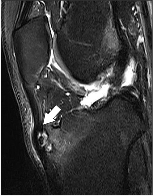
MRISagittal T2-weighted MRI of the knee: the arrow indicates thickening of the patellar tendon and bone marrow oedema at the tibial tuberosity, with a high-signal ununited fragment, a typical appearance of Osgood-Schlatter disease.
Source : Lee et al., Knee surgery & related research, 2016, figure 2 · CC BY-NC
Osgood-Schlatter disease is one of the most common causes of anterior knee pain in the growing adolescent: its prevalence is close to 1 young person in 10 at pubertal age 3. Good news for the physiotherapist and the family alike: the diagnosis is above all clinical. It rests on the history and a careful examination, not on imaging. As an international consensus of 251 professionals summarises it, the most widely endorsed diagnostic criterion is quite simply pain over the tibial tubercle 12.
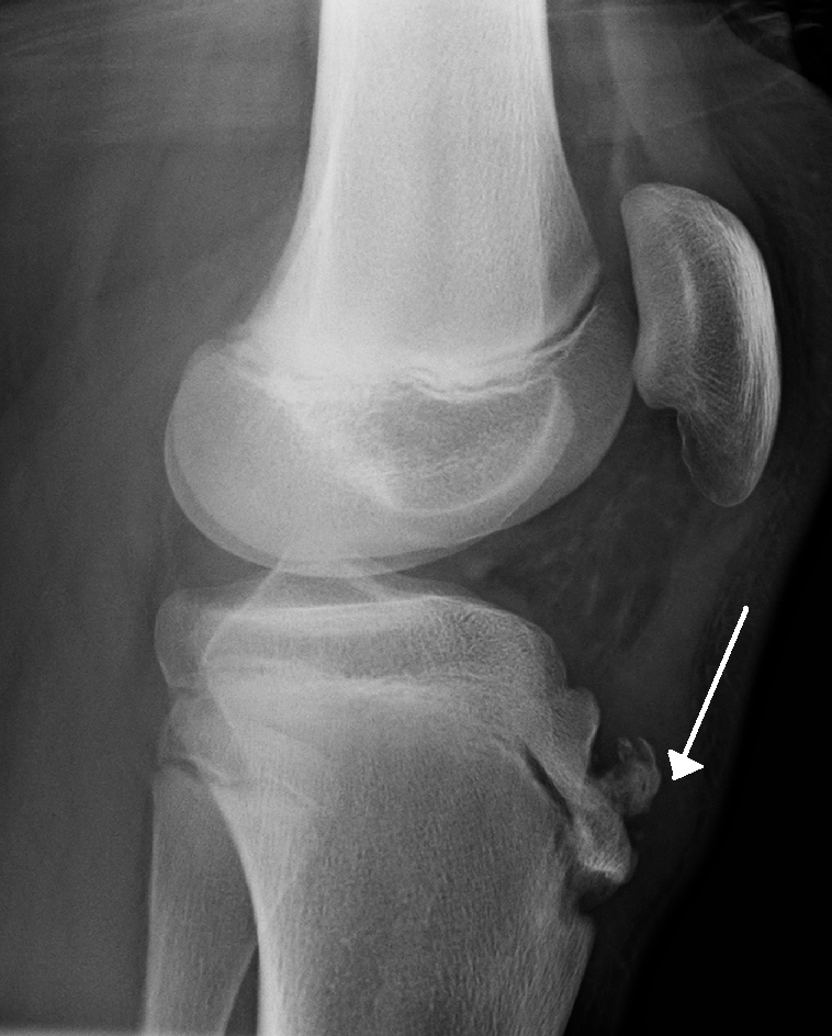
X-rayLateral knee radiograph in a patient with Osgood-Schlatter disease: fragmentation of the tibial tuberosity indicated by an arrow, with soft tissue swelling anterior to the patellar tendon.
Source : Heilman, Wikimedia Commons · CC BY-SA
Key points
- The diagnosis is clinical : pain localised to the tibial tubercle, reproduced on palpation and on resisted knee extension 12.
- Imaging is not necessary for the diagnosis; it serves only to rule out a differential diagnosis in an atypical or severe presentation 1.
- Typical setting: sporting adolescent in a growth spurt, peak around 11-12 years, bilateral involvement in 20 to 30% of cases 52.
- Shortening of the rectus femoris is the most strongly associated factor (OR 7.15), and should be looked for systematically 3.
Recognising the typical setting
The context is a strong pointer. Classically this is an adolescent in the middle of a growth spurt, more often a boy, and usually sporting. Incidence in general practice is estimated at 3.8 per 1000 person-years among 8-18 year olds, higher in boys (4.9) than in girls (2.7), with onset peaking around 12 years in boys and 11 years in girls 5. The link with sport is clear: in Kujala's historical cohort, 21.2% of active sporting adolescents had had the disease against only 4.5% of non-athletes, with first symptoms on average at 13.1 years, in the middle of maturation 6.
Pathophysiology sheds light on the examination: this is a traction apophysitis of the tibial tubercle. During growth, bone lengthens faster than the muscle-tendon unit gains flexibility, which increases the tension transmitted by the patellar tendon and the rectus femoris (quadriceps) to the still immature apophyseal cartilage; over time this repeated traction can progress to a cartilaginous avulsion 12. This is why repetitive sporting movement (running, jumping, changes of direction) is implicated, and why quadriceps flexibility deserves particular attention: shortening of the rectus femoris is the factor most strongly associated with the disease (OR 7.15; 95% CI 2.86-17.86), far ahead of sports participation alone (OR 1.94), and 74.6% of the pupils in the de Lucena cohort showed such shortening 3.
Clinical examination and provocation tests
The history reveals anterior knee pain of gradual onset, sited precisely over the tibial tubercle (and not over the joint line or the kneecap). It is typically worsened by running, jumping, climbing stairs, kneeling and squatting, and eased by rest 2. Localised swelling and a bony prominence are often seen, sometimes redness or tenderness to the slightest contact (the knee that “does not like” resting on the ground).
On examination, three concordant findings are usually enough:
- Palpation of the tibial tubercle : it is painful and often prominent. This is the cardinal sign, to be compared with the opposite side, bearing in mind that involvement may be bilateral.
- Resisted knee extension : the pain is reproduced when extension against resistance is requested, which loads the extensor mechanism over the apophysis 12. Active or passive flexion can also wake the pain.
- Testing for shortening of the rectus femoris : Ely's test, heel-to-buttock distance or extensor mechanism flexibility. Its frequency and the strength of its association make it a useful part of the examination, with directly therapeutic implications 3.
This combination (exact location, reproduction on palpation and on loading the extensor mechanism, compatible setting) forms the diagnostic foundation. An important fact that keeps the picture in proportion: the presence of symptoms does not imply severe involvement. In elite young footballers, point prevalence reached 17%, but 80% of symptomatic players lost no playing time 7. A history of Sever's disease (calcaneal apophysitis) was also strongly associated with Osgood-Schlatter disease in this population (OR 16.8), one more reason to ask about the other apophyses in an adolescent with multiple growing pains.
When (not) to order imaging
The message is clear and worth carrying to families: in a typical form, imaging is not necessary to make the diagnosis 12. In general practice, management rests first on advice and education (55.1% of consultations), and imaging was used in only 19.5% of cases 5. Piling up investigations in a routine presentation adds nothing and can cause needless worry.
Imaging does, however, keep one precise role: ruling out a differential diagnosis in an atypical or severe presentation, or one that does not behave like Osgood-Schlatter disease. Plain radiography is then the first-line investigation to exclude a fracture, a tumour or an infection 1. Ultrasound and MRI can complete the work-up case by case. It should be remembered, though, that radiological abnormalities (fragmentation, ossicle) have no simple diagnostic or prognostic value: an ununited ossicle was found in 32% of adolescents scanned at 24 months 9, and no ultrasound marker of bone maturity was associated with the disease in young footballers 7. In other words, the image does not make the diagnosis; the clinical picture does.
Which signals should, conversely, prompt imaging and sometimes referral? Night or rest pain that wakes the child, deterioration in general health, fever, diffuse swelling or a joint effusion, pain not localised to the tubercle, an acute injury with loss of function (raising the fear of an avulsion fracture), or a course that does not follow the expected path: all are flags that fall outside a simple traction apophysitis and justify further work-up.
The differential diagnoses to rule out
Making the clinical diagnosis also means knowing what has to be excluded. The literature lists as the main differentials, to be ruled out by imaging where needed: fracture (in particular an avulsion fracture of the tubercle after an acute injury), tumour, infection (osteomyelitis), patellar tendinitis/tendinopathy and Hoffa's disease 1.
To these can be added the other growth-cartilage conditions of the lower limb, which appear at the same age and sometimes in the same place: Sinding-Larsen-Johansson disease, whose painful point is at the lower pole of the patella and not over the tibial tubercle, osteochondritis dissecans, which produces an effusion where an apophysitis does not, and above all slipped capital femoral epiphysis, which causes knee pain in a substantial share of cases although the lesion is at the hip. These four entities and the reasoning that separates them are covered in lower-limb growth conditions in children and adolescents.
| Diagnostic possibility | What should raise concern | Level of evidence |
|---|---|---|
| Osgood-Schlatter | Selective pain over the tibial tubercle, worsened by resisted extension, in a growing sporting adolescent | High |
| Fracture / avulsion of the tubercle | Acute injury, sudden loss of function, inability to extend actively | Moderate |
| Infection (osteomyelitis) | Fever, rest and night pain, local inflammation, deterioration in general health | Moderate |
| Bone tumour | Non-mechanical, persistent pain, a mass, systemic signs | Low |
| Patellar tendinopathy / Hoffa's disease | Pain that is rather tendinous or from the fat pad, away from the tubercle | Moderate |
The “level of evidence” column describes how robust the data supporting each possibility are in the cited literature, not clinical severity.
One point deserves an honest qualification: the boundary with patellar tendinopathy is not watertight over time. In adults who had Osgood-Schlatter disease in adolescence, the risk of “jumper's knee” was very strongly raised (OR 70.4; 95% CI 32.9-155.0), and 85% still had a bony prominence of the tubercle 11. Adolescent apophysitis and adult tendon pain lie on the same extensor mechanism continuum, worth keeping in mind without turning it into a source of worry at the initial diagnosis.
Making the diagnosis also means informing about the prognosis
The diagnosis does not stop at the name of the disease: it includes what is said about it to the adolescent and the parents. The course is most often favourable and self-limiting once growth is complete, with no premature arrest of the tibial growth plate 8. But honesty is needed: resolution is not always quick. In a prospective cohort, 37% of adolescents still had pain at 2 years 9, and a study at about 4 years found 60.5% of young people still in pain 10. Discomfort on kneeling can persist in the majority 8, and residual symptoms are possible in adulthood in about 10% of cases 1.
Giving a reliable clinical diagnosis while setting out a realistic prognosis from the start (“this is very probably Osgood-Schlatter disease, it is benign, but it can take time to settle”) avoids two symmetrical pitfalls: the trivialising that disappoints when pain drags on, and the alarmism that pushes towards complete sporting rest, which is neither necessary nor recommended in place of progressive load management. On this last point caution remains in order: a systematic review stresses that the evidence is still of low to moderate quality and that no randomised trial has compared specific exercises with placebo or usual care 4. The diagnosis itself is solid; it is management that still calls for better-quality studies.
⏳ How does it progress? What to tell parents
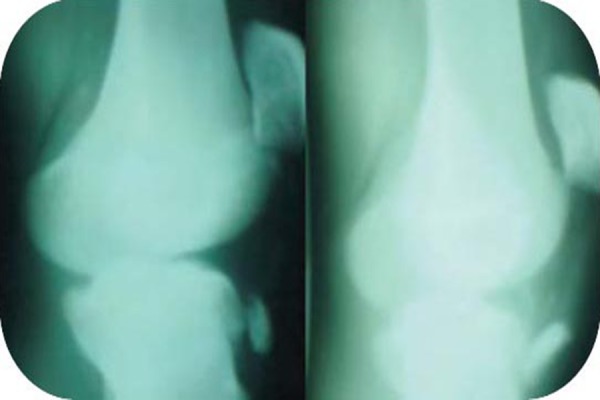
X-rayRadiographs of both knees showing bilateral avulsion fracture of the tibial tuberosity, a rare complication occurring on pre-existing Osgood-Schlatter disease.
Source : Narayana Gowda et al., Journal of orthopaedic case reports, 2012, figure 1 · CC BY-NC-SA
⏳ “It goes away with growth”: true, but not that fast, and not always without a trace
The course is most often favourable, but resolution is neither quick nor universal. A realistic message about waiting is worth more than a false promise.
Against the idea of a quick resolution. Pain at 24 months: prospective cohort, Holden et al., 2021 (PMID 34435066). Pain at ~4 years (median follow-up 3.75 years): Guldhammer et al., 2019 (PMID 31700938). Persistent prominence in adults: Krommes et al., 2025 (PMID 40439870). Reassure, yes, but without promising it will vanish in a few weeks.
The question that comes up at every consultation is almost always the same: “Will it go away?” The honest answer is yes, most of the time , but rarely overnight, and not always completely. Osgood-Schlatter disease is a traction apophysitis of the tibial tubercle: during the growth spurt, bone growth outstrips the stretching capacity of the muscle-tendon unit, which increases the tension exerted by the patellar tendon and the quadriceps on the still immature apophyseal cartilage 21. This mechanism gives both the key to the prognosis and its limit: as long as growth is not finished the area stays sensitive; once bone maturation is complete, traction no longer acts on fragile cartilage. This is why what we say to parents has to hold two threads at once: reassuring without lying.
A self-limiting course in the great majority of cases
The core message is solid and deserves to be stated plainly: Osgood-Schlatter disease is for the most part a spontaneously resolving condition, which usually disappears once the affected area finishes growing 113. Conservative treatment (education, load management, strengthening) remains effective in more than 90% of patients, and surgery is considered only in the rare disabling forms that persist after growth-plate closure 13. One reassuring point that families often do not know: in the reference study on the natural history of the untreated disease (69 knees, 50 patients reviewed later on), no premature arrest of the tibial growth plate was observed 8. In other words, contrary to a common fear, Osgood-Schlatter disease does not damage the growth of the leg and does not lead to deformity.
One point calls for frankness, though: that recovery is not always as quick as families are often told. The picture of a few weeks' discomfort that fades on its own does not match what recent cohorts show. Setting a realistic horizon from the start avoids disappointment, wandering from consultation to consultation, and the temptation to stop all sport “until it passes”.
How long does it last? Some honest markers
Reported durations vary a great deal from one adolescent to another, and it is precisely this variability that has to be named. Kujala's historical study of young athletes gives concrete and rather encouraging markers: first symptoms appear on average around 13.1 years, complete cessation of training, when it happens, lasts on average 3.2 months, and discomfort interfering with full training persists on average 7.3 months 6. These are useful orders of magnitude to give parents: we think in months, not days, but rarely in years for the most troublesome phase.
Modern prospective cohorts do, however, qualify the idea of a systematically short resolution. In a follow-up of 51 adolescents aged 10 to 14, 37% still reported knee pain related to Osgood-Schlatter disease two years later, with a median symptom duration of 42 months in those who remained in pain 9. A follow-up study at about four years (median follow-up 3.75 years) found persistent pain in 60.5% of adolescents 10. These data call into question the assumption of a quick recovery for everyone: a sizeable minority of adolescents goes through a long symptomatic phase.
How can these messages be reconciled without losing parents? By separating two things: the final prognosis (near-certain recovery once growth is complete) and the pace of the journey (often several months, sometimes more, with ups and downs across growth spurts and sporting seasons). One simple sentence sums the situation up well: “It will heal, but you have to learn to live with it for a while, by managing effort.” Since involvement is bilateral in 20 to 30% of cases 21, it is also worth warning that the second knee may speak up, without that signalling any worsening.
Residual pain: talking about it frankly
This is the subject where we must neither dramatise nor understate. Several studies converge: nearly 40% of patients report residual pain at long-term follow-up, which can sometimes become chronic 1. In Krause's natural-history study, although 76% of patients reported no activity limitation later on, 60% still had discomfort when kneeling, a concrete detail that speaks to families 8. The bony prominence of the tubercle, for its part, often persists: it is a permanent “bump” in a proportion of cases, with no functional consequence in itself.
The very long-term prognosis is therefore not always entirely silent, and honesty demands saying so. In a Danish national cohort of adults who had Osgood-Schlatter disease in adolescence, a persistent bony prominence and tibial tubercle symptoms were present in three adults out of four, with knee quality-of-life scores (KOOS) significantly lower than those of a healthy population; about half retrospectively reported a symptom duration of at least two years during adolescence 11. These figures should not alarm, since the great majority of these adults lead normal lives with normal physical activity, but they do justify taking seriously the adolescent's complaint rather than brushing it aside with “it's nothing, it will pass”.
An important qualification to avoid any misunderstanding: these long-term data describe group trends, not the fate of one particular child. Above all they are an invitation not to neglect extensor mechanism flexibility, since shortening of the rectus femoris is the factor most strongly associated with the disease 3 , and to support the adolescent actively rather than leaving them simply waiting.
Sport: neither total stoppage nor headlong flight
Parents' immediate fear is that sport will have to stop. The data argue instead for adapted continuation. In elite young footballers, the point prevalence of Osgood-Schlatter disease reached 17%, but 80% of symptomatic players lost no playing time 7 : the presence of symptoms therefore does not force a stop. Strict rest is neither necessary nor recommended as a default strategy; load management comes first. It remains honest to say that high-quality evidence is still limited here, since a systematic review finds no randomised trial comparing specific exercises with placebo or usual care, with an overall methodological quality rated “low to moderate” 4. Clinical consensus, on the other hand, is strong: patient education (99%) and exercise therapy (92%) are its pillars 12.
In practical terms, an active approach gives good results. In a cohort of 51 adolescents, a 12-week protocol combining a pain-guided activity ladder (to dose patellar tendon load), progressive knee strengthening and a graduated return to sport achieved 80% success at 12 weeks and 90% at 12 months, with the worst pain of the week falling from a median score of 7/10 to 2/10 14. The key message to pass on to families: moderate, tolerable pain during effort is not dangerous, as long as it settles afterwards and does not worsen from week to week. Running, jumping and changes of direction are reduced temporarily, not removed for life, and can be relieved by swimming or cycling 1. Corticosteroids and NSAIDs are not recommended in this condition 1.
Key points for parents
- It almost always heals once growth is complete, without damaging the growth of the leg 813.
- Count in months, not days : the troublesome phase often lasts several months, and a minority of adolescents take longer 69.
- Residual pain is possible : about 40% keep some discomfort at long-term follow-up, often when kneeling; the bony “bump” may stay 18.
- No total stoppage of sport : we adapt the load and strengthen, rather than stopping everything 714.
- Moderate, tolerable pain is acceptable as long as it settles and does not worsen week after week 14.
🎯 What can physiotherapy do?
Osgood-Schlatter disease is a traction apophysitis of the tibial tubercle: during the growth spurt, bone grows faster than the muscle-tendon unit gains flexibility, and the repeated traction of the quadriceps (above all the rectus femoris) transmitted through the patellar tendon acts on the still immature apophyseal cartilage 12. This mechanism sets out the whole logic of rehabilitation: reduce the traction stress, restore the flexibility and strength of the extensor mechanism, and above all dose the load rather than forbid it. Physiotherapy is not automatic, since in general practice it concerns only about 13% of consultations, far behind advice and education (55%), but it is the centrepiece as soon as pain limits activity 5.
Education and load management: the foundation
This is the most solid and most consensual point. An international survey of 251 professionals (general practitioners, physiotherapists, sports physicians, surgeons) found near-consensus on patient education (99%) and exercise therapy (92%) as pillars, and on training load management (97%) as the leading criterion for resuming, ahead of pain intensity (87%) and psychological factors (86%) 12. Educating, here, means explaining to the adolescent and the family that the disease is benign in its mechanism, that moderate pain during effort does not damage the knee, and that strict rest is neither necessary nor recommended.
The most telling argument comes from the sporting field: in elite young footballers, point prevalence reached 17%, but 80% of symptomatic players lost no playing time 7. In other words, being in pain does not mean having to stop. The recommended strategy is therefore to modulate: reduce running, jumping and changes of direction until improvement, replacing them with swimming and cycling, rather than imposing a total stop 1. The physiotherapist helps to find that tolerance threshold and to raise it progressively.
Key points
- Education + exercise form the foundation validated by consensus 12.
- The goal is not complete rest : we manage the load, we do not remove sport 71.
- Pain guides the dosing: tolerable if it settles and does not worsen from week to week.
- Corticosteroids and NSAIDs are not advised in this condition 1.
- The prognosis is good but not always trivial: something to say honestly to families.
Exercise: progressive knee strengthening
The most useful clinical data come from a prospective cohort of 51 adolescents aged 10 to 14, in pain for a mean of 21 months. The 12-week protocol combined three ingredients: a progressive activity ladder dosing patellar tendon load through pain monitoring, home knee-strengthening exercises, and a graduated return to sport. The result: 80% success at 12 weeks, rising to 90% at 12 months, with the worst weekly pain falling from a median of 7/10 to 2/10, a strength gain of 32% in knee extension and 24% in hip abduction 14.
This result steers practice: rather than the classic “rest and wait”, we offer active work on the quadriceps and the lower-limb chain, calibrated below the pain threshold and increased step by step. The authors explicitly present this approach as an alternative to the passive strategies often prescribed 14. One caveat, though: this is a cohort with no control group, which limits how certain we can be about the specific effect of strengthening.
Quadriceps flexibility and hamstring work
The strongest risk factor identified in the literature is not sport itself but stiffness: in a population sample of 956 adolescents, shortening of the rectus femoris was associated with an odds ratio of 7.15 (95% CI 2.86-17.86), far above regular sports participation (OR 1.94), and 74.6% of the pupils showed muscle shortening 3. This finding, consistent with the mechanics of quadriceps traction, justifies giving a place to extensor mechanism flexibility; quadriceps stretching is in fact listed among the usual conservative measures 1.
Hamstring work follows the same logic of balancing the extensor mechanism and the posterior chain, but let us be clear about the status of the evidence: no study confirmed here isolates the specific effect of quadriceps stretching or hamstring strengthening against a placebo. The association between stiffness and the disease is a strong pathophysiological and epidemiological argument 3, not a demonstration that improving flexibility cures. We therefore offer this work as a reasonable component of an overall programme, without overselling it.
Ice, straps, orthoses: what are the adjunct measures worth?
These are symptomatic adjuncts, not disease-modifying treatments. The reference systematic review is blunt here: out of 13 primary studies, only 2 randomised trials, a methodological quality rated “low to moderate”, and no randomised trial comparing specific exercises with a placebo or usual care 4. In other words, neither ice, nor infrapatellar straps, nor stretching has high-quality evidence establishing its superiority. They may give occasional relief and remain compatible with adapted continuation of activity, but they do not replace load management and exercise.
One clear marker on the drug side, by contrast: prescribing corticosteroids and NSAIDs is explicitly advised against in this condition and should be avoided 1 – a message the physiotherapist can pass on to families tempted by self-medication.
Return to sport: graduated and pain-guided
Resumption is not decided on a date but on steps. In Rathleff's cohort, return to sport came at the end of the protocol, once the steps of the activity ladder had been cleared: it rose from 16% at 12 weeks to 69% at 12 months 14. The message given to the adolescent is twofold: moderate, tolerable pain during exercise is not dangerous as long as it settles after effort and does not worsen from week to week; and load is reintroduced in stages, never abruptly.
Historical duration markers help frame family expectations: in Kujala's study, complete cessation of training lasted on average 3.2 months and discomfort interfering with full training 7.3 months, with symptoms starting around 13.1 years 6. These figures are a reminder that patience is part of the treatment.
Staying honest about the prognosis
The course is most often favourable once growth is complete, and conservative treatment succeeds in more than 90% of patients, with surgery considered only for disabling symptoms persisting after growth-plate closure 13. But the idea of a resolution that is always quick and complete deserves qualifying in front of families. A prospective cohort followed for 24 months showed that 37% of adolescents still had pain related to the disease at the end of follow-up, with a median symptom duration of 42 months in those still in pain 9. Further out, a study at about 4 years found 60.5% of adolescents still in pain 10, and among adults who had had Osgood-Schlatter disease, 85% retained a bony prominence and 73% residual symptoms, with lowered KOOS scores and a greatly increased risk of patellar tendinopathy 11. It is also worth recalling that 20 to 30% of cases are bilateral and that about 10% of symptoms may persist into adulthood 12.
Physiotherapy therefore has a clear and useful role (educating, dosing the load, strengthening, supporting the return to sport) with an honestly modest level of evidence: the principles are solid and consensual, but well-conducted controlled trials are still lacking to rank the options 4. The best service to the adolescent remains helping them keep moving below the pain threshold, rather than immobilising them.
⚖️ Should sport be stopped?
This is the first question the adolescent and the parents ask: since sport triggered the pain, should everything stop until it passes? The answer, in the light of recent data, is nuanced but clear: complete cessation of sport is neither compulsory nor the best choice in most cases. Osgood-Schlatter disease is steered far better by fine, pain-guided load management than by strict, prolonged rest. It still has to be explained, because the reflex “rest = cure” remains deeply rooted.
Why sport is implicated… without being banned
A reminder of the mechanism: Osgood-Schlatter disease is a traction apophysitis of the tibial tubercle. During the growth spurt, bone lengthens faster than the muscle-tendon unit gains flexibility, which increases the tension exerted by the quadriceps (above all the rectus femoris) through the patellar tendon on the still immature apophyseal cartilage 12. Every run, every jump, every change of direction pulls on this fragile area.
Logically, sport is a well-established risk factor. In a population sample of 956 Brazilian adolescents aged 12 to 15, regular sports participation was associated with a nearly doubled risk (odds ratio 1.94; 95% CI 1.22-3.10) 3. Kujala's historical study is even more telling: 21.2% of active young athletes had had the disease, against only 4.5% of non-athletes 6.
But “risk factor” does not mean “to be banned”. The factor most strongly associated in the Brazilian study was not sport itself but shortening of the rectus femoris (odds ratio 7.15; 95% CI 2.86-17.86), present in nearly three adolescents out of four 3. In other words, the lever is not to remove sport but to act on the flexibility and tolerance of the extensor mechanism.
A symptom does not mandate stopping
The most counter-intuitive finding comes from the elite sporting field. In elite young footballers (U13-U19), the point prevalence of Osgood-Schlatter disease reached 17%, but 80% of symptomatic players lost no playing time 7. They carried on training and playing despite clinical symptoms being present. This illustrates a condition that often has little functional impact: the pain exists, but it does not forbid participation.
This finding shifts the question. It is not a matter of knowing whether the adolescent can play sport, but how much and how, depending on what the knee tolerates. Total rest, for its part, deprives the adolescent of a structuring activity, does not strengthen tissue and has never been shown to be superior.
What the load-management approach says
The validated alternative is called pain-guided load management. The principle: we do not put the knee to rest, we gradually adjust the demand on the patellar tendon using pain as a thermometer, while actively strengthening the knee.
Rathleff's prospective cohort (51 adolescents aged 10 to 14) gives the reference protocol. Over 12 weeks it combined:
- a progressive activity ladder (an “activity ladder”) calibrating the load applied to the patellar tendon;
- an exercise programme to strengthen the knee at home;
- an incremental return to sport, reintroduced once the steps of the ladder had been cleared 14.
The results argue for the active approach: 80% success at 12 weeks, 90% at 12 months, with the worst weekly pain falling from a median score of 7/10 to 2/10, a strength gain of +32% in knee extension, +24% in hip abduction and vertical jump improved by +19% 14. The authors explicitly present this strategy as an alternative to the passive approaches (rest or watchful waiting) that are often prescribed.
Pain as a compass, not as a prohibition
The practical core of this approach is pain monitoring. The message to give to the adolescent and the parents is precise: pain that is moderate and tolerable during effort is not dangerous, as long as it settles after activity and does not worsen from week to week 14. That is what separates intelligent load adjustment from simply “gritting one's teeth”.
In practice, training is modulated according to the pain response: we climb a rung of the ladder if pain stays low and stable, and drop back a rung if it rises. In the same cohort, return to sport rose from 16% at 12 weeks to 69% at 12 months – a progressive reintroduction of load, not an abrupt resumption 14.
When symptoms are sharp, the adaptation takes a simple and well-accepted form: temporarily reduce or suspend running, jumping and changes of direction until improvement, replacing them with activities that load the extensor mechanism little, such as swimming and cycling, while stretching the extensor musculature 1. Note that corticosteroids and NSAIDs are not advised in this condition 1.
Key points
- No strict rest: pain-guided load management does better than complete cessation 14.
- Pain is a compass: moderate discomfort that settles after effort and does not worsen is acceptable; training is modulated accordingly.
- We adapt rather than remove: temporarily reduce running and jumping, switch to swimming or cycling, strengthen the knee and stretch the quadriceps 13.
- A symptom ≠ a stop: 80% of symptomatic young footballers carry on playing 7.
- Graduated return to sport: in stages, once the activity ladder has been cleared, not all at once 14.
Strict rest or load management: what the data show
| Criterion | Strict sporting rest | Active load management | Level of evidence |
|---|---|---|---|
| Tissue strengthening | None (deconditioning) | Strength gains +24 to +32% 14 | Moderate |
| Favourable outcomes | Not shown to be superior | 80% at 12 weeks, 90% at 12 months 14 | Moderate |
| Maintaining activity | Participation interrupted | 80% with no loss of playing time 7 | Moderate |
| Clinician uptake | Rest cited in 21% of consultations | Education 99%, exercise 92%, load 97% 125 | Consensus |
Indicative comparison based on the studies cited; no randomised trial directly compares strict rest with load management.
What clinicians think, and the limits to know
Field practice points the same way. In general practice, management rests first on advice and education (55.1% of consultations), far ahead of rest (21.0%) 5. And in an international survey of 251 professionals (general practitioners, physiotherapists, sports physicians, surgeons), a near-consensus emerges: patient education 99%, exercise therapy 92%, and for deciding on the return to activity, training load management 97%, pain intensity 87%, psychological factors 86% 12.
We must nonetheless stay honest about the level of evidence. A systematic review of conservative treatments (13 studies, only 2 randomised trials) rates the methodological quality as “low to moderate” and stresses that no randomised trial compares specific exercises with a placebo or usual care 4. In other words: load management is better than complete cessation, clinical experience and cohorts support it strongly, but high-quality evidence is still lacking. We therefore steer consistently, without claiming a certainty we do not have.
A prognosis not to be underestimated
Finally, avoiding strict rest does not mean playing the disease down. Contrary to the idea of a quick resolution, 37% of adolescents in a prospective cohort still had pain at 2 years 9, and at about 4 years of follow-up, 60.5% still reported discomfort 10. In adults who had had Osgood-Schlatter disease, a persistent bony prominence was present in 85% of cases and residual pain in 73% 11. This sometimes long prognosis is precisely one more argument for active and sustained management rather than passive rest, which does not prepare the knee for future loads, and a reason to be transparent with families about what lies ahead.
⚽ How is the return to sport managed?
The question the adolescent, the parents and the coach invariably ask is not “can he carry on with sport?” but “how do we resume without making things worse or dragging them out?”. The good news, long underestimated: in Osgood-Schlatter disease, complete cessation of sport is most often neither necessary nor recommended. The modern pivot of management is pain-guided load management, rather than strict rest or watchful waiting. But one still has to know the markers that make that resumption safe.
Carrying on rather than stopping: what the clinical data say
The most telling argument comes from the sporting field itself. In elite young footballers (U13-U19), the point prevalence of Osgood-Schlatter disease reached 17%, but 80% of symptomatic players lost no playing time 7. In other words, the presence of symptoms does not by itself mandate being sidelined. This reality of low impact should reassure families without denying the pain.
Total rest is not, in fact, the reference treatment. Practical recommendations favour reducing (not removing) running, jumping and changes of direction until improvement, these movements being temporarily replaceable by swimming and cycling, while working on extensor mechanism flexibility 1. Corticosteroids and NSAIDs, for their part, are explicitly advised against in this condition 1. We must nonetheless stay honest about the level of evidence: a systematic review counts only 2 randomised trials among 13 studies, with a methodological quality rated “low to moderate” and no trial comparing a specific exercise with a placebo 4. We are therefore describing a coherent and consensual approach, not a truth demonstrated at the highest level of evidence.
The pain threshold: the rule that guides everything
The operating principle fits in one sentence. Pain that is moderate and tolerable during exercise is not dangerous, provided it settles after effort and does not worsen from week to week 14. It is this slider, and not the total absence of pain, that allows a step up or requires a step down in load. In the reference cohort, patellar tendon load was steered by a progressive activity ladder coupled with pain monitoring; this protocol brought the worst weekly pain from a median score of 7/10 to 2/10 in 12 weeks 14.
Key points: steering the resumption by pain
- Pain that is acceptable during and after effort (usual marker ≤ 3-4/10) and settles at rest: we can keep going, or even progress.
- Pain that rises, persists the next day or worsens week after week : we drop back a step, without stopping altogether.
- The target is not “zero pain before resuming”, but pain that is stable and decreasing under an increasing load.
- We first reduce what loads the apophysis most: running, jumping, changes of direction; we replace them with cycling and swimming 1.
Progression criteria: a ladder, not a switch
Resumption is conceived as a graduated climb, rung by rung, and not as a binary green light. In Rathleff's protocol 14, the graduated return to sport came only after the steps of the activity ladder had been cleared (return to competition being introduced once step 8 was reached). In practice, progression combines three levers:
- Knee strengthening and hip strengthening: the reported gains were +32% in knee extension and +24% in hip abduction, with +19% in jump height, signs of a better capacity to absorb load 14.
- Extensor mechanism flexibility : shortening of the rectus femoris is the factor most strongly associated with the disease 3, which justifies the physiotherapist monitoring and working on quadriceps flexibility.
- Progressive reintroduction of impact : running, then jumping, then pivoting are added step by step, validating each rung against the pain threshold before moving on to the next.
This progressive reintroduction of load came with a return to sport in 69% of adolescents at 12 months 14. This figure also calls for patience: full resumption does not happen within a few weeks for everyone.
What timescales should be given?
This is where what we say must stay nuanced, because timescales vary enormously. Kujala's historical markers 6 give useful orders of magnitude: complete cessation of training lasted on average 3.2 months, and discomfort interfering with full training persisted on average 7.3 months. But these are only averages.
Two findings call for humility. First, the course is not always quick: in a prospective cohort followed for 24 months, 37% of adolescents still had knee pain at 2 years, with a median symptom duration of 42 months in those who remained in pain 9. Second, on the feasibility of active management: success was 80% at 12 weeks and 90% at 12 months, but the actual return to sport concerned only 16% of the young people at 12 weeks against 69% at 12 months 14. The honest message to give: we often feel better before playing fully again, and the trajectory is counted in months, sometimes in years.
Recurrence and residual symptoms: speaking plainly
Osgood-Schlatter disease is most often self-limiting once growth is complete, and conservative treatment succeeds in more than 90% of patients 13. But “self-limiting” does not mean “without a trace”. In the seminal natural-history study, 76% of patients reported no activity limitation, yet 60% still had discomfort when kneeling 8. In the long term, among adults who had the disease in adolescence, a bony prominence persisted in 85% and pain in the same area in 73%, with lowered knee quality-of-life scores (KOOS) 11. About 10% of cases keep symptoms into adulthood 1.
In practical terms, “recurrence” is often a reactivation linked to load being raised too quickly during a growth phase. The right course is not to start again from scratch, but to drop back one step on the activity ladder as soon as pain rises back above the tolerable threshold, then to climb more slowly. Involvement is bilateral in 20 to 30% of cases 2 : pain appearing in the other knee is not necessarily a worsening, but a known expression of the disease.
A message for parents
The physiotherapist's role here is as much educational as technical: in practice, patient education is near-consensual (99% of professionals), as is exercise therapy (92%) 12. With parents, a clear frame can be set:
- This is not serious in the orthopaedic sense : no case of premature growth-plate arrest has been observed in the natural history of the disease 8.
- Sport is not forbidden : most symptomatic young people carry on playing 7 ; the aim is to adjust, not to ban.
- But let us be patient and honest : pain can last for months, come back with growth spurts, and leave a small lump or residual discomfort when kneeling 911.
- Tolerable pain does no damage : it serves as a guide, not an alarm signal, as long as it is decreasing 14.
A message for the coach
The coach is the daily regulator of load: it is the coach who, in agreement with the physiotherapist, applies the activity ladder on the field. Three concrete instructions:
- Modulate, do not exclude. Temporarily reduce running volume, jumping and pivoting during painful peaks; keep technical work, mobility and strengthening 1.
- Follow the pain slider. A player can train with moderate pain that settles afterwards; the load must be lightened if it rises session after session 1412.
- Avoid sudden jumps in load after a break (holidays, injury) or in the middle of a growth spurt: this is the typical setting for reactivation. The build-up is done in validated steps.
Key points: the course to hold on return to sport
- Load management > strict rest, steering by tolerable and decreasing pain 14 (but high-quality evidence is still limited) 4.
- Graduated progression : knee and hip strengthening, quadriceps flexibility, reintroduction of impact in steps.
- Variable timescales : often several months, sometimes > 2 years 69. Give a range, not a date.
- Recurrence = load raised too quickly : drop back a step, do not stop everything; bilateral involvement is common (20-30%).
- Speak plainly to parents : benign for growth, but residual discomfort is possible in adulthood 811.
🗂️ What do concrete case reports teach us?
Large cohorts give figures; the consultation, for its part, plays out on one knee, a competition calendar and a family's worry. To link the two, here is a published case, with its PubMed identifier: an elite athlete followed over twenty months. They are built solely from data confirmed by the literature, in order to illustrate clinical reasoning, load dosing and a logic of return to sport. They carry no evidential weight: they are teaching aids.
Published case: a 12-year-old karateka followed for twenty months
The case. Gaweł and Zwierzchowska report a 12-year-old karateka at Olympic level, with Osgood-Schlatter disease that was bilateral, and who had begun sport-specific training at the age of 415. The authors' aim was to identify the aetiopathogenic factors of the bilateral form and those that support the effectiveness of management15.
The assessment. Palpation by the orthopaedic surgeon, then objective assessment with ultrasound, the wall slide test, MRI, and height and weight measurements15.
The management. It lasted twenty months, in five stages, combining physical modalities and physiotherapy15.
What this case brings that two invented adolescents did not. First, a timescale : twenty months, in a supervised and adherent athlete. That is a long way from the “few weeks of load management” sometimes announced to families, and it is the kind of figure that a teaching vignette tends to shorten without meaning to. Then a predisposing background : sport specialisation begun at 4 years of age, that is to say exactly the profile of early, repeated exposure whose causal role is under discussion.
An isolated case does not demonstrate the effectiveness of the protocol followed, and it does not replace the data presented above. What it does fix is the order of magnitude of the time involved and the reality of the sporting field.
What these two cases bring to light
This case sketches the reasoning applied at two levels of severity:
| Feature | Case 1: Lucas (a form with little impact) | Case 2: Emma (an established form) | Level of evidence |
|---|---|---|---|
| Diagnosis | Clinical: tubercle pain plus resisted extension | Clinical, bilateral (20-30% of cases) | High |
| Imaging | Not necessary (nothing atypical) | Not necessary at the outset | Moderate |
| Sporting load | Modulated, no strict stop | Pain-guided ladder | Moderate |
| Physiotherapy lever | Quadriceps flexibility plus strengthening | Progressive strengthening plus graduated return | Moderate |
| Return to sport | Maintained and adapted from the outset | Graduated, at the end of the protocol | Moderate |
Illustrative summary built on the confirmed data 7114123.
We must nonetheless stay honest about the level of evidence. A systematic review of conservative treatments for Osgood-Schlatter disease (13 studies, of which only 2 randomised trials) concludes that methodological quality is “low to moderate” and notes that no randomised trial compares specific exercises with a placebo or usual care 4. In other words: load management and strengthening are coherent approaches, supported by cohorts and a broad clinical consensus, but not yet shown to be superior by high-level trials. The right stance is therefore not to decide beyond the data, but to individualise, and to tell the patient and the family what we know and what we do not yet know.
🧭 How is this applied in practice?
Osgood-Schlatter disease is one of the most common knee complaints in the sporting adolescent: about one young person in ten of pubertal age is affected 3, with a peak around 11-12 years 5. The good news: physiotherapy management is simple in principle. It rests on three widely consensual pillars (educating, dosing the load, strengthening) and abandons the old reflex of strict rest. Here is how to translate that in the clinic.
A five-step management algorithm
Clinical reasoning can unfold as a reproducible sequence, from first contact to return to sport.
1. Confirm the diagnosis, clinically. The diagnosis is essentially clinical: pain over the tibial tubercle, reproduced on palpation and on resisted knee extension, worsened by running, jumping, kneeling and squatting 12. Imaging is not needed to make the diagnosis; plain radiography keeps its place only to rule things out in a severe or atypical presentation 1. Remember to examine both knees: involvement is bilateral in 20 to 30% of cases 2.
2. Assess the modifiable factors. Two factors dominate the risk: regular sports participation during puberty (odds ratio 1.94) and above all shortening of the rectus femoris, one head of the quadriceps 3. This last figure, very high, justifies assessing extensor mechanism flexibility systematically and making it a therapeutic target.
3. Educate, from the very first session. This is the foundation: in the international consensus of 251 professionals, patient education attracts 99% agreement 12. Explain what a traction apophysitis is: the repeated pull of the quadriceps and the patellar tendon on the growth cartilage of the tubercle, aggravated by the growth spurt in which bone grows faster than the muscle-tendon unit lengthens 12. Reassure without playing things down.
4. Dose the load rather than stopping it. We temporarily reduce running, jumping and changes of direction until improvement, offering alternatives such as swimming and cycling 1, but we do not prescribe a total, passive stop. The concrete tool is a pain-guided activity ladder: the young person moves from one step to the next as long as pain stays tolerable and settles. NSAIDs and corticosteroids are not advised in this condition 1.
5. Strengthen and rehabilitate, then a graduated return to sport. In Rathleff's protocol 14, progressive knee strengthening is added to the activity ladder; the return to sport is graduated, once the steps of the ladder have been cleared, rather than an abrupt resumption. This protocol gave 80% success at 12 weeks and 90% at 12 months, with strength gains in knee extension (+32%) and hip abduction (+24%), the worst pain of the week falling from a median score of 7/10 to 2/10.
| Lever | What it means in practice | Level of evidence |
|---|---|---|
| Patient education | Explain the apophysitis, reassure, set realistic expectations about duration | Strong consensus (99%), moderate evidence |
| Load management | Pain-guided activity ladder; swimming and cycling as substitutes | Prospective cohort, 80–90% success |
| Progressive strengthening | Quadriceps and hip, increasing tolerated load | Prospective cohort 14 |
| Extensor mechanism flexibility | Stretching of the rectus femoris and quadriceps | Plausible (strong risk factor), weak evidence |
| Complete strict rest | To be avoided as a default prescription | Not recommended |
Conservative protocols rest mainly on cohorts and expert consensus; no randomised trial compares specific exercises with a placebo or usual care 4.
The key messages to pass on to the young person and the family
A large part of the effect lies in what we say. Four simple messages structure the therapeutic alliance.
“Sport is not forbidden.” In elite young footballers, 80% of symptomatic players lost no playing time 7 : the presence of pain does not mandate stopping. We adapt, we do not remove.
“Moderate pain is not a danger.” Tolerable discomfort during exercise is acceptable as long as it settles and does not worsen week after week 14. This is the principle of pain monitoring, which replaces fear of movement.
“It can take a long time, and that is normal.” Contrary to the received idea of a quick resolution, 37% of adolescents still had pain at 2 years 9, and in Kujala's historical study 6, complete cessation of training lasted on average 3.2 months and discomfort interfering with full training 7.3 months. Announcing weeks to months, rather than days, avoids disappointment and therapeutic wandering.
“Working on quadriceps flexibility makes sense.” Shortening of the rectus femoris is the factor most associated with the disease 3 : giving the young person a concrete, active direction strengthens their sense of control.
Common mistakes to avoid
Mistake 1: prescribing strict rest. Complete rest is still often prescribed by reflex 5, whereas dosed load does better. An active approach (activity ladder, strengthening, graduated return) is presented by the authors as a credible alternative to passive approaches such as rest or watchful waiting 14. Immobilising deprives the tendon and the muscle of the stimulation they need and deconditions the young person without speeding up recovery.
Mistake 2: setting up a nocebo narrative. Talking about “bone tearing away” or a “lesion that worsens with every step”, or banning all sport, creates a counterproductive fear of movement. Yet the natural history is rather reassuring: in the seminal study, 76% of patients reported no activity limitation later on and no case of premature growth-plate arrest was observed 8. The message must protect without frightening.
Mistake 3: promising a guaranteed recovery that leaves no trace. The opposite of nocebo, excessive reassurance, is also a mistake. We must stay honest: nearly 40% of patients keep residual pain at follow-up 1, 60% discomfort when kneeling 8, and in adults who had had Osgood-Schlatter disease, a persistent bony prominence and tubercle symptoms are common 11. Symptoms are possible in adulthood in about 10% of cases 1.
Mistake 4: piling up imaging and passive treatments. Imaging adds nothing to the clinical diagnosis in typical forms 12, and NSAIDs, like corticosteroids, are not advised 1. Refocus on education and exercise, the two most consensual treatments 12.
Strict rest is neither necessary nor recommended: dose the load, do not switch it off.
Honesty about the level of evidence
It has to be said clearly to the reader: the evidence base remains limited. A systematic review of conservative treatments (13 studies, only 2 randomised trials) rates methodological quality as “low to moderate” and stresses that no randomised trial compares specific exercises with a placebo or usual care 4. The excellent results of load management come from cohorts, not from controlled trials. In other words, the direction is solid and consensual (educating, dosing, strengthening rather than immobilising), but the ideal protocol (dosing, precise exercises, duration) remains to be defined. Presenting that uncertainty is part of honest practice.
Key points
- Clinical diagnosis (pain over the tibial tubercle, resisted extension); imaging only to rule out a differential. Think of both knees (20–30% bilateral).
- Three consensual pillars: education (99%), load management, progressive strengthening; no strict rest, no NSAIDs and no corticosteroids.
- Pain-guided activity ladder: moderate pain that settles is acceptable; we adapt the sport, we do not stop it (80% of symptomatic athletes lose no playing time).
- Target the rectus femoris (a major risk factor, OR 7.15) through flexibility and strengthening.
- Favourable outcomes in 80–90% of young people with active management, but the course is sometimes long (months to years) and residual discomfort is possible: stay honest with the family.
- Low to moderate level of evidence: the direction is clear, the optimal protocol remains to be established.
Bibliography
13 of the 14 sources are indexed on PubMed and checked individually (clickable PMID); the others are official guidance. Click a superscript note marker in the text: the bibliography opens and highlights the source.
- Corbi F, Matas S, Álvarez-Herms J, Sitko S, Baiget E, Reverter-Masià J, López-Laval I (2022). Healthcare (Basel). PMID 35742062. doi:10.3390/healthcare10061011.
- Smith JM, Varacallo M (StatPearls) (2023). StatPearls (NCBI Bookshelf). Read it.
- de Lucena GL, dos Santos Gomes C, Guerra RO (2011). The American Journal of Sports Medicine. PMID 21076014. doi:10.1177/0363546510383835.
- Neuhaus C, Appenzeller-Herzog C, Faude O (2021). Physical Therapy in Sport. PMID 33744766. doi:10.1016/j.ptsp.2021.03.002.
- van Leeuwen GJ, et al. (2022). The British Journal of General Practice. PMID 34990396. doi:10.3399/BJGP.2021.0386.
- Kujala UM, Kvist M, Heinonen O (1985). The American Journal of Sports Medicine. PMID 4025675. doi:10.1177/036354658501300404.
- Schultz M, Tol JL, Veltman L, van der Kaaden L, Reurink G (2022). Physical Therapy in Sport. PMID 35305497. doi:10.1016/j.ptsp.2022.02.024.
- Krause BL, Williams JPR, Catterall A (1990). Journal of Pediatric Orthopedics. PMID 2298897.
- Holden S, Olesen JL, Winiarski LM, Krommes K, Thorborg K, Hölmich P, Rathleff MS (2021). Orthopaedic Journal of Sports Medicine. PMID 34435066. doi:10.1177/23259671211022239.
- Guldhammer C, Rathleff MS, Jensen HP, Holden S (2019). Orthopaedic Journal of Sports Medicine. PMID 31700938. doi:10.1177/2325967119878136.
- Krommes K, Bjerre A, Thorborg K, Nielsen MF, Hölmich P (2025). Sports Medicine. PMID 40439870. doi:10.1007/s40279-025-02214-5.
- Lyng KD, Rathleff MS, Dean BJF, Kluzek S, Holden S (2020). Scandinavian Journal of Medicine & Science in Sports. PMID 32562293. doi:10.1111/sms.13751.
- Ladenhauf HN, Seitlinger G, Green DW (2020). Current Opinion in Pediatrics. PMID 31714260. doi:10.1097/MOP.0000000000000842.
- Rathleff MS, Winiarski L, Krommes K, Graven-Nielsen T, Hölmich P, Olesen JL, Holden S, Thorborg K (2020). Orthopaedic Journal of Sports Medicine. PMID 32284945. doi:10.1177/2325967120911106.
- Gaweł E, Zwierzchowska A (2021). Therapeutic interventions in Osgood-Schlatter disease: A case report. Medicine (Baltimore). PMID 34918694. doi:10.1097/MD.0000000000028257.
❓ Frequently asked questions
What is Osgood-Schlatter disease and why does it occur?
It is a traction apophysitis of the tibial tubercle. During the growth spurt, bone growth outstrips the stretching capacity of the muscle-tendon unit: repeated contractions of the rectus femoris (quadriceps), transmitted through the patellar tendon, exert sustained traction on the apophyseal cartilage of the tibial tubercle and may go as far as a cartilaginous avulsion 12.
How common is it and who is affected?
In a population sample of 956 adolescents aged 12 to 15, prevalence was 9.8% (11.0% of boys, 8.3% of girls) 3. It is markedly more common in athletes: 21.2% of active sporting students versus 4.5% of inactive ones, with a mean age at onset of 13.1 years 6. In general practice, incidence is 3.8 per 1000 person-years among 8-18 year olds, with a peak around 11-12 years 5.
What are the risk factors?
Two factors stood out in multivariate analysis: regular sports participation during puberty (OR 1.94; 95% CI 1.22-3.10) and above all shortening of the rectus femoris, one head of the quadriceps (OR 7.15; 95% CI 2.86-17.86), which underlines the value of working on extensor mechanism flexibility 3. A history of Sever's disease was also strongly associated (OR 16.8) 7.
How is the diagnosis made?
The diagnosis is essentially clinical, based on symptoms: pain over the tibial tubercle reproduced on palpation and on resisted knee extension, worsened by running, jumping, kneeling and squatting. Imaging is not required for the diagnosis; plain radiography is the first-line investigation used to rule out differential diagnoses (fracture, tumour, infection, tendinitis, Hoffa's disease). Involvement is bilateral in 20 to 30% of cases 21.
Should sport be stopped, and how is it treated?
Not necessarily strict rest: among elite young footballers, 80% of symptomatic players lost no playing time 7. Treatment is conservative and centred on load management: reduce running, jumping and changes of direction until improvement (swimming and cycling can take their place), stretch the extensor mechanism; corticosteroids and NSAIDs are not advised 1. A 12-week protocol combining a pain-guided activity ladder with knee strengthening achieved 80% success at 12 weeks and 90% at 12 months 14. Patient education (99%) and exercise therapy (92%) are matters of consensus 12.
What is the long-term prognosis?
The course is most often favourable once growth is complete, but it is not always trivial. A cohort followed for 24 months showed that 37% still had knee pain at 2 years 9, and about 40% report residual pain at long-term follow-up 1. Among adults who had Osgood-Schlatter disease as adolescents, a bony prominence and tubercle symptoms persisted in roughly three out of four, with lowered KOOS scores 11. This is something to share honestly with families.

